SARS-COV-2 INFECTION INDUCES PROTECTIVE IMMUNITY AND LIMITS TRANSMISSION IN SYRIAN HAMSTERS
←
→
Page content transcription
If your browser does not render page correctly, please read the page content below
Research Article

SARS-CoV-2 infection induces protective immunity and
limits transmission in Syrian hamsters
Prabhuanand Selvaraj1,*, Christopher Z Lien1,* , Shufeng Liu1, Charles B Stauft1, Ivette A Nunez1, Mario Hernandez2,
Eric Nimako2, Mario A Ortega2, Matthew F Starost3, John U Dennis2 , Tony T Wang1

A critical question in understanding the immunity to SARS-COV-2                         recovered individual from becoming an active transmitter if re-
is whether recovered patients are protected against re-challenge                        exposed. Such information is critical needed for decision-making of
and transmission upon second exposure. We developed a Syrian                            public health policies and evaluating efficacy of vaccine candi-
hamster model in which intranasal inoculation of just 100 TCID50                        dates. To address this important question, we used a Syrian
virus caused viral pneumonia. Aged hamsters developed more                              hamster model to evaluate the protective efficacy of SARS-CoV-2
severe disease and even succumbed to SARS-CoV-2 infection,                              infection against secondary challenge and transmission.
representing the first lethal model using genetically unmodified
laboratory animals. After initial viral clearance, the hamsters
were re-challenged with 105 TCID50 SARS-CoV-2 and displayed
more than 4 log reduction in median viral loads in both nasal                           Results
washes and lungs in comparison to primary infections. Most
importantly, re-challenged hamsters were unable to transmit                             Induction of interstitial pneumonia in Syrian hamsters by 100
virus to naı̈ve hamsters, and this was accompanied by the                               TCID50 SARS-CoV-2
presence of neutralizing antibodies. Altogether, these results
show that SARS-CoV-2 infection induces protective immunity that                         Syrian hamsters of 7–9 wk old that were intranasally inoculated with
not only prevents re-exposure but also limits transmission in                           105 TCID50 (50% tissue culture infectious dose) SARS-CoV-2 had
hamsters. These findings may help guide public health policies                           weight loss for up to 7 d (Fig 1A). Lower inocula (from 104 to 10 TCID50)
and vaccine development and aid evaluation of effective vaccines                        caused lesser or no weight loss (Fig 1A). To accurately describe
against SARS-CoV-2.                                                                     active viral replication, we measured levels of subgenomic viral
                                                                                        mRNA (sgmRNA) by real-time PCR (11) in nasal washes (NW) of
DOI 10.26508/lsa.202000886 | Received 19 August 2020 | Revised 20 January               hamsters inoculated with 105 TCID50 at 2, 3, 5, and 7 days post-
2021 | Accepted 21 January 2021 | Published online 11 February 2021                     inoculation (d.p.i.). We also assessed viral RNA (vRNA) and sgmRNA
                                                                                        in lung tissue and organs at 3 and 5 d.p.i. Interestingly, high levels of
                                                                                        sgmRNA were observed in NW with a median peak of 4.87 (range
Introduction                                                                            3.73–5.38) log10 sgmRNA copies/5 μl on 2 d.p.i, but the median
                                                                                        quickly subsided to below detection limit on 5 d.p.i. (Fig 1B). On
The outbreak of the severe acute respiratory syndrome coronavirus                       3 d.p.i., high levels of sgmRNA and vRNA could be detected in both
2 (SARS-CoV-2) has quickly turned into a global pandemic (1). SARS-                     NW and in the lungs (Fig 1C and D). On Day 5 p.i., although high vRNA
CoV-2 is described to have an R0 value of 2–5 (2) and is well adapted                   levels were primarily detected in the upper and lower respiratory
to transmission among humans (3 Preprint) through aerosol and                           tract (Fig 1E), the sgmRNA level in respiratory tract (nasal turbinate,
possibly the fecal–oral route (4). Infectious virus has been isolated                   trachea, and lung) declined to just above the detection limit,
from oro- or naso-pharyngeal swabs (5), sputum (5), and stool                           whereas it remained substantial in intestine, spleen and kidney (Fig
samples (6) of individuals with severe coronavirus disease 2019                         1F). Viral pneumonia was also confirmed by HE staining of lung
(COVID-19). Limited evidence suggests that recovered individuals                        tissues at 2–7 d.p.i (Figs 1G–N and S1). The infected lung was
are likely to be protected from reinfection (7), which has also been                    characterized as many multifocal consolidations composed of
experimentally demonstrated in animal models (8, 9, 10). However,                       foamy macrophages, atypical pneumocytes hyperplasia, interstitial
it remains unclear whether postinfection immunity will prevent a                        lymphocytic, and neutrophilic infiltrates. Scattered areas of airway

1
 Division of Viral Products, Center for Biologics Evaluation and Research, US Food and Drug Administration, Silver Spring, MD, USA 2Division of Veterinary Services, Center
for Biologics Evaluation and Research, US Food and Drug Administration, Silver Spring, MD, USA 3Division of Veterinary Resources, Diagnostic and Research Services
Branch, National Institutes of Health, Rockville Pike, MD, USA

Correspondence: tony.wang@fda.hhs.gov
*Prabhuanand Selvaraj and Christopher Z Lien contributed equally to this work

© 2021 Selvaraj et al.                                                                  https://doi.org/10.26508/lsa.202000886     vol 4 | no 4 | e202000886        1 of 8
Figure 1. Viral load and histopathological changes in female Syrian hamsters intranasally challenged with SARS-CoV-2
.(A) Weight change profiles of hamsters that have been intranasally infected by indicated doses of SARS-CoV-2 USA/WA1-2020. (B) Subgenomic viral mRNA (sgmRNA) detected in nasal
washes of SARS-CoV-2 challenged hamsters (N = 3) on 2, 5, 7 dpi. (C, D) SgmRNA and viral RNA (vRNA) (D) detected in nasal washes and the lungs of SARS-CoV-2–challenged hamsters (N = 6)
on 3 dpi. (E, F) vRNA and sgmRNA detected in various organs of SARS-CoV-2–challenged hamsters (N = 4) on 5 dpi. The detection limit (25 copies for sgmRNA and 100 copies for vRNA per
reaction) was shown with the dotted line. (G, H, I, J) Hematoxylin and eosin (H&E) staining of the lungs of 105 TCID50 SARS-CoV-2–challenged hamsters on 2, 5, and 7 dpi. (K, L, M, N) Lung
tissues from hamsters challenged with indicated doses of virus were estimated for the percentage of consolidated areas. Scale bar in (G, H, I, J, K, L, M, N) = 500 μm. (O) Neutralizing antibody
titers of each hamster at the end of the study. Blue dot denotes the hamster that received 100 TCID50; Green dot represents the hamster that received 10 TCID50.

Prior infection of SARS-CoV-2 limits transmission          Selvaraj et al.                          https://doi.org/10.26508/lsa.202000886          vol 4 | no 4 | e202000886             2 of 8
proteinaceous fluid were seen (Fig S1). Notably, the percentage of              neutrophils, and necrosis of terminal bronchiolar epithelial cells (Fig 2F).
consolidated areas in the lung directly correlated with the amount             Massive amount of viral genomic RNA was detected in the lung of the
of the initial inoculum. Shown in Fig 1M, a viral inoculum of as low as        deceased hamster (Fig 2G and H). Last, infection of aged male hamsters
100 TCID50 was able to cause 10–15% consolidated areas in the lung.            (13 mo old) resulted in 100% death (n = 4) between day 5 and 8 post-
By contrast, inoculation of 10 TCID50 did not result in observable             challenge (Fig 2J). Notably, a full panel necropsy did not find lesions in
viral pneumonia in one out of two hamsters. Serum-neutralizing                 other vital organs (pancreas, small intestine, large intestine, brain, and
antibody (nAB) titers rose from Day 2 to 5 dpi with mean nAB titers of         kidney). Examination of the infected hamster brains did not show the
160 to 320 at days 5 and 7, respectively (Fig 1O).                             presence of virus genomic RNA (Fig 2I). This finding contrasts with the
   On day 2 p.i., numerous individual necrotic respiratory epithelial          reported lethal encephalitis seen in a subset of infected K18-hACE2 mice
cells were seen in the middle nasal cavity (Fig S1A–C). On Day 5 p.i.,         (14 Preprint, 15, 16, 17). In general, the pathology in aged hamsters re-
the nasal cavity showed neutrophilic infiltrate, edema, degenera-               sembles what has been observed in patients with severe COVID-19 who
tion and loss of submucosal glands and nerves. Olfactory mucosal               usually have pneumonitis and/or acute respiratory distress syndrome
cells in the nasal cavity were abnormally arranged. Loss of cilia and          with increased pulmonary inflammation, thick mucus secretions in the
edema of submucosa was also noticed. In the lung, only a few                   airways, elevated levels of serum pro-inflammatory cytokines, extensive
isolated neutrophilic infiltrates and mild type II pneumocyte hy-               lung damage, and microthrombosis. Neutrophilia predicts poor out-
perplasia were observed on Day 2 p.i. without obvious viral                    comes in patients with COVID-19 (18), and the neutrophil-to-lymphocyte
pneumonia (Fig S1H and I). On Day 5 & 7 p.i., 30–60% areas in the              ratio is an independent risk factor for severe disease (19). It is unclear
lung were consolidated and filled with abundant atypical and                    whether the deceased hamster also developed cytokine storm, which is
lesser typical pneumocyte and terminal bronchiolar epithelial                  frequently seen in severe human COVID-19 patients (20). Post-mortem
hyperplasia and hypertrophy (Fig S1J–L). Airways in these foci had             examination of COVID-19 patients has revealed signs of vascular dys-
mild to moderate neutrophilic, lymphocytic, and foamy macro-                   function (21), which was also seen in hamsters.
phage infiltrates. In infected trachea, mild mucosal hyperplasia and
attenuation with loss of cilia were observed (Fig S1D–G). Lesions              Protection of reinfection and prevention of transmission by
were not found in brain including olfactory bulbs (Fig S1M–R), heart,          postinfection immunity
liver, kidney, small intestine, spleen, and salivary gland.
   As a model for studying SARS-CoV-2 pathogenesis, Syrian                     To access transmission from reinfected animals, a group of 10 hamsters
hamsters can also be easily infected through close contact with                that were previously infected and recovered were re-inoculated intra-
another infected hamster. As shown in Fig S2, when a naı̈ve hamster,           nasally 4 wk later with 105 TCID50 SARS-CoV-2 (Fig 3A). Although we have
termed as contact hamster, was intranasally given MEM and sub-                 demonstrated that a hamster inoculated with 104 TCID50 virus was able to
sequently housed in the same clean cage with a hamster that was                transmit virus to a naı̈ve hamster, we chose a higher inoculum of 105
intranasally inoculated with 104 TCID50 SARS-CoV-2 (defined as the              TCID50 to mimic a more extreme situation. The circulating neutralizing
transmitter), despite no observed weight loss (Fig S2A), the contact           antibody titers of these 10 hamsters range from 80 to 320 (Fig 3B). Re-
hamster developed viral pneumonia (Fig S2C–H), and became                      inoculation of these hamsters did not result in any weight loss (Fig 3C).
seroconverted (Fig S2B). The presence of virus-infected cells in the           NWs were collected from at 1, 2, 3, and 7 d after reinfection. For most NW
lung was confirmed by detecting SARS-CoV-2 genomic RNA using                    samples, the median levels of sgmRNA in NWs collected at all time points
RNAscope (Fig S2I–J). This is consistent with previous reports (12).           except 1 dpi were below the detection limit and the median sgmRNA level
                                                                               on 1 dpi was about four logs lower than that seen during primary in-
Lethal infection of aged Syrian hamsters                                       fections (Fig 3D), suggesting a significant reduction in active virus rep-
                                                                               lication in the nose. In consistent with this finding, comparison of the
Older people have been disproportionally affected by COVID-19, with            amount of infectious virus in the NW also showed a 3–4 log reduction in
markedly higher death rate (13). To explore whether this phenomenon is         the secondary infection (Fig 3E). The sgmRNA in the lungs was also
recapitulated in hamsters, we first intranasally inoculated four aged           undetectable on both Day 3 and 7 postinfection, although a very low level
female hamsters (10, 13, and 20 mo old) with 105 TCID50 SARS-CoV-2. Two        of vRNA could be detected in the lungs (Fig 3F). Pathology examination of
10-mo-old female hamsters were intranasally inoculated with plain              the lungs also showed no viral pneumonia, confirming protection of
medium as negative controls. Aged hamsters showed marked weight loss           these hamsters from virus-induced disease (Fig 3G).
(around 20%) within 7 d (Fig 2A) and outward signs of sickness such as             To understand the impact of prior infection on the ability of re-
sneezing, shortness of breath, shivering, and lethargy (Fig 2B and C). The     exposed hamsters to transmit virus, four re-inoculated hamsters
illness was especially obvious when breathing rates were analyzed, in          were paired with four naı̈ve hamsters (on Day 1 post–re-inoculation)
which the infected aged hamsters were breathing two to three times             for 24 h and then with another two aged naı̈ve hamsters on Day 2
faster than uninfected animals (Fig 2C). Sustainable levels of subgenomic      post-re-inoculation for 24 h in new clean cages. Over a period of 7 d,
viral mRNA (sgmRNA) were detected from NW up to 7 d postinfection,             the contact hamsters showed no signs of sickness, no weight loss,
indicating a prolongated period of active virus replication in the nose (Fig   and no detectable viral sgmRNA or RNA in the nose or in the lungs,
2D). The oldest hamster (20 mo old) died on Day 6 p.i. Postmortem              and no pathology in the lung (Fig 4A–E). By contrast, high amount of
histopathology examination indicated that the animal had severe, diffuse       vRNA was detected from two control contact hamsters that were
bronchointerstitial pneumonia along with myocardial disease and                exposed to hamsters which had no prior infection and subsequently
thrombus formation in the left atrium leading to death (Fig 2E). Worth         challenged with 105 TCID50 SARS-CoV-2 (Fig 4C). At the end of the
mentioning is that there were airway infiltrates of macrophages and             study, none of the contact hamsters exposed to re-inoculated

Prior infection of SARS-CoV-2 limits transmission   Selvaraj et al.            https://doi.org/10.26508/lsa.202000886   vol 4 | no 4 | e202000886    3 of 8
Figure 2. Prolonged virus replication in nasal cavity of aged Syrian hamsters.
(A) Weight change profile of aged hamsters after intranasal inoculation of 105 TCID50 SARS-CoV-2 isolate USA-WA1/2020. The date of birth was indicated to the right of
each hamster ID. WH031 and 32 represent negative controls inoculated with media. (B) Clinical scores of hamsters. Y-axis represents day post-infection (dpi). Day 0 is the
day when inoculum was given. X-axis represents the animal ID with WH031 and 032 being uninfected control. (C) Breathing rate was counted by two independent observers
and averaged. (D) sgmRNA levels in uninfected (black solid circles) and infected hamsters (colored solid circles) on 2,5,7,9,14 dpi. (A) Each color corresponds to the same
colored animal ID in (A). (E) Pathology of the heart of the hamster that died. The thrombus formation was noticed in the left atrium (indicated by white arrows and a small
black box) with a closeup image shown to the right. Scale bar = 200 μm. (F) Pathology of the lung of the hamster that succumbed to infection. Scale bar: left = 5 mm; right =
60 μm. (G, H, I) RNAscope images of the uninfected hamster lung (negative), the lung and the brain of WH036. Red dots indicate the presence of viral genomic RNA. (G, H,
J) Scale bar: 200 μm in (G), 60 μm in (H), 300 μm in (J). (J) Survival curves of four aged hamsters.

hamsters developed neutralizing antibodies, implying that they have                      disease manifestation in the older human population. In our study,
never been infected (Fig 4F). Altogether, these data suggest that                        infected hamsters only occasionally recorded increases in body
hamsters that have been previously infected are severely impaired in                     temperature. Young hamsters, even when losing 15–20% body
transmitting virus upon re-exposure.                                                     weight, did not show outward signs of sickness. Aged hamsters, by
                                                                                         contrast, exhibited sneezing, shortness of breath, and noticeable
                                                                                         lethargy upon infection. Aged hamsters in our study succumbed to
                                                                                         infection between Day 5 and 8 p.i. To the best of our knowledge, this
Discussion                                                                               is the first lethal model of SARS-CoV-2 using genetically unmodified
                                                                                         laboratory animals. The cause of death of aged hamsters is most
While several studies showing the promise of hamsters, non-                              likely due to severe pneumonia plus thrombus formation in the left
human primates, and ferrets to study SARS-CoV-2, disease mani-                           atrium. At this time, we are uncertain whether the thrombus for-
festation in laboratory animals ranges from mild to moderate. Here,                      mation in the left atrium was directly related to SARS-CoV-2 in-
we report that aged hamsters develop much more pronounced                                fection. Future investigation using additional aged hamsters are
disease and even fatality upon challenge, recapitulating the                             warranted. Notably, all infected hamsters showed recruitment of

Prior infection of SARS-CoV-2 limits transmission    Selvaraj et al.                     https://doi.org/10.26508/lsa.202000886     vol 4 | no 4 | e202000886         4 of 8
Figure 3. Post-infection immunity protects re-challenge.
(A) Overall study design of the study. 10 hamsters Fig 1A recovered from infection were reinoculated with 105 TCID50 SARS-CoV-2 isolate USA-WA1/2020 on Day 28 post
primary infection. (B) Neutralizing antibody titers of each hamster at the time of re-inoculation. (C) Weight change profile of all re-inoculated hamsters. (D) sgmRNA
detected from nasal washes collected on 0, 1, 2, 3, and 7 dpi. (E) Virus titers of nasal washes collected from first (red) and second (green) infections. (F) Viral RNA (red) and
sgmRNA levels (blue) in the lungs on 3, 7 dpi. (G) A representative HE image of the lung from a re-inoculated hamster. Scale bar = 60 μm.
both macrophages and neutrophils to the infected lungs. Around Materials and Methods
10–15% of COVID-19 patients progresses to acute respiratory distress
syndrome triggered by a cytokine storm. It would be interesting to Viruses and cells
measure other immune correlates in infected aged hamsters, but the
relative lack of reagents for conducting immunological assays in Vero E6 cell line (Cat. no. CRL-1586) was purchased from American Type
hamsters prevented us from measuring cytokine profiles. Never- and Cell Collection and cultured in eagle’s MEM supplemented with 10%
theless, aged hamsters offer a superior immunocompetent animal fetal bovine serum (Invitrogen) and 1% penicillin/streptomycin and
model to study severe form of COVID-19 and will be valuable ex- L-glutamine. The SARS-CoV-2 isolate USA-WA1/2020 was obtained from
perimental tools in developing vaccine and public health strategies Biodefense and Emerging Infectious Research Resources Repository (BEI
aimed at ending the global COVID-19. Resources), National Institute of Allergy and Infectious Diseases (NIAID),
The most important finding of this study is that prior infection of National Institutes of Health (NIH), and had been passed three times on
hamsters induced protective immunity, accompanied by the presence of Vero cells and one time on Vero E6 cells before acquisition. It was further
neutralizing antibodies, in the lung with significantly reduced active virus passed once on Vero E6 cells in our laboratory. The virus has been
replication in the nose upon re-exposure and hence restricted trans- sequenced verified to contain no mutation to its original seed virus.
mission. This finding provides important new insights into our under-
standing of postinfection immunity against SARS-CoV-2. To date, no
Virus titration
human reinfections with SARS-CoV-2 have been confirmed; there is also
no evidence that clinically recovered individuals continue transmitting
SARS-CoV-2 isolate USA-WA1/2020 was titered using the Reed &
SARS-CoV-2 to others. However, it is possible that the virus may replicate
Muench Tissue Culture Infectious Dose 50 Assay (TCID50/ml) system
in the nose of recovered individuals upon re-exposure such that these
(22). Vero cells were plated the day before infection into 96 well plates
persons may facilitate transmission of the virus to the rest of the
at 1.5 × 10⁴ cells/well. On the day of the experiment, serial dilutions of
population. In this scenario, prior infection may have little impact to stop
virus were made in media and a total of six to eight wells were infected
the continuous transmission of the virus. In our study, although active
with each serial dilution of the virus. After 48 h incubation, cells were
virus replication was sporadically detected from NW of a few re-
fixed in 4% PFA followed by staining with 0.1% crystal violet. The TCID50
inoculated hamsters, the overall level of active replication was four
was then calculated using the formula: log (TCID50) = log(do) + log (R) (f
logs lower than those in primary infection. We hypothesize the amount of
+ 1). Where do represents the dilution giving a positive well, f is a
virus shed from these hamsters was so low that the probability of
number derived from the number of positive wells calculated by a
transmitting and infecting another naı̈ve hamster becomes finite. It will
moving average, and R is the dilution factor.
be highly desirable for a vaccine to achieve similar effects, that is, to
protect against both lower respiratory tract disease and upper respiratory
tract disease. In fact, a recent SARS-CoV-2 DNA vaccine conferred pro- SARS-CoV-2 pseudovirus production and neutralization assay
tection with significant reductions in median viral loads in bron-
choalveolar lavage and nasal swabs in immunized rhesus macaques Human codon-optimized cDNA encoding SARS-CoV-2 S glycoprotein
compared with sham controls (11). Obviously, our study was carried out in (NC_045512) was synthesized by GenScript and cloned into eukaryotic cell
hamsters and hence did not address the question of whether SARS-CoV- expression vector pcDNA 3.1 between the BamHI and XhoI sites. Pseu-
2 may replicate more efficiently in humans. Nonetheless, our findings dovirions were produced by co-transfection of Lenti-X 293T cells with
support assessing virus replication in the upper respiratory tract, par- psPAX2, pTRIP-luc, and SARS-CoV-2 S expressing plasmid using Lipof-
ticularly in the nose, in preclinical studies to help with the evaluation of ectamine 3000. The supernatants were harvested at 48 and 72 h post-
potential vaccine efficacy before clinical trials. transfection and filtered through 0.45-mm membranes.
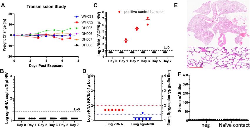
Prior infection of SARS-CoV-2 limits transmission Selvaraj et al. https://doi.org/10.26508/lsa.202000886 vol 4 | no 4 | e202000886 5 of 8Figure 4. Post-infection immunity limits transmission.
The overall study design is depicted in Fig 3A. Four hamsters were paired with four naı̈ve hamsters in clean cages on Day 29 (1 d after re-inoculation) and then another
two naı̈ve hamsters on Day 30 (2 d after re-inoculation of the transmitters). Each cage housed two hamsters (one re-inoculated and one naı̈ve). (A) Weight change profile
of contact hamsters. (B, C) sgmRNA and viral RNA (C) in nasal washes collected on 0, 1, 2, 3, 5, and 7 d post-contact. (C) Red solid circles in (C) denote two contact hamsters
which were exposed to hamsters that were without prior infection but challenged with 105 TCID50 virus. Black solid circles indicate contact those hamsters that were
exposed to the re-challenged hamsters. (D) Viral loads detected in the lungs. (E) Representative HE images of the lung from a contact hamster. Scale bar = 200 μm.
(F) Neutralizing antibody titers of each hamster at the end of the study.
For the serum neutralization assay, 50 μl of SARS-CoV-2 S pseudo- treated water provided in bottles, and all animals were acclimatized
virions were preincubated with an equal volume of medium containing at the BSL3 facility for 4–6 d or more before the experiments. The
serum at varying dilutions at room temperature for 1 h, then virus– study protocol details were approved by the White Oak Consolidated
antibody mixtures were added to 293T-hACE2 cells in a 96-well plate. Animal Care and Use Committee and carried out in accordance with
After a 3 h incubation, the inoculum was replaced with fresh medium. the PHS Policy on Humane Care & Use of Laboratory Animals.
Cells were lysed 48 h later, and luciferase activity was measured using For challenge studies, young (5–9 wk old) and aged (10–20 mo old)
luciferin-containing substrate. Controls included cell only control, virus Syrian hamsters were anesthetized with intraperitoneal injection of
without any antibody control and positive control sera. The end point anesthetic cocktail (50 mg/kg ketamine + 0.15 mg/kg dexmedetomi-
titers were calculated as the last serum dilution resulting in at least 50% dine). Intranasal inoculation was performed by pipetting 105 TCID50 or
SARS-CoV-2 neutralization. The amount of pseudovirions used in this desirable doses of SARS-CoV-2 in 50–100 μl volume dropwise into the
assay has been determined to give rise to a target input 5 × 10⁵–107 RLU/ nostrils of the hamster under anesthesia. To facilitate recovery, each
ml, under which condition the neutralization law is observed. hamster received 3.3 mg/kg diluted atipamezole solution intraperi-
toneally immediately after intranasal inoculation. Hamsters were
Hamster challenge experiments weighed and assessed daily. NW were collected by pipetting ~160 μl
sterile phosphate buffered saline into one nostril when hamsters were
Male and Female outbred Syrian hamsters were purchased from anesthetized by 3–5% isoflurane. For tissue collection and blood
Envigo at 4–5 wk of age. Aged hamsters from the same source were collection, hamsters were euthanized by intraperitoneal injection of
previously purchased and held at The US Food and Drug Adminis- pentobarbital at 200 mg/kg on days 2, 5, 7 as necessary.
tration (FDA) vivarium. All experiments were performed within the
biosafety level 3 (BSL-3) suite on the White Oak campus of the U.S. RNA isolation from NW and tissues
Food and Drug Administration. The animals were implanted sub-
cutaneously with IPTT-300 transponders (BMDS), randomized, and RNA was extracted from 50 μl NW or 0.1-g tissue homogenates using
housed two per cage in sealed, individually ventilated rat cages QIAamp vRNA mini kit or the RNeasy 96 kit (QIAGEN) and eluted with
(Allentown). Hamsters were fed irradiated 5P76 (Lab Diet) ad lib, 60 μl of water. 5 μL RNA was used for each reaction in real-time RT-
housed on autoclaved aspen chip bedding with reverse osmosis- PCR.
Prior infection of SARS-CoV-2 limits transmission Selvaraj et al. https://doi.org/10.26508/lsa.202000886 vol 4 | no 4 | e202000886 6 of 8Real-time PCR assay of SARS-CoV-2 viral and subgenomic RNA Acknowledgements
Quantification of SARS-CoV-2 vRNA was conducted using the SARS-CoV-2 We thank Drs. Carolyn Wilson, Jerry Weir, and Robin Levis for their support of
(2019-nCoV) CDC qPCR Probe Assay (IDTDNA) using iTaq Universal Probes this study and Drs. Marian Major and Surender Khurana for critical reading of
One-Step Kit (Bio-Rad). The standard curve was generated using 2019- the manuscript. We thank Ms. Katherine Shea and Dr. Hongquan Wan for
nCoV_N_Positive Control (IDTDNA). The detection limit of the vRNA was assistance with pathology slides and Mr. Andy LaClair and Ms. Michele
Howard for assistance with working in the BSL-3 laboratory. We are par-
determined to be 100 copies/reaction. Quantification of SARS-CoV-2 E
ticularly grateful to US Food and Drug Administration White Oak Division of
gene subgenomic mRNA (sgmRNA) was conducted using Luna Universal Veterinary Services staff and contractors who assisted in the hamster
Probe One-Step RT-qPCR Kit (New England Biolabs) on a Step One Plus studies. The following reagent was deposited by the Centers for Diseases
Real-Time PCR system (Applied Biosystems). The primer and probe Control and Prevention and obtained through Biodefense and Emerging
sequences were: SARS2EF: CGATCTCTTGTAGATCTGTTCT; PROBE: FAM- Infections Research Resources Repository, NIAID, NIH: SARS-Related Coro-
navirus 2, Isolate USA-WA1/2020, NR-52281. The work described in this
ACACTAGCCATCCTTACTGCGCTTCG- BHQ-1; SARS2ER: ATATTGCAGCAGTACG- manuscript was supported by US FDA intramural grant funds. The funders
CACACA. To generate a standard curve, the cDNA of SARS-CoV-2 E gene had no role in study design, data collection and analysis, decision to publish,
sgmRNA was cloned into a pCR2.1-TOPO plasmid. The copy number of or preparation of the manuscript. The content of this publication does not
sgmRNA was calculated by comparing with a standard curve obtained necessarily reflect the views or policies of the Department of Health and
Human Services, nor does mention of trade names, commercial products, or
with serial dilutions of the standard plasmid. The detection limit of the
organizations imply endorsement by the US Government.
sgmRNA was determined to be 25 copies/reaction. Values below de-
tection limits were mathematically extrapolated based on the standard
curves for graphing purpose. When graphing the results in Prism 8, values Author Contributions
below the limit of detection were arbitrarily set to half of the LoD values.
P Selvaraj: data curation, formal analysis, and methodology.
CZ Lien: conceptualization, investigation, methodology, and project
Histopathology analyses
administration.
S Liu: data curation and formal analysis.
Tissues (hearts, livers, spleens, duodenums, brains, lungs, kidneys, trachea,
CB Stauft: data curation.
salivary gland, and nasal turbinates) were fixed in 10% neural buffered
IA Nunez: data curation.
formalin overnight and then processed for paraffin embedding. The 4-μm
M Hernandez: methodology.
sections were stained with hematoxylin and eosin for histopathological
E Nimako: investigation and methodology.
examinations. Images were scanned using an Aperio ImageScope.
MA Ortega: investigation and methodology.
MF Starost: investigation and methodology.
In-situ hybridization JU Dennis: investigation and methodology.
TT Wang: conceptualization, data curation, and formal analysis.
To detect SARS-CoV-2 genomic RNA in FFPE tissues, in situ hybridization
(ISH) was performed using the RNAscope 2.5 HD RED kit, a single plex Conflict of Interest Statement
assay method (Cat. no. 322373; Advanced Cell Diagnostics) according
to the manufacturer’s instructions. Briefly, Mm PPIB probe detecting The authors declare that they have no conflict of interest.
peptidylprolyl isomerase B gene (housekeeping gene) (Cat. no. 313911,
positive-control RNA probe), dapB probe detecting dihydrodipicolinate
reductase gene from Bacillus subtilis strain SMY (a soil bacterium) (Cat.
no. 310043, negative-control RNA probe), and V-SARS-Cov-2-S (Cat. no.
References
854841) targeting SARS-CoV-2 positive-sense (genomic) RNA. Tissue
1. WHO (2020) WHO Director-General’s opening remarks at the media
sections were deparaffinized with xylene, underwent a series of ethanol
briefing on COVID-19
washes and peroxidase blocking, and were then heated in kit-provided
2. Sanche S, Lin YT, Xu C, Romero-Severson E, Hengartner N, Ke R (2020) High
antigen retrieval buffer and digested by kit-provided proteinase. Sec-
contagiousness and rapid spread of severe acute respiratory syndrome
tions were exposed to ISH target probes and incubated at 40°C in a coronavirus 2. Emerg Infect Dis 26: 1470–1477. doi:10.3201/eid2607.200282
hybridization oven for 2 h. After rinsing, ISH signal was amplified using
3. Zhan SH, Deverman BE, Chan YA (2020) SARS-CoV-2 is well adapted for
kit-provided pre-amplifier and amplifier conjugated to alkaline phos- humans. What does this mean for re-emergence? BioRxiv 10.1101/
phatase and incubated with a fast-red substrate solution for 10 min at 2020.05.01.073262 (Preprint posted May 2, 2020).
room temperature. Sections were then stained with 50% hematoxylin 4. Xu Y, Li X, Zhu B, Liang H, Fang C, Gong Y, Guo Q, Sun X, Zhao D, Shen J, et al
solution followed by 0.02% ammonium water treatment, dried in a 60°C (2020) Characteristics of pediatric SARS-CoV-2 infection and potential
dry oven, mounted, and stored at 4°C until image analysis. evidence for persistent fecal viral shedding. Nat Med 26: 502–505.
doi:10.1038/s41591-020-0817-4
5. Wolfel R, Corman VM, Guggemos W, Seilmaier M, Zange S, Muller MA,
Niemeyer D, Jones TC, Vollmar P, Rothe C, et al (2020) Virological
Supplementary Information assessment of hospitalized patients with COVID-2019. Nature 581:
465–469. doi:10.1038/s41586-020-2196-x
Supplementary Information is available at https://doi.org/10.26508/lsa. 6. Zhang Y, Chen C, Zhu S, Shu C, Wang D, Song J, Song Y, Zhen W, Feng Z, Wu
202000886. G, et al (2020) Isolation of 2019-nCoV from a stool specimen of a
Prior infection of SARS-CoV-2 limits transmission Selvaraj et al. https://doi.org/10.26508/lsa.202000886 vol 4 | no 4 | e202000886 7 of 8laboratory-confirmed case of the coronavirus disease 2019 (COVID-19). 15. Winkler ES, Bailey AL, Kafai NM, Nair S, McCune BT, Yu J, Fox JM, Chen RE,
China CDC Weekly 2: 123–124. doi:10.46234/ccdcw2020.033 Earnest JT, Keeler SP, et al (2020) SARS-CoV-2 infection of human ACE2-
7. Kirkcaldy RD, King BA, Brooks JT (2020) COVID-19 and postinfection transgenic mice causes severe lung inflammation and impaired
immunity: Limited evidence, many remaining questions. JAMA 323: function. Nat Immunol 21: 1327–1335. doi:10.1038/s41590-020-0778-2
2245–2246. doi:10.1001/jama.2020.7869 16. Golden JW, Cline CR, Zeng X, Garrison AR, Carey BD, Mucker EM, White LE,
8. Chandrashekar A, Liu J, Martinot AJ, McMahan K, Mercado NB, Peter L, Shamblin JD, Brocato RL, Liu J, et al (2020) Human angiotensin-
Tostanoski LH, Yu J, Maliga Z, Nekorchuk M, et al (2020) SARS-CoV-2 converting enzyme 2 transgenic mice infected with SARS-CoV-2 develop
infection protects against rechallenge in rhesus macaques. Science 369: severe and fatal respiratory disease. JCI Insight 5: e142032. doi:10.1172/
812–817. doi:10.1126/science.abc4776 jci.insight.142032
9. Chan JF, Zhang AJ, Yuan S, Poon VK, Chan CC, Lee AC, Chan WM, Fan Z, Tsoi 17. Oladunni FS, Park JG, Pino PA, Gonzalez O, Akhter A, Allue-Guardia A, Olmo-
HW, Wen L, et al (2020) Simulation of the clinical and pathological Fontanez A, Gautam S, Garcia-Vilanova A, Ye C, et al (2020) Lethality of
manifestations of coronavirus disease 2019 (COVID-19) in a golden SARS-CoV-2 infection in K18 human angiotensin-converting enzyme 2
Syrian hamster model: Implications for disease pathogenesis and transgenic mice. Nat Commun 11: 6122. doi:10.1038/s41467-020-19891-7
transmissibility. Clin Infect Dis 71: 2428–2446. doi:10.1093/cid/ciaa32 18. Yang AP, Liu JP, Tao WQ, Li HM (2020) The diagnostic and predictive role
10. Imai M, Iwatsuki-Horimoto K, Hatta M, Loeber S, Halfmann PJ, Nakajima of NLR, d-NLR and PLR in COVID-19 patients. Int Immunopharmacol 84:
N, Watanabe T, Ujie M, Takahashi K, Ito M, et al (2020) Syrian hamsters as 106504. doi:10.1016/j.intimp.2020.106504
a small animal model for SARS-CoV-2 infection and countermeasure 19. Liu Y, Du X, Chen J, Jin Y, Peng L, Wang HHX, Luo M, Chen L, Zhao Y (2020)
development. Proc Natl Acad Sci U S A 117: 16587–16595. doi:10.1073/ Neutrophil-to-lymphocyte ratio as an independent risk factor for
pnas.2009799117 mortality in hospitalized patients with COVID-19. J Infect 81: e6–e12.
11. Yu J, Tostanoski LH, Peter L, Mercado NB, McMahan K, Mahrokhian SH, doi:10.1016/j.jinf.2020.04.002
Nkolola JP, Liu J, Li Z, Chandrashekar A, et al (2020) DNA vaccine 20. Sinha P, Matthay MA, Calfee CS (2020) Is a “cytokine storm” relevant to COVID-
protection against SARS-CoV-2 in rhesus macaques. Science 369: 19? JAMA Intern Med 180: 1152–1154. doi:10.1001/jamainternmed.2020.3313
806–811. doi:10.1126/science.abc6284 21. Menter T, Haslbauer JD, Nienhold R, Savic S, Deigendesch H, Frank S,
12. Sia SF, Yan LM, Chin AWH, Fung K, Choy KT, Wong AYL, Kaewpreedee P, Turek D, Willi N, Pargger H, Bassetti S, et al (2020) Postmortem
Perera R, Poon LLM, Nicholls JM, et al (2020) Pathogenesis and examination of COVID-19 patients reveals diffuse alveolar damage with
transmission of SARS-CoV-2 in golden hamsters. Nature 583: 834–838. severe capillary congestion and variegated findings in lungs and other
doi:10.1038/s41586-020-2342-5 organs suggesting vascular dysfunction. Histopathology 77: 198–209.
13. Garg S, Kim L, Whitaker M, O’Halloran A, Cummings C, Holstein R, Prill M, doi:10.1111/his.14134
Chai SJ, Kirley PD, Alden NB, et al (2020) Hospitalization rates and 22. Reed LJ, Muench H (1938) A simple method of estimating fifty percent
characteristics of patients hospitalized with laboratory-confirmed endpoints. Am J Epidiomol 27: 493–497. doi:10.1093/
coronavirus disease 2019 - COVID-NET, 14 states, March 1-30, 2020. oxfordjournals.aje.a118408
MMWR Morb Mortal Wkly Rep 69: 458–464. doi:10.15585/mmwr.mm6915e3
14. Rathnasinghe R, Strohmeier S, Amanat F, Gillespie VL, Krammer F,
Garcı́a-Sastre A, Coughlan L, Schotsaert M, Uccellini M (2020) License: This article is available under a Creative
Comparison of transgenic and adenovirus hACE2 mouse models for Commons License (Attribution 4.0 International, as
SARS-CoV-2 infection. BioRxiv doi:10.1101/2020.07.06.190066 (Preprint described at https://creativecommons.org/
posted July 6, 2020). licenses/by/4.0/).
Prior infection of SARS-CoV-2 limits transmission Selvaraj et al. https://doi.org/10.26508/lsa.202000886 vol 4 | no 4 | e202000886 8 of 8You can also read