The AUTOTAC chemical biology platform for targeted protein degradation via the autophagy-lysosome system
←
→
Page content transcription
If your browser does not render page correctly, please read the page content below

ARTICLE
                   https://doi.org/10.1038/s41467-022-28520-4               OPEN

                  The AUTOTAC chemical biology platform for
                  targeted protein degradation via the autophagy-
                  lysosome system
                  Chang Hoon Ji1,2,10, Hee Yeon Kim 1,2,10, Min Ju Lee1,2,10, Ah Jung Heo1,2, Daniel Youngjae Park1, Sungsu Lim3,
                  Seulgi Shin3,4, Srinivasrao Ganipisetti5, Woo Seung Yang3, Chang An Jung2, Kun Young Kim2, Eun Hye Jeong2,
                  Sun Ho Park2, Su Bin Kim1, Su Jin Lee1, Jeong Eun Na2, Ji In Kang 6, Hyung Min Chi7, Hyun Tae Kim2,
                  Yun Kyung Kim 3,4 ✉, Bo Yeon Kim 6,8 ✉ & Yong Tae Kwon 1,2,9 ✉
1234567890():,;

                  Targeted protein degradation allows targeting undruggable proteins for therapeutic appli-
                  cations as well as eliminating proteins of interest for research purposes. While several
                  degraders that harness the proteasome or the lysosome have been developed, a technology
                  that simultaneously degrades targets and accelerates cellular autophagic flux is still missing.
                  In this study, we develop a general chemical tool and platform technology termed
                  AUTOphagy-TArgeting Chimera (AUTOTAC), which employs bifunctional molecules com-
                  posed of target-binding ligands linked to autophagy-targeting ligands. AUTOTACs bind the
                  ZZ domain of the otherwise dormant autophagy receptor p62/Sequestosome-1/SQSTM1,
                  which is activated into oligomeric bodies in complex with targets for their sequestration and
                  degradation. We use AUTOTACs to degrade various oncoproteins and degradation-resistant
                  aggregates in neurodegeneration at nanomolar DC50 values in vitro and in vivo. AUTOTAC
                  provides a platform for selective proteolysis in basic research and drug development.

                  1 CellularDegradation Biology Center and Department of Biomedical Sciences, College of Medicine, Seoul National University, Seoul 03080, Korea.
                  2 AUTOTAC     Bio Inc., Changkkyunggung-ro 254, Jongno-gu, Seoul 03080, Korea. 3 Convergence Research Center for Brain Science, Brain Science Institute,
                  Korea Institute of Science and Technology (KIST), Seoul 02792, Korea. 4 Division of Bio-Medical Science & Technology, KIST School, University of Science
                  and Technology (UST), Seoul 02792, Korea. 5 Brown Cancer Center, University of Louisville, 529 S Jackson Street, Louisville, KY 40202, USA. 6 Anticancer
                  Agents Research Center, Korea Research Institute of Bioscience and Biotechnology, Ochang, Cheongju 28116, Korea. 7 Department of Chemisty, Pohang
                  University of Science and Technology, Pohang 37673, Korea. 8 Department of Biomolecular Science, KRIBB School, University of Science and Technology
                  (UST), Daejeon 34113, Korea. 9 SNU Dementia Research Center, College of Medicine, Seoul National University, Seoul 110-799, Republic of Korea. 10These
                  authors contributed equally: Chang Hoon Ji, Hee Yeon Kim, Min Ju Lee. ✉email: yunkyungkim@kist.re.kr; bykim@kribb.re.kr; yok5@snu.ac.kr

                  NATURE COMMUNICATIONS | (2022)13:904 | https://doi.org/10.1038/s41467-022-28520-4 | www.nature.com/naturecommunications                               1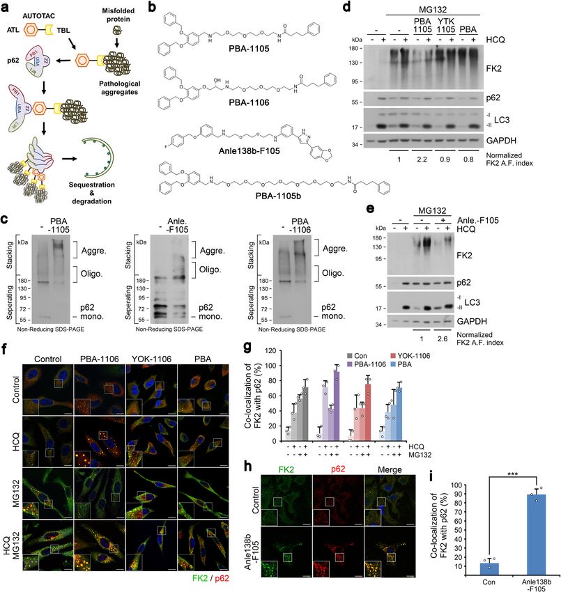
ARTICLE                                                               NATURE COMMUNICATIONS | https://doi.org/10.1038/s41467-022-28520-4

I
    n the central dogma, the genetic information in DNA is                 branch of this pathway uses Arg, Lys, His (type 1), Phe, Tyr, Trp,
    transcribed into RNA, which in turn is translated to protein.          Leu, and Ile (type 2) as N-terminal degrons, among which the Arg
    Recent advances in genetic bioengineering, exemplified by               N-degron can be generated via ATE1 R-transferase-mediated
CRISPR, TALEN, and siRNA, have enabled selective destruction               conjugation of L-Arg to Asp, Glu, or oxidized Cys. Recently, we
and functional silencing of DNA and RNA1,2. In contrast to the             discovered that the Arg/N-degron pathway mediates not only
universally applicable nature of DNA and RNA editing, there are            UBR-dependent proteasomal clearance but also macroautophagic
currently no general tools by which proteins are selectively               proteolysis, wherein the archetypal autophagic cargo adaptor p62/
recognized and targeted for destruction. Functional silencing of           SQSTM1 acts as an N-recognin that binds type-1 and type-2 N-
proteins via small molecule inhibitors, while highly penetrant,            degrons via its ZZ domain21. Binding of the Nt-Arg residue
rapid, and straightforward, is limited to approximately only a fifth        through the p62-ZZ domain conformationally activates p62 into
of the entire human proteome3. Such a technology, if available,            an autophagy-compatible form, accelerating its self-oligomeriza-
will be readily applied as research tools and agents to selectively        tion, interaction with LC3, and autophagosome biogenesis,
downregulate target proteins in cultured cells and in living               facilitating autophagic targeting of p62-cargo complexes in a
organisms such as mice and flies. Selective degradation of disease-         multi-step manner21–23.
associated proteins also provides an attractive opportunity to                Here, we developed a generally applicable degrader, AUTO-
develop degrader-type drugs.                                               TAC, by which a broad range of cellular proteins can be selec-
   Targeted protein degradation (TPD) is the latest of emerging            tively recognized and targeted to autophagic membranes for
modalities in drug discovery and development and typically                 lysosomal degradation. Central to the mode of action in
employs heterobifunctional chimeric molecules comprised of a               AUTOTAC is the ability of the p62-binding moiety to induce a
target binder linked to a degradation-inducing moiety, which offer         conformational activation of otherwise inactive p62 into an
an attractive therapeutic means to eradicate disease-associated            autophagy-compatible form. Upon binding to the p62 ligand, p62
proteins, especially those belonging to the once-considered                exposes PB1 and LIR domains, which respectively facilitates p62
undruggable proteome3–7. Current TPD technology, as exempli-               self-polymerization in complex with targets and its interaction
fied by PROteolysis-TArgeting Chimera (PROTAC), is limited to               with LC3 on autophagic membranes. Thus, AUTOTACs can
inducing the ubiquitination of target substrate for its degradation8.      induce the degradation of a broad range of cellular proteins
Despite its advantages, however, ubiquitin (Ub)-dependent                  including their misfolded aggregates.
PROTAC is largely confined to a limited set of target proteins and
of E3 ligases, due to technical difficulties in forming a substrate-
PROTAC-E3 ternary complex9. These technical challenges, which              Results
to date have ruled out the possibility of a pan-ubiquitinating and         Development of the AUTOTAC platform. In contrast to the
promiscuous E3 ligase for PROTACs, limit the meaningful clinical           UPS in which each of the 800 E3 ligases possesses a specific
targets of PROTAC to a handful of otherwise short-lived ubi-               clientele of substrates, selective autophagy employs only a handful
quitinated substrates, especially oncoproteins8,10,11. Moreover,           of receptors for intracellular cargoes, amongst which p62 plays a
Ub-dependent PROTAC may not be an ideal TPD platform to                    dominant role. If a bifunctional molecule binds both a target and
degrade misfolded and aggregation-prone pathological protein               p62 and activate the otherwise inactive p62, any given proteins
species, such as those of neurodegenerative proteinopathies, as            could be targeted for autophagic degradation in principle. We
these protein species are typically resistant to unfolding and             therefore developed autophagy-targeting ligands (ATL; p62-
subsequent degradation by the proteasome due to size                       binding moieties) that bind and activate p62 into an
limitations12,13. Recent advances in the field of autophagy-based           autophagy-compatible form (Fig. 1a and Supplementary
degraders or molecular glues, such as AUTAC, ATTEC, and                    Fig. 1a–e). 3D structure modeling, followed by SAR (structure-
LYTAC have been successful in degrading a number of targets via            activity relationship) studies, was employed on the ZZ domain of
the lysosome14–17. However, LYTAC is applicable to only extra-             p62. Three compounds, YOK-2204, YOK-1304 and YTK-105
cellular proteins, ATTEC directly targets mutant Htt or lipid              (Fig. 1b), showed high docking scores based on low/negative
droplets to the autophagosome without prior sequestration, and             energy of the stable system (−5.8, −5.5, and −4.0 kcal/mol for
AUTAC utilizes S-guanylation that is still dependent on ubiqui-            YOK-1304, YOK-2204, and YTK-105, respectively) (Supple-
tination of the target14–17. These challenges of the existing TPD          mentary Fig. 2a–f) when modeled to the p62-ZZ domain. Specific
modalities necessitate further development of a generally applic-          residues of the ZZ domain critical for N-degron recognition, such
able TPD platform independent of Ub and the proteasome.                    as Phe168, Arg139, Ile127, Asp129, Asp147, and Asp149, were
   Selective macroautophagy is a catabolic process by which                also identified (Supplementary Fig. 2a–f).
unwanted or harmful cytoplasmic constituents including pro-                   In vitro pulldown assays using biotinylated p62 ligands
teins, aggregates, and organelles are specifically sequestered by           confirmed that p62 bound YT-8-8, YOK-2204, and YTK-105,
autophagosomes for lysosomal hydrolysis18. Selective targeting of          as opposed to the negative control V-BiP-biotin peptide (Fig. 1c
autophagic cargoes calls for specific receptors such as p62 and             and Supplementary Fig. 1e). The efficacy of ATLs in inducing p62
other Sequestosome-like receptors (SLRs) that simultaneously               polymerization, a critical step in cargo collection, was demon-
recognize Ub chains on protein cargoes and the lipidated LC3 in            strated using in vitro oligomerization assays (Fig. 1d). In co-
the autophagosomal inner membrane via the UBA and the LIR                  immunostaining analyses, p62 ligands induced the formation and
domain, respectively19. While recent efforts—such as harnessing            co-localization of not only p62 and LC3 punctate structures
S-guanylation-inducing molecular tags (AUTAC) or targeting                 (Fig. 1e–g and Supplementary Fig. 2g), but also those of p62 and
extracellular and secreted/membrane proteins directly to the               the lysosomal marker LAMP1 (Supplementary Fig. 2h, i). Next,
lysosome via peptidic tags (LYTAC)—attempt to address the                  we confirmed that p62-ZZ ligands accelerated the formation of
degradation of PROTAC-resistant cargoes, no degrader technol-              p62-positive and WIPI2-positive (canonical marker of omega-
ogy yet exists for directly sequestering and targeting proteins and        somes and phagophores) punctate structures as early as 6 h
their aggregates using autophagy cargo receptors (e.g., p62).              (Supplementary Fig. 2l, m) and lasting at least 24 h (Supplemen-
   In the N-degron pathway, specific single N-terminal amino                tary Fig. 2j, k). These data suggest that ATLs not only activate and
acids of proteins, termed N-degrons, are recognized by                     target p62 to autophagic membranes but also facilitate autopha-
N-recognins to induce substrate degradation20. The arginylation            gosome biogenesis to receive incoming p62-cargo complexes for

2                                NATURE COMMUNICATIONS | (2022)13:904 | https://doi.org/10.1038/s41467-022-28520-4 | www.nature.com/naturecommunications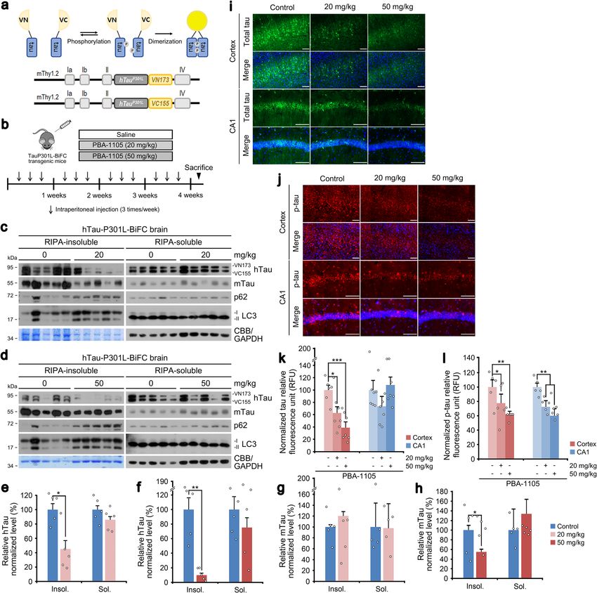
NATURE COMMUNICATIONS | https://doi.org/10.1038/s41467-022-28520-4                                                                     ARTICLE

Fig. 1 Nt-Arg-mimicking p62-ZZ ligands activate p62-dependent selective macroautophagy. a A model illustrating the mode of action of autophagy-
targeting ligands. b Chemical structures of autophagy-targeting ligands. c In vitro pulldown assay in HEK293T cells of the 12-mer V-BiP peptide or
autophagy-targeting ligands. d In vitro p62 oligomerization assay in HEK293T cells incubated with the p62-ZZ ligands in (b). e ICC of HeLa cells treated
with p62-ZZ ligands in (b) (2.5 μM, 24 h). Scale bar, 10 μm. f, g Quantification of (e) for puncta formation and co-localization, respectively (n = 3
biologically independent experiments each counting 50 cells). h A schematic/formulae for autophagy flux index. i Autophagic flux assay in HeLa cells
treated with YOK-1304 or YTK-105 (2.5 μM, 24 h) in the presence or absence of HCQ (10 µM, 24 h). Data are presented as mean values ± SD where
relevant. P-values (from a two-sided unpaired t test): ***P < 0.000956 (for p62 punctate formation), ***P < 0.000539 (for LC3 punctate formation),
***P < 0.000925 (for p62-LC3 co-localization). Source data are provided with this paper.

eventual lysosomal degradation. When autophagic flux indices                    ZZ domain that is crucial for N-degron recognition, was severely
were compared based on the ratio of substrate levels in the                    crippled (Supplementary Fig. 2q). These results validate p62-ZZ
presence or absence of late-step autophagic inhibitors, p62-ZZ                 ligands as ATLs capable of selectively interacting with and
ligands enhanced autophagic turnover of p62 and LC3, indicative                activating p62.
of increased autophagy flux (Fig. 1h, i and Supplementary Fig. 2n,
o), in a proteasome-independent manner (Supplementary Fig. 2o).
Importantly, the model p62 ligand YOK-2204 interacted with p62                 Targeted degradation of soluble proteins by AUTOTAC. We
but not NBR1, a very structurally and functionally similar                     then used these p62-ZZ ligands to synthesize AUTOTACs,
autophagic cargo receptor also containing a ZZ domain                          composed of target-binding ligands (TBLs) linked to p62-binding
(Supplementary Fig. 2p). Moreover, the interaction between                     moieties via a repeating polyethylene glycol (PEG) moiety (Fig. 2a
YOK-2204 and mutant p62D129A, carrying a point mutation in its                 and Supplementary Fig. 3a–e). To test the degradative efficacy of

NATURE COMMUNICATIONS | (2022)13:904 | https://doi.org/10.1038/s41467-022-28520-4 | www.nature.com/naturecommunications                               3

ARTICLE                                                                 NATURE COMMUNICATIONS | https://doi.org/10.1038/s41467-022-28520-4

Fig. 2 Targeted autophagic degradation of endogenous oncoproteins by AUTOTAC. a A model illustrating oncoproteins-targeting AUTOTAC. b
Chemical structures of oncoprotein-targeting AUTOTAC. c In vitro p62 oligomerization assay in HEK293T cells incubated with PHTPP-1304,
VinclozolinM2-2204, or Fumagillin-105. d Western blot (WB) in HEK293T cells treated with PHTPP-1304 at the indicated concentrations (24 h). e
Densitometry of (d) (n = 3). f, g WB in ACHN and MCF7 cells, respectively, treated with PHTPP-1304 at the indicated concentrations (24 h). h WB in
MCF7 cells treated with PHTPP-1304, PHTPP or YOK-1304 (1 µM, 24 h). i WB in LNCaP cells treated with VinclozolinM2-2204 at the indicated
concentrations. j Densitometry of (i) (n = 3). k WB in LNCaP cells treated with VinclozolinM2-2204, Vinclozolin or YOK-2204 (1 µM, 24 h). l WB in
HEK293T cells treated with Fumagillin-105, Fumagillin or YTK-105 at the indicated concentrations (24 h). m Densitometry of (l) (n = 3). n WB in U87-MG
cells treated with Fumagillin-105 at the indicated concentrations (24 h). o WB in U87-MG cells treated with Fumagilin-105, Fumagilin or YTK-105 (1 µM,
24 h). When indicated, n = 3 biologically independent experiments. Data are presented as mean values ± SD where relevant. Source data are provided with
this paper.

AUTOTACs, we sought out to degrade estrogen receptor beta                    vinclozolinM2, a metabolite of vinclozolin, and fumagillol, a
(ERβ) using its nonsteroidal and synthetic ligand, PHTPP                     hydrolyzed product of fumagillin (Fig. 2b). Fumagillin covalently
(Fig. 2a, b). PHTPP-based AUTOTAC (PHTPP-1304) induced                       binds MetAP2 via a spiro-epoxide moiety24, and vinclozolin
self-oligomerization of p62 (Fig. 2c) and degradation of ERβ at              derivatives and metabolites inhibit androgen binding to AR25,26.
half-degrading concentration values (DC50) of ~2 nM in                       As expected, AR and MetAP2 AUTOTACs efficiently induced
HEK293T cells (Fig. 2d, e) and

NATURE COMMUNICATIONS | https://doi.org/10.1038/s41467-022-28520-4                                                                ARTICLE

o). These results validate AUTOTAC as robust degraders for                     degraders was assessed using wound healing assays, PHTPP-1304
targeted proteolysis of intracellular oncoproteins.                            more efficiently inhibited cell migration compared with p62-
   Co-localization analysis in LNCaP cells showed that                         binding moiety or TBL (Supplementary Fig. 4i, j). Analogous
vinclozolinM2-2204 induced the formation of AR+LC3+ autop-                     assays targeting MetAP2 revealed that fumagillin-105 more effi-
hagic membranes (Fig. 3a, b). PHTPP-1304 in ACHN cells                         ciently inhibited the migration of U87-MG glioblastoma cells as
facilitated dosage-dependent formation of p62+ERβ+ puncta                      early as 4 h and up to 24 h post-scratch (Fig. 3k, l). Finally, flow
subject to autophagic flux when treated with the late-step                      cytometry showed that prolonged exposure to fumagillin-105
autophagic flux inhibitor hydroxychloroquine (Fig. 3c, d).                      induced programmed cell death as marked by the sub-G1 sub-
Additionally, PHTPP-1304 treatment induced the punctate                        population (Supplementary Fig. 4k). These data demonstrate the
formation and co-localization of ERβ and the omegasome marker                  therapeutic advantage of AUTOTACs against cancer cell growth
WIPI2-GFP (Supplementary Fig. 4a). Consistently, fumagillin-                   and progression.
105 treatment drastically up-regulated the autophagic flux of
MetAP2, leading to lysosomal degradation via autophagosomes
(Fig. 3e). Crucially, the degradation of ERβ by PHTPP-1304 was                 AUTOTAC enables the targeting of Ub-conjugated misfolded
completely abolished by RNA interference of either p62 or ATG5                 protein aggregates to the lysosome. Most proteins are misfolded
(Fig. 3f), which were corroborated by immunostaining analyses of               or damaged at least once during their limited lifespans and thus
ERβ or MetAP2 puncta formation present only in wild-type but                   necessitate their turnover via the UPS or autophagy. Soluble
not p62−/− or ATG5−/− mouse embryonic fibroblasts (Supple-                      misfolded proteins are primarily degraded through the UPS,
mentary Fig. 4b–d). These results demonstrate that AUTOTAC                     which involves unfolding into nascent polypeptides and cleavage
drives lysosomal degradation of target proteins via p62-                       by the proteasome12,29,30. However, the proteasome has an inner
dependent macroautophagy.                                                      diameter as narrow as 13 Å whose pore is inaccessible to oligo-
   As an autophagic cargo adaptor, p62 contains a LIR domain for               mers and aggregates and clogged by partially misfolded sub-
LC3 interaction on autophagic membranes and a UBA domain                       strates, leaving autophagy as possibly the last line of defense
that binds poly-Ub chains (Supplementary Fig. 4e). Thus, we                    against pathogenic aggregates31,32. We therefore applied
determined whether AUTOTAC-based proteolysis depends on                        AUTOTAC for UPS-resistant misfolded proteins and their oli-
ubiquitination of target substrate and recognition of Ub chains                gomeric/aggregated species (Fig. 4a).
via the p62-UBA domain. Importantly, AUTOTAC-driven                               We first searched for a chemical chaperone that selectively
degradation of MetAP2, ERβ, and AR remained not only intact                    recognizes the exposed hydrophobic regions as a universal
but was even enhanced under Ubb interference (Fig. 3g, h and                   signature of misfolded proteins. Screening of various compounds
Supplementary Fig. 4f), possibly as a consequence of compensa-                 identified 4-phenylbutyric acid (PBA), an FDA-approved drug
tory crosstalk between the UPS and autophagy. Following this                   and chemical chaperone that improves proteostasis and amelio-
vein of reasoning, we speculated that AUTOTAC-mediated                         rates misfolding-induced ER stress33. PBA-1105 and PBA-1106
degradation would be even more apparent in conditions of                       AUTOTACs (Fig. 4b) efficiently activated p62 and triggered its
proteasome impairment. Indeed, degradation of ERβ upon                         self-oligomerization (Fig. 4c). Immunoblotting analyses showed
PHTPP-1304 treatment with proteasomal inhibition in MCF7                       that PBA-1105 increased the autophagic flux of Ub-conjugated
cells exhibited subnanomolar DC50 and Dmax values (Supple-                     aggregates under prolonged proteasomal inhibition (Fig. 4d, flux
mentary Fig. 4g). Moreover, this robust degradation persisted up               indices 1 vs. 2.2), which was increasingly sustained for at least
to at least 8 h post-washout of PHTPP-1304 (Supplementary                      48 h (Supplementary Fig. 5a). When their intracellular distribu-
Fig. 4h), suggesting that AUTOTACs may display sustained                       tion was visualized using immunofluorescence analyses, PBA-
degradative efficacy and be recycled from the lysosome. Taken                   1106 facilitated the formation of Ub+ cytosolic puncta, the vast
together, these results suggest that AUTOTAC does not require                  majority of which colocalized with p62+ puncta (Fig. 4f, g) as well
ubiquitin-dependent and PPI-driven cooperativity for its sus-                  as p62+LC3+ autophagic membranes (Supplementary Fig. 5b).
tained autophagic proteolysis.                                                 Specifically, while PBA-1106 treatment alone compared to
                                                                               control seemingly did not affect the number of p62 punctate
                                                                               structures, the increase in p62 punctate structures was drastically
Therapeutic efficacy of AUTOTAC in cancer signaling. To                         apparent in conditions of autophagy inhibition (14 ± 4.2 vs.
assess the therapeutic efficacy of autophagy-targeting degraders,               6 ± 2.4 p62 puncta structures). Strikingly, no such efficacy was
we compared the downstream signaling of AR and ERβ in cells                    observed with PBA or p62-binding ligand alone (Fig. 4d, f, g and
treated with either AUTOTACs or their cognate TBLs. Dihy-                      Supplementary Fig. 5b). Moreover, siRNA-mediated knockdown
drotestosterone (DHT) and estradiol (E2) are natural agonists                  of either p62 or ATG7 abolished PBA AUTOTAC-dependent
that respectively bind AR and ERβ and induce their dimerization                degradation of Ub-conjugated protein aggregates (Supplementary
and nuclear translocation, resulting in transcriptional activation             Fig. 5c).
of downstream proteins27,28. When the levels of EGFR and p-                       In neurodegeneration and other proteinopathies, aggregation-
Akt/Akt were measured in DHT-activated cells, vinclozolinM2-                   prone misfolded proteins inherently form oligomers that
2204 inhibited AR pathways approximately 4-folds more effi-                     aggregate into fibrillary species. The screening of TBLs that
ciently than its TBL (Fig. 3i). Similarly, PHTPP-1304 inhibited                selectively recognize the oligomeric signature of proteins and
ERβ signaling 10-folds more efficiently than its TBL as deter-                  their aggregates yielded Anle138b, a phase 1 clinical trial
mined by the levels of EGFR, p-ERK/ERK and p-Akt/Akt in E2-                    compound that binds oligomers and aggregates of neurodegen-
stimulated LNCaP cells (Fig. 3j). Next, we examined the potency                erative proteinopathies34,35. Anle138b-F105 facilitated p62 self-
of AUTOTACs in cancer cell growth and progression. WST-                        polymerization (Fig. 4c) and autophagic flux of Ub-conjugated
based viability assays in ACHN cells showed that PHTPP-1304                    oligomeric/aggregated proteins (Fig. 4e) via p62-associated
exerted a ~5-fold higher cytotoxicity (IC50, 3.3 μΜ) than those of             macroautophagy (Fig. 4h, i). In the same vein, Anle138b-F105
its p62-binding ligand (>20 μΜ for YOK-1304) or TBL (18 μΜ                     treatment induced the punctate formation and co-localization of
for PHTPP) (Fig. 3m). VinclozolinM2-2204 also exhibited higher                 Ub-conjugated protein aggregates with the omegasome marker
cytotoxicity (IC50, 4.7 μΜ) as compared with its p62 ligand (>100              WIPI2-GFP (Supplementary Fig. 5d) and the lysosome marker
μΜ) and TBL (>100 μΜ) (Fig. 3n). When the efficacy of                           LAMP1 (Supplementary Fig. 5e). Similar to PBA-based

NATURE COMMUNICATIONS | (2022)13:904 | https://doi.org/10.1038/s41467-022-28520-4 | www.nature.com/naturecommunications                         5
ARTICLE                                                                  NATURE COMMUNICATIONS | https://doi.org/10.1038/s41467-022-28520-4

Fig. 3 Targeted degradation using AUTOTAC inactivates oncogenic signaling. a ICC of LNCaP cells treated with Vinclo.-2204 (2.5 µM, 24 h). Scale bar,
10 μm. b Quantification of (a) (n = 3 biologically independent experiments each counting 50 cells). c ICC of ACHN cells treated with PHTPP-1304 at the
indicated concentrations and HCQ (10 μM) (24 h). Scale bar, 10 μm. d Quantification of (c) (n = 3 biologically independent experiments each counting 50
cells). e WB in U-87 MG cells treated with fumagillin-105 (1 μM, 24 h) with or without bafilomycin A1 (200 nM, 6 h). f WB in HEK293T cells treated with
PHTPP-1304 (0.1 μM, 24 h) under siRNA-mediated knockdown of p62 and ATG5 (40 nM, 48 h). g WB in HeLa cells treated with fumagillin-105 (0.1 μM,
24 h) following RNA interference of Ubb (40 nM, 48 h). h Identical to (g), but with PHTPP-1304 for ERβ. i WB in LNCaP cells treated with vinclozolinM2-
2204 (2.5 μM) or inclozolin (10 μM) with or without DHT (15 nM) (24 h). j Identical to (i) but with PHTPP-1304 (0.5 μM) or PHTPP (5 μM) with or
without E2 (10 nM) (24 h). k Wound healing assay in ACHN cells treated with PHTPP-1304, PHTPP, or YOK-1304 (5 μM) at the indicated time points. l
Quantification of (k) (n = 2 biologically independent experiments). Scale bar, 100 μm. m, n Cell viability assay of ACHN and LNCaP cells treated with the
indicated compounds and concentrations (n = 2 biologically independent experiments). Data are presented as mean values ± SD where relevant. P-values
(from a two-sided unpaired t test): ***P < 0.000276, **P < 0.00161. Source data are provided with this paper.

6                                   NATURE COMMUNICATIONS | (2022)13:904 | https://doi.org/10.1038/s41467-022-28520-4 | www.nature.com/naturecommunications

NATURE COMMUNICATIONS | https://doi.org/10.1038/s41467-022-28520-4                                                                      ARTICLE

Fig. 4 Targeted autophagic delivery and degradation of misfolded protein cargoes using AUTOTACs with aggregate-binding warhead. a A model
illustrating the mode of action of aggregate-targeting AUTOTAC. b Chemical structures of PBA-1105, PBA-1106, Anle138b-F105 and PBA-1105b. c In vitro
p62 oligomerization assay in HEK293T cells incubated with the indicated compounds in (b). d WB in HEK293T cells treated with MG132 (1 μM, 24 h),
HCQ (10 μM, 24 h), PBA-1105, YTK-1105, and PBA (1 μM, 24 h). e Identical to (d), but with Anle-F105. f ICC of HeLa cells treated with HCQ (10 μM, 24 h),
MG132 (2 μM, 18 h) or both in the presence of PBA-1106, YOK-1106 or PBA (1 μM, 24 h). Scale bar, 10 μm. g Quantification of (f) (n = 3, 50 cells). h ICC of
HeLa cells treated with Anle138b-F105 (1 μM, 24 h). Scale bar, 10 μm. i Quantification of (h) (n = 3, 50 cells). When indicated, n = 3 biologically
independent experiments each counting 50 cells. Data are presented as mean values ± SD where relevant. P-values (from a two-sided unpaired t test):
***P < 7.66E−05. Source data are provided with this paper.

AUTOTAC, autophagic proteolysis of Ub-conjugated protein                       and respiratory failure36. The many mutant and pathological
aggregates by Anle138b-F105 treatment was nullified by p62 or                   protein species attributed to this disorder share a common trait in
ATG7 interference (Supplementary Fig. 5f).                                     that they misfold, aggregate, and accumulate along with ubiquitin
   Desminopathies are systemic disorders caused by dysfunctional               and other amyloidogenic proteins into insoluble granulo-
mutations in desmin or alphaB-crystallin, which cripple the                    filamentous material36. While wild-type desmin is not known
intracellular filamentous network in cardiac and skeletal muscle                to aggregate whatsoever, aggregation and accumulation of mutant
cells and eventually induce muscle weakness, including cardiac                 desmin (which prevents its normal turnover via the UPS) is

NATURE COMMUNICATIONS | (2022)13:904 | https://doi.org/10.1038/s41467-022-28520-4 | www.nature.com/naturecommunications                                 7
ARTICLE                                                              NATURE COMMUNICATIONS | https://doi.org/10.1038/s41467-022-28520-4

known to disrupt protein homeostasis, including but not limited           tau species, which act as a seedbed for tau aggregation. While
to chaperone deficiency, proteasome impairment, and mitochon-              okadaic acid treatment impaired the autophagic flux of mutant
drial dysfunction36. Thus, to generalize the efficacy of the               tauP301L (Supplementary Fig. 6g, lanes 1 and 2 vs. 5 and 6),
AUTOTAC platform to misfolded protein aggregates, we tested               presumably due to hyper-sequestration of tau, PBA-1106
the degradative efficacy of PBA-1105 and Anle138b-F105 against             significantly rescued autophagic flux of these otherwise non-
wild-type desmin and mutant, aggregation-prone desminL385P.               digestible species (Supplementary Fig. 6g, lanes 3 and 4 vs. 6 and
Notably, all the tested AUTOTACs (i.e., PBA-1105, PBA-1105b,              8). These results were consistent with our observation that PBA-
and Anle138b-F105) induced the degradation of exogenously                 1105 treatment led to an increase in the number of GFP-
expressed mutant desminL385P but not wild-type desmin in a                quenched RFP-GFP-hTauP301L pre-formed inclusion bodies
concentration- (Supplementary Fig. 5g, h) and macroautophagy-             following their hyperphosphorylation and aggregation via prior
dependent (Supplementary Fig. 5i) manner. Collectively, our data          treatment with okadaic acid, signaling their lysosomal digestion
validate AUTOTAC-facilitated autophagic proteolysis of other-             (Supplementary Fig. 6h, i). Detergent-based insoluble/soluble
wise non-degradable, Ub-conjugated and UPS-resistant oligo-               fractionation of okadaic acid-induced hyperphosphorylated tau
meric or aggregated proteins by recognizing their exposed                 further confirmed that PBA-1105 not only rescued but drastically
hydrophobic motifs or oligomeric signature.                               accelerated the autophagic flux of both phosphorylated and total
                                                                          tau species (Fig. 5m, autophagy flux indices). Importantly, co-
                                                                          immunoprecipitation analyses revealed that Anle138b-F105
Degradation of pathological aggregates of neurodegenerative               treatment induced not only the degradation of tauP301L but
diseases using AUTOTAC. Neurodegenerative diseases such as                also its interaction with mutant p62 lacking the UBA domain,
Alzheimer’s disease are associated with an ever-increasing accu-          which is normally required for the interaction (Supplementary
mulation of degradation-resistant misfolded hallmark protein              Fig. 6j). These data suggest that AUTOTAC provides a platform
aggregates. Traditional approaches for developing ligands that            to target aggregation-prone misfolded proteins in neurodegen-
alter the activity of neurodegeneration-associated targets are not        erative proteinopathies for lysosomal degradation in an ubiquitin-
applicable for pathological aggregates, leaving degraders as pos-         independent manner.
sibly the only therapeutic means. Since PROTAC-based approa-                 Next, we tested whether AUTOTAC is applicable for other
ches are inherently incapable of degrading large oligomers and            neurodegeneration-associated proteins such as mutant hunting-
aggregates, we tested whether AUTOTACs can target                         tin. HeLa cells were engineered to stably express wild-type (Q25)
aggregation-prone P301L tau mutant that forms neurofibrillary              or mutant (Q97) huntingtin based on their nuclear localization/
tangles because of its prion-like seeding behavior37.                     export signals (Htt-NLS-GFP or Htt-NES-GFP). Autophagy flux
   In vitro pulldown assays confirmed that the chemical                    assays showed that PBA AUTOTACs induced lysosomal
chaperone 4-PBA bound tauP301L stably expressed in SH-                    degradation of both nucleus- and cytosol-resident mutant
SY5Y cells (Supplementary Fig. 6a). PBA-1105 and PBA-1106                 huntingtin (Htt-NES-Q97 and Htt-NLS-Q97) at DC50 of 0.1–1
AUTOTACs induced autophagic degradation of stably expressed               μM (Supplementary Fig. 7a, b). Similar degradation efficacy was
mutant tau at DC50 of ~1–10 nM and Dmax,24 hr of 100 nM,                  observed with transiently expressed mutant huntingtin, HDQ103
followed by a hook effect at higher concentrations (Fig. 5a–d and         (Supplementary Fig. 7c). PBA AUTOTACs exhibited no
Supplementary Fig. 6b). In contrast to PBA AUTOTACs,                      significant degradative efficacy against wild-type huntingtin,
virtually no degradation was observed with their TBL or p62-              HttQ25 (Supplementary Fig. 7d, e), further supporting their
ZZ ligands (Fig. 5a–d and Supplementary Fig. 6b). We next                 specificity to mutant Htt. As expected, their p62-binding ligands
confirmed that AUTOTAC-mediated degradation may not be                     exhibited little efficacy against neither wild-type nor mutant Htt
critically dependent on linker length by synthesizing and                 proteins (Supplementary Fig. 6b, e). Similar to PBA-based
confirming the anti-tauP301L degradative efficacy of PBA-                   degraders, Anle138b-F105 also showed DC50 of ~3 nM against
1105b, which carries a drastically longer PEG-based linker than           the nuclear subpopulation of Htt (HttQ97-NLS-GFP) 24 h post-
PBA-1105 (Fig. 4b and Supplementary Fig. 6c). Similar to PBA-             treatment (Supplementary Fig. 7f–i). When visualized using
based degraders, Anle138b-F105 targeted tauP301L for lysosomal            immunostaining analyses, Anle138b-F105 selectively promoted
degradation at DC50 of ~3 nM, as opposed to its TBL or p62                autophagic targeting of mutant HttQ103 as determined by co-
ligand (Fig. 5e–h). Autophagy-based targeted degradation was              localization of HttQ103 and LC3 in cells treated with hydroxy-
obvious as early as 30 min and reached a sustained maximal effect         chloroquine (Supplementary Fig. 7j, k). Given that nuclear LC3
from 3 hrs onwards (Fig. 5i). Anle138b-F105-mediated degrada-             does not localize to autophagic membranes unless it is retro-
tion of mutant tau species persisted up to at least 8 h post-             translocated and post-translationally modified in the cytosol,
washout (Supplementary Fig. 6d). These data raise the likelihood          AUTOTACs likely bind and activate nuclear p62 (or cytosolic
that AUTOTACs are linker length-insensitive and exhibit                   p62 that translocates to the nucleus) and deliver TBL-bound
sustained efficacy against tau oligomers and aggregates.                   mutant HttQ97 to the cytosol for proteolysis. These data
   We next confirmed whether AUTOTACs exert their efficacy by               demonstrate that AUTOTAC is generally applicable for a broad
directly targeting oligomeric and aggregated species. Co-                 range of pathogenic aggregates in neurodegeneration.
localization analysis showed that PBA-1105 AUTOTAC selec-
tively induced the sequestration and autophagic targeting of
tauP301L inclusion bodies in contrast to YTK-1105 or PBA                  AUTOTAC mediates the eradication of tau aggregates from
(Fig. 5j, k). Consistently, in vivo aggregation assays revealed that      mouse brains. Increasing evidence points to soluble tau oligomers
PBA-1105 effectively eliminated high-molecular weight tau                 as causative agents in tau pathology due to their neurotoxic effect
aggregates (Fig. 5l). Similarly, detergent-based fractionation of         and a prion seed-like behavior for self-propagation38. To date, there
tauP301L into insoluble or soluble species revealed that only             are no general methods for targeted degradation of pathological
PBA-1105, in contrast to its TBL or p62-binding moiety,                   protein oligomers and aggregates in neurodegeneration. We have
promoted autophagic degradation of not only detergent-soluble             previously developed hTauP301L-BiFC transgenic mice that express
but critically also -insoluble species (Supplementary Fig. 6e, f).        human tauP301L in the brain by employing bimolecular fluores-
Next, we used the phosphatase inhibitor okadaic acid to examine           cence complementation to visualize soluble tau oligomers39 (Fig. 6a).
the efficacy of PBA-based degraders against hyperphosphorylated            We assessed the efficacy of PBA-1105 AUTOTAC, which showed

8                               NATURE COMMUNICATIONS | (2022)13:904 | https://doi.org/10.1038/s41467-022-28520-4 | www.nature.com/naturecommunications
NATURE COMMUNICATIONS | https://doi.org/10.1038/s41467-022-28520-4 ARTICLE Fig. 5 Selective degradation of pathological aggregation-prone tau species by aggregate-binding AUTOTAC. a–c WB in SH-SY5Y-tauP301L cells treated with PBA-1105, PBA, or YTK-1105 at the indicated concentrations. d Densitometry of (a, b, and c) (n = 3 biologically independent experiments). e–g Same as (a–c) but with Anle138b-F105, Anle138b, or YTK-105. (h) Densitometry of (e, f, and g) (n = 3 biologically independent experiments). i WB in SH- SY5Y-tauP301L cells treated with PBA-1105 (0.1 μM) at the indicated time points. j ICC of HeLa cells expressing recombinant TauP301L-GFP and treated with the indicated compounds (1 μM, 24 h) and HCQ (10 μM, 24 h). Scale bar, 10 μm. k Quantification of (j) (n = 3 biologically independent experiments each counting 50 cells). l In vivo oligomerization assay in SH-SY5Y-tauP301L cells treated with okadaic acid (15 nM, 24 h) and the indicated compounds (0.1 μM, 24 h). m Triton X-100-fractionation assay in SH-SY5Y-tauP301L cells treated with a combination of HCQ (10 μM, 24 h), okadaic acid (15 nM, 24 h) or PBA-1105 (0.1 μM, 24 h). Data are presented as mean values ± SD where relevant. P-values (from a two-sided unpaired t test): **P < 0.00821. Source data are provided with this paper. good albeit rapidly metabolized exposure (Supplementary Table 1), oligomeric htauP301L-BiFC bodies using Sudan Black B staining on to eradicate misfolded tau aggregates from their brains. The mice the cross sections of murine brains. Notably, PBA-1105 AUTOTAC were intraperitoneally injected (saline, 20, or 50 mg/kg) three times induced a drastic reduction of total tau oligomers on both the cortex per week for one month (Fig. 6b). Brain hemispheres were subjected and the CA1 region of the hippocampus sections in a dosage- to RIPA-based insoluble/soluble fractionation, followed by immu- dependent manner as determined by both the number of tau bodies noblotting analyses of both endogenous wild-type murine tau and and their fluorescence signals (Fig. 6i, k). Moreover, AT8 staining human tauP301L. We first confirmed that PBA-1105 AUTOTAC revealed a marked eradication of soluble bodies of phosphorylated did not degrade endogenous wild-type murine tau in either tau-BiFC in the cortex as well as the CA1 region (Fig. 6j, l). These detergent-soluble or -insoluble fractions (Fig. 6c, d, g, h). In sharp results indicate that AUTOTAC provides a platform to eradicate contrast, PBA-1105 induced marked clearance of detergent-insoluble pathological aggregates from the brain. tauP301L in a dosage-dependent manner (Fig. 6c–f). This reduction in insoluble tau aggregates correlated to an increase in RIPA- insoluble LC3 levels (Fig. 6c, d). In contrast to insoluble species, Discussion levels of soluble tau species showed little if any reduction (Fig. 6c–f), In this study, we developed AUTOTAC as a generally applicable demonstrating substrate specificity of AUTOTAC towards chemical platform that enables targeted degradation of a variety aggregation-prone tauP301L mutant. Next, we visualized soluble of cellular proteins. Our previous work has established the ability NATURE COMMUNICATIONS | (2022)13:904 | https://doi.org/10.1038/s41467-022-28520-4 | www.nature.com/naturecommunications 9
ARTICLE                                                                   NATURE COMMUNICATIONS | https://doi.org/10.1038/s41467-022-28520-4

Fig. 6 Chaperone-based AUTOTAC ameliorates mutant tau pathology in brain-specific murine model. a Schematic of hTauP301L-BiFC murine model
construction. b Injection timeline and details of PBA-1105 in hTauP301L-BiFC murine model. c, d RIPA-insoluble fractionation assay in brain tissues of
hTauP301L-BiFC mice intraperitoneally injected with PBA-1105 (20 or 50 mg/kg). e, f Normalized densitometry of (c) and (d) for hTau levels, respectively
(n = 5 mice). g, h Same as (e) and (f) but for mTau levels, respectively (n = 5 mice). i, j Immunohistochemistry of BiFC fluorescence for total hTau levels or
AT8 fluorescence for total phosphorylated levels in hTauP301L-BiFC mice injected with PBA-1105 as outlined in (b). Scale bar, 100 μm. k, l Quantification of
BiFC or AT8 punctate fluorescence signals in i and j, respectively (n = 7 mice). Data are presented as mean values ± SEM where relevant. P-values (from a
two-sided unpaired t test): *P < 0.0111 (for insoluble hTau, 20 mg/kg), **P < 0.00105 (for insoluble hTau, 50 mg/kg), *P < 0.0442 (for insoluble mTau,
50 mg/kg). Source data are provided with this paper.

of a p62-binding ATL to activate an otherwise inactive p62 to an                also oligomeric species of aggregation-prone proteins, including
autophagy-compatible form via a conformational change. Upon                     hallmark substrates of neurodegenerative proteinopathies. Ther-
binding to ATL, p62 exposes its PB1 domain for self-                            apeutic efficacy of a misfolded protein-targeting AUTOTAC was
polymerization in complex with TBL-bound cargoes (effectively                   further confirmed in a brain-specific murine model expressing
sequestering target cargoes) and its LIR domain for interaction                 transgenic human mutant pathological tau. Additionally,
with LC3 on autophagic membranes. Here, we report the proof-                    AUTOTACs required neither the ubiquitination of the target
of-concept development of AUTOTACs built upon ATL-based                         protein nor the p62-mediated recognition of said ubiquitin chains
p62 binding and activation, through which AUTOTACs can                          on the target protein for its sustained degradation. These results
mediate targeted degradation of not only monomeric oncopro-                     substantiate AUTOTACs as generally applicable heterobifunc-
teins (whose oncogenic signaling was functionally silenced) but                 tional chimeric degraders for Ub-independent and p62-mediated

10                                   NATURE COMMUNICATIONS | (2022)13:904 | https://doi.org/10.1038/s41467-022-28520-4 | www.nature.com/naturecommunications
NATURE COMMUNICATIONS | https://doi.org/10.1038/s41467-022-28520-4                                                                        ARTICLE

Fig. 7 Speculative model of AUTOTAC and its mechanism-of-action. Connecting a protein-of-interest target-binding ligand (TBL) to a p62-binding
autophagy-targeting ligand (ATL) using an intermediate linker generates the chimeric AUTOTAC degrader. Recognition of the target protein occurs in
tandem with the binding and activation of p62 via its ZZ domain, which is self-polymerized in complex with cargoes. Additionally, such interaction initiates
a macroautophagy induction cascade in a p62-dependent manner. AUTOTACs show sustained degradative efficacy post-lysosomal degradation of the
target protein, suggesting that it is recycled.

autophagic clearance of a broad range of intracellular target                  necessitates extremely specific linker lengths and types for ternary
proteins.                                                                      complex formation, subject to change for each of the numerous
   Thus, we speculate that AUTOTACs would mediate targeted                     E3 ligase-substrate combinations3,7,8. Moreover, despite the initial
degradation through the following multi-step mechanisms                        promising outlook on hijacking E3 ligases to ubiquitinate non-
(Fig. 7). First, AUTOTAC brings a target to p62 via its TBL and                native substrates, it is becoming increasingly clear that the current
ATL, forming a ternary complex. Second, normally inactive p62 is               spectrum of E3 ligases used in PROTAC technology exhibits
structurally activated for self-oligomerization, forming target-p62            restricted substrate specificities, and that a pan-ubiquitinating E3
oligomeric complexes. Third, AUTOTAC facilitates Ub-and                        ligase is yet to be found16,40. Our data imply that AUTOTACs
proteasome-independent degradation of the target-p62 com-                      may not be critically reliant upon a specific linker length and do
plexes via macroautophagy. Fourth, AUTOTACs are recycled                       not require ubiquitination of the target substrate for its degra-
from the lysosome towards other targets in the cytosol, providing              dation. These lines of evidence suggest that AUTOTACs may not
a sustained nature of degradation.                                             require protein-protein interaction-mediated positive coopera-
   Among proteinopathies with gain-of-function toxicity, a large               tivity for ternary complex formation, at least for TBLs with high
subset is defined as protein misfolding/aggregation disorders                   affinity towards their respective substrates. That said, however,
wherein misfolded proteins, from their monomeric to oligomeric                 some low-affinity TBL-target combinations might benefit from
and aggregated species, are pathological29,30. Due to the inherent             p62-target interaction, including recognition of ubiquitin chains
size and substrate-conformation limitations of a proteasome, any               on a substrate. Additionally, the variety of structurally distinct
conformationally stable subspecies of a protein beyond its                     p62-ZZ ligands used in AUTOTACs to successfully eradicate
monomeric form is not only non-degradable by the UPS but can                   equally numerous and diverse target proteins highlight p62 as a
even clog the pores of proteasomal subunits12,31. While several                pan-autophagy receptor, capable of targeting non-native or
recent efforts using PROTACs have succeeded in degrading                       autophagy-resistant substrates such as MetAP2 or hyperpho-
pathological and aggregation-prone tau, it is most likely that the             sphorylated mutant tau. Thus, our work opens a line of clinical
targeted species were monomeric and that the anti-aggregate                    research exploring alternative avenues of targeting the human
efficacy, if any, is more of a prevention than a treatment5. In this            proteome for degradation.
sense, AUTOTACs provide a direct means to target not only the                     Critical remaining questions from our study involve the
monomeric but also the oligomeric and aggregated species of                    pharmacological and mechanistic properties of AUTOTACs. For
these pathological hallmark proteins.                                          example, it remains to be seen whether and how AUTOTACs can
   Another advantage that AUTOTAC offers over current TPD                      be recycled for multiple rounds of degradation. While
modalities is in its promiscuity. Notably, the requirement of                  AUTOTAC-mediated silencing of a target oncoprotein and its
ligand-induced proximity to achieve spatial and temporal co-                   downstream signaling is several folds more effective than that of
localization between E3 and target substrate using PROTACs                     the target-binding inhibitor alone, and AUTOTACs do indeed

NATURE COMMUNICATIONS | (2022)13:904 | https://doi.org/10.1038/s41467-022-28520-4 | www.nature.com/naturecommunications                                  11
ARTICLE                                                                           NATURE COMMUNICATIONS | https://doi.org/10.1038/s41467-022-28520-4

exhibit sustained efficacy, it remains to be seen whether                                Modified Eagle’s Medium, MEM medium or RPMI-1640 medium with 10% Fetal
AUTOTACs act catalytically and/or escape the lysosome, not                              Bovine Serum and antibiotics (100 units/mL penicillin and 100 μg/mL strepto-
                                                                                        mycin) in a 5% CO2 incubator. For stable cell lines, the expression of the intended
unlike the cytotoxic drug moieties of antibody-drug conjugates41.                       tagged target protein(s) was confirmed by immunoblotting and/or
Additionally, the off-target and selectivity issues of the AUTO-                        immunocytochemistry.
TAC platform have yet to be fully investigated and should be
addressed in follow-up studies. While our data show selective                           Western blotting. Adherent cells were washed with phosphate-buffered saline
interaction with p62 over NBR1, which carries a similar ZZ                              (PBS) and cell pellets were lysed in SDS-based sample buffer (277.8 mM Tris-HCI,
domain, the lipophilic nature of the current generation of ATLs                         pH 6.8, 4.4% LDS, 44.4% (v/v) glycerol) containing beta-mercaptoethanol. Alter-
may require further optimization to minimize off-target binding.                        natively, cell pellets or protein supernatants were lysed in 5X Laemmli sample
                                                                                        buffer. Whole-cell lysates were separated using SDS-PAGE, and transferred onto
Another question concerns the autophagic sequestration mode of                          polyvinyllidene difluoride membranes at 100 V for 2 h at 4 °C. The membrane was
action possibly unique to AUTOTACs. Given the propensity of                             incubated with a blocking solution consisting 4% skim milk in PBS solution for
p62 for self-oligomerization, it will be interesting to determine                       30 min at room temperature and incubated with primary antibodies overnight,
whether and how much sequestration of a target protein con-                             followed by incubation with host-specific HRP-conjugated secondary antibodies
                                                                                        (1:10000 dilution). For signal detection, the membrane was developed with a
tributes to the overall efficacy of AUTOTACs in biologically                             mixture of ECL solution (Thermo Fisher Scientific) using X-ray films. Densito-
inactivating said target. Although the general efficacy of                               metry of developed bands was measured and analyzed with ImageJ (NIH,
AUTOTACs has yet to be fully evaluated, our results suggest that                        Bethesda).
activating the p62-ZZ domain in an Arg/N-degron-dependent
manner for targeted proteolysis can provide an avenue of research                       Immunocytochemistry. Cells were cultured on coverslips coated with poly-L-
and therapeutic investigation into a myriad of diseases.                                lysine (Sigma) to observe cellular localization of proteins. Using 4% paraf-
                                                                                        ormaldehyde in PBS (pH 7.4), the cells were fixed for 15 min at room temperature
                                                                                        and washed three times for 5 min with PBS. After fixing, the cells were permea-
Methods                                                                                 bilized with 0.5% Triton X-100 in PBS solution for 15 min and washed three times
Compounds, plasmids, and other reagents. The chemical synthesis and analy-              with PBS for 5 min. The cells were blocked containing 2% BSA in PBS solution for
tical data of Nt-Arg-mimicking compounds are described in the Supplementary             1 h at room temperature. Subsequently, the cells were incubated with primary
Methods.                                                                                antibody diluted in 2% BSA/PBS solution overnight at 4 °C, followed by washing
    The recombinant neurodegenerative hallmark protein plasmid was constructed          the cells three times for 10 min with PBS and incubated with Alexa Fluor-
as follows. The GFP tagged Htt-103 plasmid was constructed into pcDNA3.1/myc-           conjugated secondary antibody diluted in 2% BSA/PBS for 30 min at room tem-
His plasmid (Thermo Fisher Scientific) at EcoRI/XhoI sites using PCR                     perature. Using a DAPI-containing mounting medium (Vector Laboratories), the
amplification. The TauP301L plasmid was a gift from Min Jae Lee (Seoul National          coverslips were mounted on glass slides. Confocal images were taken by laser
University, Korea). These plasmids were transiently transfected using                   scanning confocal microscope 510 Meta (Zeiss) and analyzed by Zeiss LSM Image
Lipofectamine 2000 or Lipofectamine 3000 (Thermo Fisher Scientific). Other               Browser (ver. 4.2.0.121). Subsequently, cells were deemed to exhibit significant co-
reagents used in this study were bafilomycin A1, hydroxychloroquine, (Sigma);            localization if more than ten clear puncta structures of the respective proteins
MG132 (Enzo).                                                                           showed association or full co-localization. Quantification results are shown as
                                                                                        mean ± S.D. or S.E.M. of three independent experiments
Transfection. Plasmids were transfected into HeLa and HEK293T cells using the
Lipofectamine 2000 Transfection Reagent according to the manufacturer’s                 In vitro p62 oligomerization. HEK293T cells were transiently transfected with a
instructions (Invitrogen).                                                              plasmid encoding p62-myc/his fusion proteins. Cells were resuspended in lysis
                                                                                        buffer [50 mM HEPES (pH 7.4), 0.15 M KCl, 0.1% Nonidet P-40), 10% glycerol,
                                                                                        containing a mixture of protease inhibitors and phosphatase inhibitor (Abcam)].
RNA interference analysis. Cells in a 12-well plate (0.5 × 10⁶ per well) were           To lyse the cells, 10 cycles of freezing and thawing was done, followed by cen-
transfected with 40 nM siRNA using RNAiMax reagent (Thermo Fisher Scientific).           trifugation at 13,000 × g for 20 min at 4 °C. Using a BCA assay, the protein
The sequences of pre-designed Silencer Select siRNAS (Thermo Fisher Scientific)          concentration of the supernatant was measured. A total of 1 μg of protein was
against p62, and of pre-designed siRNA (Bioneer) against siubb or siATG5 are as         incubated with 1 mM of p62-ZZ ligands in the presence of 100 μM bestatin (Enzo)
follows: sip62 (sense, 5′-GUGAACUCCAGUCCCUACA-3′; antisense, 5′-                        at room temperature for 2 h. After incubation, each sample was mixed with non-
UGUAGGGACUGGAGUUCAC-3′), siubb (sense, 5′-CCAGCAGAGGCUCAU-                              reducing 4X LDS sample buffer, heated at 95 °C for 10 min, and resolved using
CUUU-3′; antisense, 5′-AAAGAUGAGCCUCUGCUGG-3′) and siATG5 (sense,                       4–20% gradient SDS-PAGE (Bio-Rad). To monitor the conversion of p62 mono-
5′- CCUUUCAUUCAGAAGCUGUtt-3′; antisense, 5′-ACAGCUUCUGAAU-                              mers into oligomers or aggregates, IB assay was performed using anti-myc
GAAAGGtc -3′).                                                                          antibody.

Antibodies. The antibodies used in this study are as follows: mouse monoclonal          In vivo oligomerization. HEK293T cells were transfected with P301L-tau-EGFP
anti-p62 (Abcam, ab56416, 1:10,000), rabbit polyclonal anti-LC3 (Sigma, L7543,          plasmid using Lipofectamine 2000. HEK293T and SHYSY5Y-tau cells were treated
1:10,000), rabbit polyclonal anti-ATE1 (Sigma, HPA038444, 1:1000), mouse                with p62-ZZ ligands for 24 h. After incubation on ice for 30 min for supernatant
monoclonal anti-FK2 specific to Ub-conjugated proteins (Enzo, BML-PW8810,                collection, the cells were lysed by a cycle of freezing/thawing and centrifuged at
1:3000), rabbit polyclonal anti-GAPDH (BioWorld, AP0063, 1:20,000), rabbit              13,000 g for 10 min. Protein concentration was determined using the Pierce BCA
polyclonal anti-b-actin (BioWorld, AP0060, 1:20,000), mouse monoclonal anti-            Protein Assay Kit (Thermo Fisher Scientific). Non-reducing 4X LDS sample buffer
MetAP2 (Santa Cruz, sc-365637, 1:2000), rabbit polyclonal anti-ERβ (Invitrogen,         was added to sample lysate, followed by boiling at 100 °C for 10 min and samples
PA1-310B, 1:2000), rabbit polyclonal anti-Androgen Receptor (Cell Signaling,            were loaded on a 3% stacking and 8% separating SDS-PAGE. Immunoblotting
3202, 1:5000), rabbit polyclonal anti-EGFR (Cell Signaling, 4265, 1:2000), rabbit       assays were carried out using anti-GFP antibody (Sigma) to visualize the oligomeric
polyclonal anti-p-Akt (Cell Signaling, 9271, 1:2000), rabbit polyclonal anti-Akt        complexes of Tau.
(Cell Signaling, 2920, 1:1000), rabbit polyclonal anti-p-ERK (Cell Signaling, 9101,
1;1000), rabbit polyclonal anti-ERK (Cell Signaling, 9102, 1:1000), rabbit polyclonal
anti-ATG5 (Novus, NB110-53818, 1:1000), mouse monoclonal anti-Ub (Santa                 In vitro pulldown assay. A set of synthetic 12-mer peptides corresponding to the
Cruz, sc-8017, 1:2000), mouse monoclonal anti-GFP (Santa Cruz, sc-9996, 1:2000),        N-terminal sequences of the V-BiP (V19-EEDKKEDVGTK-biotin) and V-RGS4
mouse monoclonal anti-Tau5 (Invitrogen, AHB0042, 1:5000), rabbit polyclonal             (V2-KGLAGLPASCLK-biotin) was C-terminally biotin-conjugated. Alternatively,
anti-p-Tau (Invitrogen, 44–752G, 1:5000). The following secondary antibodies            biotinylated versions of YOK-1304, YT-8-8, YTK-1105, YOK-2204, and PBA were
were used: alexa fluor 488 goat anti-rabbit IgG (Invitrogen, A11034, 1:1000), alexa      synthesized. To cross-link the above peptides with resin beads, biotin-conjugated
fluor 488 goat anti-mouse IgG (Invitrogen, A11029, 1:1000), alexa fluor 555 goat          peptides, and small molecule were mixed with high-capacity streptavidin agarose
anti-rabbit IgG (Invitrogen, A32732, 1:1000), alexa fluor 555 goat anti-mouse IgG        resin (Thermo Fisher Scientific) at a ratio of 0.5 mg of peptide per 1 mL of settled
(Invitrogen, A32727, 1:1000), anti-rabbit IgG-HRP (Cell Signaling, 7074, 1:10,000),     resin and incubated on a rotator at 4 °C overnight. After washing five times with
and anti-mouse IgG-HRP (Cell Signaling, 7076, 1:10,000).                                PBS, the peptide/small molecule-bead conjugates were diluted with PBS at a 1:1
                                                                                        ratio. To prepare protein extracts, cells were collected by centrifugation and lysed
                                                                                        by freezing and thawing at least 10 times in hypotonic buffer [10 mM KCl, 1.5 mM
Cells and cell culture. HeLa, HEK293, HEK293T, U-87 MG, ACHN, MCF7 and                  MgCl2, and 10 mM HEPES (pH 7.9)] with a protease inhibitor mix (Sigma). After
LNCaP cell lines were obtained from ATCC. SH-SY5Y-tauP301L-GFP was                      centrifugation at 15,000 rpm 4 °C for 15 min, proteins were quantified using a BCA
obtained from Innoprot (P30722). HeLa-NLS/NES-Htt-Q25/Q97-eGFP cell lines               protein assay kit (Thermo Fisher Scientific). Total protein (200 μg) diluted in
were a gift from Min Jae Lee (originally created by Min Jae Lee’s lab at Seoul          300 μL of binding buffer [0.05% Tween-20, 10% glycerol, 0.2 M KCl, and 20 mM
National University, Korea). The above cell lines were cultured in Dulbecco’s           HEPES (pH 7.9)] were mixed with 50 μL of peptide/small molecule-bead resin and

12                                       NATURE COMMUNICATIONS | (2022)13:904 | https://doi.org/10.1038/s41467-022-28520-4 | www.nature.com/naturecommunications
NATURE COMMUNICATIONS | https://doi.org/10.1038/s41467-022-28520-4 ARTICLE
incubated at 4 °C for 2 h on a rotator. The protein-bound beads were collected by O.C.T (Leica, Germany) and serially cut in the coronal plane into 30-μm thick
centrifugation at 2400 × g for 3 min and washed five times with binding buffer. The sections on a cryostat microtome (Leica). Tissue slices were transferred to PBS
beads were resuspended in 25 μL of SDS sample buffer, heated at 95 °C for 5 min, containing 0.05% sodium azide as a preservative and stored at 4 °C.
and subjected to SDS/PAGE and immunoblotting.
Sudan Black B stain and BiFC image acquisition. To reduce autofluorescence of
Co-immunoprecipitation. To study protein interactions, co-immunoprecipitation brain tissues, Sudan Black B stain was performed. Brain tissue slices were mounted
assays were performed. For exogenous co-IP, HEK293T cells were transfected with onto glass slides and were stained with Sudan Black B solution (70 % ethanol
indicated constructs using Lipofectamine 2000. For both endogenous and exo- containing 0.05% Sudan Black B) for 10 min. Then, to eliminate the excessive stain,
genous co-IP, cells were treated after 24 h with specified reagents for indicated the slides were dipped in PBS containing 0.1% Triton X-100 three times and
incubation times. The cell pellets were scraped and pelleted by centrifugation, were washed with distilled water after. For nuclei counter-stain, brain tissues were
resuspended and lysed in immunoprecipitation buffer [50 mM Tris-HCl pH 7.5, stained with 0.5 μg/mL Hoechst in distilled water for 30 min. BiFC fluorescence
150 mM NaCl, 0.5% Triton X-100, 1 mM EDTA, 1 mM phenylmethylsulfonyl (λex = 460−490 nm and λem = 500−550 nm) of brain slices were imaged using
fluoride (PMSF; Roche) and protease inhibitor cocktail (Sigma)] for 30 min on ZEISS® Axio Scan.Z1 (Zeiss, Germany). Fluorescence and total area of each puncta
rotator at 4 °C. Next, the supernatant and remaining pellet were passed through a were measured using ImageJ (NIH, Bethesda).
26-gauge 1 mL syringe 15 times and centrifuged at 13,000 g at 4 °C and collected
for the supernatant, to which we added normal mouse IgG (Santa Cruz) and Statistics and reproducibility. For all data shown, stated values represent the
Protein A/G-Plus agarose beads (Santa Cruz) to preclear the lysate at 4 °C on a mean ± S.D or S.E.M. of at least three independent experiments (unless otherwise
rotor overnight. Cell lysate was then incubated with M2 FLAG-affinity Gel agarose stated). For each experiment, sample size (n) was determined as stated in the figure
beads (Sigma) at 4 °C on a rotor for 3 h. The gel beads were washed four times with legends. For all experiments, p-values were determined using two-tailed, unpaired
IP buffer, resuspended in 2X Laemmli Sample Buffer, separated by SDS-PAGE and student’s t test (degree of freedom = n − 1) with Prism 6 software (GraphPad).
analyzed by immunoblotting with specified antibodies.
Reporting summary. Further information on research design is available in the Nature
Triton X-100-based insoluble/soluble fractionation. SH-SY5Y-tau BiFC cells Research Reporting Summary linked to this article.
were treated with PBA-1105, PBA and YTK-1105 to determine the degraded fraction of
Tau. Using a cell lysis buffer (20 mM HEPES pH 7.9, 0.2 M KCl, 1 mM MgCl2, 1 mM
EGTA, 1% Triton X-100, 10% glycerol, protease inhibitor and phosphatase inhibitor), Data availability
the cells were collected and incubated on ice for 15 min, followed by centrifugation at Data generated in this study are provided in the article and its associated files. Source
13,000 g for 10 min at 4 °C. The supernatant was collected as the soluble fraction and Data are provided with this paper. All other data are available from the corresponding
the pellet as the insoluble fraction. Using PBS, the insoluble fraction was washed 4 times authors on request. Source data are provided with this paper.
and lysed with a SDS-detergent lysis buffer (20 mM HEPES pH 7.9, 0.2 M KCl, 1 mM
MgCl2, 1 mM EGTA, 1% Triton x-100, 1% SDS, 10% glycerol, protease inhibitors and
phosphatase inhibitors). 5X Laemmli sample buffer was added to the soluble and Received: 30 November 2020; Accepted: 25 January 2022;
insoluble samples and boiled for 10 min at 100 °C and loaded on a SDS-PAGE gel. Published online: 16 February 2022
Wound healing assay. To analyze cell migration in two dimensions, U-87 MG or
ACHN cells were plated to a monolayer. Cells were scratched with a sterile 10 μl
pipette tip and the debris was removed using medium. Cells were treated with
compounds and incubated for different time to monitor cell behavior. Photographs References
were obtained using a microscope at different time point. 1. Boettcher, M. & McManus, M. T. Choosing the right tool for the job: RNAi,
TALEN, or CRISPR. Mol. Cell 58, 575–585 (2015).
2. Christofi, T. & Zaravinos, A. RNA editing in the forefront of
Cell viability assay. Cell viability was quantified using the water-soluble tetrzo- epitranscriptomics and human health. J. Transl. Med. 17, 319 (2019).
lium salt-based EZ-Cytox cell viability assay kit (Dojindo Laboratory) according to 3. Moon, S. & Lee, B. H. Chemically induced cellular proteolysis: an emerging
the manufacturer’s instructions. Briefly, following siRNA-mediated knockdown of therapeutic strategy for undruggable targets. Mol. Cells 41, 933–942 (2018).
control or ATE1 (48 h), HeLa cells in a 96-well plate were treated with the indicated 4. Fisher, S. L. & Phillips, A. J. Targeted protein degradation and the enzymology
compounds. Subsequently, assay reagent solution (10 µL) was added to each well of degraders. Curr. Opin. Chem. Biol. 44, 47–55 (2018).
and cells were incubated for 4 h at 37 °C in a CO2 incubator. Optical density (OD) 5. Silva, M. C. et al. Targeted degradation of aberrant tau in frontotemporal
values were measured at 450 nm using a Evolution 350 UV-Vis Spectrophotometer dementia patient-derived neuronal cell models. Elife 8, https://doi.org/
(Thermo Fisher Scientific). 10.7554/eLife.45457 (2019).
6. Hu, J. et al. Discovery of ERD-308 as a highly potent proteolysis targeting
Flow cytometry. Cell death and cell cycle arrest were quantified by staining cells chimera (PROTAC) degrader of estrogen receptor (ER). J. Med Chem. 62,
with propidium iodide for flow cytometry with a BD FACSCalibur (BD Bios- 1420–1442 (2019).
ciences) according to the manufacturer’s instructions. Briefly, 1 × 10⁶ cells were 7. Schapira, M., Calabrese, M. F., Bullock, A. N. & Crews, C. M. Targeted protein
incubated with fumagillin-105 (1 µM, 48 h) or negative control DMSO. Cells were degradation: expanding the toolbox. Nat. Rev. Drug Discov. 18, 949–963 (2019).
collected by centrifugation and fixed in 70% ethanol at 4 °C for 24 h. Cells were 8. Pettersson, M. & Crews, C. M. PROteolysis TArgeting Chimeras (PROTACs)
washed with PBS and stained with propidium iodide (10 µg/mL) with RNAse —Past, present and future. Drug Discov. Today Technol. 31, 15–27 (2019).
treatment at 37 °C for 30 min. DNA content at each cell cycle checkpoint was 9. Roth, S., Fulcher, L. J. & Sapkota, G. P. Advances in targeted degradation of
measured and analyzed using BD CellQuest Pro (BD Biosciences) and ModFit LT endogenous proteins. Cell Mol. Life Sci. 76, 2761–2777 (2019).
Systems (Verity Software House). 10. Ohoka, N. et al. Development of small molecule chimeras that recruit AhR E3
ligase to target proteins. ACS Chem. Biol. 14, 2822–2832 (2019).
Animals. hTau-P301L- BiFC and ICR mice were bred and housed in a 12:12 light- 11. Zhao, C. et al. A novel nickel complex works as a proteasomal deubiquitinase
dark cycle, pathogen-free, temperature- and humidity-controlled facility with food inhibitor for cancer therapy. Oncogene 35, 5916–5927 (2016).
and water available at Korea Institute of Science and Technology. Animal protocols 12. Thibaudeau, T. A., Anderson, R. T. & Smith, D. M. A common mechanism of
followed the principles and practices outlined in the approved guidelines and also proteasome impairment by neurodegenerative disease-associated oligomers.
received ethical approval by the Institutional Animal Care and Use Committee Nat. Commun. 9, 1097 (2018).
(IACUC) of the Korea Institute of Science and Technology. 13. Takalo, M., Salminen, A., Soininen, H., Hiltunen, M. & Haapasalo, A. Protein
aggregation and degradation mechanisms in neurodegenerative diseases. Am.
J. Neurodegener. Dis. 2, 1–14 (2013).
Administration of PBA-1105 toTauP301L-BiFC mice. To evaluate the effect of 14. Li, Z. et al. Allele-selective lowering of mutant HTT protein by HTT-LC3
PBA-1105 on tau aggregation in vivo, PBA-1105 was intraperitoneally adminis-
linker compounds. Nature 575, 203–209 (2019).
tered to 9-month-old TauP301L-mice (n = 7 per group) with 20 or 50 mg/kg
15. Banik, S. M. et al. Lysosome-targeting chimaeras for degradation of
dosage. PBS containing 30% polyethylene glycol (PEG) was used as a vehicle.
extracellular proteins. Nature 584, 291–297 (2020).
Twelve total administrations were performed for 4 weeks, three times a week.
16. Takahashi, D. et al. AUTACs: cargo-specific degraders using selective
autophagy. Mol. Cell 76, 797–810 e710 (2019).
Brain tissue preparation. Mice were anesthetized by intraperitoneal injection of 17. Fu, Y. et al. Degradation of lipid droplets by chimeric autophagy-tethering
2% avertin (2,2,2-Tribromoethanol). Mice were then perfused with 0.9% saline. compounds. Cell Res. 31, 965–979 (2021).
Brains were rapidly extracted and fixed with PBS containing 4% paraformaldehyde 18. Kaur, J. & Debnath, J. Autophagy at the crossroads of catabolism and
for 48 hr. For, cryprotection, the brains were infiltrated with PBS containing 30% anabolism. Nat. Rev. Mol. Cell Biol. 16, 461–472 (2015).
sucrose at 4 °C until they sunk. For cryosectioning, the brains were embedded with
NATURE COMMUNICATIONS | (2022)13:904 | https://doi.org/10.1038/s41467-022-28520-4 | www.nature.com/naturecommunications 13You can also read