CSF1R Inhibition Combined with GM-CSF Reprograms Macrophages and Disrupts Protumoral Interplays with AML Cells
←
→
Page content transcription
If your browser does not render page correctly, please read the page content below

cancers
Article
CSF1R Inhibition Combined with GM-CSF Reprograms
Macrophages and Disrupts Protumoral Interplays with
AML Cells
Tatiana Smirnova *,† , Caroline Spertini † and Olivier Spertini *

                                          Service and Central Laboratory of Hematology, Centre Hospitalier Universitaire Vaudois, University of Lausanne,
                                          1011 Lausanne, Switzerland; caroline.spertini@chuv.ch
                                          * Correspondence: tatiana.smirnova@chuv.ch (T.S.); olivier.spertini@chuv.ch (O.S.)
                                          † These authors contributed equally to the study and are regarded as joint first authors on this manuscript.

                                          Simple Summary: Acute myeloid leukemia is a blood disease whose long-term treatment is not
                                          satisfactory due to frequent (>50%) relapse despite complete initial remission. By adopting protu-
                                          moral properties, macrophages in the leukemia cell microenvironment may promote resistance to
                                          treatment leading to relapse. Our goal was to assess whether macrophages in contact with leukemia
                                          cells have an impact on blast survival and sensitivity to chemotherapy. We observed that leukemia
                                          cells can educate macrophages to support their survival and proliferation and reduce their sensitivity
                                          to therapy, as long as the CSF1 receptor remains active and they are in close contact. By inhibiting the
                                          activity of the CSF1 receptor, in the presence of GM-CSF, we could modify the macrophage phenotype
                                          and increase blast apoptosis and sensitivity to treatment. Our results indicate that leukemia therapies
                                          should not only target blasts but also their microenvironment and specifically macrophages and their
                                receptor for CSF1.


Citation: Smirnova, T.; Spertini, C.;
                                          Abstract: Relapse is a major issue in acute myeloid leukemia (AML) and while the contribution
Spertini, O. CSF1R Inhibition
                                          of gene mutations in developing drug resistance is well established, little is known on the role of
Combined with GM-CSF Reprograms
                                          macrophages (MΦs) in an AML cell microenvironment. We examined whether myeloblasts could
Macrophages and Disrupts
Protumoral Interplays with AML
                                          educate MΦs to adopt a protumoral orientation supporting myeloblast survival and resistance to
Cells. Cancers 2021, 13, 5289. https://   therapy. Flow cytometry analyses demonstrated that M2-like CD163+ MΦs are abundantly present,
doi.org/10.3390/cancers13215289           at diagnosis, in the bone marrow of AML patients. We showed that myeloblasts, or their conditioned
                                          medium, polarize monocytes to M2-like CD163+ MΦs, induce the secretion of many protumoral
Academic Editor: Christian Buske          factors, and promote myeloblast survival and proliferation as long as close intercellular contacts are
                                          maintained. Importantly, pharmacologic inhibition of the CSF1 receptor (CSF1R), in the presence of
Received: 3 September 2021                GM-CSF, reprogrammed MΦ polarization to an M1-like orientation, induced the secretion of soluble
Accepted: 15 October 2021                 factors with antitumoral activities, reduced protumoral agonists, and promoted the apoptosis of
Published: 21 October 2021
                                          myeloblasts interacting with MΦs. Furthermore, myeloblasts, which became resistant to venetoclax
                                          or midostaurin during their interplay with protumoral CD163+ MΦs, regained sensitivity to these
Publisher’s Note: MDPI stays neutral
                                          targeted therapies following CSF1R inhibition in the presence of GM-CSF. These data reveal a crucial
with regard to jurisdictional claims in
                                          role of CD163+ MΦ interactions with myeloblasts that promote myeloblast survival and identify
published maps and institutional affil-
                                          CSF1R inhibition as a novel target for AML therapy.
iations.

                                          Keywords: AML; macrophages; M-CSF; CSF1 receptor; GM-CSF; CD163; microenvironment; orien-
                                          tation; drug resistance; cytokines

Copyright: © 2021 by the authors.
Licensee MDPI, Basel, Switzerland.
This article is an open access article
                                          1. Introduction
distributed under the terms and
conditions of the Creative Commons             AML is a very aggressive disease with poor long-term survival [1]. Seventy to ninety
Attribution (CC BY) license (https://     percent of adult patients under the age of 60 achieve complete remission; however, 50–70%,
creativecommons.org/licenses/by/          with intermediate or high genetic risk, relapse within 5 years. The European LeukemiaNet
4.0/).                                    (ELN) 2017 recommendations for AML treatment are presently based on risk stratification

Cancers 2021, 13, 5289. https://doi.org/10.3390/cancers13215289                                               https://www.mdpi.com/journal/cancers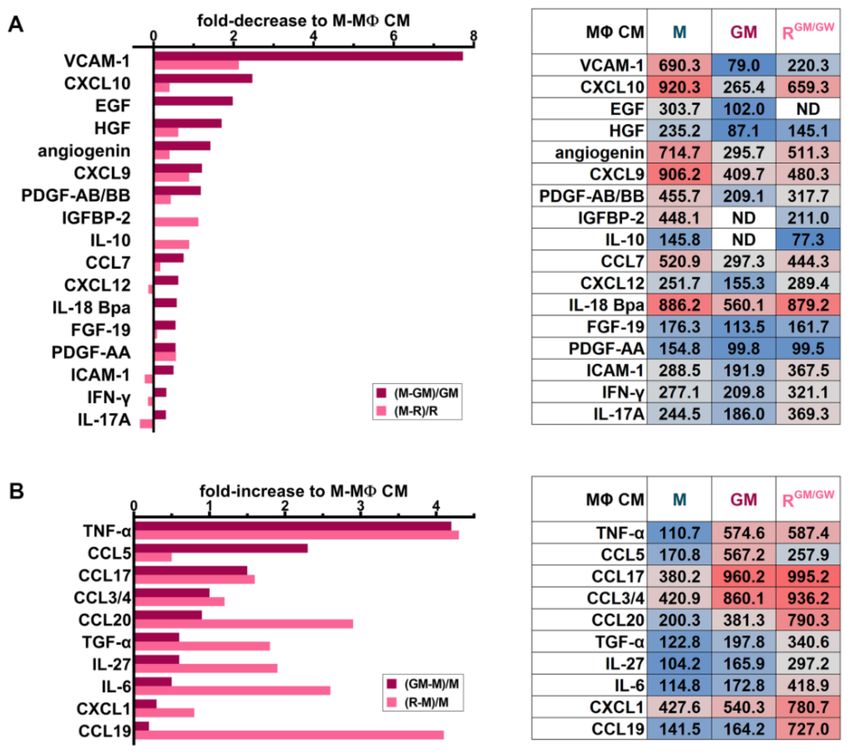
Cancers 2021, 13, 5289                                                                                          2 of 22

                         by genetics and also consider the possibility to target the AML microenvironment as novel
                         therapies [2].
                               Chemotherapy and hematopoietic stem cell transplantation most often allow the
                         achievement of sustained complete remission. Moreover, targeted therapies improve
                         the AML response to chemotherapy and favorably affect survival [3]. However, they
                         remain restricted to a small number of AML categories. Over time and regardless of the
                         treatment, the tumor microenvironment strongly contributes to promoting the growth and
                         survival of drug-resistant subclones, leading to relapse [4,5]. Several clinical studies have
                         demonstrated that heavy tumor infiltration by MΦs is of poor prognosis: tumor-associated
                         macrophages (TAMs) promote tumor cell growth, resistance to treatment, and escape from
                         immune surveillance [4,6].
                               TAMs are often called M2-like protumoral MΦs while the classical M1-like MΦs have
                         inflammatory, antitumor properties [7,8]. However, MΦ plasticity is complex [9,10], and
                         the M1/M2 nomenclature does not correspond to observations made in tumors, where
                         mixed phenotype MΦs, expressing both M1 and M2 markers, are observed. In the tumor
                         microenvironment, malignant cells activate and educate MΦs, which secrete cytokines and
                         promote angiogenesis, matrix remodeling, metastasis, immunosuppression, cancer cell
                         survival, and drug resistance [9,11–13].
                               Tumor escape from immune surveillance is further promoted by the expression of
                         the “do not eat me” signal, CD47 [11,14] and the increased expression of programmed cell
                         death protein-1 (PD1) by tumor-infiltrating T-cells and of its ligand PD-L1 (programmed
                         death-ligand 1) by TAMs [15]. While the involvement of TAMs in the solid cancer mi-
                         croenvironment is established [16,17], little information is available on their role in human
                         AML. In mice, heterogeneity in MΦ polarization was observed in an MLL-AF9 AML
                         model [18,19]. Myeloblasts can recruit MΦs to support their proliferation in bone marrow
                         (BM) and spleen [20].
                               The scavenger receptor CD163 is a marker of TAMs with protumoral characteris-
                         tics [7,12,13]. Several studies reported that CD163 is a poor prognosis marker in solid
                         tumors [13] and in AML [21]. CD163 expression appears to be required to allow mouse and
                         human MΦs to adopt a protumoral phenotype and support malignant cell proliferation.
                         Thus, in a mouse model of sarcoma, tumor growth was abrogated in CD163 deficient
                         mice [22]. CD163-expressing TAMs may contribute to create an immunosuppressive mi-
                         croenvironment. In melanoma, the depletion of CD163+ MΦs promotes tumor infiltration
                         by activated T lymphocytes and tumor regression [7].
                               Targeting TAM polarization is a novel important therapeutic approach in aggressive
                         cancers [16,23]. The colony-stimulating factor-1 (CSF-1, also called M-CSF), which controls
                         the survival, proliferation, differentiation, and polarization of MΦs by binding to its
                         receptor, the CSF1R, is a promising therapeutic target in aggressive tumors and possibly in
                         AML [24]. In multiple myeloma and mantle cell lymphoma, the inhibition of CSF1R can
                         reprogram TAMs and reverse drug-resistance [25–28]. However, whether MΦs contribute
                         to AML cell proliferation and/or survival in creating a protumorigenic niche associated
                         with a protumoral MΦ orientation is not established.
                               Using human AML cell lines and primary myeloblasts, we examined the impact of
                         AML cells on allogeneic and autologous MΦs. We showed that the co-culture of primary
                         myeloblasts with healthy donor (HD) monocytes or with BM cells in their microenviron-
                         ment induces a protumoral MΦ orientation (AML-MΦ), which supports blast cell prolif-
                         eration and survival. Furthermore, using CSF1R inhibitors and granulocyte-macrophage
                         colony-stimulating factor (GM-CSF), we succeeded in reprogramming protumoral AML-
                         MΦs and in reversing drug-resistance induced by myeloblast cross-talk with MΦs.

                         2. Materials and Methods
                         2.1. Patient Samples
                              Heparinized blood and BM samples were obtained from 42 newly diagnosed AML
                         patients at CHUV (Centre Hospitalier Universitaire Vaudois, Lausanne, Switzerland).
Cancers 2021, 13, 5289                                                                                        3 of 22

                         Informed consent was obtained from all subjects involved in the study. After red cell
                         lysis with ammonium chloride, the samples were processed for leukocyte immunostaining
                         and analysis by flow cytometry (FC) [29]. Leukemia diagnosis was based on WHO 2016
                         classification [30] and ELN 2017 risk stratification by genetics [2] and are indicated in
                         Table S1. The study was conducted in accordance with the Declaration of Helsinki and
                         approved by the Commission Cantonale d’Ethique sur la recherche sur l’être humain
                         CER-VD (protocol 2017-01509, 9 November 2017).

                         2.2. Immunophenotypic Analyzes by Flow Cytometry
                              Cell proliferation, apoptosis, and immunophenotypic analyzes were performed us-
                         ing a Beckman Coulter Gallios flow cytometer. Patient samples were analyzed with a
                         10-color/24-antibody panel [29] according to ELN recommendations [31]. MΦs were de-
                         tached from culture dishes in phosphate-buffered saline (PBS) containing 10 mM ethylene-
                         diaminetetraacetic acid (EDTA), for 10 min at 37 ◦ C, washed with Roswell Park Memorial
                         Institute (RPMI) 1640 and characterized using CD45, CD14, CD163, CD80, CD206, and
                         CSF1R; additional blast and lymphocyte markers were used for patient BM and peripheral
                         blood (PB) sample and BM co-culture analyses (Table S2).

                         2.3. Healthy Donor-Derived Macrophages
                               The monocytic fraction was isolated by centrifuging HD PB anticoagulated with citrate
                         phosphate dextrose-adenine 1 and diluted 1/2 with PBS for 10 min at 1000 g to prepare a
                         buffy coat. The concentrated leukocytic fraction was then centrifuged at 400 g for 40 min
                         on a Ficoll-Paque PLUS (GE Healthcare, Dietikon, Switzerland) gradient. Mononuclear cells
                         were collected and plated in RPMI + 10% heat-inactivated fetal bovine serum (FBS) = plain
                         medium (PM), see Figure S1 for the schematic protocol; monocyte-derived MΦs exposed to
                         10 ng/mL recombinant human M-CSF (ImmunoTools, Friesoythe, Germany) were called M-
                         MΦs and those exposed to 1000 U/mL GM-CSF (Miltenyi Biotec, Solothurn, Switzerland):
                         GM-MΦs [25,32].
                               After overnight culture, the wells were rinsed to deplete non-adherent cells; >95% of
                         adherent cells were CD14+ monocytes. The medium was changed, with the added factors,
                         every 2 days, for 7–9 days, when CD163 expression was checked by FC. Alternatively,
                         to obtain HDAML -MΦs, HD monocytes were stimulated for 7–9 days with (1) primary
                         myeloblast or cell line conditioned medium (CM) diluted 1:1 with PM; with (2) AML
                         cell lines (1–5 × 10⁴ /well) or primary blasts (0.5–1 × 10⁶ /well), co-cultured in direct
                         contact with HD monocytes; or (3) separated from MΦs by a 0.4 µm-pore membrane insert
                         (Transwell (TW), Falcon, Dietikon, Switzerland).
                               For MΦ reprogramming experiments, HD MΦ monolayers initially activated with
                         M-CSF (M-MΦ) or blast CM (HDAML -MΦ) were washed with PBS and cultured for
                         7–9 more days in PM containing GM-CSF and GW2580 (1 µM, Sigma-Aldrich, Buchs,
                         Switzerland) [25] or PLX3397 (100 nM, MedChemExpress, Luzern, Switzerland), renewed
                         every 2 days. The change in MΦ CD163 expression after reprogramming was assessed by
                         FC, as described above.

                         2.4. Cell Lines and Cell Culture
                              HL-60 (AML with maturation), U937 (monoblastic leukemia), NB4 (acute promye-
                         locytic leukemia), OCI-AML3 (NPM1 mutated) cell lines were cultured in plain medium
                         (PM) [29] and MV-4-11 (monoblastic AML with FLT3-ITD mutation) in Iscove’s Modified
                         Dulbecco Medium + 10% FBS.

                         2.5. PKH Labeling and Apoptosis Detection
                              Cells were labeled with 10 µM PKH26 (Sigma-Aldrich) according to the manufac-
                         turer’s protocol and immediately added to rinsed wells containing adherent MΦs for
                         assays. A fraction of the labeled cells were fixed in 1% paraformaldehyde and used to
                         calculate the normalized PKH mean fluorescence intensity (MFI), as PKH MFI at day 4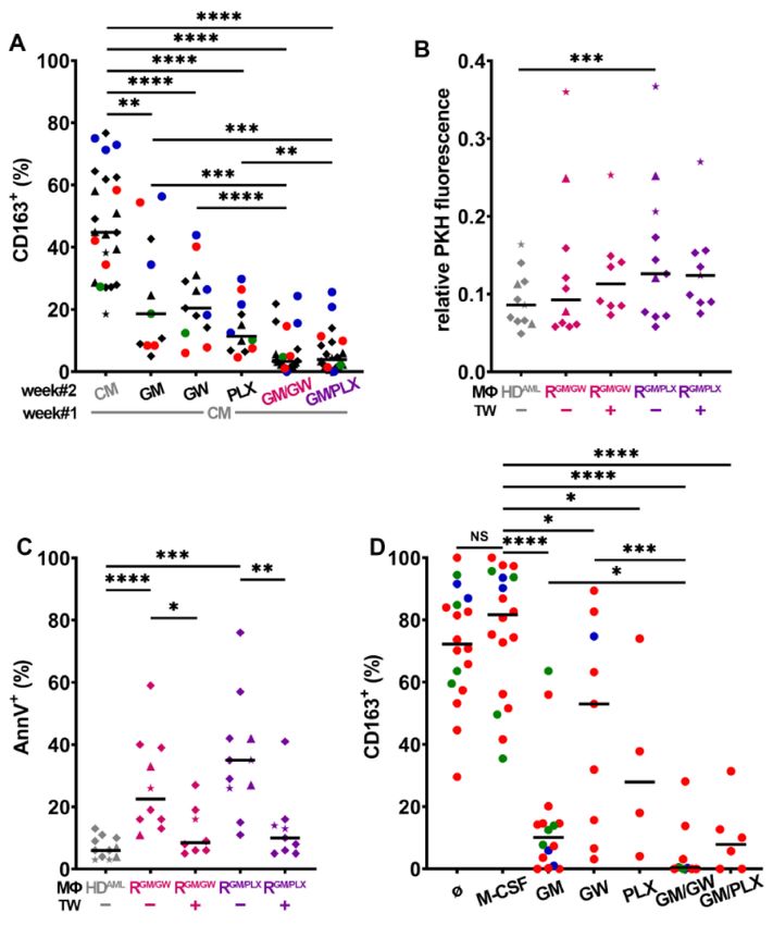
Cancers 2021, 13, 5289                                                                                       4 of 22

                         divided by PKH MFI at day 0. Blast cell apoptosis was assessed by FC, analyzing cells
                         stained with annexin V (AnnV, eBioscience, Zug, Switzerland).

                         2.6. Cleaved Caspase-3 Detection
                              Cells were cultured in 100% PM or in 75% PM supplemented with 25% MΦ-CM. After
                         48 h, the cells were lysed, and the cell lysates were subjected to SDS-PAGE, transferred
                         to nitrocellulose membranes, immunoblotted with anti-cleaved caspase-3 antibody (Cell
                         Signaling Technology) followed by horseradish peroxidase-linked goat anti-rabbit im-
                         munoglobulin G (IgG) antibody (Cell Signaling Technology), and revealed with Luminata
                         Forte chemiluminescence (Millipore, Schaffhausen, Switzerland) [29]. As a loading control,
                         membranes were also immunoblotted with anti-α-tubulin antibody (Sigma-Aldrich) fol-
                         lowed by horseradish peroxidase-conjugated sheep anti-mouse IgG antibody (Amersham,
                         Dietikon, Switzerland) and revealed with Luminata Crescendo chemiluminescence (Milli-
                         pore). Blocking antibodies were from InvivoGen (anti-IL-6) and R&D Systems (anti-tumor
                         necrosis factor α (anti-TNF-α)).

                         2.7. Cytokine Arrays
                              The CM of the indicated leukemia cell lines, primary myeloblasts or MΦs were
                         collected and centrifuged twice at 1500 rpm for 10 min, twice for 10 min at 2500 rpm,
                         and filtered with 0.22 µm low protein binding syringe filter. CM was collected at day 7,
                         from M- or GM-MΦs cultured in medium containing M- or GM-CSF, respectively. CM
                         from reprogrammed MΦs (RGM/GW -MΦs) was obtained after one week of culture with
                         M-CSF, followed by another week in medium supplemented with GM-CSF and GW2580.
                         CM from M-, GM-, and RGM/GW -MΦs were obtained from at least three different HD
                         and pooled for cytokine assays. The CM from primary AML blasts (mononuclear cell
                         suspension containing >95% blast cells) co-cultured with MΦ were also analyzed and
                         processed as above.
                              Cytokine array assays were conducted according to the manufacturer’s protocol (R&D
                         Systems, Human XL Cytokine Array kit #ARY022B). X-ray films were digitalized with
                         ImageScanner III (GE Healthcare) and dot pixel density was measured with ImageQuant
                         software (Molecular Dynamics, Glattbrugg, Switzerland). The mean of duplicates was
                         normalized to the mean pixel density of the six reference spots.

                         2.8. Myeloblast Resistance to Venetoclax and Midostaurin
                              HD MΦs were polarized for 7 days with M-CSF and then either reprogrammed for
                         7 more days in medium containing GW2580 or PLX3397 and GM-CSF or kept in M-CSF-
                         containing medium. MΦ orientation and reprogramming was confirmed by FC. Medium
                         was then removed, and MΦs were washed with PBS. Following this, 150 × 103 NB4
                         or MV-4-11 were added to MΦ monolayers in PM with inhibitors (venetoclax from LC
                         Laboratories, or midostaurin from Sigma-Aldrich). After 48 h, cells were removed from
                         the wells, stained for AnnV and/or 7-aminoactinomycin D (7-AAD), and analyzed by FC.
                         Experiments were repeated using MΦs from at least three different HD.

                         2.9. Statistical Analysis
                             Statistical significance of differences between groups was examined with the Mann-
                         Whitney (M-W) test between two nonparametric unpaired groups, the paired t-test between
                         parametric matched samples, or the Wilcoxon matched-pairs signed rank (WMP) test
                         between nonparametric matched samples. p values < 0.05 were considered as significant.

                         3. Results
                         3.1. Presence of CD163-Expressing Macrophages in BM from AML Patients
                              BM and/or PB were obtained from 42 adult patients with newly diagnosed AML
                         (Table S1). MΦs and myeloblasts were identified by multiparameter FC with a panel of
                         antibodies (Table S2, see Methods for details) [29,31]. BM and PB analyses showed that
Cancers 2021, 13, 5289                                                                                                    5 of 22

                         independently of ELN risk categories [2], the majority of MΦs in AML patients expressed
                         CD163 (Figure 1A), a marker frequently associated with a protumoral phenotype [18–21].
                         By contrast, CD80, a marker typically associated with the inflammatory phenotype and
                         whose expression and functions remain poorly characterized in AML, was expressed at
                         much lower frequency [25,27].

                         Figure 1. CD163 expression on MΦs is modulated by stimuli and depends on CSF1R activity
                         (A) Expression frequency of CD163 and CD80 in BM and/or PB of 42 AML patient MΦs (within
                         CD14+ /CD45+ gate) at diagnosis. Each patient is color-coded according to ELN genetic risk
                         (green = favorable, blue = intermediate, and red = high risk). The median is indicated by a black
                         horizontal line. (B) Expression frequency of CD163 in HD MΦs after one week of culture in PM
                         supplemented with M- or GM-CSF, n = 30. (C) CD163 expression allogeneic HDAML -MΦs obtained
                         from monocytes cultured with AML cell lines (F = HL-60, N = NB4,  = U937, and H = OCI-AML3)
                         or primary patient blasts (dots color-coded according to genetic risk) either in direct contact or
                         separated with a TW. AML conditioned medium (CM) was also tested. The median is indicated
                         by a black horizontal line, n = 13–21. (D) HD Mo were cultured for 7 days with primary patient
                         blasts (blue) or leukemia cell lines (symbols) CM, supplemented with GW2580 (GW) or PLX3397
                         (PLX) before analyzing CD163 expression by FC. CM from: F = HL-60, N = NB4, and  = U937,
                         blue dot = patient#18; n = 3–6 HD. (E) Relative quantification of selected cytokines secreted by patient
                         blasts with three different genetic risks and two leukemia cell lines. *** p < 0.001 and **** p < 0.0001
                         (M-W test).

                         3.2. Myeloblasts Polarize HD Monocytes to Macrophages
                            We first monitored the differentiation of HD Mo into MΦs by adding either M- or
                         GM-CSF in PM for one week. Monocytes cultivated in M-CSF differentiated into M2-like
                         CD163high MΦs, whereas those cultured in GM-CSF differentiated into M1-like CD163low
                         MΦs (Figure 1B). To determine whether myeloblasts can polarize protumoral MΦs and
Cancers 2021, 13, 5289                                                                                          6 of 22

                         induce CD163 expression at their surface, we performed co-cultures of HD Mo with human
                         AML cell lines or primary myeloblasts obtained from AML patients. After 7 days, HD Mo
                         differentiated into MΦs (HDAML -MΦs; Figure 1C, contact), whose CD163 expression was
                         similar to that of HD MΦs cultured in medium supplemented with M-CSF.
                               To determine if direct contact is required for myeloblasts to polarize monocytes into
                         MΦs, they were separated with a 0.4 µm-pore membrane-insert (TW). CD163 expres-
                         sion on HDAML -MΦs was consistently observed even in the absence of direct contact
                         (Figure 1C). Similar results were obtained by stimulating monocytes with CM from AML
                         cell lines or primary myeloblasts. As control, the co-culture of normal hematopoietic stem
                         progenitor cells (HSPC) with HD Mo, under the same conditions as AML cells, did not
                         induce CD163 expression at the surface of monocytes (Figure S2A). In addition, culture in
                         PM or with other inflammatory factors (lipopolysaccharide, TNF-α, IL-1β) did not induce
                         CD163 expression; moreover, boiled U937 CM also failed to induce CD163 expression
                         (Figure S3). These results suggest that soluble factors released by myeloblasts contribute to
                         inducing MΦ polarization.
                               Several other polarization markers were analyzed: the expression level of CD80 was
                         expectedly higher in GM- than in M-MΦs (Figure S4A) but exhibited some variability
                         among HDAML -MΦs (Figure S4B). M- and GM-MΦs exhibited variable expression of
                         CD206 (Figure S4C), a marker typically associated with protumoral MΦ activation in
                         different tumor types including AML [33]. CD206 was also expressed by most HDAML -MΦs
                         (Figure S4D), both after contact with blasts or with their CM only.
                               As M-CSF plays a crucial role in polarizing M-MΦs, we assessed whether inhibiting its
                         receptor could prevent the up-regulation of CD163 expression induced by myeloblasts CM.
                         Indeed, GW2580 and PLX3397, when added into CM, significantly prevented CD163 upregu-
                         lation (Figure 1D), confirming a major role for CSF1R signaling in inducing CD163 expression.
                               To understand what factors in AML may be important in contributing to alternative
                         MΦ polarization and the induction of CD163 expression, we performed semi-quantitative
                         cytokine array analyses using myeloblast CM from three AML patients with favorable,
                         intermediate, or high genetic risk (patients #3, #18, and #23, respectively) and from two
                         AML cell lines (HL-60 and U937; Figure 1E). The assay can detect 105 factors, indicated
                         in Table S3, which shows the pixel density of dots revealed on membranes illustrated in
                         Figure S5A), some of which may contribute to promote cancer growth, cell migration,
                         angiogenesis, and metastasis, such as CXCL5, the urokinase-type plasminogen activator
                         receptor (uPAR), the matrix metalloproteinase 9 (MMP-9), serpin E1 [34], and the growth
                         differentiation factor-15 (GDF-15, a novel targetable immune checkpoint [35]).
                               VEGF (vascular endothelial growth factor), and other angiopoietic factors, with
                         CXCL5 [36], M-CSF [37], and CD163-expressing MΦs promote angiogenesis [38]. The
                         hepatocyte growth factor (HGF) is a key player in AML cell proliferation [24], drug-
                         resistance, and poor survival [39,40]. The insulin-like growth factor (IGF) and its binding
                         protein insulin-like growth factor binding protein 2 (IGFBP-2), secreted by HL-60 and U937
                         cells, have important functions in promoting blast proliferation/survival and resistance
                         to chemotherapy [41–43]. Cytokines, such as IL-18 and the IL-18-binding protein, gained
                         increasing attention for their roles in MΦ activation syndrome [44]. Furthermore, M-CSF,
                         macrophage migration inhibitory factor (MIF), IL-8, and CCL2 may promote a protumoral
                         MΦ polarization [25,45] contributing to forming a protective niche for leukemia stem
                         cells [17].
                               CXCL10 could contribute to either a protumoral or an antitumoral activation, depend-
                         ing on the context [46]. The relative levels of cytokine secretion strongly differ between
                         patients and/or leukemia cell lines, which is consistent with the heterogeneity of AML
                         genetics, phenotypes, and biology and may contribute to the variability in the expression
                         of markers associated to HDAML -MΦ polarization, such as CD163, CD80, and CD206.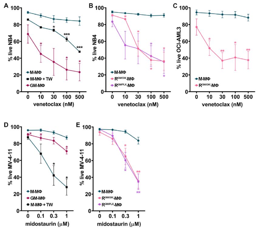
Cancers 2021, 13, 5289                                                                                             7 of 22

                         3.3. HD Macrophages Promote Primary Myeloblast Survival
                              Since myeloblasts can polarize monocyte-derived MΦs (Figure 1C), we examined
                         whether co-culturing primary human myeloblasts with HD monocytes (HD Mo) would
                         affect blast cell survival and proliferation. Myeloblasts obtained from nine newly-diagnosed
                         patients were cultured alone or with freshly isolated HD Mo, in direct contact or separated
                         by a TW, for 7 days. Myeloblasts labeled with PKH26 did not reveal significant changes
                         in proliferation when they interacted with HD Mo (T. S., personal observation, 2020).
                         However, the co-culture of six patient cells in direct contact with HD Mo significantly
                         improved their survival at day 7, compared to culture in plastic (Figure 2A).

                         Figure 2. CD163+ MΦs support myeloblast survival and proliferation. (A) Frequency of AnnV+
                         AML blasts after 7 days of culture alone (plastic), or with monocytes either in direct contact
                         (HD Mo) or separated by a TW. n = 6 patients (#9, 11, 17, 18, 26, and 42), color-coded ac-
                         cording to ELN genetic risk. *** p < 0.001 (paired t-test). (B) Selected cytokines that were
                         increased in CM when primary myeloblasts (favorable, green vs. high risk, red) were cul-
                         tivated for 7 days with HD Mo vs. alone are illustrated. (C,D) PKH-labeled leukemia cell
                         lines were cultured for 4 days on a monolayer of M- or GM-MΦs and analyzed for prolifera-
                         tion (C) and survival (D). * p < 0.05 (M-W test), *** p < 0.001, and **** p < 0.0001 (WMP test).
                         (F = HL-60,  = MV-4-11, N = NB4, and  = U937).

                              This survival advantage was dependent on direct contact with the MΦs, as it was
                         abrogated when blasts and MΦs were separated by TW inserts. These observations suggest
                         that the survival of certain sub-types of AML could be dependent on direct interactions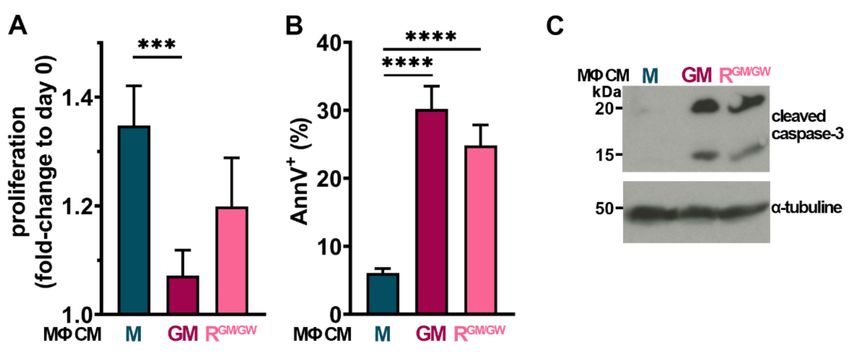
Cancers 2021, 13, 5289                                                                                          8 of 22

                         with MΦs. In contrast, the co-culture of normal HSPC, under the same conditions, with
                         HD Mo did not affect their survival (Figure S2B).
                               The cross-talk between myeloblasts, MΦs, and other components of the BM microenvi-
                         ronment induces the secretion of multiple proinflammatory and protumoral growth factors,
                         which promote the remodeling of the leukemia stem cell niche and may stimulate leukemia
                         cell proliferation and/or survival [47]. The identification of the involved soluble molecules
                         (SM) and/or their receptors is important as they may be therapeutic targets. SM resulting
                         from the interplay between HD Mo co-cultured with primary myeloblasts from patients #3
                         and #23 were identified after 7 days using a cytokine array (Figure S5B, Table S3).
                               The results were compared to those of myeloblasts cultured without monocytes
                         (Figure S5A) and both conditions were illustrated in Figure 2B. Common and distinct
                         characteristics are shared by AML#3 with favorable vs. #23 with adverse genetic risk.
                         Several survival and proliferation factors were clearly upregulated when myeloblasts were
                         in contact with HD Mo as opposed to cultured on plastic, such as IGFBP-2, IL-10, and
                         epidermal growth factor (EGF) [48] (Figure 2B).
                               HGF was secreted at higher levels in co-culture with AML#23. M-CSF was upregulated
                         in co-culture with AML#3, involved in paracrine cross-talks previously observed to drive
                         solid tumor invasion [32,49]. Adhesion receptors VCAM-1 (vascular cell adhesion protein
                         1) and ICAM-1 (intercellular adhesion molecule-1) were both increased in the co-culture
                         medium from patient#23. Several proangiogenic factors were strongly upregulated by
                         co-culture, including angiogenin, PDGF-AA (platelet-derived growth factor), -AB/BB,
                         and fibroblast growth factor 19 (FGF-19). Moreover, CXCL10, CXCL12, and CCL7, which
                         may promote tumor progression, were also highly induced [32,50,51]. IL-17A, known to
                         promote multiple myeloma cell proliferation, was highly secreted by primary myeloblasts
                         co-cultured with MΦs [52].

                         3.4. CD163+ MΦs Activated with M-CSF Support Myeloblast Survival and Proliferation
                              As MΦs can contribute to creating a protumoral blast niche in BM microenvironment,
                         we tested more specifically the effect of MΦ orientation on blast survival and proliferation.
                         Myeloblasts were co-cultured for 4 days with HD MΦs, which had been oriented during
                         one week in medium containing GM-CSF (GM-MΦs) or M-CSF (M-MΦs). Co-cultures
                         were performed in PM, either in direct contact or separated with a TW.
                              The analysis of PKH26 dilution at day 4 showed that myeloblasts co-cultured with
                         the CD163− GM-MΦs exhibited a significantly lower proliferative activity, correlated to
                         higher PKH26 fluorescence, compared to those cultured on CD163+ M-MΦs (Figure 2C).
                         Moreover, a significantly higher proportion of AnnV+ myeloblasts was detected, when
                         they were cultured in contact with GM- than with M-MΦs (Figure 2D). Interestingly, in the
                         absence of direct contact between myeloblasts and GM-MΦs, blasts in TW inserts largely
                         escaped from apoptosis. By contrast, the proportion of AnnV+ blasts co-cultured with
                         M-MΦs did not significantly differ in the presence or absence of contact.

                         3.5. CSF1R Inhibition and Exposure to GM-CSF Reprograms CD163+ M- and HDAML -MΦs
                              As the upregulation of CD163 expression on HDAML -MΦs can be prevented by the
                         inhibition of CSF1R activity (Figure 1D), we assessed whether selective CSF1R tyrosine
                         kinase inhibitors GW2580 [25] or PLX3397 [53] could modify MΦ polarization. As illus-
                         trated in Figure 3A, high CD163 expression on M-MΦs was maintained after 7 additional
                         days of culture in M-CSF (week#2, Figure 3A). CSF1R inhibition or addition of GM-CSF
                         during week#2 lowered CD163 expression, with a stronger effect when GM-CSF was used
                         in combination with either CSF1R inhibitor. In parallel, all treatments also significantly
                         increased CD80 surface expression (Figure S6A).
                              The effect of M-MΦ reprogramming on myeloblast proliferation was assessed by
                         comparing the proliferation of HL-60, MV-4-11, NB4, U937, and OCI-AML3 myeloblasts
                         co-cultured for 4 days with M- vs. R-MΦs. Compared to myeloblasts in direct contact
                         with CD163+ M-MΦs, the co-culture with R-MΦs (reprogrammed with GM-CSF combined
Cancers 2021, 13, 5289                                                                                             9 of 22

                         with either GW2580 = RGM/GW , or PLX3397 = RGM/PLX ), significantly slowed down their
                         proliferation and promoted myeloblast apoptosis (Figure 3B,C). Interestingly, a significant
                         decrease in myeloblast apoptosis was observed when they were separated from R-MΦs by
                         TW inserts. We next assessed the survival of primary myeloblasts of all three risk categories
                         cultured on R-MΦs (Figure 3D): their proliferation was not significantly affected by R-MΦs
                         (T. S., personal observation, 2020), but their survival was strongly and significantly reduced
                         compared to survival on M-MΦs.

                         Figure 3. R-MΦs induce myeloblast apoptosis. (A) Expression frequency of CD163+ MΦs
                         whose culture medium has been supplemented with M-CSF for 2 weeks or switched to reorient-
                         ing medium with GM-CSF (GM) and/or GW2580 (GW) and/or PLX3397 (PLX) for one week,
                         n = 11–26. (B,C) PKH-labeled leukemia cell lines were cultured for 4 days on monolayers of M- or
                         RGM/GW - or RGM/PLX -MΦs +/− TW and analyzed for proliferation (B) and survival (C), n = 14–30.
                         F = HL-60,  = MV-4-11, N = NB4,  = U937, and H = OCI-AML3. (D) Primary patient
                         myeloblasts color-coded according to ELN genetic risk were co-cultured for 4 days on mono-
                         layers of M- or RGM/GW - or RGM/PLX -MΦs and analyzed for survival; AnnV positivity is ex-
                         pressed as fold-change compared to culture on M-MΦs. n = 11–12. * p < 0.05, ** p < 0.01,
                         *** p < 0.001, and **** p < 0.0001 (using M-W test (A–C) or WMP test (D)).
Cancers 2021, 13, 5289 10 of 22
We then investigated the impact of HDAML -MΦ reprogramming: after 7 days of re-
orientation, single treatments significantly lowered CD163 expression, whereas the combi-
nation of GW2580 or PLX3397 with GM-CSF almost abolished it (Figure 4A); in parallel,
reorientation increased CD80 expression (Figure S6B). The co-culture of HL-60, NB4, and
U937 cells with HDAML R-MΦs, lowered their proliferative activity (Figure 4B) and signif-
icantly induced their apoptosis (Figure 4C). As observed with R-MΦs (Figure 3C), the
separation of myeloblasts from HDAML R-MΦs by a TW, protected them from apoptosis
(Figure 4C). These findings reveal a major role for HDAML -MΦ polarization, and most
likely for adhesive interactions, in controlling myeloblast survival.
Figure 4. Reprogrammed AML macrophages induce myeloblast apoptosis. (A) After one
week of stimulation in CM from U937 (), HL-60 (F), NB4 (N), or primary patient blasts
(colored dots), HDAML -MΦs were either left in the same CM or reoriented in PM with in-
dicated supplements for one additional week and CD163 expression was analyzed by FC.
n = 11–24. (B) Myeloblast proliferation and (C) apoptosis induced after 4 days of co-culture
on HDAML -MΦs. F = HL-60, N = NB4, and = U937. (D) Primary BM patient samples
color-coded according to genetic risk were cultured for 7 days in PM + indicated supple-
ments and analyzed by FC. (GM/GW = GM-CSF + GW2580, GM/PLX = GM-CSF + PLX3397).
* p < 0.05, ** p < 0.01, *** p < 0.001, and **** p < 0.0001 (using M-W test except between paired groups
that were compared with WMP test).Cancers 2021, 13, 5289 11 of 22
The impact of MΦ reprogramming using CSF1R inhibitors combined to GM-CSF was
then assessed on autologous primary BM MΦs from AML patients. BM cells were plated at
diagnosis, and MΦs were co-cultured with myeloblasts, leukocytes, and stroma for 7 days
under the conditions indicated in Figure 4D. Autologous BM AML-MΦs maintained their
CD163 positivity in PM, as in medium supplemented with M-CSF.
CSF1R inhibition with GW2580 was least efficient, while GM-CSF supplementation
reduced CD163 expression, but not as strongly as when combined with GW2580. The
combination of PLX3397 with GM-CSF also efficiently inhibited CD163 surface expression.
In parallel, we monitored CD80 expression, which significantly increased with GM-CSF
either alone or combined with GW2580 or PLX3397 (Figure S6C). Finally, we measured
primary patient blast apoptosis in four different BM patient co-cultures in medium with
M-CSF vs. reprogramming conditions; twice as many apoptotic blast cells were found in
both reprogramming media compared to what was observed in M-CSF (Figure S6D).
Innate and adaptive immune checkpoint deregulation have been shown to play im-
portant roles in myeloid malignancies. We, therefore, analyzed the expression of CD47,
which couples to SIRPα on MΦs [14,54], and of PD-L1 [15]. Interestingly, PD-L1 expression
was unchanged or even increased on CD163low HD or autologous primary MΦs exposed
to CSF1R inhibitors, compared to CD163high cells cultured with M-CSF (Figure S7A,B).
Further FC analyses showed that the reprogramming of autologous MΦs co-cultured with
primary myeloblasts did not change CD47 on myeloblasts nor SIRPα expression on MΦs
(Figure S7C,D).
Taken together, our results indicate that CSF1R plays a critical role in the polarization
of HD M-MΦs and of allogeneic HDAML - or autologous AML-MΦs toward a protumoral
M2-like phenotype and that inhibiting the CSF1R activity, in the presence of GM-CSF,
efficiently reverses MΦ orientation. This is evidenced by a down-regulation of CD163 and
an increase of CD80 expression, which has a direct impact on myeloblast proliferation
and survival.
3.6. Macrophage Reprogramming Reverses Drug Resistance
We next assessed whether MΦ orientation affects the sensitivity of myeloblasts to
targeted therapy. We first examined whether MΦ polarization has an effect on myeloblast
sensitivity to venetoclax, a BCL-2 inhibitor. Myeloblasts were cultured in direct contact
with HD M- vs. GM-MΦs (week#1) or M- vs. R-MΦs (week#2) in presence of vehicle
or escalating doses of venetoclax. After 48 h, the percentage of live cells (AnnV− and
7-AAD− ) was measured by FC.
We observed that the co-culture of NB4 cells with M-MΦs induced blast resistance to
venetoclax, as compared to blasts cultured alone (T. S., personal observation, 2020). Direct
contact contributed to the resistance of myeloblasts to venetoclax, as NB4 cells became
significantly more sensitive to it, when they were separated from M-MΦs by TW inserts
(Figure 5A). By contrast, venetoclax strongly induced NB4 cell apoptosis, even at low
concentrations, when blasts were cultured on GM-MΦs (Figure 5A). Importantly, this was
also the case when NB4 were cultured on R-MΦs (reprogrammed with either GW2580 or
PLX3397, and GM-CSF, RGM/GW -, or RGM/PLX -MΦs; Figure 5B). MΦ reprogramming with
GW2580 and GM-CSF also significantly sensitized OCI-AML-3 myeloblasts to venetoclax
(Figure 5C).
FLT3-mutated AML have a poor prognosis [55]. To determine the impact of MΦ
orientation and reprogramming on myeloblasts sensitivity to midostaurin (a FLT3 inhibitor
used in AML patients [56]), we co-cultured the MLL-AF4 and FLT3-ITD mutated MV-
4-11 AML cells with M- vs. GM- (week#1) or M- vs. R-MΦs (week#2). MV-4-11 cells
co-cultured in direct contact with M-MΦs were resistant to midostaurin (Figure 5D). In
contrast, when myeloblasts were separated from M-MΦs by a TW insert, midostaurin
efficiently induced MV-4-11 apoptosis, suggesting an important role for cell adhesion
in promoting resistance to midostaurin (Figure 5D). As observed with venetoclax, the
co-culture of MV-4-11 myeloblasts on GM- or R-MΦs strongly increased their sensitivityCancers 2021, 13, 5289 12 of 22
to midostaurin (Figure 5D,E). Taken together, our results suggest a critical role of MΦ
orientation in promoting drug-resistance to midostaurin and venetoclax.
Figure 5. MΦ reprogramming reverses myeloblast resistance to targeted therapy. AML cell lines were
cultured with indicated MΦ monolayers for 48 h, and the frequency of live (AnnV/7-AAD-negative)
cells was measured by FC in the presence of escalating doses of venetoclax (A–C), or midostaurin
(D–E). (A) Frequency of live NB4 cells after culture with GM- or M-MΦs either in direct contact
or separated by a TW. (B) Frequency of live NB4 cells cultured with MΦ whose culture medium
has been supplemented with M-CSF for 2 weeks (M-MΦs) or switched to reorienting medium
supplemented with GM-CSF (GM) and GW2580 (GW) or PLX3397 (PLX) (RGM/GW - or RGM/PLX -MΦs)
during week#2. (C) Frequency of live OCI-AML3 as above (B) cultured with M- or RGM/GW -MΦs.
(D) Frequency of live (7AAD-negative) MV-4-11 cells after culture with GM- or M-MΦs either in
direct contact or separated by a TW. (E) The % of live MV-4-11 cells cultured with MΦs whose
culture medium has been supplemented with M-CSF for 2 weeks (M-MΦs) or switched to reorienting
medium (RGM/GW - or RGM/PLX -MΦs). Experiments were performed using monocytes isolated from
3–13 different HD. Data points represent the mean ± SEM; * p < 0.05, ** p < 0.01, and *** p < 0.001 to
M-MΦs (M-W test).
3.7. Factors Secreted by GM- and R-MΦs Play a Role in Myeloblast Apoptosis
In order to assess whether factors secreted by MΦs could also have an impact on
myeloblasts, HL-60, NB4, OCI-AML3, and U937 cells were cultured with MΦ CM. Of the
four tested cell lines, only U937 displayed sensitivity to MΦ CM: after 24 h, GM-MΦ CM
significantly impaired their proliferation, while RGM/GW -MΦ CM strongly decreased it
(Figure 6A). Moreover, both GM- and RGM/GW -MΦ CM induced >25% apoptosis in U937
cells (Figure 6B).
Effects on proliferation and apoptosis were already detectable at 6 h (C. S., personal
observation, 2020). Moreover, after 48 h of incubation with MΦ CM, cleaved caspase-3
was detected by western blot (Figure 6C; uncropped version Figure S8); of note, caspase-3Cancers 2021, 13, 5289 13 of 22
cleavage was already detectable at 24 h, but less markedly. M-MΦ CM had no impact
on apoptosis induction and caspase-3 cleavage. These data indicate at least a partial
contribution to apoptosis of both inflammatory/cytotoxic molecules contained in polarized
GM- and RGM/GW -MΦ CM.
Figure 6. GM- and R- MΦ CM impair U937 proliferation and induce their apoptosis. (A,B) U937 cells
were cultured in PM supplemented with 25% MΦ CM and counted after 24 h, n = 5–8 (A) or analyzed
by FC with AnnV staining, n = 7–13 (B). The mean + SEM is plotted for each condition. *** p < 0.001,
and **** p < 0.0001 (M-W test). (C) Lysates from 106 U937 cells cultivated for 48 h with indicated
MΦ CM were immunoblotted with anti-cleaved caspase-3 antibody (upper panel) or anti-α-tubulin
(lower panel) antibody as control.
3.8. Identification of Soluble Molecules Secreted by MΦs
To facilitate the identification of SM predominantly secreted by M-, GM-, and RGM/GW -
MΦs, we illustrated several relevant cytokines as their fold-decrease (Figure 7A) or -increase
(Figure 7B) in GM- or R-MΦ CM relative to their quantification in M-MΦ CM, while the
short tables indicate the SM relative levels (data from all analyzed SM are shown in
Table S3 and revealed membranes are shown in Figure S5C). M-MΦs secrete prosurvival
factors, such as HGF, and EGF, and high levels of VCAM-1, which has been demonstrated
to play an essential role in the BM stromal compartment in mediating leukemia cell
chemoresistance [57].
Other M-MΦ SM detected in the array may play a role, such as CXCL12 or IL-8 (Figure 7A
and Table S3), as they contribute to support myeloblast survival and proliferation [58,59] and
to orient MΦs toward immunosuppressive and proangiogenic functions [60]. Furthermore,
M-MΦ reprogramming with GM-CSF and GW2580 reduced the secretion of many factors
responsible for survival, angiogenesis, and cross-talk with stromal cells, for example
HGF, angiogenin, IGFBP-2, IL-10, and PDGF-AB/BB. The tumor-promoting chemokine
CCL7 [61] is also more abundantly secreted by M-MΦs than GM- or R-MΦs. CXCL9 and
CXCL10, which may exhibit pro- or anti-tumoral activities, depending on the splice variant
of their receptor CXCR3 [62], were reduced in CM of both GM- and R-MΦs [63].
The inflammatory cytokine TNF-α is highly secreted in GM- and RGM/GW -MΦ CM
(Figure 7B). Other factors, with context-dependent antitumoral or immune cell chemotactic
activity, such as CCL5 [64], CCL17 [65], CCL3/4, CCL20 [66], CXCL1, and CCL19 [67],
were also predominantly detected in CM of GM- and RGM/GW -MΦs. Compared to MΦs
stimulated with M-CSF, GM- and R-MΦs secreted increased levels of IL-6 and IL-27,
which may also contribute to inhibiting myeloblast proliferation and promoting blast
apoptosis [39,68].
We next determined the effect of MΦ reprogramming on soluble factor secretion
resulting from the interplay between HD MΦs co-cultured with primary myeloblasts from
patient #3 and #23. HD Mo were first stimulated for one week with CM from patient
#3 or #23. On day 7, after confirming the induction of CD163 expression in HDAML -
MΦs, primary blasts of patients #3 or #23 were added to the respective MΦ monolayersCancers 2021, 13, 5289 14 of 22
for one more week (week#2), in PM vs. PM supplemented with GM-CSF and GW2580
(Figure S5D). The level of each SM was illustrated as the ratio of its level in PM supple-
mented with GM-CSF and GW2580 divided by that secreted by co-cultures in PM (key
cytokines in Figure 8).
Figure 7. Cytokine secretion profiles differ with MΦ polarization status. HD monocytes were
stimulated for 7 days with M- or GM-CSF and CM was collected and analyzed. Alternatively, CM
of MΦs whose culture medium has been supplemented with M-CSF for 1 week and switched to
reorienting medium supplemented with GM-CSF (GM) and GW2580 (GW) for one more week was
also collected. (A) A selection of cytokines is illustrated, which are decreased in GM- (dark red) and
R-MΦ (pink) CM compared to M-MΦ CM, according to the formula in graph legend. (B) Selected
increased cytokines in GM- (dark red) or R-MΦ (pink) CM compared to M-MΦ CM are illustrated
and calculated according to the formula in the graph legend. For both A and B, the relative optical
density of the selected cytokines are indicated in the tables and colored from blue (low) to grey
(medium) to red (high) according to their mean pixel intensity.
Consistent with analyses of GM- and R-MΦ CM shown in Figure 7, the relative levels
of TNF-α increased in both co-cultures of AML #3 and #23 with HD MΦs in the presence
of GM-CSF and GW2580. The secretion of interferon γ, a key inflammatory molecule, also
increased in both co-cultures under reprogramming conditions, as were CCL5, CCL17,
CCL3/4, CCL19 and CCL20. IL-6 was highly secreted in CM of AML #3 co-cultured with
HDAML -MΦs in reprogramming conditions. IL-27 exhibited an opposite trend between
patients. Interestingly, a number of other SM known for their protumoral activity, such
as IL-10, EGF, HGF, IGFBP-2, PDGF-AB/BB, CXCL12, FGF-19, IL-18 Bpa, and VCAM-
1, slightly increased or did not exhibit any significant relative change in their levels in
this experiment.
3.9. TNF-α Induces Apoptosis in U937 Cells
TNF-α is highly increased in GM- and R-MΦ CM (Figure 7B); while it could have a
major function in modulating the surrounding microenvironment cross-talks, and inducing
the expression and secretion of other inflammatory factors, it was shown to be cytotoxic to
U937 cells [69]. We, therefore, tested the effect of escalating doses of recombinant TNF-αCancers 2021, 13, 5289 15 of 22
on U937 cells. After 24 h, we confirmed that TNF-α triggered apoptosis of U937 cells in a
dose-dependent manner (Figure 9A). We next investigated whether adding a neutralizing
antibody against TNF-α in MΦ CM would affect the induction of apoptosis. As illustrated
on Figure 9B, adding a monoclonal antibody against TNF-α in the CM strongly decreased
apoptosis, whereas adding an antibody against IL-6, an inflammatory factor which is also
increased in both MΦ CM, had no impact.
Figure 8. The secretion of proinflammatory cytokines is increased in co-cultures of primary AML
blasts with reprogrammed HDAML -MΦs. HD Mo were stimulated for 7 days with CM from primary
blasts from patients #3 or #23 and primary blasts from the same patients were then added to the
respective HDAML -MΦs with or without GM-CSF + GW2580 for 7 more days (week#2). At the end
of week#2, CM from the four distinct co-cultures were collected and analyzed in cytokine arrays.
(A) Fold change in relative secretion of selected cytokines secreted by blasts from patient #3 (favorable
risk, green bars) and #23 (high risk, red bars) co-cultured with reprogrammed HDAML MΦs in PM
containing GM-CSF and GW2580, divided by their respective relative levels in control co-cultures
of AML blasts with HDAML MΦs in PM. (B) Relative optical density of the selected cytokines
illustrated in the graph colored from blue (low) to grey (medium) to red (high) according to their
mean pixel density.
Figure 9. TNF-α in MΦ CM can trigger apoptosis in U937 cells. (A) U937 cells were cultured for 24 h
with increasing concentrations of TNF-α and apoptosis was monitored by FC. (B) U937 cells were
cultured for 24 h with 25% indicated MΦ CM and anti-TNF-α (30 µg/mL) or anti-IL-6 (300 ng/mL)
antibodies. AnnV positivity was analyzed by FC.Cancers 2021, 13, 5289 16 of 22
4. Discussion
AML form a heterogeneous group of diseases, frequently of poor prognosis, with a
high risk of relapse after completion of therapy. In patients with solid tumors and leukemia,
the presence of TAMs has been reported as a bad prognosis factor, which may be related
to the change in MΦ polarization inside tissues infiltrated by malignant cells, promoting
tumor growth and survival [23]. As little information is available on AML-associated
MΦs, we examined their impact on myeloblast survival, proliferation, and resistance to
venetoclax or midostaurin.
In addition, we examined the possibility of changing the MΦ orientation by inhibiting
the CSF1R. Our results show that (1) BM AML MΦs frequently express the protumoral
orientation marker CD163 at diagnosis; (2) myeloblasts can educate HD MΦs to express
CD163 in a CSF1R-dependent manner and promote their growth and survival; (3) nu-
merous SM are secreted that support myeloblasts proliferation, survival, migration, and
angiogenesis; (4) resistance to venetoclax and midostaurin is promoted by myeloblast
interactions with MΦs; and (5) MΦ orientation can be reprogrammed by inhibition of
CSF1R combined with GM-CSF exposure, leading to the reversal of resistance to targeted
therapies and myeloblast apoptosis.
MΦs are highly plastic cells that can change their phenotype and function in response
to microenvironmental stimuli, which modulate their polarization [70]. In vitro, they
adopt either an antimicrobial and antitumoral activity when they are exposed to cytokines
secreted by Th1 lymphocytes (such as TNF-α or interferon γ) or an anti-inflammatory
phenotype promoting angiogenesis and tumor growth when they are exposed to Th2
lymphocytic cytokines (such as IL-4, -5, and -13) [71]. In tumors, depending on local cy-
tokines secreted by malignant cells and tissue environment, TAM orientation can be shared
between M1 and M2 status, with multiple phenotypes between these two extremes [15].
In the majority of AML patients, we observed that the BM and PB MΦs significantly
express the M2-like marker CD163 at diagnosis (Figure 1A). Interestingly, similar observa-
tions were reported in B- and T-cell acute lymphoblastic leukemia, suggesting that leukemia
cells and their microenvironment may dysregulate MΦ function [72]. In a minority of
AML patients, we also observed MΦs expressing CD80, suggesting that a continuum of
phenotypes between M1- and M2-like extremes are present at diagnosis, whose function
may change in response to local signals.
The co-culture of AML cell lines or primary myeloblasts with HD Mo highly induced
CD163 expression on HDAML -MΦs (Figure 1C), mimicking the effect of M-CSF (Figure 1B).
This induction did not require direct contact between myeloblasts and MΦs as it was also
observed with exposure to AML CM, suggesting that it may depend on soluble factors,
mainly cytokines [73]. However, cytokines known to induce the expression of CD163,
such as IL-4, -13, and -10 [74], were not detected in AML CM (Table S3). Alternatively,
we hypothesized that it may depend on CSF1/CSF1R pathway activation, as in solid
cancers [15] and lymphoma [27,28], which was confirmed by the abrogation of CD163
expression induced by AML CM supplemented with GW2580 or PLX3397 (Figure 1D).
Previous observations reported a sensitivity of primary AML cells to CSF1R inhibition,
in favorable-risk patients, through the secretion of cytokines, in particular HGF, by CSF1R-
expressing supportive cells [24]. Inhibiting CSF1R in order to reprogram protumoral MΦ
was crucial in our experiment, but MΦs were simultaneously cultured with GM-CSF to
induce an M1-like polarization. GM-CSF, as a key driver of inflammatory reaction, con-
tributes to the antitumoral effect of R-MΦs. It may do so by enhancing the MΦ phagocytic
activity, the production of reactive oxygen species, and the secretion of proinflammatory
cytokines that affect T-lymphocyte response against malignant cells [75,76].
M-MΦs (Figure 3A), HDAML -MΦs (Figure 4A), and primary autologous MΦs in their
BM microenvironment (Figure 4D) were efficiently reprogrammed by CSF1R inhibition
leading to a drastic decrease in CD163 expression; addition of GM-CSF potentiated this
down-regulation. In co-culture assays, R-MΦs efficiently induced primary myeloblasts and
AML cell line apoptosis, but as with GM-MΦs, myeloblasts were partially rescued fromCancers 2021, 13, 5289 17 of 22
apoptosis in the absence of direct contact, suggesting a role for cell adhesion molecules.
However, SM also participated in the proapoptotic effect of R-MΦs. CM from GM- or
R-MΦs induced caspase-3 cleavage and the apoptosis of >25% of U937 monoblasts.
Interestingly, mAb blocking studies indicated that TNF-α may contribute to inducing
U937 apoptosis. High TNF-α levels were detected in CM of GM- and R-MΦs (Figure 7B)
and recombinant TNF-α induced U937 cell apoptosis. In myeloblasts, TNF-α could activate
the apoptosis cascades through binding to its receptor TNFR1, FADD (Fas-associated
protein with death domain) recruitment, and caspase-8 and -3 activation or by activation
of the mitochondrial pathway leading to caspase-9 and -3 activation [77].
In a mouse model of chronic lymphocytic leukemia, targeting MΦs using an anti-
CSF1R mAb efficiently inhibited disease progression; interestingly, the leukemic cell death
was dependent on TNF-α signaling and tumor microenvironment reprogramming toward
an antitumoral phenotype [78]. In vivo, TNF-α could have not only an effect on myeloblasts
but also a major impact on MΦ orientation by counterbalancing the emergence of M2-like
AML-MΦs by inhibiting IL-13, and possibly IL-4, secreted by eosinophils in tumors [79].
Moreover, TNF-α can induce the secretion of other proinflammatory effectors, like
GM-CSF, which activates interferon regulatory factor 4 (IRF4) and induces the biosynthesis
of CCL17 in MΦs, a cytokine loop with proinflammatory properties [80] promoting MΦ
M1-like orientation and myeloblasts apoptosis. However, the CM of GM- or R-MΦs had
no proapoptotic effect on three other AML cell lines; this may be due to the ability of
TNF-α to simultaneously activate pathways leading to cell apoptosis or survival and
proliferation [77].
Other mechanisms may contribute in vivo to the proapoptotic effect of R-MΦs. Figure 7
shows that the cytokine profile of R-MΦs differs from that of GM-MΦs by the secretion
of higher levels of chemokines, such as CCL20, CCL19, IL-6, and IL-27. The crucial role
played by CCL19 in the immune response is illustrated by its ability with IL-7 to promote
tumor infiltration by T cells and dendritic cells and to improve the therapeutic effect of
CAR-T cells against solid tumors [81] or multiple myeloma [82].
The proinflammatory molecule IL-6 may also contribute to immune response by
promoting T-cell-mediated immune defense [83] and was even reported to stimulate
myeloblast differentiation by MΦs [84]. Finally, blocking CSF1R in vivo inhibits monocyte
recruitment into the tumor environment, leading to MΦ depletion. MΦ reprogramming
induced by CSF1R inhibition may also strengthen immune defense by stimulating T-cell
recruitment in a malignant cell environment [85].
Drug resistance is a major problem in relapsed/refractory AML. The combination
of the BCL-2 inhibitor venetoclax with hypomethylating agents or low dose cytarabine
demonstrated high initial efficiency in patients with de novo AML, who were ineligible for
intensive chemotherapy [86]. However, the response to treatment is most often lost after
several months, with poor survival of relapsing patients [87].
Several mechanisms of resistance to venetoclax have been reported [88] involving the
up-regulation of antiapoptotic molecules Mcl-1 and Bcl-xL, mutations in genes controlling
different kinase pathways, transcription factors, epigenetic modifiers, and tumor suppres-
sors. Similarly, drug resistance affects the sensitivity of FLT3-mutated AML to midostaurin.
Like for venetoclax, the mechanisms of resistance may be complex and involve the intrinsic
properties of AML cells as well as of the microenvironment [89].
The ability of reprogrammed macrophages to increase the sensitivity of myeloblasts
to midostaurin or venetoclax suggests a role for macrophage polarization in promoting
resistance to these targeted therapies. Interestingly, close contacts between macrophages
and myeloblasts were required to promote resistance to midostaurin or venetoclax, which
may suggest the involvement of adhesion receptors. The presence of high soluble VCAM-1
levels in M-MΦ CM and its strong decrease in the CM of GM- and R-MΦs may suggest
the involvement of VCAM-1/CD49d, which were previously reported to trigger NF-κB
signaling and promote chemoresistance [57].Cancers 2021, 13, 5289 18 of 22
Aiming to modulate the MΦ phenotype in AML is a promising therapeutic approach.
Currently, clinical development of CSF1R inhibitors, including PLX3397, for AML treatment
is at the early stages [90]. The efficacy of PLX3397, a FLT3 and CSF1R inhibitor, was
demonstrated in relapsed/refractory FLT3-ITD-mutated AML with a safety profile similar
to that of other FLT3 inhibitors [91]. Its impact on CSF1R inhibition is unclear in relapsing
FLT3-ITD AML, and its clinical activity is predominantly related to its ability to inhibit
FLT3 [91]. CSF1R inhibition alone may not be sufficient to overcome resistance mechanisms
in vivo [92,93], but it may represent a general approach to target the microenvironment of
AML cells and may be promising when used in combination with inhibitors of the immune
checkpoints, angiogenesis, or with the adoptive T-cell transfer, which are undergoing
clinical investigations [94].
To reprogram human MΦs in AML, combining CSF1R inhibition with GM-CSF might
be more efficient for inducing an antitumoral response. It may however not be sufficient
to restore MΦ phagocytic activity and antigen-presenting capacity in vivo. Indeed, PD-L1
expression is unchanged or higher on CD163− MΦs exposed to CSF1R inhibitors, than
on CD163+ cells. PD1/PD-L1 inhibition may improve the antitumoral activity of R-MΦs
and conversely, CSF1R inhibition may improve the effect of anti-PD1/PD-L1 monoclonal
antibody in relapsed/refractory AML.
In addition to the T-cell immune checkpoints, CD47 is the dominant MΦ checkpoint,
which is overexpressed in myeloid malignancies and inhibits phagocytosis through “do
not eat me” signals upon its binding to SIRPα on MΦs [54]. As observed for PD1/PD-L1,
the reprogramming of AML-MΦs co-cultured with primary myeloblasts did not change
CD47 expression on myeloblasts nor SIRPα expression on macrophages. As the com-
bination of the anti-CD47mAb, magrolimab, with 5-azacitidine strongly improves the
antitumoral activity of MΦs [54], considering a combination of CD47 blockade and CSF1R
inhibitors, with 5-azacitidine, might be a way to improve the in vivo antitumoral activity
of reprogrammed MΦs.
5. Conclusions
Relapse and resistance to treatment remain major issues in the treatment of AML. We
showed here that protumoral CD163+ MΦs predominate, at diagnosis, in the BM of AML
patients and that the induction of CD163 expression on MΦs by myeloblasts was depen-
dent on CSF1R. We observed that numerous SM were secreted by MΦs and myeloblasts,
which promoted an M2-like MΦ orientation, leading to increased blast survival and drug
resistance in the presence of close intercellular contacts. Finally, inhibiting CSF1R, in the
presence of GM-CSF, reprogramed MΦ orientation, reversed the resistance to targeted
therapies, and promoted myeloblast apoptosis. CSF1R inhibition may serve as a novel way
to target the microenvironment of myeloblasts and improve the efficiency of AML therapy.
Supplementary Materials: The following are available online at https://www.mdpi.com/article/10.3
390/cancers13215289/s1, Figure S1: Schematic protocol of MΦ differentiation process, Figure S2: HSPC
do not upregulate CD163 expression on MΦs, Figure S3: Inflammatory or inactivated factors do not
upregulate CD163 expression on HD MΦs, Figure S4: CD80 and CD206 are frequently expressed by HD
and HDAML -MΦs, Figure S5: Autoradiographies of cytokine array membranes used for densitometry,
Figure S6: CD80 expression is increased in R-MΦs, Figure S7: PD-L1 is increased on R-MΦs, while SIRPα
is unchanged, Figure S8: Uncropped western blot of Figure 6C, Table S1: FAB, WHO 2016 and ELN
2017 genetic risk classifications from included patients, Table S2: Antibodies for FC, Table S3: Relative
quantification of all cytokines from membranes depicted on Figure S5.
Author Contributions: Conceptualization, T.S. and O.S.; methodology, C.S., T.S. and O.S.; investiga-
tion, T.S. and C.S.; project administration, T.S.; Validation, C.S., T.S. and O.S.; resources, T.S. and O.S.;
data curation, C.S. and T.S.; writing—review and editing, C.S., T.S. and O.S.; funding acquisition, T.S.
All authors have read and agreed to the published version of the manuscript.
Funding: This research was supported by a grant from Fond’Action contre le cancer and the Founda-
tion Dr. Henri Dubois-Ferrière Dinu Lipatti.You can also read