The Interaction of the Gut Microbiota with the Mucus Barrier in Health and Disease in Human - MDPI
←
→
Page content transcription
If your browser does not render page correctly, please read the page content below
Review

The Interaction of the Gut Microbiota with the Mucus
Barrier in Health and Disease in Human
Anthony P. Corfield
 Mucin Research Group, School of Clinical Sciences, Bristol Royal Infirmary, Level 7, Marlborough Street,
 Bristol BS2 8HW, UK; mdapc@bristol.ac.uk

 Received: 29 June 2018; Accepted: 30 July 2018; Published: 2 August 2018

 Abstract: Glycoproteins are major players in the mucus protective barrier in the gastrointestinal
 and other mucosal surfaces. In particular the mucus glycoproteins, or mucins, are responsible for
 the protective gel barrier. They are characterized by their high carbohydrate content, present in
 their variable number, tandem repeat domains. Throughout evolution the mucins have been
 maintained as integral components of the mucosal barrier, emphasizing their essential biological
 status. The glycosylation of the mucins is achieved through a series of biosynthetic pathways
 processes, which generate the wide range of glycans found in these molecules. Thus mucins are
 decorated with molecules having information in the form of a glycocode. The enteric microbiota
 interacts with the mucosal mucus barrier in a variety of ways in order to fulfill its many normal
 processes. How bacteria read the glycocode and link to normal and pathological processes is
 outlined in the review.

 Keywords: gastrointestinal; glycoprotein; glycosylation; glycan; glycocode; microbiota; mucus;
 mucin; mucosal

1. Introduction
     The mucosal protective barrier is a feature of higher animals and has been developed and
maintained throughout evolution [1,2]. The family of mucus glycoproteins, the mucins, are an
integral part of this barrier and also feature throughout evolution [3,4]. A principal character of the
mucins is their glycosylation, a high proportion of their molecular weight consists of carbohydrate in
the form of oligosaccharides, or glycan chains [5–8]. The glycans are made up of a sequence of
monosaccharides and are biosynthesized and degraded by enzymes that recognize the glycan
structures and their linkages. The sequences generated and expressed are known and predictable,
due to their mode of synthesis. They form a glycocode [9] where the sequence is recognized by
proteins that play a role in mucosal protection, resident and pathogenic microorganisms, transient
food borne bacteria interactions, and innate and adaptive immune responses [10]. This glycocode is
species and tissue specific and is closely linked to the microbiota associated with individual mucosal
surfaces [10–12]. The expression of the mucins in the mucosal defensive barrier is dynamic and is
known to adapt to mucosal changes, in order to maintain optimal protection. A number of diseases
have been identified which relate to aberrant glycosylation of the mucins and have been used as
biomarkers for these pathological conditions [13–15]. The known diseases include genetic based
abnormalities [16,17] in addition to tissue specific and environmentally effected changes which
would influence mucins and lead to mucus which does not function effectively and results in
reduced mucosal protection and the appearance of pathological features [7,18–26].
     This review will identify the principal characteristics of the mucosal protective barrier in the
gut, with regard to the role of the mucins and their glycosylation.

Microorganisms 2018, 6, 78; doi:10.3390/microorganisms6030078               www.mdpi.com/journal/microorganisms
Microorganisms 2018, 6, 78                                                                        2 of 58

2. The Structure of the Mucus Barrier
      The mucus barrier is the primary defensive layer at the surface of mucosal surfaces throughout
the body of higher animals. It is a multi-component structure, which is integrated to ensure both
protection and communication. This is achieved in several ways depending on the individual
components.
      The mucosal cells themselves are characteristic and many additional elements are derived
directly from them [6,27–30]. The oral cavity and the oesophagus comprise squamous epithelial cell
layers, while in the lower gastrointestinal (GI) tract a single layer of columnar epithelial cells are
dominated by enterocytes. In addition, the intestine is well innervated and the enteric nervous
system mediates gut motility, fluid exchange, blood flow, secretion, and barrier permeability
through paracrine processes, while juxtacrine mechanisms occur via cell-cell contacts formed at the
gap junctions. Both of these mechanisms are calcium dependent.
      The adherent mucus is synthesized and secreted by the Goblet cells, located in all parts of the
intestinal tract. Recent work has emphasized the range of Goblet cells found in the GI tract, together
with specific functions relating to the mode of mucus secretion [28–31]. The function of the Goblet
cells varies depending on their location in the small intestinal or colorectal crypts. The identification
of a “sentinel” Goblet cell at the mouth of colonic crypts serves to underline the concept that Goblet
cells vary depending on their intestinal location [32]. The number of vesicles found in these cells,
together with the release of mucus into the crypt lumen is mediated to ensure channeled release and
formation of mucus fibrils. This has recently been demonstrated in the lung for MUC5B [33,34] and
is assumed to function in a similar manner in the gut with MUC2. The mucus product found at
mucosal surfaces throughout the body is derived from the gel-forming mucins and is well
recognized as part of the mucosal barrier with a characteristic thickness. The mucus thickness in the
GI tract has been extensively analyzed and reported [35,36].
      A contrasting viewpoint regarding mucus thickness has recently been reported, proposing that
the mucosal contents govern the thickness of the mucus layer and this is region specific, occurring
largely in the distal colon [37]. The observations in this case show no evidence for an adherent
mucosal gel layer where fecal content is present. Instead mucus is attached to the fecal pellet and is
absent from the surface of the epithelium. A functional role for the mucus, as proposed in the
two-layer model, would therefore be redundant.

3. The Mucin Gene Family and Their Role in the Gut
      The family of mucin genes currently includes 21 members. Their macromolecular structure is
organized through disulfide bridges and some of the mucins also contain isopeptide linkages. They
have been divided into two basic groups on the basis of their biological functions, secreted mucins
and the membrane-associated mucins. The secreted members include the gel-forming mucins,
important for mucus barrier formation at mucosal surfaces, and also secreted, non-gel forming
mucins. The membrane-associated mucins are essentially components of the cell-surface glycocalyx
and it is here that their glycans contribute to the carbohydrate rich surface involved in many
interactions between cells and the external environment. Those mucins commonly found in the gut
include the gel-forming mucins MUC2 (jejunum, ileum, and colon), MUC5AC (stomach), MUC5B
(in submandibular and other salivary glands), MUC6 (stomach and ileum) and the non-gel forming
mucin MUC7 (sublingual and submandibular glands). Membrane-associated mucins in the gut
include MUC1, MUC3A/B, MUC4, MUC12, MUC13, MUC15, MUC17, MUC20, and MUC21. Each
mucin has a typical protein domain structure which correlates with their secreted or
membrane-associated nature. More information on the individual mucins can be found in the
literature [6,8,38–43]. The major feature of all mucins is the proline-threonine-serine (PTS) rich
domain, which contains the serine and threonine residues that form the glycosidic links to GalNAc,
the first monosaccharide in the O-linked glycan chains, typical of mucins. The PTS domains are
expressed as tandem repeats, thus generating a domain, which carries a large number of glycans.
The size and pattern of these PTS domains varies between mucins. The main features of these
molecules are shown in Tables 1 and 2.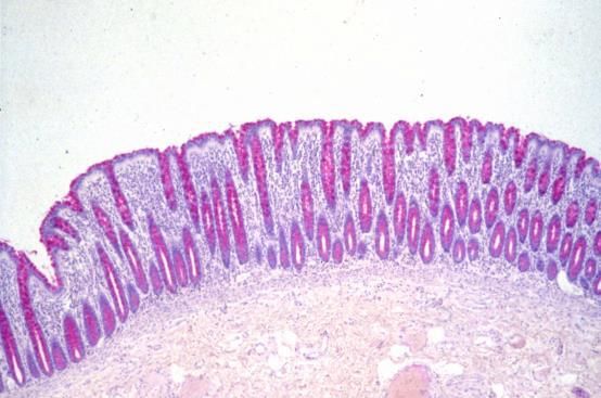
Microorganisms 2018, 6, 78                                                                             3 of 58

                                   Table 1. The Mucin (MUC) Gene Family.

   MUC                              Tandem        N-Terminal Signal             Gastrointestinal Tract
                 Chromosome
   Gene                            Repeat Size         Sequence                       Location
                                      Membrane Associated Mucins
                                                                                 Stomach, duodenum,
  MUC1                 1q21             20                     √
                                                                                     ileum, colon
MUC3A/B                7q22              17                    √                 Small intestine, colon
 MUC4                  3q29              16                    √                 Small intestine, colon
 MUC12                 7q22              28                    √                         Colon
 MUC13                3q21.2             27                    √                 Small intestine, colon
 MUC15               11p14.3           none                    √                 Small intestine, colon
 MUC16               19p13.2            156                    √                    Not expressed
                                                                                 Stomach, duodenum,
  MUC17                7q22             59                     √
                                                                                         colon
  MUC20               3q29               18                     √                        Colon
  MUC21               6p21               15                     √                        Colon
                                         Secreted gel-forming mucins
 MUC2                11p15.5             23                     √               Jejunum, ileum, colon
MUC5AC               11p15.5             8                      √                       Stomach
MUC5B                11p15.5             29                     √                   Salivary glands
 MUC6                11p15.5            169                     √                   Stomach, ileum
 MUC19                12q12              19                     √               No reports for GI tract
                                       Secreted non gel-forming mucins
  MUC7              4q13-q21             23                     √                   Salivary glands
  MUC8               12q243            13/41                    √                   Not expressed
  MUC9                1p13               15                     √                   Not expressed
      The chromosome location, size of the tandem repeat domain, confirmation of an N-terminal
      sequence, and expression pattern in the gastrointestinal tract are shown.

     The mucins are essentially glycosylated polymer proteins, which have been evolved to function
as part of the mucosal protective barrier and as cell membrane components presenting a
characteristic glycoarray at the cell surface [5,6,39,44–47].
     Studies on the evolution of both the mucins and protein glycosylation clearly demonstrate that
these are biologically significant features. The origin of the mucins can be traced back to phyla
associated with the early metazoan period [3,4,48], while the glycans show a similar evolutionary
profile within the eukaryotes [1,49–52]. In contrast, the prokaryotes show a diverse range of protein
glycans that vary from the eukaryotes in their structure and mode of metabolism [53]. This
evolutionary data highlights the physiological consequences of mucin glycosylation and gives a
perspective in relation to the current emphasis placed on DNA and protein sequence information.

                                       Table 2. Mucin peptide domains.

 Peptide Domain
                               Mucin             Mucin Type                Peptide Domain Function
      Type
                                                                        Non-glycosylated multiple copy
Cysteine rich CYS        MUC2, MUC5AC,                                  domains adjacent or interrupting
                                                   Secreted
    domains              MUC5B, MUC19                                tandem repeat domains. Important for
                                                                       various mucin–mucin interactions.
                         MUC2, MUC5AC,
  Cysteine Knot          MUC5B, MUC6,              Secreted                Involved in dimerization.
                            MUC19
 Von Willebrand          MUC2, MUC5AC,                              Mediate oligomerisation located at N- &
    Factor D             MUC5B, MUC6,              Secreted           C-terminus D3 is directly active in
 (D1, D2, D’, D3)           MUC19                                               polymerization.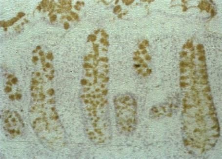
Microorganisms 2018, 6, 78                                                                              4 of 58

                                                                        Located N-terminally to the D4 is
 Von Willebrand          MUC2, MUC5AC,
                                               Secreted &               located C-terminally to the VNTR
    Factor D              MUC5B, MUC6
                                            Membrane-associated            domains, contains the GDPH
      (D4)                  MUC4
                                                                            autocatalytic cleavage site.
                                                                      Located on the cytoplasmic side of the
                         MUC1, MUC3A/B,
                                                                         cell surface membrane. Contains
                         MUC12, MUC13,
 Cytoplasmic Tail                           Membrane-associated         phosphorylation sites involved in
                         MUC16, MUC17,
                                                                     signaling. MUC3, MUC12, and MUC17
                            MUC21
                                                                             have PDZ binding motifs
       SEA
                         MUC3A/B, MUC4,
  (Sperm protein,                                                      Protein binding properties. Contains
                         MUC12, MUC13,      Membrane-associated
  Enterokinase &                                                      autocatalytic proteolytic cleavage site.
                          MUC17, MUC21
      Agrin)
       EGF               MUC1, MUC3A/B,
                                                                       Mediate interactions between mucin
    (Epidermal           MUC12, MUC13,      Membrane-associated
                                                                        subunits and ERBB receptors.
  Growth Factor)            MUC17
                         MUC1, MUC3A/B,
                          MUC4, MUC12,
                                                                      Membrane-spanning sequence typical
 Transmembrane           MUC13, MUC16,      Membrane-associated
                                                                           for membrane proteins
                         MUC17, MUC20,
                            MUC21
      GDPH
                             MUC2, MUC4,       Secreted &            Autocatalytic site cleaving between GD
   autocatalytic
                              MUC5AC        Membrane-associated                 and PH residues
  proteolytic site
                         MUC1, MUC3A/B,
    Proteolytic           MUC4, MUC12,                                Found in MUCs with the SEA domain
                                            Membrane-associated
   cleavage site         MUC13, MUC16,                                          and in MUC16
                            MUC17
      The major mucin peptide domains are indicated for each of the secreted and membrane-associated
      mucin genes. An indication of their function is summarized. In addition to the conventional mucin
      forms, there are similar molecules that have been given names such as mucin-like, see previous
      papers [54–56]. These molecules are different to the mucin family shown in Table 1 and are not
      considered further in this review.

4. Bacterial Species in the Human Gastrointestinal Tract
     The GI microbiota shows characteristic patterns throughout the tract and this has implications
for the nature of interactions between the bacterial cells and the mucosal surface glycoarrays. Oral
cavity species include Streptococcus, Prevotella, Porphyromonas, and Fusobacterium strains [57,58],
stomach accommodates Streptococcus, Lactobacillus, Staphylococcus, and Peptostreptococcus [59], while
an abundance of more than 1000 species are found in the small intestine and colon [60,61]. These are
largely anerobes, with 2–3 times more than facultative anaerobes and aerobes. The most common
species are in the Firmicutes and Bacteroidetes, with fewer Proteobacteria, Fusobacteria, Cyanobacteria,
Verrucomicrobia, and Actinobacteria strains. Ethnicity has also been shown to influence the GI tract
microflora [62], this needs to be considered when comparisons between different population groups
are made.
     The ability of the human enteric microbiota to turn over mucus in the intestinal mucosa
depends on the production of a series of hydrolytic enzymes, which degrade the mucus glycans to
yield monosaccharides which serve as an energy source for the microbiota. The glycohydrolases
adapted to the blood group of each individual and this has been demonstrated for mucin
oligosaccharide degrader (MOD) strains [63,64].
     Among other bacterial species that have special relevance for the mucins is the anaerobe
Bifidobacteria, which are abundant in early life, especially in breast fed infants [65,66]. They are able
to digest a range of host and diet derived glycans, including mucus and mucins. Evidence
supporting this feature is the selective expression of carbohydrate transport systems and many
proteins, which catalyze the degradation and metabolism of a variety of carbohydrates including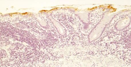
Microorganisms 2018, 6, 78                                                                       5 of 58

low molecular weight oligosaccharides [65], polysaccharides such as glycogen, pullulan, starch,
maltodextrin, and amylopectin [65] and mucins [67].
     Lactobacillus species play a significant role in normal gut glycan metabolism and have been
widely used as probiotics [68–70]. In addition, binding to intestinal mucus and mucins has been
demonstrated [71,72]. A similar situation exists in the female reproductive tract, where the mucus
layer in the vagina is normally colonized by Lactobacillus strains, and where reduction or loss of these
species results in abnormal colonization, largely Garderella spp., and the development of bacterial
vaginosis occurs and can be treated by probiotic Lactobacillus administration [73–75].
     An important group of bacteria that have major roles in the metabolism of mucins in the gut are
Akkermansia spp. [76,77]. Originally isolated from the gut flora in 2004 with mucin as a sole carbon
source it was named after the Dutch microbiologist Antoon Akkermans [78]. Akkermansia spp. has
been identified as human gut species present from early childhood [76,78–80]. In accord with its
location in the mucus layer of the gut many strains have carbohydrate metabolic proteins in their
genome and therefore are well able to metabolize and utilize mucus and its monosaccharides from
the secreted gel-layer [76,81]
     A fundamental trait of these bacteria is cross-feeding, whereby the carbohydrate metabolic
capacity of individual species at any one location contributes to the energy requirements of all
species present. This means that although some strains may not express all enzymes necessary for
generation of monosaccharide substrates the total flora is able to achieve this and provide
monosaccharides for all strains present [82–84].
     Developmental aspects are important and age related variations are found throughout life [85–
88]. The expression of mucin glycosylation during development has been followed in mammalian
species and the fruit fly Drosophila melanogaster, widely used as a research organism [89]. In the fruit
fly, detection of O-glycans showed limited and precise tissue patterns in embryonic tissues and
larval imaginal disks [90]. In mammals similar developmental expression of O-glycans in organs and
tissues has been detected [91] and the maintenance of UDP-GalNAc:polypeptide
alpha-N-acetylgalactosaminyltransferases (ppGalNAcT’s) through evolution from Drosophila to
mammals strongly suggests that O-glycans have been specifically selected and conserved for the
biological roles linked to developmental events [92].
     Patterns of intestinal mucin gene expression during different stages of fetal developments have
been reported and reviewed [93,94], but give no indication of glycosylation arrays. Early
histochemical studies of mucins in the human fetal intestine showed similar sialylation and
sulphation patterns to adult colonic tissue [95–97]. However, a closer chemical examination of the
O-glycans in fetal intestinal tissues showed relevant variations to the adult state. Although most of
the O-glycan structures were the same as those found in adults, with variation of the linkage to the
peptide through the different core structures shown in Table 3, largely core 2, and some core 1, 3 and
4 based structures, but no Sda glycans (Neu5Acα2-3(GalNAcβ1-4)Galβ1-3/4GlcNAcβ1-3GalNAc-R)
were observed and the acidic gradient, due to sialylated and sulphated O-glycans was not detected.
A constant pattern of O-glycans was found along the length of the intestine, in contrast to the
variation as seen in the adult colon [98].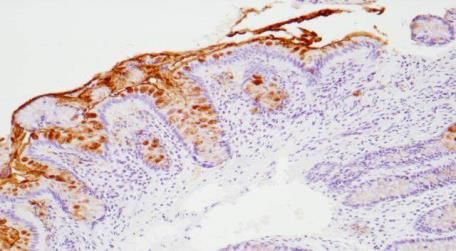
Microorganisms 2018, 6, 78                                                                    6 of 58

                              Table 3. Mucin Core and Backbone Repeat Glycan Structures.

                                   Core Type                             Structure

                                        1

                                                                      Galβ1-3GalNAc

                                        2

                                                               Galβ1-3(GlcNAcβ1-6)GalNAc

                                        3

                                                                    GlcNAcβ1-3GalNAc

                                        4

                                                             GlcNAcβ1-3(GlcNAcβ1-6)GalNAc
                                Backbone Repeat                        Structure

                                     Type 1

                                                                      Galβ1-3GlcNAc

                                     Type 2

                                                                      Galβ1-4GlcNAc

                             Poly N-acetyllactosamine
                                      type 2

                                                                  (Galβ1-4GlcNAcβ1-3-)n

                   Branched N-acetyllactosamine type 2

                                                                    Galβ1-4GlcNAcβ1-6
                                                                                   Galβ1
                                                                    Galβ1-4GlcNAcβ1-3
      The range of basic mucin glycan core and backbone structures are shown. The details of the
      abbreviations and symbols are indicated at the end of the paper.
Microorganisms 2018, 6, 78 7 of 58
The question that arises from these results is whether the developmental regulation of intestinal
O-glycans relates to the bacterial flora present. In the amnion and fetus, there is essentially no
bacterial presence and normal colonization initiates at birth. This suggests that there is a
programmed glycomic response to the introduction of bacteria to the gut and certain O-glycan
structures, in particular the Sda antigen, and their location in the gut are relevant to the development
and establishment of a stable and normal flora and an effective and dynamic mucus barrier.
At the early stages of life there is evidence that glycosylation plays an important role in
establishing the stability and protection of the GI tract. This is apparent at birth, during lactation,
through weaning, and the subsequent progression to adulthood. Much of the data derived for this
concept has come from the dietary profile of children from birth onwards. It has been reported that
the glycosylation of milk proteins varies during lactation, this has been shown for the major family
of milk glycoproteins, the caseins in both man and cow [99,100] and also for human milk lactoferrin
[101,102]. In keeping with this concept, the pattern of low molecular weight oligosaccharides present
in mothers-milk is known to change during lactation [103,104]. The oligosaccharides are thought to
play a role as prebiotics, as inhibitors of pathogenic or detrimental bacterial binding to the
developing gut mucosa and in order to promote colonization of beneficial stains and to establish a
normal flora [105–109].
With the completion of the lactation period and the change in the diet leading into weaning a
series of changes is initiated which subsequently results in the establishment of the adult pattern of
intestinal microbiota. This has been noted in human and animal studies [87,110–112]. The aging
process has a profound influence on the composition and homeostasis of the human microbiota and
also impacts on mucin glycosylation [113,114] and host immune system [115,116]. A reduction of the
salivary mucins MUC5B and MUC7 was found [113] and a reduction in the diversity of the
microbiota was observed [117,118]; a decrease in A. muciniphilia has also been reported [76]. In
contrast, a greater array of species was detected in another study [119].
The diet has been identified as a strategic factor maintaining the flora [120]. Many of the
diseases associated with advanced age also correlate with changes in the gut microbiota, mucus
expression, and glycosylation [121]. In elderly patients with Clostridium difficile, a lower microbial
diversity was found [122], while a wider variety of micobiota was found in aged IBD patients. H.
pylori infection was found to correlate with histological and serological changes in the elderly [123].
Specific probiotics have been adopted to stabilize and maintain the microbiota in older individuals
[124].
5. Mucin Glycosylation and the Sugar Code
5.1. Bulk Properties—Gel Formation and Viscoelasticity
Before considering the sequence of the mucin glycans it is necessary to address the primary
physical properties of the mucins in vivo. These are the characteristics that contribute to the barrier
function of the secreted mucus and are evident in the mucus layers found in the GI tract. The
secreted mucins form viscoelastic gels through generation of molecular networks. The gel forming
mucins display rheological properties through bulk mucus flow. They are both viscous and elastic,
fundamental properties due to covalent and reversible interactions, mediated by the concentration
of the gel forming mucins themselves, environmental salt concentration, and local pH [125]. Mucin
rheology should be regarded as a fundamental physiological property of mucins reflecting selective
molecular design throughout evolution [126–128]. Recently the biological importance of the GI
mucus barrier as a two-layer system, initially described by the Allen group [35,129], has been
demonstrated to comprise an inner, adherent gel on the surface of the mucosa, which is devoid of
enteric bacteria, and an outer, thicker layer, that is constantly being degraded and shed, but which
harbors a bacterial population [130–133].
The mucus barrier is dynamic. In order to maintain its primary functions in mucosal protection
it is continuously renewed at a rate sufficient to balance the normal destructive forces leading to the
constant erosion and loss of the outer layer.Microorganisms 2018, 6, 78 8 of 58
5.2. Mucin Glycans; Sequence, Topography and Mucosal Interactions
The glycosylation of mucins is a selective process and derives from the biological design to
yield a high molecular weight polymer than can be secreted and will form a gel or has a recognition
function and forms a part of a glycoarray at the surface of the in the glycocalyx. The formation of
viscoelastic, secreted polymers can be achieved without the range of glycan structures found in the
gel-forming secreted mucins. This suggests that the selection of mucin glycosylation is designed to
provide recognition information in addition to the physicochemical properties.
The carbohydrates are well suited, both chemically and physiologically, to generate a broad
variety of glycan structures that have sequential identity and therefore information [134,135]. Unlike
the nucleic acids and proteins, which have linear structures only, the glycans can form branched
structures in addition to linear chains. The basic building blocks the monosaccharides are epimers of
each other and exist as α- or β-glycosides. Thus the anomeric configuration, regiochemistry, and
stereochemistry of the glycosidic linkage are basic features of glycan chains [136]. Protein
glycosylation appears in number of well-known forms and which are outlined in Table 4.
Table 4. Protein Glycosylation Patterns.
Protein Carrier Glycan Structure
Glycoproteins
N-Glycans
Mannose 6-phosphate glycansMicroorganisms 2018, 6, 78 9 of 58
a3/6 b3 -a-Ser/Thr
a3/6 b4
b6
Glycoproteins -a-Ser/Thr
O-Glycans b3
a3/6
Mucin type O-Glycans
Linear sialylated core 3
Branched sialylated core 4
Glycoproteins
O-GlcNAcylation
Glycoproteins
C-Mannose
The main linkages of glycans to proteins are listed.
The mucins are well-known as proteins carrying a wide range of glycans of the “mucin type”.
This abundance of glycans takes the form of O-linked glycans attached to serine and threonine
(ser/thr) groups in the mucin polypeptide tandem repeat, PTS rich domains. Recent analysis has
implicated the link to either serine or threonine as a selective process with biological significance.
Comparison of serine-linked versus threonine-linked mucin O-glycans shows different properties in
their interaction with lectins, implying a potential for different functions based on the type of
O-glycan linkage. [137]. The linkage sugar is N-Acetyl-D-galactosamine (GalNAc) and, as noted
below, the transfer of this initial sugar is catalyzed by a family of
N-acetyl-D-Galactosaminyltransferases, which show specificity with regard to the mucin peptide
sequence, including the proximity of other ser/thr attachment sites and whether they are already
substituted by a GalNAc residue. The chemical and biochemical complexity of this glycosylation
step emphasizes the biological importance of this initial event and coordinates the mucin for its
physiological role at its site of biosynthesis and secretion [138–140].
Extension of the initial GalNAc generates a series of mucin core structures. Eight core structures
have been identified, of which only four show widespread abundance. These are shown in Table 3.
The structure of cores 2 and 4 demonstrates the potential for the formation of branched
structures, in contrast to the nucleic acids and proteins. The branching option expands the viable
range of O-glycan structures and correlates well with the extensive scope of glycans carried by
mucins. The core structures may remain as short oligosaccharides, but the majority are extended.
Larger glycan structures are achieved through the action of a range of well-established pathways as
shown in Figure 1.Microorganisms 2018, 6, 78 10 of 58
Figure 1. Biosynthetic Pathways leading to Mucin Core 1–4 Structures. Abbreviations and
monosaccharide symbols are given at the end of the paper.
The extension process enables the formation of larger and more branched glycans. Some of the
peripheral glycan structures are shown in Table 5. The scope for formation of these glycans in the
mucins includes transfer of L-fucose, N-acetylneuraminic acids (sialic acids), acetylation, sulphation,
and methylation [5,20,49,141–144].
N-glycosylation is also a significant feature of mucin glycosylation, but fewer N-glycan chains
are found compared with the O-glycans. They occur principally in the membrane-associated mucins,
but show discrete patterns in MUC1, MUC4, and MUC16. MUC1 contains N-glycans in both the PTS
and SEA domains, while in MUC4 these are only found in the EGF domain and MUC16 expresses
N-glycans in its PTS region [40].
The location of N-glycans on mucin peptide and other glycoproteins is determined by
recognition of a tripeptide sequence, asparagine-X-serine, where X is any other amino-acid, except
proline. Considerable structural variation of the N-glycans occurs in nature, and this range of
glycans is derived from three main core forms, as shown in Figure 1. The cores are extended to create
the series of N-glycans found in nature and accordingly in the mucins. Different numbers of
antennae are known, bisecting GlcNAc is also present in certain cases and an internal fucose,
attached to the GlcNAc linked to asparagine also occurs. Oligo-mannose forms, and complex forms
terminated with a sialyl-N-acetyl-lactosamine trisaccharide and hybrid forms are common (see
Figure 1). As with the O-glycans, noted above, the N-glycans possess a variety of different peripheral
substitutions, leading to the profusion of N-glycans that have been detected and reported in
glycoproteins. The N-glycans play important roles in mucin peptide processing, which occurs
during biosynthesis [6,145–147].
An unusual type of glycosylation involving a single alpha-mannose unit attached through a C–
C (carbon–carbon) linkage to peptide tryptophan residues located in mucin peptide WXXW motifs
has been reported [148]. This novel form of glycosylation has been identified in the CysD domains of
the secreted mucins, MUC2 (2 units), MUC5AC (9 units), and MUC5B (7 units). It has been proposed
that these units function in protein folding, subcellular localization and trafficking [149,150].
C-mannosylation in MUC2, MUC5AC, and MUC5B is required for maturation and secretion.
Deficient C-mannosylation of mucins results in their inability to exit the Endoplasmic Reticulum
(ER) and leads to ER stress [43].Microorganisms 2018, 6, 78 11 of 58
An important feature of glycosylation is its tissue and cell specificity. As stated above there are
many glycan structures associated with mucins and it is clear that that same mucins are expressed in
different organs, tissues, and cells. A good example of this is MUC5AC, which is expressed in the
respiratory tract [151], stomach [152,153], gallbladder [154], conjunctiva and tear film [155,156],
middle ear [157,158], prostate [159], and the female reproductive tract [160,161].
The need to provide optimal protection at different mucosal surfaces imposes a design and
synthetic requirement for mucin glycosylation. The defensive processes necessary will depend on
the mucosal surface in question and this fits well with the opportunity to biosynthesize mucin
glycan sequences, which are adapted to the needs of each mucosal surface. It is known that the
glycobiome, which has the ability to glycosylate individual proteins to yield distinct and discrete
glycoforms, will have ideal function at their site of synthesis [5]. A good example is the glycosylation
of MUC2 in the human GI tract. Regional patterns of MUC2 glycosylation occur from the small
intestine through to the rectum, largely through sialylation and glycosulphation [162,163]. These
patterns were constant when examined in more than 50 normal individuals [164] and in patients
with ulcerative colitis, where aberrant mucin glycosylation is associated with the disease, recovery is
accompanied with a return to the normal healthy glycosylation profile. In addition to the GI tract,
characteristic mucin glycosylation profiles have been found in the oral cavity [165], the pancreas
[166], the ocular surface and conjunctiva [155,167], the respiratory tract [168], human sperm [169],
and the female reproductive tract [170].
It is clear that the variety of glycans found in mucins is a molecular design feature adopted and
optimized throughout evolution. The glycocode is therefore well suited to the biological
requirements of mucins as mucosal barrier components displaying dynamic, sequence based
information.
As well as the wide ranging patterns of glycosylation found in mammals and especially in man,
there are a number of human features which indicate that the sugar code is an integral part of our
normal existence giving us unique labels at an individual level and establishing molecular
recognition which govern interactions with our environments. The human blood group system is
well known to rely on glycan sequences for its recognition [171–173]. The human blood groups
found on proteins include the ABO(H) antigens, the Lewis antigens [174], the Sda antigen [175,176],
and the i and I blood groups [177]. Much of the immunochemistry was established through the work
of Karl Landsteiner [178], Elvin Kabat [179], Walter Morgan, and Winifred Watkins [172]. The
development and conservation of the human blood group system has been confirmed through
evolutionary study [180] and serves to emphasize the biological relevance and magnitude of this
recognition system. These structures are carried on glycan chains of type 1 (Galβ1-3GlcNAc),
common in O-glycans, or type 2 (Galβ1-4GlcNAc), mostly found in N-glycans and type 3 (β1-3
GalNAc-αser/thr-) associated with mucins. Some of the key glycan structures are shown in Table 5.Microorganisms 2018, 6, 78 12 of 58
Table 5. Key Glycan Structures found in Mucins.
Type of Glycan Structure
Blood group H
type 1
Fucα1-2Galβ1-3GlcNAcβ1-
(Galβ1-3GlcNAc)
Blood group A
type 2
GalNAcα1-3Galβ1-4GlcNAcβ1-
(Galβ1-4GlcNAc) | α1-2
Fuc
Blood group B
type 1
Galα1-3Galβ1-3GlcNAcβ1-
(Galβ1-3GlcNAc) | α1-2
Fuc
Galα1-3GlcNAcβ1-
| α1-4
Lewisa Fuc
and
Lewisx
Galβ1-4GlcNAcβ1-
| α1-3
Fuc
Lewisb
Galβ1-3GlcNAcβ1-
| α1-2 | α1-4
Fuc FucMicroorganisms 2018, 6, 78 13 of 58
Lewisy
Galβ1-4GlcNAcβ1-
| α1-2 | α1-3
Fuc Fuc
Neu5Acα2-3Galβ1-3GlcNAcβ1-
Sialyl Lewis a | α1-4
and Fuc
Sialyl Lewisx
Neu5Acα2-3Galβ1-4GlcNAcβ1-
| α1-3
Fuc
Sialyl-Tn
Neu5Acα2-6GalNAc-α-O-Ser/Thr
Neu5Acα2-3Galβ1-3GalNAc-α-O-Ser/Thr
Monosialylated-T-antigen
Neu5Ac
| α2-6
Galβ1-3GalNAc-α-O-Ser/Thr
Monosialylated core 3Microorganisms 2018, 6, 78 14 of 58
Neu5Ac
| α2-6
Galβ1-4GlcNAcβ1-3 GalNAc
Neu5Ac
| α2-3
GalNAcβ1-4Galβ1-3GlcNAcαβ1-3GalNAc
Sda antigen
Type 1 & 2 chains
Neu5Ac
| α2-3
GalNAcβ1-4Galβ1-4GlcNAcαβ1-3GalNAc-
Some examples of important glycan structures commonly found in mucin glycans are shown.
The blood group antigens are essentially expressed on the surface membranes of erythrocytes
red cells. However, the expression of the same structures on mucosal glycoproteins is regulated
through the glycosyltransferase FUT2, also known as the secretor gene. Individuals who have this
fucosyltransferase transfer fucose to glycoproteins to give a α1-2 linkage [181,182]. These individuals
are able to express blood group antigens on mucosal surfaces and are termed secretors. In contrast,
those who have no FUT2 gene do not show the blood group antigens on their cellular glycoproteins
and are known as non-secretors [181,183]. In addition to the molecular identification specified by the
FUT2 gene it is also directly related to disease and is known to mediate infection and susceptibility
[183,184]. The ABO blood group antigens expressed on erythrocytes (red cells) have been shown to
modulate the pattern and arrangement of sialylated glycans on the erythrocyte surface [185]. A
further feature of blood group activity and secretor status has been demonstrated for salivary
MUC5B. The non-secretors had a higher sialylated form of MUC5B, with increased sialyl-Lewisa,
compared with the secretors [186]. Thus demonstrating that mucin glycosylation depends on both
blood group and secretor status.
The Sda antigen is commonly found in the normal colon [175] and its formation is regulated by
the addition of β1-4GalNAc by the B4GALNT2 glycosyltransferase [187,188]. This contrasts with the
Lewis and sialyl-Lewis antigens, which are normally only found at low levels. The biosynthetic
pathway leading to the Sda antigen includes the intermediate structure sialyl-N-acetyllactosamine
(Neu5Acα2-3Galβ1-3/4GlcNAcβ1-) and this represents an important branch point in the pathway as
it may be converted to the Sda antigen, sialyl-Lewisa, or sialyl-Lewisx, as shown in Figure 2.Microorganisms 2018, 6, 78 15 of 58
BIOSYNTHETIC ROUTES TO Sda ANTIGEN
1-4 2-3 1-4
1- R 1- R 1- R
2-3 1-3 2-3 1-3 1-4 1-3
Neu5Ac -3(GalNAc 1-4)Gal 1-3GlcNAc-R Neu5Ac -3Gal 1-3(Fuc1-4)GlcNAc-R Neu5Ac -3Gal 1-4(Fuc1-3)GlcNAc-R
a a x
Sd antigen Sialyl-Lewis Sialyl-Lewis
ST3Gal4
B4GNT2 FUT3 ST3Gal6
FUT3,FUT11
2-3 1-3/4
1- R
Neu5Ac 2-3Gal 1-3/4GlcNAc-R
Sialyl N-acetyllactosamine
Normal O-glycosylation pathways from core 1
1-3
ser/thr
Figure 2. Biosynthetic Routes to the Sda antigen. The sequential steps leading to the Sda antigen from
core 1, via sialyl-N-acetyllactosamineare shown. The individual glycosyltransferases for each step are
indicated. The red arrow indicates the major pathway, while the blue arrows indicate competing
steps to the sialyl-Lewisa and sialyl-Lewisx antigens. Abbreviations and monosaccharide symbols are
given at the end of the paper.
As demonstrated by chemical analysis on normal colonic mucus Sd a antigen is the major
structure found [162,163]. Unpublished data from our laboratory has demonstrated that the normal
colonic mucus Sda antigen sialic acids are O-acetylated. Figure 3 shows the Sda antigen, detected
with the KM694 antibody is sensitive to saponification with mild alkali [189–191] and adds a further
regulatory asset to the antigen as the O-acetylated sialic acid is resistant to sialidase action.Microorganisms 2018, 6, 78 16 of 58
HISTOLOGICAL DETECTION OF O-ACETYLATED
SIALIC ACIDS AND Sda ANTIGEN
a) mPAS, direct, x 400
b) mPAS with saponification x 100
c) Sda antigen, direct x 100
d) Sda antigen, saponification x 100.
Figure 3. Histological Detection of O-acetylated Sialic acids and Sda antigen. The O-acetylated sialic
acids detected by the mPAS stain, directly (a) and with saponification, (b) this shows a longitudinal
section of the mucosa, in contrast to a, c, and d. Also note the difference in magnification. Direct
staining for the Sda antigen with the KM694 antibody (c), and with saponification (d) is shown.
O-Acetylation of sialic acids is well known to be a major modification in human colonic mucus.
The demonstration of individual glycoproteins as carriers of O-acetylated sialic acids has not been
widely studied. The human colonic mucins are a major carrier of O-acetylated sialic acids. The Sda
antigen is one of many sialylated glycans carried by the mucins and is a focus of attention in this
review.
It is also known that a small proportion of the general population do not express O-acetyl sialic
acids and are known as sialic acid non-O-acetylators. These individuals can be detected using the
mPAS (mild periodic acid/Schiff) stain with and without prior saponification. The biological
adaptation to the absence of these sialic acids has not been examined. There is no indication whether
they are more susceptible to gastrointestinal disease, or whether a natural adaptation occurs, as in
the case of blood group secretors and non-secretors [186], with a corresponding glycobiological
modification.Microorganisms 2018, 6, 78 17 of 58
6. Mucin Glycans as Biological Arrays Linked to Function
The mucins represent the presentation of an array of glycan structures at whichever site they
are expressed, as noted above. At many mucosal surfaces this mucin glycoarray interacts with the
bacterial flora present under normal conditions. Much recent work in this area has identified
microbiota which interact with different mucosal surfaces and which are adapted to each specific
mucosa. The human gut has been widely examined [12,192–198].
The mucins are designed to provide defense at mucosal surfaces in many different ways and
this reflects the adaptability of these molecules for this function. The basic organization of mucin
protein domain composition and their glycosylation allows adaptation to the demands posed at each
mucosal surface. As the production of the mucins is dynamic it is ideally adapted to respond to
developmental and environmental changes that are expected. In the GI tract this is apparent at birth,
during lactation, weaning, and in adulthood.
As mentioned above the mucosal barrier in the GI tract shows a mucus gel layer of differing
thickness, depending on the location in the tract. The stomach and colon have a gel layer of about
700 µm, while the small intestinal thickness ranger between 150 and 300 µm [6,35,36]. The colonic
barrier consists of two secreted mucus layers; these are essentially composed of MUC2. A major
feature of these two layers is the distribution of bacterial populations. The inner, adherent secreted
mucus is free of microbes, while the outer layer is colonized by the enteric gut bacterial flora. The
sophistication of this system is apparent with the identification of different types of Goblet cells,
which synthesize and secrete the mucus along the crypt in the human colon. Indeed a “sentinel”
Goblet cell has been identified, positioned at the top of each colonic crypt. Endocytosis of TLR
generates MUC2 secretion, together with an intercellular gap junction signal, which induces MUC2
secretion in adjacent Goblet cells and thus regulates the entry of bacteria into the crypt [28,32].
In contrast to the continuous, two-layer system, a recent report has presented data showing that
the luminal contents of the distal colon have an influence on the location of mucus [37]. The report
shows that mucus covers the feces, but not the distal colonic epithelium. As a result it confines the
enteric microbiota to the surface of the feces and prevents it remaining in the vacant distal colon.
Further work is required to confirm or refute this observation and it underlines the importance of
regular review and interpretation of existing data.
The apical glycocalyx is ubiquitous to all cell types and is essential for normal cell interaction
with neighboring cells and the external environment. It provides a platform for communication and
links with signaling pathways within the cells. In common with the secreted mucins it has a
characteristic composition at each mucosal surface. The membrane-associated monomeric mucins
form a significant proportion of the molecular makeup of the glycocalyx and accordingly create a
cell surface anchored glycoarray. Typically MUC1, MUC4, MUC12, MUC16, and MUC20 are found,
with MUC1 present in most mucosal surface membranes [5,6,40,45,46,199–202].
7. Screening for Mucin Glycans and Mucin Glycan Engineering
The progress made in understanding mucin structure, organization, synthesis and degradation
relied on improvements in technology. In addition, access to glycomic based databases has provided
a reliable and constantly growing source of information for structural and functional aspects [203].
The two most widely used databases are CaZy, the Carbohydrate-Active enZYmes (CaZy) Database
(http://www.cazy.org), and The Consortium for Functional Glycomics
(http://functionalglycomics.org). A consequence of the increased biological interest in glycans has
been a focus on the chemistry–glycobiology frontier and the need to understand chemical and
physical aspects of all glycans [204].
The detection, isolation, and characterization of glycans has been improved through the
production of reagents together with chemical, biophysical and biochemical methodology [205]. The
techniques best suited to and most widely used in glycan isolation, detection, and assessment are
Affinity Chromatography, which employs an immobilized binding protein on a suitable support
such as Affi-Gel or Sepharose [206–208]. This can be used simply to bind the target glycan and
separate it from all other compounds in a tissue or cell preparation. It can also be used to calculateMicroorganisms 2018, 6, 78 18 of 58
the strength of binding and generate a kd value when compared with a glycan not bound by the
protein. The strength of binding can also be calculated using isothermal titration calorimetry
[209,210]. The change in enthalpy is measured, in a microcalorimeter, for varying concentrations of
the glycan at constant glycan binding protein concentration, and used to calculate the k d value.
Surface plasmon resonance has also been widely adopted to follow the kinetics of reaction and relies
on the reflection of polarized light as the glycan is allowed to flow over the immobilized glycan
binding protein [211,212]. Again values for the kd of the reaction can be obtained. Fluorescence
polarization techniques also allow measurement of kd values. Characterization of non-covalent
interactions between glycans and specific proteins can be measured using mass spectrometric and
NMR methods.
The profusion of techniques existing for detection of glycans has led to the design of strategies
for analysis of glycosylation patterns. The classical methodology for optimal glycan structural
analysis is mass spectroscopy or NMR if sufficient probe is available, usually after HPLC separation
of released glycans [213–218].
The recognition of specific glycan sequences can be monitored using proteins that bind to such
glycans. Many of these have been used to probe for the presence and cellular and subcellular
location of glycan motifs in tissues and cell lines, which express mucins.
The majority of these proteins are lectins or adhesins, isolated from microbial, plant and animal
sources, readily available commercially and used widely as standard reagents [219–226]. There is a
large literature on this topic and it is not the main focus for this review, however a brief overview
with a small selection of references serves to indicate the important links in relation to mucin
glycosylation and its biological recognition. An overview of current knowledge can be found in
Essentials of Glycobiology, Third Edition [227].
R-type lectins are a superfamily of proteins, which contain a carbohydrate binding module
(CBM, see below), and which bind to β-galactose or N-acetylgalactosamine. This is a large family
and includes the GalNAc transferases involved in mucin synthesis, mannose receptors, bacterial
lectins, invertebrate lectins, bacterial hydrolases, plant toxins, and Drosophila lectin [228].
L-type lectins are derived from leguminous plants, with glycan binding proteins from other
eukaryotic organisms; they bind to a range of different glycans. Concanavalin A binds to glucose
and mannose, while Sambucus nigra and Maackia amurensis lectins show affinity for sialylated
oligosaccharides [229].
The P-type lectins recognize mannose-6-phosphate (M6P) carried on N-glycans. Glycoproteins
that carry the M6P motif are generated through a series of steps and are delivered to the lysosomes.
M6P acts as a translocation signal for lysosomal proteins.
C-type lectins are the largest and most diverse family. They are calcium dependent, with
homology in their CBMs and include the collectins, selectins, endocytic receptors, and proteoglycans
and may be either secreted or membrane bound. Fundamental conserved determinants implicated
in glycan binding are the EPN motif promoting Man, Glc, Fuc, and GlcNAc recognition and the
WND motif for Gal and GalNAc [230].
I-type lectins have binding domains which have homology with the immunoglobulin
superfamily. They include the selectin family, which bind α2-3, α2-6, and α2-8 linked sialic acids.
The specificity varies between the selectins and also includes Neu5Ac or Neu5Gc identification and
O-acetylation patterns [231,232].
The galectins are typical β-galactose binding proteins found in vertebrate and invertebrate
forms and sharing CBM homology. They exist as three major groups, (1) prototypical having only
one CBM and which bind as homodimers, (2) chimera type, a single CBM with an attached proline
rich peptide, and (3) tandem repeat which have two CBMs linked by a peptide [233,234].
Certain viral strains have also been used to screen for sialic acids and their O-acetylated forms
[235–238]. Viral proteins that show hemagglutinin binding properties and those that have specific
esterase activity for 4-O-acetylated sialic acids have been reported [238]. Recently a series of
virolectins from nidovirus strains have been isolated and used to probe for O-acetylated sialic acids
[239]. Dual function hemagglutinin-esterase envelope proteins were found to show very selective,Microorganisms 2018, 6, 78 19 of 58
differential binding patterns when used in soluble form. Discrimination between 4-O-Ac, 9-O-Ac,
7,9-diOAc, and 4,9-diOAc was possible and differential expression was revealed in human and
mouse tissue arrays. This shows a pattern of sialic acid O-acetylation, which is programmed, exists
at an organ, tissue, and cellular level and implicates O-acetylated sialic acids in cell development,
homeostasis, and other functions [239]. This aspect of glycan structure and metabolism has relevance
for the colonic mucins and in particular the Sda antigen.
A family of mucus binding proteins (MUBs) have been characterized in lactic acid bacteria,
which are cell surface anchored effector molecules containing multiple mub domains. The precise
pattern of glycan binding has not yet been resolved [225,240].
Carbohydrate binding modules (CBMs) are non-enzymatic domains found in many proteins
that attach to glycan sequences in polysaccharides and glycoconjugates [241–243]. Over 69 families
have been identified, indicating a wide range of glycan sequence recognition
The design and use of array technology has offered a powerful method to examine the presence
and function of glycan structures and this includes options to search for mucin related glycan
epitopes, a few examples from a large literature are given as follows; [244–253]. Of particular interest
are those arrays that correlate mucin glycan epitopes with bacterial binding [254–259]. Although a
considerable range of glycans can be displayed and screened using this technique the conformation
of the glycans on the surface of the chips remains a problem. Attachment of individual glycans can
be achieved using a number of different methods and on different chip surfaces, see previous papers
[244,245,247,249,252,253,255,260–265], however, this does not necessarily achieve the molecular
conformation found in vivo when attached to proteins. Some improvements have been made using
known glycans attached to peptides, where the normal in vivo conformation is more likely to be
preserved [266–268]. A further problem is the density of attachment, which may not mirror the in
vivo situation. Single glycan attachment, or clustered glycan attachment must conform with the
biological arrangement in order to yield binding results that have genuine in vivo relevance [269].
Attempts have been made to address clustering, which is a feature of mucin O-glycans in the tandem
repeat PTS domains of the mucins [250,270], but an array of O-glycans as found in mucins remains
difficult to mimic. In spite of these problems, valuable information has been gleaned from glycan
array screening.
As mucins represent a primary target for bacteria in the GI tract and other mucosal surfaces the
production of a mucin microarray has been adopted for rapid throughput screening purposes
[170,258,271,272]. Preparation of such mucin arrays relies on the prior purification of mucins from
appropriate sources. As noted earlier, the preparation of mucins is demanding due to their high
molecular weight and separation from other contaminant proteins, glycoproteins, and glycolipids.
The available sources are also limited as many normal human mucosal tissues or their secretions
cannot be obtained for ethical reasons and disease tissue will deliver abnormal mucin products. The
use of cell culture is also dependent on the nature of the mucins produced by the cells. Most cell lines
that produce and secrete mucins are cancer derived and as a result yield products that are also
influenced by mutations and other cancer related changes including glycosylation. Finally, the
attachment of mucins to the microarray plates will result in multiple attachment sites [258] and the
conformation of the attached mucin is unlikely to mimic the in vivo situation, although no imaging
studies have been reported. Atomic Force Microscopy (AFM) has provided images of purified
mucins [273–275], but these also do not provide an ideal match for the in vivo mucins at mucosal
surfaces. Force microscopy has been used for screening glycans structures. A range of different
microscopic techniques have evolved and used to monitor glycans in various molecules including
the mucins. This is an area where microscope design has driven the sensitivity and resolution of
molecular imaging as well as yielding values for binding affinities [273–280].
A general appreciation of the biological significance of glycomics and the applications of
glycoproteomics has grown in recent years [281]. This has led to increased awareness of glycan
structure as a biological phenomenon requiring thorough assessment for all glycoconjugates, and
glycoproteins and mucins in particular. It has opened the way for the involvement of syntheticMicroorganisms 2018, 6, 78 20 of 58
chemical approaches to the strategic design of biological molecules with therapeutic application.
This is not further detailed in this review.
Two recently developed technologies are worth mentioning at this point and although there is
currently only limited application to glycobiology it is certain that they will attract attention in the
immediate future. Firstly, the CRISPR-Cas9 genome editing methodology [282,283] has been used to
the cell-specific delivery of the asialoglycoprotein receptor to hepatic cells [284]. The binding of the
receptor to the cell surface, uptake through endocytosis, endosomal escape through endosome
acidification and subsequent nuclear import has been achieved [284] and illustrates the power of this
technology for application to mucosal surfaces. Secondly, the process of 3D bioprinting is being used
in a variety of situations [285–291] and is an obvious target for mucosal surface bioengineering
strategies. Significant interest in the pharmaceutical industry and development for high throughput
screening bodes well for expansion of this technology in glycomics.
The wealth of glycomics information generated also prompted the development of methods to
store and access the data. Glycoinformatics for processing and accessing the glycomics data has been
reported [292–294].
8. Metabolism of Mucin Glycans
The metabolism of mucin glycans encompasses synthesis, degradation, and recycling. The
synthesis of mucin O-glycans can be mapped to well-defined pathways in the ER and Golgi
compartments of the cell, where the glycosyltransferases add the monosaccharides, one by one, to
the growing O-glycan chain attached to the mucin peptide serine and threonine residues. The
specificity of the glycosyltransferases governs the nature of the glycan chains synthesized and the
complement of glycosyltransferases present in each cell determines the O-glycan core structures,
backbone extensions and peripheral sialylation, fucosylation, and sulphation patterns. The absence
of individual glycosyltransferases results in glycan structures, which may be shorter, less extended,
or showing variations in sialylation, fucosylation, and sulphation. These events are dictated at the
genetic level and form the basis for the type of O-glycans synthesized in any one cell [143,295,296].
The glycosyltransferases require an activated form of each monosaccharide to be transferred in
addition to the growing O-glycan acceptor. Each monosaccharide exists as a nucleotide-sugar, and
these donor molecules (See Table 6) are formed through standard pathways [297]. Active sulphate is
also a substrate utilized in these pathways, while in the sialic acids, O-acetylation is mediated
through acetyl-CoA transfer and O-methylation through S-adenosylmethionine and a
methyltransferase.
Table 6. Nucleotide Forms for Transfer to Mucins.
Nucleotide Nucleotide
Transfer
Nucleotide Transport Transport Comment
Moiety
ER Golgi
Golgi location. Also
Neu5Ac CMP-Neu5Ac − +
transfers Neu5Gc
Fuc GDP-Fuc + + ER and Golgi location
Gal UDP-Gal − + Only in Golgi
Man GDP-Man − + Only in Golgi
GlcNAc UDP-GlcNAc + + ER and Golgi location
GalNAc UDP-GalNAc + + ER and Golgi location
Sulphate PAPS * − + Only in Golgi
Acetate Acetyl-CoA + + ER and Golgi location
Acyl Acyl-CoA ? ? Not known
Methyl S-adenosyl-methionine ? ? Not known
Phosphate ATP + + ER and Golgi location
The Table shows the nucleotide monosaccharide forms active as substrates for the
glycosyltransferases. In addition the donors for transfer of sulfate, acetate, acyl, methyl, and
phosphate groups found in the glycosylation pathways mucin is listed. The location of the nucleotideMicroorganisms 2018, 6, 78 21 of 58
transfer is indicated where known. Detail of the monosaccharide metabolic pathways is shown in
Figure 4. * PAPS, 3′-phosphoadenosine-5′-phosphosulphate.
The pathways leading to the nucleotide sugars derive from the hexose monophosphate pool,
Glc-6P, and Fruc-6P. The hexosamine pathway is initiated by the amination of Fruc-6P with
glutamine by glutamine:fructose amidotransferase (GFAT), which is feedback inhibited by
UDP-GlcNAc, the end product of the pathway. UDP-GlcNAc is then further metabolized on the
sialic acid pathway through two key enzymes, UDPGlcNAc 2-epimerase and ManNAc kinase,
which act together in a bifunctional complex and lead to the formation of ManNAc-6P from
UDP-GlcNAc [298–300]. This enzyme is feedback inhibited by the end product of this pathway,
CMP-Neu5Ac [298]. UDP-GlcNAc may also be converted to UDP-GalNAc through the action of
UDP-GlcNAc 4-epimerase and both of these nucleotide sugars are substrates for glycosyltransfer.
The kinase generating GlcNAc-6P is subject to feedback inhibition by UDP-GlcNAc [301]. Free
ManNAc enters the pathways after its conversion to GlcNAc by a specific GlcNAc 2-epimerase.
Recycling or salvage pathways for monosaccharides ensure that optimal use is made of the
monosaccharides generated during glycan degradation. The enzymes involved in these steps
process monosaccharide intermediates and feed back into the main stream of metabolic pathways
generating the nucleotide sugars. In this way D-GlcN, D-GalN, and D-ManN are re-N-acetylated to
generate GlcNAc, GalNAc, and ManNAc. These N-acetylhexosamines are subsequently
phosphorylated at the 1 position for GlcNAc and GalNAc, or at the 6 position for GlcNAc, GalNAc,
and ManNAc. The phosphorylated sugars are part of the pathways leading to UDP-GlcNAc,
UDP-GalNAc, and CMP-Neu5Ac. Free sialic acid is cleaved to ManNAc and pyruvate by the action
of acylneuraminate pyruvate lyase [302–304], while ManNAc is recycled after epimerization to
GlcNAc, or phosphorylation to ManNAc-6P. The control of these pathways is well integrated by end
product feedback inhibition as noted above and shown in Figure 4.
Figure 4. Feedback Inhibition on Glycan Activation Pathways.
The Figure shows the known biosynthetic pathways relating to the formation and
recycling/salvage of monosaccharides found in glycans. The nucleotide sugars are the end products
of each pathway and are shown in red e.g., UDP-Glc. The individual reactions, which are subject to
feedback inhibition, are shown with a red arrow, →. The individual monosaccharides found in
glycan structures, and which are activated to the nucleotide sugars through the metabolic pathways,
are shown in blue e.g., Glc. The black text and black arrows show the intermediate monosaccharides
on the pathways and their conversion steps in the pathways. Abbreviations are as listed at the end of
the paper.
UDP-GlcNAc is a crucial intermediate. It serves directly as a substrate for glycosyltransferases
or it may be epimerized at the 2-position to generate N-acetyl-D-mannosamine, with loss of the UDP
group, to enter the sialic acid pathway. It may also be 4-epimerised to yield UDP-GalNAc, anotherYou can also read