TNF-α and IFN-γ Participate in Improving the Immunoregulatory Capacity of Mesenchymal Stem/Stromal Cells: Importance of Cell-Cell Contact and ...
←
→
Page content transcription
If your browser does not render page correctly, please read the page content below
International Journal of
               Molecular Sciences

Review
TNF-α and IFN-γ Participate in Improving the Immunoregulatory
Capacity of Mesenchymal Stem/Stromal Cells: Importance of
Cell–Cell Contact and Extracellular Vesicles
Lucero López-García and Marta E. Castro-Manrreza *

                                            Immunology and Stem Cells Laboratory, Multidisciplinary Unit of Experimental Research Zaragoza,
                                            FES Zaragoza, National Autonomous University of Mexico, Mexico City 09230, Mexico; lucerop624@gmail.com
                                            * Correspondence: elmar_ca@yahoo.com.mx; Tel.: +52-55-2645-3554

                                            Abstract: Mesenchymal stem/stromal cells (MSCs) have an immunoregulatory capacity and have
                                            been used in different clinical protocols requiring control of the immune response. However, variable
                                            results have been obtained, mainly due to the effect of the microenvironment on the induction,
                                            increase, and maintenance of MSC immunoregulatory mechanisms. In addition, the importance of
                                            cell–cell contact for MSCs to efficiently modulate the immune response has recently been highlighted.
                                            Because these interactions would be difficult to achieve in the physiological context, the release of
                                            extracellular vesicles (EVs) and their participation as intermediaries of communication between MSCs
                                            and immune cells becomes relevant. Therefore, this article focuses on analyzing immunoregulatory
                                            mechanisms mediated by cell contact, highlighting the importance of intercellular adhesion molecule-

                                     1 (ICAM-1) and the participation of EVs. Moreover, the effects of tumor necrosis factor-alpha (TNF-α)
                                            and interferon-gamma (IFN-γ), the main cytokines involved in MSC activation, are examined. These
Citation: López-García, L.;
                                            cytokines, when used at the appropriate concentrations and times, would promote increases in the
Castro-Manrreza, M.E. TNF-α and
                                            expression of immunoregulatory molecules in the cell and allow the acquisition of EVs enriched with
IFN-γ Participate in Improving the
Immunoregulatory Capacity of
                                            these molecules. The establishment of certain in vitro activation guidelines will facilitate the design of
Mesenchymal Stem/Stromal Cells:             conditioning protocols to obtain functional MSCs or EVs in different pathophysiological conditions.
Importance of Cell–Cell Contact and
Extracellular Vesicles. Int. J. Mol. Sci.   Keywords: mesenchymal stem/stromal cells; immunoregulation; cell–cell contact; extracellular
2021, 22, 9531. https://doi.org/            vesicles
10.3390/ijms22179531

Academic Editor: Maurizio Battino
                                            1. Mesenchymal Stem/Stromal Cells (MSCs)
Received: 16 July 2021                            MSCs are multipotent stromal cells with the capacity for self-renewal and differen-
Accepted: 26 August 2021                    tiation. They were originally identified in bone marrow (BM) [1,2] and, currently, cells
Published: 2 September 2021
                                            with similar characteristics can be obtained from different tissues [3–5]. Three biological
                                            properties have been identified in BM-MSCs: (a) differentiation potential, (b) secretion of
Publisher’s Note: MDPI stays neutral
                                            trophic factors, and (c) immunoregulatory capacity. These properties have led to their use
with regard to jurisdictional claims in
                                            in different experimental cell therapy protocols designed for the treatment of inflammatory
published maps and institutional affil-
                                            and autoimmune diseases, tissue repair, and regeneration, and to favor the acceptance of
iations.
                                            transplanted cells, tissues, and organs [3,5,6]. The first clinical trials performed with MSCs
                                            bet on their differentiation potential. Currently, it is accepted that the benefit observed in
                                            cell therapy protocols is due to the interaction of the three biological properties mentioned
                                            above, mainly the association between the secretion of trophic factors and the regulation of
Copyright: © 2021 by the authors.           the immune response [3]. In particular, this last property is relevant because the regulation
Licensee MDPI, Basel, Switzerland.
                                            of inflammatory processes is essential for tissue regeneration and repair to occur.
This article is an open access article
distributed under the terms and             2. Immunoregulatory Properties of BM-MSCs
conditions of the Creative Commons
                                                MSCs generate an anti-inflammatory environment by modulating the function of
Attribution (CC BY) license (https://
                                            immune cells, such as neutrophils, natural killer (NK) cells, monocytes, macrophages,
creativecommons.org/licenses/by/
4.0/).
                                            dendritic cells (DCs), and T and B lymphocytes [7–13]. It is currently proposed that MSCs

Int. J. Mol. Sci. 2021, 22, 9531. https://doi.org/10.3390/ijms22179531                                          https://www.mdpi.com/journal/ijms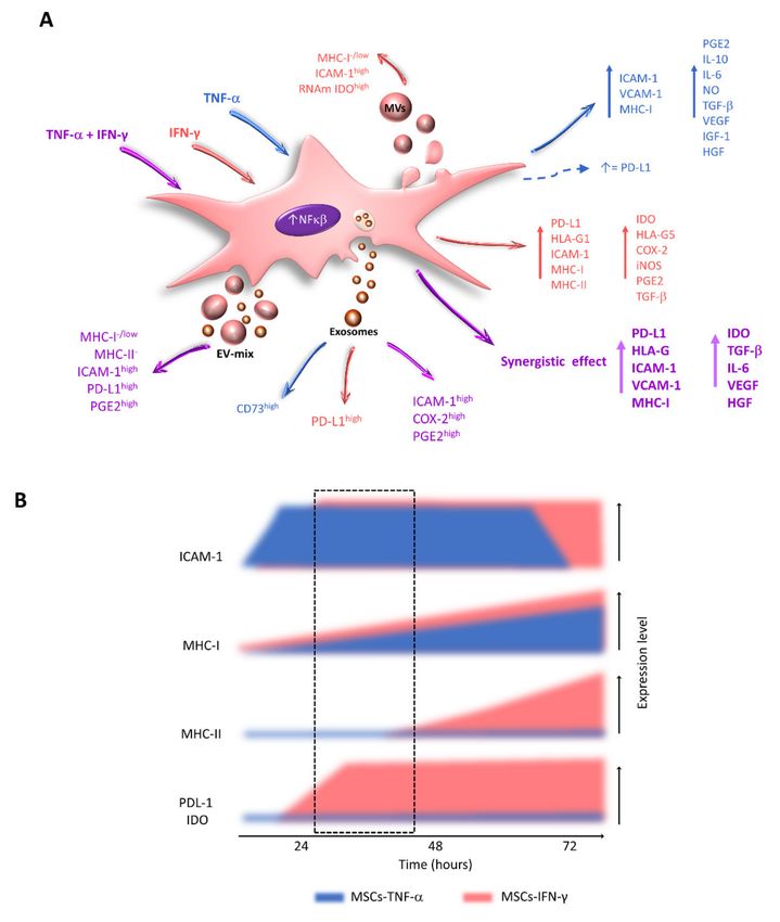
Int. J. Mol. Sci. 2021, 22, 9531                                                                                             2 of 31

                                   modulate the activation, differentiation, and effector function of immune cells by secreting
                                   factors through cell–cell contact and extracellular vesicles (Figure 1).

      Figure 1. Immunoregulatory mechanisms of activated MSCs. MSCs are generally in a resting state with low or no expression
      of the molecules involved in their immunoregulatory function. However, when exposed to proinflammatory cytokines
      such as TNF-α, IFN-γ, IL-1, and IL-17, these cells become activated. This event increases or induces the expression of
      immunoregulatory molecules in MSCs, which can be secreted or remain attached to the cell membrane. In addition, resting
      and activated MSCs release extracellular vesicles (exosomes and microvesicles), which are capable of traveling through
      body fluids and reaching distant sites, where they establish contact with immune cells.

                                        In vitro, preclinical, and clinical studies have documented the importance of an in-
                                   flammatory environment in the biological properties of MSCs. These cells are generally
                                   resting, with low or no expression of the molecules involved in their immunoregulatory
                                   function [7]. However, when MSCs are exposed to an inflammatory environment, the
                                   expression of these molecules is increased or induced. Therefore, some researchers have
                                   suggested that MSCs must be “activated” for efficient immunoregulation. The activation
                                   of MSCs is induced by inflammatory cytokines such as tumor necrosis factor-alpha (TNF-
                                   α), interferon-gamma (IFN-γ), interleukin (IL)-1, and IL-17 [8–14]. Because TNF-α and
                                   IFN-γ are the first cytokines secreted in an inflammatory response, it is relevant to analyze
                                   their effects on the expression of molecules involved in the different immunoregulatory
                                   mechanisms used by MSCs (Figure 1).

                                   3. Immunoregulation Mediated by Secreted Factors
                                       Immunoregulatory mechanisms mediated by secreted factors have likely been the
                                   most extensively analyzed to date, and they have been the subject of numerous re-
                                   views [5,15]. Therefore, only a general overview of these mechanisms is provided here.
                                   The molecules involved in these processes include prostaglandin E2 (PGE2), transform-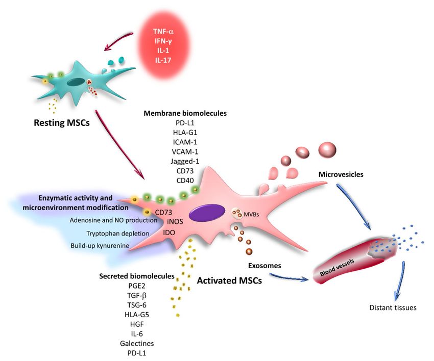
Int. J. Mol. Sci. 2021, 22, 9531                                                                                        3 of 31

                                   ing growth factor-beta (TGF-β), tumor necrosis factor-stimulated gene-6 (TSG-6), hep-
                                   atocyte growth factor (HGF), human leukocyte antigen-G5 (HLA-G5), IL-6, IL-10, and
                                   galectins [5,6,16–18]. These mechanisms also include the effects on the microenvironment
                                   induced by the intracellular enzymes indoleamine-2,3-dioxygenase (IDO) and inducible
                                   nitric oxide synthase (iNOS) [10,19,20], as well as the production of adenosine by the
                                   ectonucleotidase CD73 [21] (Figure 1).
                                         MSCs decrease NK cell proliferation, cytotoxicity, and the expression of cytokines
                                   through IDO, PGE2, TGF-β, and HLA-G5 [22–25]. In addition, it has been recently proposed
                                   that the secretome of human BM-MSCs is responsible for promoting a regulatory phenotype
                                   in macrophages [26] by inducing monocyte differentiation toward an anti-inflammatory
                                   M2 phenotype, with a lower secretion of TNF-α and IL-12 and an increased secretion of
                                   IL-6 and IL-10 [27]. These effects are due to the action of PGE2 secreted by MSCs [28–30].
                                   Through similar mechanisms, these cells affect the activation and maturation of DCs and
                                   revert mature DCs to an immature state with a lower capacity to present antigens to
                                   T lymphocytes [31,32].
                                         MSCs also affect the proliferation and differentiation of T lymphocytes by decreas-
                                   ing the generation of Th1 and Th17 cells and increasing the expansion of regulatory T
                                   lymphocytes (Tregs). The main molecules involved in these effects are PGE2, HLA-G5,
                                   and IDO [22,31,33,34], the expression of which is increased in MSCs exposed to IFN-γ.
                                   Wharton’s jelly-derived MSCs (WJ-MSCs) pretreated with this cytokine are more efficient
                                   in inducing the production of Tregs and affect the secretion of IFN-γ, TNF-α, and IL-17
                                   by activated T lymphocytes [35]. Similar results have been obtained with human adipose
                                   tissue-derived MSCs (AT-MSCs), in which treatment with IFN-γ increases their immunoreg-
                                   ulatory effects on CD4 and CD8 T lymphocytes through mechanisms mainly mediated by
                                   IDO [36,37].
                                         In addition, MSCs decrease the proliferation, activation, and maturation of B cells
                                   through the secretion of IL-10, TGF-β, PGE2, nitric oxide (ON), and IDO, affecting im-
                                   munoglobulin production (IgM, IgG, IgA, and IgE) and chemokine receptor expression
                                   (CXCR4, CXCR5, and CCR7), which reduces the migration capacity of B cells [38–40]. One
                                   of the mechanisms used by AT-MSCs to decrease B lymphocyte proliferation is tryptophan
                                   depletion mediated by the action of IDO, the expression of which is significantly increased
                                   in AT-MSCs exposed to IFN-γ [40]. In turn, murine BM-MSCs stimulated with this cy-
                                   tokine decrease the production of IL-10 by activated B lymphocytes; this effect involves
                                   the cyclooxygenase 2 (COX-2) pathway and cell–cell contact [41]. The interconnection of
                                   immunoregulatory mechanisms mediated by secreted factors and by cellular contact has
                                   also been observed in the generation of Tregs [22,42]. These data demonstrate that MSCs
                                   can alter the function of cells of the immune system through paracrine mechanisms, which
                                   are linked to mechanisms mediated by cell–cell contact.

                                   4. Immunoregulation Mediated by Cell–Cell Contact
                                        Although regulatory factor secretion plays an important role in the immunoregulatory
                                   potential of MSCs, several studies have reported the relevance of direct contact with im-
                                   mune system cells for the development of efficient immunoregulation. Molecules expressed
                                   in the MSC membrane, such as programmed cell death ligand 1 (PD-L1), human leukocyte
                                   antigen-G1 (HLA-G1), CD40, Jagged-1, intercellular adhesion molecule 1 (CD54/ICAM-1),
                                   and vascular cell adhesion molecule 1 (VCAM-1), participate in these mechanisms [43–47]
                                   (Figure 1). Furthermore, the generation of nanotubes by T lymphocytes to establish contact
                                   with MSCs has been reported [48].
                                        Using transwell systems, direct cellular contact has been identified as an essential
                                   event for placenta-derived MSCs (PL-MSCs), which express HLA-G1 upon stimulation
                                   with IFN-γ to decrease the cytotoxicity of NK cells toward the K562 tumor line [32,36,37].
                                   In addition, the participation of HLA-G1 and HLA-G5 in decreasing the proliferation of
                                   alloantigen-activated peripheral blood mononuclear cells (PBMCs) and differentiation of
                                   CD4+CD25+Forkhead box P3 (FoxP3)+ Tregs has been described. HLA-G1 is an isoform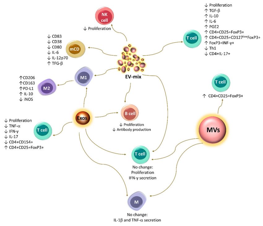
Int. J. Mol. Sci. 2021, 22, 9531 4 of 31
that remains bound to the MSC membrane, while HLA-G5 is secreted [22,49]; the latter can
stimulate IL-10 production by activated T lymphocytes. IL-10 stimulates the expression
of both isoforms in MSCs, generating a positive feedback mechanism [22,50]. HLA-G1 is
likely the first molecule involved in the establishment of this interaction. This hypothesis
is supported by a study in which basal levels of HLA-G1 mRNA were detected in MSCs
derived from the BM, AT, and fetal liver. Moreover, overexpression of this isoform in
AT-MSCs increases their immunoregulatory capacity toward activated T lymphocytes [44].
Likewise, cell–cell contact between MSCs and populations enriched in CD3+ T lympho-
cytes is essential for the increase in IL-10 levels detected in the supernatants of these
cocultures [51,52]. This process is important because IL-10 participates in the generation
of Tregs by MSCs and increases the expression of programmed cell death protein 1 (PD-1)
in CD4+CD25+ cells, which is associated with greater immunoregulatory activity [53]
(Figure 2).
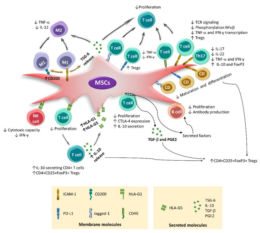
Figure 2. Immunoregulatory mechanisms mediated by cell–cell contact. The main membrane molecules involved in
the immunoregulation exerted by MSCs are shown. Cell–cell interactions, in addition to affecting the proliferation,
differentiation, and effector function of immune cells, also increase the immunoregulatory capacity of MSCs. The contact of
M1 macrophages with MSCs through ICAM-1 induces an M2 phenotype in macrophages, while in MSCs, the expression
of CD200 and TSG-6 is increased (brown arrows), which also favors the differentiation of M2 macrophages. Conversely,
ICAM-1, PD-L1, and jagged-1 decrease the secretion of cytokines and proliferation of activated T lymphocytes, as well as
the maturation and differentiation of DCs. Furthermore, CD40 affects the proliferation of T cells, while HLA-G1 induces
Tregs differentiation. HLA-G1 is also involved in the decrease in effector function of NK cells. Conversely, direct contact of
MSCs with T lymphocytes (circle with dotted lines) induces changes in T lymphocytes and stimulates the secretion of TGFβ
and PGE2, as well as factors that affect the function of B lymphocytes (dotted lines). However, it is unknown exactly which
molecules are involved in this interaction.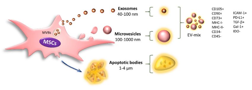
Int. J. Mol. Sci. 2021, 22, 9531                                                                                           5 of 31

                                         In BM-MSC cocultures with enriched CD4+ lymphocyte populations, the generation
                                   of CD4+CD25+FoxP3+ lymphocytes involves the participation of TGF-β and PGE2, but
                                   direct contact between the two cell types is an indispensable prerequisite [42]. Similar
                                   observations have been made in co-cultures with tonsil-derived MSCs (T-MSCs), where
                                   cell contact is essential to decrease the proliferation of CD4 T lymphocytes, as well as the
                                   differentiation of CD4+TNF-α+ and CD4+IFN-γ+ cells [47]. Likewise, our working group
                                   showed that the cell–cell interaction between activated CD3+ T lymphocytes and MSCs
                                   derived from BM or umbilical cord blood (UCB) is necessary to increase the expression
                                   of cytotoxic T lymphocyte-associated protein 4 (CTLA4), a molecule that is constitutively
                                   expressed by Tregs [51]. Interestingly, the direct contact between MSCs and CD3+ T lym-
                                   phocytes, in addition to affecting the function of these same T lymphocytes, can also affect
                                   B lymphocytes.
                                         Direct contact of human BM-MSCs with CD3+ T lymphocytes apparently inhibits the
                                   proliferation, differentiation, and production of antibodies by B cells because these effects
                                   are observed in cocultures of MSCs with CpG-activated peripheral blood lymphocytes but
                                   not in cocultures with sorted B cells. Moreover, the use of transwells in the first system
                                   reverses the inhibitory effects, while the addition of sorted T cells to the second system
                                   recovers the immunoregulatory activity of MSCs. The authors propose that the immuno-
                                   suppressive effect of MSCs on B cells is mediated by soluble factors secreted during the
                                   cell–cell interaction of MSCs with CD3 T lymphocytes [54]. However, other studies have
                                   observed that direct contact between MCSs and populations enriched in CD19+ B cells
                                   affects the functions of the latter (Figure 2). In this regard, it has been documented that
                                   resting human AT-MSCs do not decrease the proliferation of B lymphocytes but favor
                                   the differentiation of regulatory CD19+CD24hiCD38hi B lymphocytes and increase the
                                   expression of IL-10. In turn, AT-MSCs exposed to IFN-γ do not favor regulatory B lym-
                                   phocyte differentiation but reduce B lymphocyte proliferation and inhibit IgG production.
                                   These effects are more efficient when direct contact between MSCs and CD19+ B cells is
                                   allowed [40].
                                         The current evidence highlights the importance of cell–cell contact in the immunoreg-
                                   ulatory activity of MSCs by favoring the development of mechanisms mediated by soluble
                                   factors. Therefore, we must improve our knowledge of these interactions and understand
                                   the participation of the different membrane-bound molecules. In this sense, several recent
                                   reports have indicated the relevance of adhesion molecules in the interaction of MSCs with
                                   immune cells, particularly intercellular adhesion molecule-1 (ICAM-1) [19,55,56].
                                         ICAM-1 is a highly glycosylated protein that belongs to the immunoglobulin su-
                                   perfamily of cell adhesion molecules. It is expressed on fibroblasts, endothelial cells,
                                   antigen-presenting cells, and lymphocytes and participates in cell–cell and cell–matrix ad-
                                   hesion, modulating cell migration processes [57]. This adhesion molecule is a key regulator
                                   of the immune response, is involved in the differentiation of monocytes and DCs, and
                                   participates in the immunological synapse between antigen-presenting cells and T lympho-
                                   cytes, which modulates the activation and differentiation of the latter [58,59]. It has been
                                   observed that ICAM-1 promotes a decrease in the differentiation of Th17 lymphocytes [59]
                                   and the establishment of memory CD8 T lymphocytes [60,61]. In addition, endothelial
                                   cells exposed to TNF-α show increased expression of this adhesion molecule, which is
                                   important in inducing and regulating the immune response, since the contact of DCs with
                                   lymphatic endothelial ICAM-1hi cells decreases the expression of CD86 and the capacity of
                                   DCs to induce the activation and proliferation of T lymphocytes [62]. Similar mechanisms
                                   have been identified in MSCs.
                                         Resting MSCs express ICAM-1 at low levels; however, their expression is increased
                                   and induced in the presence of an inflammatory environment, which favors its interaction
                                   with immune cells [19,45,63]. ICAM-1 promotes the adhesion of murine BM-MSCs to DCs,
                                   inhibiting the maturation and differentiation of the latter [63]. Additionally, during exacer-
                                   bated inflammatory responses, ICAM-1 favors the interplay of human BM-MSCs with M1
                                   macrophages, which induces the generation of M2 anti-inflammatory macrophages [64].
Int. J. Mol. Sci. 2021, 22, 9531 6 of 31
ICAM-1 polarization has been observed in cell contact areas, which results in the formation
of an “unconventional synapse” capable of modulating the function of both cells [64].
Likewise, direct contact between M1 macrophages and murine BM-MSCs increases the im-
munoregulatory activity of these cells toward the same macrophages and the proliferation
of CD4+ T lymphocytes. In addition, the interaction of MSCs with M1 macrophages in
a contact-dependent or contact-independent manner increases the expression of CD200
in MSCs, another membrane molecule involved in the transition from M1 to M2 [65]
(Figure 2).
Human MSCs derived from BM [66], umbilical cord (UC), and TA [67] more efficiently
inhibit PBMC proliferation when direct contact occurs between the two cell types. In
cocultures of CD3+ cells activated in the presence of MSCs derived from BM, amnion, or
UCB [12,51,52,68,69], the inhibition of cell contact practically reestablishes the proliferation
of T lymphocytes. Additionally, the generation of regulatory T cell populations by MSCs
involves the participation of several membrane molecules, including HLA-G1, PD-L1,
VCAM-1, and ICAM-1 [19,22,70] (Figure 2).
The ICAM-1 blockade restores the proliferation of activated T lymphocytes in the
presence of BM-MSCs [55] or AT-MSCs [56] and inhibits the generation of FoxP3+ cells [56].
In addition, it has been proposed that the direct contact of human BM-MSCs with T lym-
phocytes through ICAM-1 and CD43 is a critical event in the immunoregulatory activity
of these cells. This interaction is capable of immediately decreasing the transcription of
TNF-α and IFN-γ in activated T lymphocytes because ICAM-1 expressed on MSCs regu-
lates T cell receptor (TCR) signaling [71]. Moreover, this adhesion molecule participates in
the interplay of human BM-MSCs with Th17 cells through a mechanism that involves the
interaction of CCR6 with its ligand CCL20 and the induction of a conformational change in
CD11a/CD18 that promotes its binding to ICAM-1. This event increases when MSCs are
exposed to TNF-α and IFN-γ due to the increased expression of ICAM-1. Subsequently,
the production of IL-17, IL-22, IFN-γ, and TNF-α by differentiated Th17 cells is decreased,
and a regulatory phenotype with increased expression of IL-10 and FoxP3 is induced [34]
(Figure 2). Importantly, the effects of MSCs on differentiated Th17 cells decrease when a
transwell is used, but they are not completely inhibited, since PGE2 is also involved in
these processes.
The importance of ICAM-1 in the immunoregulatory effects exerted by MSCs has
also been observed in preclinical studies. In a murine model of graft-versus-host disease
(GVHD), an infusion of ICAM-1+ BM-MSCs reduced the expression of inflammatory
cytokines and the percentage of Th1 lymphocytes. Additionally, it increased the migratory
capacity of MSCs and the differentiation of T cells toward a regulatory phenotype associated
with immune tolerance [63]. Likewise, in a murine model of inflammatory bowel disease,
the administration of BM-MSCs overexpressing ICAM-1 decreased the percentage of Th1
and Th17 cells, as well as the transcription of IFN-γ and IL-17. In addition, these cells
increased the differentiation of Tregs and the transcription of FoxP3, all of which are
associated with a decrease in lesions [45].
On the other hand, the expression of jagged-1, a Notch ligand, on the surface of MSCs
has been implicated in its immunoregulatory activity toward DCs and T lymphocytes.
Blocking jagged-1 with antibodies has been observed to decrease the inhibitory effect
of human BM-MSCs on CD4+ lymphocyte proliferation [46]. In murine BM-MSCs, the
participation of jagged-1 in the expansion of CD4+CD25+FoxP3+ Treg cells and the in-
duction of DCs with a semimature phenotype has been confirmed, which also favors the
differentiation of Tregs from CD4+CD25-FoxP3- populations [72] (Figure 2).
Another important molecule involved in contact-mediated immunoregulation is PD-
L1, the expression of which increases in MSCs treated with IFN-γ [9,67,73–76]. This
molecule contributes to the decrease in the differentiation and maturation of DCs [73].
In addition, it affects the activation, proliferation, and effector function of T lympho-
cytes [67,74,77,78], increasing the generation of Treg cells [73] and decreasing the secretion
of TNF-α and IFN-γ in activated T lymphocytes [79]. A recent study shows that this lastInt. J. Mol. Sci. 2021, 22, 9531 7 of 31
effect is also mediated by CD40, whose expression increases in T-MSCs activated with
TNF-α and IFN-γ [47] (Figure 2). It is important to mention that, although PD-L1 is a molecule
present in the membrane of MSCs, some studies have suggested that it can also be secreted and
that this form is capable of decreasing the expression of CD25 and IL-2 in activated T lympho-
cytes [78]. Interestingly, this working group found that PD-L1 is secreted freely or bound to
extracellular vesicles, structures that can mediate the immunoregulatory effect of MSCs.
5. Immunoregulation Mediated by Extracellular Vesicles
Given the importance of cellular contact in the immunoregulatory activity of MSCs, it
is not clear how this event might occur in the physiological context, since a poor grafting
capacity and low viability of the transplanted cells have been observed in patients over the
course of days [80]. Thus, it is currently proposed that MSCs release extracellular vesicles
(EVs), structures that have been recognized as an important mechanism of paracrine and
endocrine cellular communication [81,82], to mediate the immunoregulatory effects of
MSCs even at distant sites.
EVs are mainly classified based on their biogenesis, size, and shape into exosomes,
microvesicles, and apoptotic bodies. Exosomes are homogeneous vesicles with a size
ranging from 40 to 100 nm that are derived from multivesicular bodies (MVBs) and secreted
through fusion of the MVBs with the cell membrane. In addition, they are positive for CD63,
CD9, CD81, and TSG101. Microvesicles (MVs) are a heterogeneous population ranging
from 100 to 1000 nm in size that originate from direct protrusions of the cell membrane that
detach from the surface, and these structures retain many characteristics of their cells of
origin [83,84]. Finally, apoptotic bodies are EVs with a diameter of 1000–4000 nm that are
released from the plasma membrane when the cells undergo apoptosis. These structures
are characterized by the presence of phosphatidylserine in the outer membrane and contain
organelles, histones, and fragmented DNA [48,83,84] (Figure 3).
Figure 3. Classification of extracellular vesicles. EVs are classified into exosomes, microvesicles, and
apoptotic bodies. Exosomes and MVs are important intercellular communication mechanisms. These
structures transport different molecules inside or on the membrane, through which they modify the behavior
of their target cells. Some of the main molecules identified in EVs released by resting MSCs are shown.
All cells of an organism release exosomes and MVs, which are an important mecha-
nism of intercellular communication because they can reach the bloodstream and travel
to distant sites where they establish contact with their target cells and influence their
biological behaviors. Through proteomics, genomics, lipidomics, and metabolomics assays,
exosomes and MVs have been shown to contain proteins (ligands, receptors, adhesion
molecules, enzymes, cytokines, and growth factors), lipids, metabolites, mRNAs, and
microRNAs, through which they modulate their target cells [83–87]. Currently, there is
still controversy regarding the nomenclature and methods of obtaining these structures.
The terms exosome, MV, and EVs are used as synonyms in several reports, which may
confuse the interpretation of the results. These discrepancies highlight the importance of
characterizing and analyzing the specific function of each type of EV [88,89]. Therefore,
in each article cited in this review, we specify whether the study was performed with
exosomes, MVs, or a mixture of exosomes and microvesicles (EV-mix) based on an analysis
of the method used to obtain them (Table 1).Int. J. Mol. Sci. 2021, 22, 9531 8 of 31
Table 1. Isolation method used and type of EVs obtained.
Name Used in the Original Report
Isolation Method Structures Obtained and Size Range Study Model Ref.
Cell Source and In Vitro Conditioning Method
Extracellular vesicles Microvesicles In vitro:
Human TA-MSCs Exosome isolation reagent (Invitrogen). ≈150–500 nm T lymphocyte proliferation [37]
IFN-γ (50 ng/mL) for 48 h. Mean: 262.4 nm and differentiation.
MVs and Exo preparations:
(a) 300× g for 10 min;
(b) 2000× g for 10 min;
(c) 0.8 µm membrane filtration; MVs:
Extracellular vesicles In vitro:
(d) 12,500× g for 20 min at Mean: 400–500 nm
Human and murine TA-MSCs Proliferation and secretion [48]
room temperature (pure MV isolates); Exosomes:
Resting MSCs of cytokines by T lymphocytes.
(e) Removing the residual MVs by Mean: 80–100 nm
centrifugation at 20,500× g for 40 min;
(f) 0.22 µm membrane filtration;
(g) 100,000× g for 70 min (pure Exo isolates).
Extracellular In vitro:
membrane vesicles T lymphocyte proliferation
(a) 2000× g for 20 min; EV-mix
Human UCB-MSCs and induction of regulatory T cells. [85]
(b) 100,000× g for 1–2 h. 20–700 nm
IFN-γ (100 ng/mL) for In vivo:Ischemia-reperfusion-induced acute
24 and 48 h. kidney injury rat model.
(a) 400× g for 5 min;
Extracellular Vesicles
(b) 2000× g for 20 min; EV-mix In vitro:
Human BM-MSCs [87]
(c)10,000× g for 45 min; 152 ± 23 nm Maturation and secretion of cytokines by CDs.
Resting MSCs
(d) 100,000× g for 90 min.
(a) 300× g for 10 min; In vitro:
Microvesicles
(b) 1000× g for 20 min; EV-mix Proliferation and secretion of cytokines by T
Murine BM-MSCs [90]
(c) 10,000× g for 30 min; 50–200 nm lymphocytes.
Resting MSCs
(d) 100,000× g for 2 h. Induction of regulatory T cells.
Microvesicles In vivo:
(a) 2000× g for 20 min; EV-mix
Human WJ-MSCs Ischemia-reperfusion-induced acute kidney [91]
(b) 100,000× g for 1 h. 30–500 nm
Resting MSCs injury rat model.
In vitro:
Microvesicles (a) 1500× g for 20 min;
EV-mix Proliferation and secretion of cytokines by T
Human BM-MSCs (b) 10,000× g for 20 min; [92]
60–160 nm lymphocytes.
Resting MSCs (c) 100,000× g for 1 h.
Induction of regulatory T cells.Int. J. Mol. Sci. 2021, 22, 9531 9 of 31
Table 1. Cont.
Name Used in the Original Report
Isolation Method Structures Obtained and Size Range Study Model Ref.
Cell Source and In Vitro Conditioning Method
Extracellular vesicles
Human BM-MSCs (a) 0.2-µm membrane filtration; In vitro:
Exosomes
TNF-α (20 ng/mL) plus (b) Millipore Lab-scale TFF system equipped Cytokine secretion by activated primary rat [93]
75–165 nm
IFN-γ (20 ng/mL) with a Biomax 500 kDa Pellicon filter. splenocytes.
during the night
In vitro:
Extracellular vesicles (a) 2000× g for 20 min; EV-mix
T lymphocyte proliferation [94]
Murine TA-MSCsResting MSCs (b) 100,000× g for 2 h. 100–1000 nm
and induction of regulatory T cells.
Microvesicles
EV-mix In vitro:
Human BM-MSCs 100,000 g for 1 h at 4◦ C twice. [95]
≈200 nm Activation of murine mast cells.
Resting MSCs
Microvesicles
(a) 300× g for 20 min; EV-mix In vitro:
Human BM-MSCs [96]
(b) 100,000× g for 1 h. 50–200 nm Human type II alveolar cells.
Resting MSCs
Microvesicles
a) 1500× g for 15 min; In vitro:
Human BM-MSCs Exosomes
b) 0.22 µm membrane filtration; Uptake by human umbilical cord endothelial [97]
Hypoxia-induced 50–100 nm
c) 170,000× g for 5 h. cells.
MSCs
Extracellular vesicles
Human BM-MSCs (a) 300× g for 10 min; In vitro:
EV-mix
IFN-γ (10 ng/mL) plus (b) 2000× g for 30 min; Proliferation and secretion of cytokines by NK, [98]
≈60–400 nm
TNF-α (15 ng/mL) (c) 100,000× g for 90 min. T, and B cells.
for 40 to 48 h.
(a) 300× g for 10 min;
Exosomes In vitro:
(b) 10,000× g for 20 min; Exosomes
Human BM-MSCs Proliferation and differentiation of T and B [99]
(c) 0.2 µm membrane filtration; 65–100 nm
Resting MSCs lymphocytes.
(d) 100,000× g for 60 min.
Extracellular vesicles
(a) 2000× g for 20 min; EV-mix In vitro:
Human BM-MSCs [100]
(b) 100,000× g for 2 h. 61–121 nm Polarization of macrophages.
Resting MSCs
Exosomes
Exosome Isolation Kit Exosomes In vitro:
MSCs from carcinoma and healthy breast tissue [101]
(Invitrogen). Size not reported Polarization of macrophages.
Resting MSCsInt. J. Mol. Sci. 2021, 22, 9531 10 of 31
Table 1. Cont.
Name Used in the Original Report
Isolation Method Structures Obtained and Size Range Study Model Ref.
Cell Source and In Vitro Conditioning Method
In vitro:
Extracellular vesicles Induction of regulatory T cells.
(a) 300× g for 10 min;
Human BM-MSCs EV-mix In vivo:
(b) 2000× g for 30 min; [102]
IFN-γ (10 ng/mL) plus ≈25–500 nm A xenograft mouse model with
(c) 100,000× g for 90 min.
TNF-α (15 ng / ml) for 4 h. steroid-refractory acute graft-versus-host
disease.
(a) 300× g for 10 min;
In vivo:
Extracellular vesicles (b) 2000× g for 20 min; Exosomes
Murine model of chronic graft-versus-host
Human UC-MSCs (c) 10,000× g for 30 min; 105.1–181.1 nm [103]
disease.
Resting MSCs (d) 0.2 µm membrane filtration; Mean: 139.2 nm
Infiltration and activation of macrophages.
(e) 100,000× g for 90 min.
In vivo:
Extracellular vesicles
a) 10,000× g for 20 min; EV-mix Murine model of lung ischemia-reperfusion
Human WJ-MSCs [104]
b) 100,000× g for 1 h. 164 ± 10.4 nm injury.
Resting MSCs
Cytokine expression levels.
Single-center, randomized, placebo-controlled,
Extracellular vesicles
(a) 2000× g for 20 min; EV-mix phase II/III clinical pilot study.
Human UC-MSCs [105]
(b) 100,000× g for 60 min. 80–1000 nm Patients with grade III-IV chronic kidney
Resting MSCs
disease.
Small extracellular vesicle (a) 400× g for 10 min; In vitro:
Human WJ-MSCs (b) 2000× g for 30 min; Exosomes Activation of T lymphocytes.
[106]
IFN-γ (2.5 ng/mL) (c) 10,000× g for 1.5 h; 30–150 nm In vivo:
Time is not reported (d) 100,000× g for 90 min. Patients with acute graft-versus-host disease.
Microvesicles a) 500× g for 15 min; In vitro:
Microvesicles
Human BM-MSCs b) 2000× g for 20 min; Analysis of changes in the [107]
130–1000 nm
IFN-γ (10 ng/mL) for 72 h c) 17,000× g for 60 min. transport of HLA-I and ICAM-1.
Exosomes
Human TA-MSCs Exosomes In vitro:
ExoQuick-TC System Biosciences. [108]
IFN-γ plus TNF-α 115 ± 11.5 nm Polarization of macrophages.
(10, 20 and 40 ng/mL) for 48 h.
Microvesicles
(a) 300× g for 30 min; MVs In vitro:
Human BM-MSCs [109]
(b) 16,500× g for 20 min. ≈150 nm Induction of regulatory T cells.
IFN-γ (10 ng/mL) for 48 h, 74 h, or 4 daysInt. J. Mol. Sci. 2021, 22, 9531 11 of 31
Table 1. Cont.
Name Used in the Original Report
Isolation Method Structures Obtained and Size Range Study Model Ref.
Cell Source and In Vitro Conditioning Method
Exosomes
UC-MSCs (a) 3000× g for 30 min;
TGF-β (10 ng/mL) (b) 0.22 µm membrane filtration;PEG6000 Exosomes In vitro:
[110]
IFN-γ (1000 IU/mL) was added; 141.6 ± 23.3 nm Induction of regulatory T cells.
for 72 h. (c) 3000 rpm for 30 min.
Alone or combined
Microvesicles In vitro:
(a) 2500× g for 10 min; MVs
Human BM-MSCs Analysis of the content of [111]
(b) 14,000× g for 45 min at 10 ◦ C. 150–450 nm
Ischemic brain extract for 24 h. immunoregulatory molecules.
MVs and Exo preparations:
(a) 300× g for 10 min; MVs:
Extracellular vesicles
(b) 0.8 µm membrane filtration; Mean: 271 nm In vitro:
Murine TA-MSC [112]
(c) 12,600× g for 30 min (MVs); Exosome: Mouse-derived peritoneal macrophages.
Resting MSCs
(d) 0.22 µm membrane filtration; Mean: 90 nm
(g) 100,000× g for 70 min (Exo).
In vitro:
Extracellular Vesicles (a) 3200× g for 10 min; Activation of CD4 + cells.
EV-mix
Human UC-MSCs (b) 10,000× g for 30 min; In vivo: [113]
≈100–300 nm
Resting MSCs (c) 100,000× g for 2 h. Mouse liver ischemia/reperfusion injury
model.
(a) 300× g for 10 min; In vivo:
Small extracellular vesicles
(b) 2000× g for 10 min; EV-mix Rat model of rheumatoid arthritis
Human UC-MSCs [114]
(c) 10,000× g for 30 min; 40–200 nm T lymphocyte proliferation, apoptosis, and
Resting MSCs
(d) 100,000× g for 70 min. differentiation.
Extracellular Vesicles (a) 0.22 µm membrane filtration; In vivo:
Exosomes
Human TA-MSCs (b) 30,000× g for 20 min; Mouse model of dextran sodium [115]
90–120 nm
Resting MSCs (c) 120,000× g for 3 h. sulfate-induced colitis.
(a) 500× g for 10 min;
Extracellular vesicles (b) 2000× g for 20 min; In vivo:
Exosomes
Human PL-MSC (c) 5000× g for 30 min; Mouse model of colitis-induced with [116]
85–125 nm
Resting MSCs (d) 0.2 µm membrane filtration; trinitrobenzene sulfonic acid.
(e) 130,000× g for 2 h.
Extracellular vesicles
(a) 1200 rpm for 6 min; In vivo:
Murine BM-MSCs
(b) 0.22 mm filter; Murine model of sodium dextran.
IL-6 (20 ng/mL), EV-mix
(c) Ami-Con filters Ultra-15, regenerate Sulfate-induced colitis. [117]
TNF-α (25 ng/mL) plus ≈30–300 nm
cellulose 100,000 NMWL; Merck Millipore); Polarization of intestinal macrophages.
IL-1β (25 ng/mL)
(d) 3200× g at 4 ◦ C for 15 min. Regulatory T cell differentiation.
for 24 hInt. J. Mol. Sci. 2021, 22, 9531 12 of 31
Table 1. Cont.
Name Used in the Original Report
Isolation Method Structures Obtained and Size Range Study Model Ref.
Cell Source and In Vitro Conditioning Method
(a) 300× g for 10 min;
Exosomes
Exosomes (b) 3000× g for 10 min; In vitro:
Control:
Human G- MSCs (c) 20,000× g for 30 min; Macrophage polarization.
123 ± 3.1 nm [118]
TNF-α (100 ng/mL) (d) 120,000× g for 70 min; In vivo:
TNF-α:
for 48 h (e) Sucrose gradient; Ligature-induced periodontitis model in mice.
164 ± 7.3 nm
(f) 110,000× g for 3 h at 4 ◦ C.
Small extracellular vesicles (a) 800× g for 5 min;
human TA-MSCs (b) 2000× g for 10 min; EV-mix In vitro:
[119]
IFN-γ (10 ng/mL) plus (c) 0.22 mm pore filters; ≈80–300 nm CD4 T cell proliferation.
TNF-α (15 ng/mL) for 72 h (d) 110,000× g for 2 h.
Extracellular vesicles
DP-MSCs (a) 2000× g for 20 min; In vitro:
IFN-γ (50 ng/mL), (b) 10,000× g for 70 min; EV-mix T cell proliferation.
[120]
TNF-α (10 ng/mL) plus (c) 0.22 µm membrane filtration; ≈100–350 nm In vivo:
IL-1β (10 ng/mL) (d) 110,000× g for 120 min. Delayed-type hypersensitivity mouse model.
for 48 h
Extracellular vesicles In vitro:
Human BM-MSCs (a) Centrifugation to remove cells and cell Murine primary microglia.
EV-mix
IFN-γ (25 ng/mL) plus debris; In vivo: [121]
≈100–500 nm
TNF-α (20 ng/mL) (b) 110,000× g for 70 min. Triple-transgenic model of Alzheimer’s
for 24 or 48 h disease.
Centrifugations were carried out at 4 ◦ C unless another temperature was indicated. MVs: microvesicles; Exo: exosomes; TA: adipose tissue; BM: bone marrow; UCB: umbilical cord blood; WJ: Warton’s jelly; UC:
umbilical cord; G: gingival; DP: dental pulp; MSCs: mesenchymal stem/stromal cells.Int. J. Mol. Sci. 2021, 22, 9531 13 of 31
Resting MSCs release exosomes and MVs with positivity for the characteristic markers
of these cells (CD105, CD90, and CD73) and the absence of MHC-I, MHC-II, CD34, and
CD45 [48,90–94,122,123]; they also transport immunoregulatory molecules such as PD-L1,
Gal-1, and TGF-β [90,124] (Figure 3).
Currently, EV-mediated communication is proposed to involve the direct contact of
these structures with their target cells. In this regard, CD44 participates in the uptake of
the EV-mix by bone marrow-derived mast cells [95] and human alveolar epithelial type II
cells [96]. Likewise, phosphatidylserine transported on the surface of exosomes released
by human BM-MSCs facilitates the uptake of these structures by human umbilical vein
endothelial cells (HUVECs) [97]. Furthermore, it has been shown that these structures
can be captured by granulocytes, NK cells, mast cells, monocytes, CDs, and T and B
lymphocytes [87,93,95,98,99,125], which affects the function of these immune cells.
The EV-mix released by resting human BM-MSCs induces an M2 phenotypic switch in
monocyte-derived macrophages, along with a higher expression of CD206 and PD-L1 [100].
Similar observations have been made with exosomes released by MSCs from breast tumors,
which promote the differentiation of myeloid cells into M2-type immunosuppressive
macrophages with high expression levels of CD206, PD-L1, and IL-10 and higher L-arginase
activity [101]. In contrast, a recent study reports that exosomes and MVs released by murine
TA-MSCs are unable to decrease the secretion of pro-inflammatory cytokines (IL-1β y TNF-
α) by peritoneal macrophages stimulated with LPS [112], while the EV-mix released by
resting human BM-MSCs decreases the uptake of antigens by immature DCs, suggesting
that these structures potentially affect this key event in the maturation of DCs. Additionally,
mature DCs exposed to these structures show reduced CD83, CD38, and CD80 expression,
as well as IL-6 and IL-12p70 secretion, but increased TGF-β production [87]. It is still
necessary to determine whether the changes in the differentiation of macrophages and DCs
induced by EVs affect the ability of these cells to induce the activation, proliferation, and
differentiation of T lymphocytes.
Currently, the results regarding the effect of EVs on T lymphocytes are controversial.
In vitro tests have shown that the EV-mix released by murine and human BM-MSCs can
increase the generation of the Tregs CD4+CD25+FoxP3+ and CD4+CD25+CD127low FoxP3+,
respectively [90,102]. They also decrease the proliferation of PBMCs and stimulate the
expression of IL-10 and TGF-β [90]. Likewise, the EV-mix released by murine AT-MSCs
affects the proliferation of T lymphocytes and the generation of Th1 cells. In addition, they
promote the differentiation of a population of FoxP3+ IFN-γ+ cells, which are capable of
decreasing the proliferation of CD4 and CD8 T lymphocytes [94]. Moreover, exosomes
released by human BM-MSCs and EV-mix derived from AT-MSCs reduce the proliferation
of these cells [37,92,99] and the secretion of IFN-γ and IL-17 while increasing the production
of IL-10, IL-6, and PGE2, as well as the differentiation of Tregs [92]. In contrast, Matula
et al. (2017) observed that exosomes and MVs (fractions analyzed separately) released
by human AT-MSCs are incapable of altering the proliferation of T lymphocytes and the
secretion of IFN-γ [48]. A similar result was obtained with the EV-mix released by human
BM-MSCs [98], and it has been proposed that these structures are not taken up by CD3+
T cells [87]. However, interestingly, it has been observed that the exosomes and MVs
released by activated T lymphocytes can be taken up by MSCs and increase the secretion of
PGE2 [48]. This evidence indicates that EVs are also involved in the feedback mechanisms
established between MSCs and immune cells. The above findings highlight the importance
of further studies of these structures.
Studies conducted with an EV-mix [125] or exosomes [99] released by human BM-
MSCs have shown that these structures also exert immunosuppressive effects on B lym-
phocytes, since they decrease their proliferation and differentiation, significantly affecting
the production of IgM, IgA, and IgG [99,125].
Furthermore, the immunoregulatory capacity of EV-MSCs has been observed in
preclinical models. In animal models, the EV-mix constitutively secreted by UCB- and
WJ-MSCs attenuated the kidney damage caused by ischemia [85,91], as well as the clinicalInt. J. Mol. Sci. 2021, 22, 9531 14 of 31
manifestations in the skin of mice with chronic GVHD [103]. These effects are related
to reduced infiltration and activation of macrophages, an increase in IL-10 levels, and a
decrease in TNF-α levels in damaged tissues [91,103]. Similar results have been obtained
in a murine model of pulmonary ischemia, where the administration of an EV-mix released
by WJ-MSCs decreased tissue damage, which was related to lower concentrations of IL-
17 and TNF-α, as well as an increase in the levels of PGE2, IL-10, and the keratinocyte
growth factor in the bronchoalveolar fluid [104]. Moreover, in a mouse model of acute
GVHD, the administration of an EV-mix released by human BM-MSCs stimulated Treg
differentiation [102].
In addition, the administration of EV-mix released by resting UC-MSCs, in a mouse
liver ischemia/reperfusion injury model, significantly attenuated liver tissue damage,
with a decrease in the percentage of intrahepatic CD4+CD154+ and CD68+ cells, and less
production of TNF-α and IFN-γ [113]. A similar anti-inflammatory effect has been observed
with EV-mix administered in a rat model of collagen-induced arthritis. Attenuation of
the severity of the disease is observed, which is associated with less proliferation and
increased apoptosis of splenic T cells. In addition, the differentiation of CD4+IL-17+
cells is affected and the differentiation of T reg CD4+CD25+FoxP3+ is favored. All of
the above is accompanied by lower serum levels of IL-17 and an increase in IL-10 and
TGF-β [114]. Similarly, the administration of exosomes released by TA- or PL-MSCs in an
induced colitis mouse model reduces local and systemic inflammation. In the colon tissue,
a lower expression of IL-1β, IL-6, TNF-α, IFN-γ, IL-17, and IL-12 is observed, as well as
an increase in IL-10 and TFG-β [115,116]. Moreover, the levels of reactive oxygen species
(ROS), the expression of apoptotic proteins, and metalloproteinase (MMP-2 and MMP-9)
are reduced [116].
Currently, only one clinical trial conducted with EVs released by resting MSC has
reported results. The administration of two doses of EV-mix obtained from UCB-MSCs
to patients with grade III-IV chronic kidney damage induced an improvement in renal
function, decreasing inflammation and TNF-α levels, and increasing plasma levels of
TGF-β and IL-10 [105]. In addition, an increase in the percentage of PD-L1+ exosomes was
detected in the plasma of patients with acute GVHD treated with clinical-grade WJ-MSCs,
suggesting that these structures are potential mediators of the therapeutic effect [106].
This evidence indicates that the EVs released by MSCs may exert an immunosuppressive
effect similar to that observed for the cells. EVs have the advantage that their content
and possible therapeutic effect are not affected by the microenvironment, which is a
disadvantage associated with the use of whole cells. However, it is necessary to carry out
clinical trials to determine this, in this regard, there are currently 13 clinical trials registered
on the clinicaltrials.gov page, in which the use of EVs released by resting MSCs in the
treatment of different pathologies is proposed.
6. Effect of the Inflammatory Microenvironment on the Immunoregulatory Capacity
of MSCs
As already mentioned, the inflammatory environment modulates the immunoregula-
tory capacity of MSCs, and this phenomenon has been reported in preclinical and clinical
studies. These cells exert a greater therapeutic effect when administered to patients at
intermediate stages of the disease due to the presence of an inflammatory environment that
stimulates the immunoregulatory properties of MSCs. High serum IFN-γ levels may be a
favorable prognostic marker to predict the therapeutic success of UCB-MSCs in patients
with lupus erythematosus [126]. Similar observations have been made in the treatment of
GVHD, where the administration of MSCs significantly decreased the symptoms of the
disease in patients with an acute GVHD refractory to steroids [3,127]. In addition, exposure
of AT-MSCs to synovial fluid from patients with rheumatoid arthritis was recently shown to
induce overexpression of COX-2, IDO, IL-6, TSG-6, ICAM-1, VCAM-1, and PD-L1 mRNA;
using antibodies, the authors determined that these changes were mainly induced by the
action of TNF-α [128]. Based on these findings, numerous laboratories have focused on
analyzing the effect of proinflammatory cytokines on the different biological propertiesInt. J. Mol. Sci. 2021, 22, 9531 15 of 31
of MSCs to establish in vitro conditioning protocols that improve the immunoregulatory
potential of these cells and, therefore, their therapeutic effect. It is important to mention
that currently all clinical trials have used resting MSCs and only one study registered in
clinicaltrials.gov will use IFN-γ-primed MSCs in adult and pediatric patients undergoing
hematopoietic cell transplantation for the treatment of acute leukemia and myelodysplas-
tic syndrome (NCT04328714). However, the therapeutic effect of MSCs stimulated with
pro-inflammatory cytokines has been analyzed in various preclinical models.
In a murine model of dextran sulfate-induced colitis, the administration of BM-MSCs
treated with IFN-γ decreased mucosal damage and improved survival rates. These effects
were associated with increased migratory and immunoregulatory capacities of MSCs
since they were more efficient at inhibiting the inflammatory response mediated by Th1
cells [129]. Likewise, the administration of human BM-MSCs treated with IFN-γ decreased
tubular injury and improved renal function in a murine model of acute renal injury with
cisplatin, and the changes were associated with an increased secretion of IL-10 by activated
MSCs [130]. Similar results have been obtained using mice with GVHD, in which the
signs of the disease were prevented and their survival increased [131]. In addition, the
administration of human BM-MSCs activated with IFN-γ promotes bone regeneration in
mice with calvarial lesions. These positive effects were mainly due to an increase in the
expression of IDO by human BM-MSCs [10], which affects T lymphocyte proliferation
in vitro [132].
A conditioned medium from rat BM-MSCs exposed to TNF-α, IL-1β, and NO (cy-
tokines released by irradiated epithelial cells) was administered to rats with radiation-
induced intestinal damage, resulting in decreased structural and functional damage in the
intestine and increased survival. MSCs exposed to this inflammatory environment showed
an increased capacity to decrease apoptosis, stimulate the proliferation of intestinal epithe-
lial cells, and generate intestinal stem cells. Additionally, they reduced IL-1β, IL-6, and
TNF-α levels and increased the secretion of IL-10 and the differentiation of CD4+FoxP3+
cells, which together decreased the local and systemic inflammatory response in the ani-
mals [133]. Similarly, exposure of AT-MSCs to the plasma of patients with acute GVHD
increased their ability to induce Treg differentiation in vitro [134]. These results reveal the
importance of the design of in vitro conditioning strategies that ensure the therapeutic
success of these cells under different pathophysiological conditions. Recently, several
studies have focused on analyzing the effect of IFN-γ on MSCs; however, the importance
of TNF-α as the first stimulus has recently been highlighted.
7. Effects of TNF-α and IFN-γ on the Expression of Immunoregulatory Molecules
by MSCs
TNF-α is a pleiotropic cytokine that functions in an autocrine, paracrine, or systemic
manner; is expressed mainly by macrophages, DCs, and lymphocytes; and is involved
in numerous inflammatory, immunomodulatory, and tissue regeneration pathways. Al-
terations in its secretion or activity are associated with inflammatory and autoimmune
diseases [135]. During an inflammatory process, TNF-α is one of the first cytokines secreted
by immune system cells and can increase (prime) or decrease (desensitize or tolerate) the
ability of cells to respond to other stimuli [135–137]. Nevertheless, there are few reports
on the effect of TNF-α on MSCs. Some studies indicate that this cytokine does not alter
the expression of immunoregulatory molecules in these cells. In contrast, TNF-α has been
proposed to provide the initial stimulus for MSC priming, since it is the first cytokine
released by activated T lymphocytes [30,107,138].
Treatment of BM-MSCs with TNF-α does not affect the expression of IDO [14] but
increases the levels of PGE2, IL-10, IL-6, NO [14,31,139], and ICAM-1 [34,140]. In particular,
in human BM-MSCs, TNF-α induces the expression of ICAM-1 after 6 h of exposure,
reaching the highest levels at 24 and 48 h [107]; this event is associated with a greater
migratory capacity [140]. Likewise, at 24 h, treatment with TNF-α increases the levels of
VCAM-1 [141], vascular endothelial growth factor (VEGF), insulin-like growth factor 1
(IGF-1), and HGF [142,143], while at 48 h, significantly higher levels of IL-6 are detected [16].Int. J. Mol. Sci. 2021, 22, 9531 16 of 31
In addition, in rat UCB-MSCs, TNF-α stimulates the expression of TGF-β and IL-10 [144].
Thus, TNF-α favors the immunoregulatory activity of MSCs and, given the importance of
the mechanisms that involve cell contact, it is important for this cytokine to immediately
increase the expression of adhesion molecules (Figure 4).
Figure 4. Effect of TNF-α and IFN-γ on the expression of immunoregulatory molecules by MSCs. (A) Changes in the
expression of immunoregulatory molecules observed in MSCs stimulated with TNF-α (blue), IFN-γ (red), or TNF-α
and IFN-γ (purple) are indicated. The changes in exosome load, MVs, and EV-mix released by MSCs treated with the
indicated cytokines are also shown. (B) Temporal effect of TNF-α and IFN-γ on the expression of immunoregulatory and
immunogenic molecules by MSCs. Between 24 and 48 h of treatment with TNF-α, MSCs increase the expression of ICAM-1
but not MHC-I and MHC-II. Simultaneously, IFN-γ stimulates the expression of ICAM-1, PD-L1, and IDO but not MHC-II.
These events generate a window in which MSCs with high immunoregulatory capacity and low immunogenic potential can
be obtained (dotted-line box).Int. J. Mol. Sci. 2021, 22, 9531 17 of 31
Moreover, it has been reported that TNF-α does not affect the expression of PD-L1 [14]
or that it increases its expression but to lower levels than those observed in MSCs stimulated
with IFN-γ [75,78]. These contradictory results highlight the importance of further research
on the effects of this cytokine on MSCs (Figure 4).
IFN-γ is a key cytokine involved in the induction of innate and adaptive immune
responses. It is produced mainly by Th1 lymphocytes, CD8, and NK cells [145]. This
cytokine stimulates or increases the expression of major histocompatibility complex (MHC)
molecules on the surface of various cell types, including MSCs [25,67,146]. It also induces
the expression of IDO and PD-L1 [10,12,18,36,73,74,78,147,148] and increases the expres-
sion of HLA-G1, HLA-G5, ICAM-1 [34,43,149], COX-2, and iNOS [10,19,20], as well as
the secretion of PGE2 [14,31] and TGF-β [18,25]. These events translate into a greater
immunoregulatory capacity of MSCs. Numerous reports have described the effects of
IFN-γ, which, while providing an important stimulus, is not the only molecule to which
MSCs are exposed, indicating the need to explore the effects of other cytokines (Figure 4).
8. Effects of the Combination of TNF-α and IFN-γ on the Immunoregulatory Capacity
of MSCs
Few studies have analyzed the effect of combined cytokines on the biology of MSCs.
Doing so would be relevant since, in the physiological context, the activation of these cells
would occur in response to the presence of different stimuli, the balance of which would
dictate the MSC phenotype and function. In support of these findings, some studies have
determined that MSCs treated with mixtures of proinflammatory cytokines have a greater
immunoregulatory capacity [30,102,150].
In vitro studies have shown that TNF-α and IFN-γ exert a powerful synergistic effect
on the expression of immunoregulatory molecules in BM-MSCs [75,107,150], including
TGF-β, IL-6, VEGF [150,151], HGF [14], IDO, PD-L1, and HLA-G [75,78,150], as well
as ICAM-1, VCAM-1 [55,107], and IL-6 [150]. Similar results have been obtained with
UC-MSCs [152] (Figure 4). In addition, a proteomics study carried out with BM-MSCs
stimulated with TNF-α and IFN-γ reported a higher expression of IL-4, IL-10, IL-12, IL-
15, PD-L1, IDO, and HLA-G, as well as the chemokines CCL5, CXCL9, CXCL10, and
CXCL11 [75]. The researchers proposed that the combined treatment eliminated the
variation in the expression of cytokines and chemokines intrinsic to each donor [75].
Nevertheless, to date, no study has analyzed whether the synergistic effect of TNF-α
and IFN-γ on the expression of immunoregulatory molecules translates into a greater
immunoregulatory capacity of MSCs compared to these same cells activated with IFN-γ
alone. To date, only one study has reported that human BM-MSC spheroids stimulated
with this cytokine mixture could more efficiently decrease the production of TNF-α by
macrophages than MSCs stimulated with the same cytokine [150].
Other studies have analyzed the effect of TNF-α and IFN-γ on MSCs. However, due
to the experimental design of these works, it is not possible to confirm the presence of a
synergistic effect. In this regard, it has been observed that the stimulation of TA-MSCs
with both cytokines increases the expression of immunoregulatory molecules, such as IDO,
PGE2, IL-10, IL-6, IL-18, CCL-2 [108], TSG-6 [153], and HLA-G5 [154]. Likewise, in BM-
MSCs, they increase the expression of TGF-β [155], ICAM-1 and VCAM-1 [102,156], IDO,
TSG-6 [157,158], PD-L1 [102], HLA-G5, and factor H, a primary complement inhibitor [159].
It has been determined that human BM-MSCs activated with TNF-α and IFN-γ in-
crease the expression of IDO, which induces the conversion of monocytes to IL-10-secreting
CD206+ M2 immunosuppressive macrophages; these features contribute to the immunoreg-
ulatory effect of MSCs on T lymphocyte proliferation [157]. BM-MSCs and TA-MSCs treated
with both cytokines also directly decrease the proliferation of T lymphocytes [154]. The
few studies conducted to date highlight the need to determine whether the synergistic
effect induced by TNF-α and IFN-γ on the expression of immunoregulatory molecules
truly increases the immunoregulatory activity of these cells, which would indicate a need
to use TNF-α in conditioning protocols.You can also read