Transplantation of Wnt5a-modified NSCs promotes tissue repair and locomotor functional recovery after spinal cord injury
←
→
Page content transcription
If your browser does not render page correctly, please read the page content below
Li et al. Experimental & Molecular Medicine (2020) 52:2020–2033
                                    https://doi.org/10.1038/s12276-020-00536-0                                                                            Experimental & Molecular Medicine

                                     ARTICLE                                                                                                                                                    Open Access

                                    Transplantation of Wnt5a-modified NSCs promotes
                                    tissue repair and locomotor functional recovery
                                    after spinal cord injury
                                    Xiang Li1, Zhiming Peng1, Lingli Long2, Xiaofang Lu3, Kai Zhu1, Ying Tuo4, Ningning Chen5, Xiaoyang Zhao1,
                                    Le Wang1,6 and Yong Wan 1,6

                                      Abstract
                                      Traditional therapeutic strategies for spinal cord injury (SCI) are insufficient to repair locomotor function because of the
                                      failure of axonal reconnection and neuronal regeneration in the injured central nervous system (CNS). Neural stem cell
                                      (NSC) transplantation has been considered a potential strategy and is generally feasible for repairing the neural circuit
                                      after SCI; however, the most formidable problem is that the neuronal differentiation rate of NSCs is quite limited.
                                      Therefore, it is essential to induce the neuronal differentiation of NSCs and improve the differentiation rate of NSCs in
                                      spinal cord repair. Our results demonstrate that both Wnt5a and miRNA200b-3p could promote NSC differentiation
                                      into neurons and that Wnt5a upregulated miRNA200b-3p expression through MAPK/JNK signaling to promote NSC
                                      differentiation into neurons. Wnt5a could reduce RhoA expression by upregulating miRNA200b-3p expression to
                                      inhibit activation of the RhoA/Rock signaling pathway, which has been reported to suppress neuronal differentiation.
                                      Overexpression of RhoA abolished the neurogenic capacity of Wnt5a and miRNA200b-3p. In vivo, miRNA200b-3p was
                  1234567890():,;
1234567890():,;
                  1234567890():,;
1234567890():,;

                                      critical for Wnt5a-induced NSC differentiation into neurons to promote motor functional and histological recovery
                                      after SCI by suppressing RhoA/Rock signaling. These findings provide more insight into SCI and help with the
                                      identification of novel treatment strategies.

                                    Introduction                                                                                        reducing tissue damage and improving patient quality of
                                      Spinal cord injury (SCI) is considered to be a refractory                                         life4,5. However, none of these therapeutic strategies are
                                    disease with devastating physical, psychosocial, and voca-                                          sufficient to repair the interruption of the neuroanatomical
                                    tional implications for patients and caregivers despite                                             circuit. Consequently, the failure of axonal connections
                                    enormous advances in medical and surgical treatments1,2.                                            and neural regeneration in the injured central nervous
                                    The incidence of SCI around the world is on average 40–80                                           system (CNS) remains a challenge for researchers1.
                                    per million, with 250,000 to 500,000 injuries occurring                                                Cell transplantation has been considered a potential
                                    each year globally and imposing a large financial burden1,3.                                         strategy to repair the neural circuit after SCI6,7. Several
                                    Therapeutic interventions include surgical decompression,                                           studies have reported that neural stem cell (NSC) trans-
                                    therapeutic hypothermia, and pharmacotherapy aimed at                                               plantation results in partial repair due to the neuronal
                                                                                                                                        differentiation of NSCs8,9. However, in most SCI cases,
                                                                                                                                        only a few exogenous NSCs differentiate into neurons,
                                    Correspondence: Le Wang (wangle3@mail.sysu.edu.cn) or
                                                                                                                                        while most of them differentiate into astrocytes, which is a
                                    Yong Wan (wanyong@mail.sysu.edu.cn)
                                    1
                                     Department of Spine Surgery, The First Affiliated Hospital, Sun Yat-Sen                             disadvantage in spinal cord repair10,11. Therefore, the
                                    University, 510080 Guangzhou, Guangdong, China
                                    2
                                                                                                                                        challenging points are increasing the differentiation rate
                                     Translational Medicine Center, The First Affiliated Hospital, Sun Yat-Sen
                                                                                                                                        of neurons and promoting the differentiated NSCs to
                                    University, 510080 Guangzhou, Guangdong, China
                                    Full list of author information is available at the end of the article                              reconnect the neural circuit.
                                    These authors contributed equally: Xiang Li, Zhiming Peng, Lingli Long

                                    © The Author(s) 2020
                                                        Open Access This article is licensed under a Creative Commons Attribution 4.0 International License, which permits use, sharing, adaptation, distribution and reproduction
                                                        in any medium or format, as long as you give appropriate credit to the original author(s) and the source, provide a link to the Creative Commons license, and indicate if
                                    changes were made. The images or other third party material in this article are included in the article’s Creative Commons license, unless indicated otherwise in a credit line to the material. If
                                    material is not included in the article’s Creative Commons license and your intended use is not permitted by statutory regulation or exceeds the permitted use, you will need to obtain
                                    permission directly from the copyright holder. To view a copy of this license, visit http://creativecommons.org/licenses/by/4.0/.

                                    Official journal of the Korean Society for Biochemistry and Molecular Biology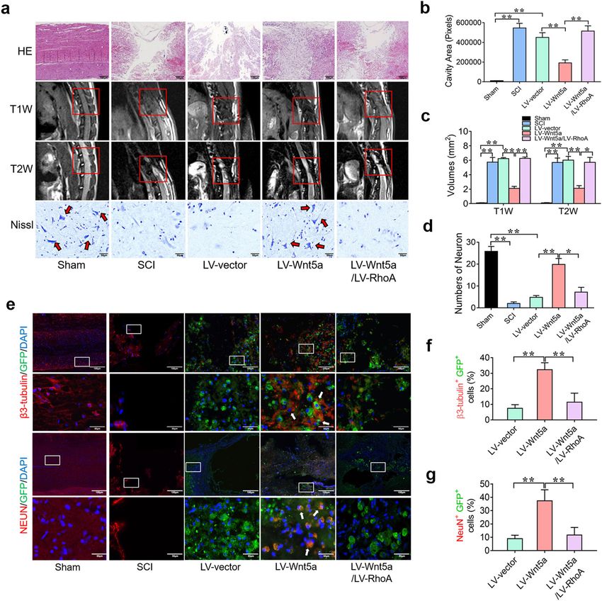
Li et al. Experimental & Molecular Medicine (2020) 52:2020–2033                                                                       2021

   One of most important mechanisms of cell transplan-                         Dawley (SD) rats (Laboratory Animal Center of Sun Yat-
tation therapy is promoting neuronal-oriented differ-                          Sen University, Guangzhou, China)27–29. NSCs were pla-
entiation to rebuild the neural circuit1. Wnt signaling is                     ted in a T25 culture flask (Corning, Acton, MA, 430639)
critical in modulating many biological processes, includ-                      containing maintenance medium consisting of Dulbecco’s
ing neuronal differentiation and regeneration. Many stu-                       modified Eagle’s medium/F12 nutrient mixture, 2% B27,
dies of SCI have demonstrated that canonical Wnt/                              1% penicillin/streptomycin, 1% L-glutamine (Invitrogen,
β-catenin signaling has a neuroprotective effect and                           Carlsbad, CA, 11320033), 20 ng/mL fibroblast growth
induces axonal and neural regeneration effectively12–15;                       factor-2 (FGF-2) and 20 ng/mL epidermal growth factor
however, Wnt/β-catenin has been reported to induce                             (EGF) (PeproTech, Rocky Hill, NJ, 96–400–29, 96-AF-
tumorigenesis under specific conditions, and the neural                         100–15). NSCs were cultured at 37 °C in 5% CO2 and
effect of β-catenin is suppressed by other pathways, such as                   were passaged via weekly digestion with Accutase (Milli-
Notch signaling16–20. Thus, the clinical application of Wnt/                   pore, Bedford, MA, SCR005) in the medium described
β-catenin signaling is greatly limited. Recently, noncanonical                 above. All NSCs used in this study were between passages
Wnt proteins, such as Wnt4, Wnt5a, and Wnt11, have                             2 and 4.
gained growing attention as attractive factors for neuronal                      To induce neuronal differentiation, cells were plated at
differentiation. Noncanonical Wnt signaling, including the                     a density of 2 × 10⁵ cells/well in 6- or 12-well tissue cul-
Wnt/Ryk, Wnt/Ca+, and Wnt/JNK pathways, has been                               ture plates and allowed to adhere for 24 h at 37 °C, at
reported to have a positive effect on neuronal differentia-                    which time cells were switched to neuronal differentiation
tion21–23, but the therapeutic effect and mechanism in                         medium consisting of basic medium supplemented with
promoting neuronal differentiation are still unclear.                          2% B27, 1% penicillin/streptomycin, and 1% l-glutamine.
   MicroRNAs (miRNAs) are small noncoding RNA mole-                            The medium was changed every 2–3 days29.
cules that have been shown to play critical roles in reg-
ulating gene expression at the posttranscriptional level in                    Animal experiments
many cellular processes, including neural development,                           Adult female SD rats (weighing 200–220 g, supplied by
tumor metastasis, cell proliferation, apoptosis, and differ-                   the Experimental Animal Center of Sun Yat-Sen Uni-
entiation24. Previous studies have reported that RhoA is the                   versity, Guangzhou, China) were used to establish an SCI
target gene of miRNA200b-3p (mi200b-3p) and that RhoA                          animal model27–29. Behavioral and histological analyses
expression is downregulated by mi200b-3p25. In addition,                       were performed using previously described standard
the RhoA/Rock1 pathway has been recognized as a negative                       methods30–33 and detailed in online supplementary
neurogenesis pathway that has adverse effects on neuronal                      materials. All procedures performed on experimental
differentiation and tissue repair in SCI26. Thus, confirming                    animals were approved by the Animal Care and Use
the mechanism of action of mi200b-3p/RhoA in neuronal                          Committee of Sun Yat-Sen University and were con-
differentiation is necessary but has yet to be achieved.                       ducted in accordance with the Guide to the Care and Use
  The purpose of the current study was to investigate the                      of Experimental Animals by the National Research
neurogenic capacity of Wnt5a and the potential of Wnt5a                        Council (1996, United States).
for application in NSC transplantation in SCI and explore
the underlying mechanism, which may provide useful                             Results
information for translational applications.                                    miRNA200b-3p is critical for Wnt5a promoting neuronal
                                                                               differentiation
Methods and materials                                                            We first determined the neuronal differentiation capa-
   A detailed description of all materials and methods used                    city of Wnt5a and mi200b-3p in NSCs. NSCs were treated
is provided in the Supplementary Materials and Methods,                        with Wnt5a (10 ng/mL) or transfected with the mi200b-
as follows: NSC isolation, culture, and transfection, len-                     3p mimic. The immunofluorescence results showed that
tiviral vector construction, transduction, transfection of                     the number of β3-tubulin- and MAP2-positive cells was
mi200b-3p mimics and inhibitors, pharmaceutical inhi-                          significantly increased by Wnt5a and mi200b-3p, while
bition, real-time quantitative reverse transcription PCR                       the number of GFAP-positive cells was not increased
(RT-qPCR), western blot analysis, luciferase reporter                          (Supplementary Figs. S1a, b and S2a, b). Similar results of
assay, immunofluorescence, surgical procedures and cell                         RT-qPCR and WB revealed the expression of neurogenic
transplantation, functional assessment, histological ana-                      markers, including β3-tubulin, MAP2 and GFAP, at the
lyses and statistical analyses.                                                mRNA and protein levels (Supplementary Figs. S1c, d and
                                                                               S2c, d). These results suggest that Wnt5a and mi200b-3p
NSC isolation and culture                                                      were able to promote neuronal differentiation.
  NSCs were obtained from the fetal brain of embryonic                           We next determined whether mi200b-3p was involved
day 14 rats, which were extracted from pregnant Sprague-                       in Wnt5a-induced neurogenesis. We first examined the

Official journal of the Korean Society for Biochemistry and Molecular Biology
Li et al. Experimental & Molecular Medicine (2020) 52:2020–2033 2022
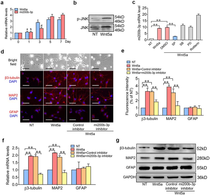
Fig. 1 miRNA200b-3p is critical for Wnt5a to promote neuronal differentiation. a RT-qPCR analyses of Wnt5a and mi200b-3p expression with
neuronal differentiation over time. b Western blot analyses of p-JNK expression in NSCs treated with Wnt5a. c RT-qPCR analyses of mi200b-3p in NSCs
treated with different pathway inhibitors and then stimulated with Wnt5a. d and e Immunofluorescence staining of NSCs transfected with an
mi200b-3p inhibitor and then stimulated with Wnt5a. f and g RT-qPCR and western blot analyses of neurogenic marker expression in NSCs
transfected with an mi200b-3p inhibitor and then stimulated with Wnt5a. SP: JNK inhibitor; PD: ERK inhibitor; SB: p38 inhibitor; Rottlerin: PKCδ
inhibitor (the data are presented as the mean ± SD from one representative experiment of three independent experiments performed in triplicate.
a, b
P < 0.01 compared between 3 days, 5 days, and 7 days; c, dP < 0.05 compared between 5 days and 7 days; **P < 0.01 compared between groups.).
expression of Wnt5a and mi200b-3p in the process of Wnt5a for 24 h. mi200b-3p expression was then detected
neuronal differentiation. The RT-qPCR results showed with RT-qPCR, and the results showed that inhibiting the
that both Wnt5a and mi200b-3p expression significantly JNK pathway significantly decreased mi200b-3p expres-
increased with the duration of neuronal differentiation sion at the mRNA level (Fig. 1c), suggesting that Wnt5a
(Fig. 1a). The WB results confirmed that Wnt5a could upregulated mi200b-3p expression through the MAPK/
promote the activation of MAPK/JNK pathways (Fig. 1b). JNK pathway. To further determine whether mi200b-3p
Furthermore, NSCs were treated with pharmaceutical was involved in Wnt5a-induced neurogenesis, a target-
inhibitors of MAPKs and PKCδ and then stimulated with specific inhibitor was transfected into NSCs to suppress
Official journal of the Korean Society for Biochemistry and Molecular Biology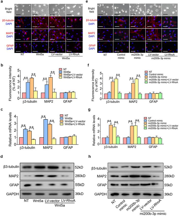
Li et al. Experimental & Molecular Medicine (2020) 52:2020–2033                                                                          2023

mi200b-3p expression, and then the cells were treated                          Wnt5a induces NSC differentiation into neurons to
with Wnt5a. The immunofluorescence results showed                               promote spinal cord repair in SCI
that the number of β3-tubulin- and MAP2-positive cells                           To investigate whether Wnt5a can enhance the ther-
was significantly decreased by the mi200b-3p inhibitor,                         apeutic benefit of NSC transplantation in vivo. LV-
while the number of GFAP-positive cells was not                                Wnt5a- and LV-shmi200b-3p-transfected NSCs were
increased (Fig. 1d, e). Similar results of RT-qPCR and WB                      injected into the injury site to assess the effect of LV-
showed the expression of neurogenic markers, including                         Wnt5a-transfected NSC transplantation on functional
β3-tubulin, MAP2 and GFAP, at the mRNA and protein                             recovery. As shown in Fig. 4, the rats in the sham group
levels (Fig. 1f, g). These results suggest that mi200b-3p                      could grab and step easily using the hindlimb, whereas the
was critical to Wnt5a-induced neuronal differentiation.                        rats in the SCI group could hardly grab or stand up. As
                                                                               expected, the rats in the LV-Wnt5a group could grab
Wnt5a suppresses RhoA/Rock signaling by upregulating                           mildly and step slowly using their hindlimb, whereas the
mi200b-3p                                                                      rats in the LV-Wnt5a/LV-shmi200b-3p group could
  RhoA is a highly conserved gene in many species and                          hardly grab or stand up, like those in the SCI group (Fig. 4a).
modulates many biological processes, including inter-                          To determine whether behavioral function had been
cellular adhesion, cell polarity, neural maintenance and                       repaired, we used the BBB score to evaluate hindlimb
differentiation, as well as gene expression34 (Fig. 2a).                       locomotor activity after SCI, and as expected, hindlimb
Activation of RhoA/Rock1 signaling in neural stem cells                        locomotion was zero immediately after the operation for
inhibits their survival and differentiation26. To investigate                  all rats. Over the course of 2 months, the rats in the LV-
whether the neurogenic effect of Wnt5a depends on                              Wnt5a group exhibited significantly higher BBB scores
inhibition of the RhoA/Rock1 pathway, NSCs were trea-                          than those in the SCI group; however, the rats in the LV-
ted with Wnt5a (10 ng/mL) for 3 days, and the RT-qPCR                          Wnt5a/LV-shmi200b-3p group exhibited the same BBB
and WB results showed that Wnt5a suppressed RhoA and                           scores as those in the SCI group (Fig. 4b). To further
Rock1 gene expression (Fig. 2b, c). Moreover, we con-                          confirm sensory and motor functional recovery, we per-
firmed that RhoA is the target gene of mi200b-3p and that                       formed electrophysiological analysis. The SCEP in the LV-
the binding site is from 290–329 bp (Fig. 2d). The western                     Wnt5a group was stronger, the latency was shorter, and
blot and luciferase activity results showed that the                           the amplitude was higher than that in the SCI and LV-
mi200b-3p mimic suppressed RhoA and Rock1 gene                                 vector groups. However, the SCEP in the LV-Wnt5a/LV-
expression, while the mi200b-3p inhibitor increased RhoA                       shmi200b-3p group was weaker, the latency was longer,
and Rock1 expression at the protein level and transcrip-                       and the amplitude was lower than that in the LV-Wnt5a
tional activity in NSCs (Fig. 2e, f). To further determine                     group (Fig. 4c–e). These results suggest that Wnt5a
whether mi200b-3p is required for the inhibitory effect of                     promoted motor functional recovery after spinal cord
Wnt5a on RhoA and Rock1 expression, an mi200b-3p                               injury in an mi200b-3p-dependent manner.
inhibitor was used to suppress mi200b-3p expression in                           To further determine the tissue repair effect of Wnt5a-
NSCs before Wnt5a stimulation. The results showed that                         NSC transplantation in vivo, the lesion cavity was calcu-
with successful suppression of mi200b-3p, the inhibitory                       lated on H&E staining at 8 weeks after the operation to
effect of Wnt5a on the expression of RhoA and Rock1 was                        detect tissue repair. The total size of the lesion cavity in
significantly reduced (Fig. 2g, h). These results suggest                       the LV-Wnt5a group was significantly smaller than that in
that Wnt5a suppressed RhoA/Rock1 expression in an                              the SCI group. However, the total size of the lesion cavity
mi200b-3p-dependent manner.                                                    in the LV-Wnt5a/LV-shmi200b-3p group was larger than
                                                                               that in the LV-Wnt5a group (Fig. 5a, b). We also used
Overexpression of RhoA sabotaged the neuroinductive                            MRI to measure the volume of the injured spinal cord.
effects of Wnt5a/mi200b-3p                                                     The volume of the injured spinal cord cavity was identi-
   To further confirm that Wnt5a and mi200b-3p promote                          fied as a hypointense region in T1-weighted images and a
neuronal differentiation by suppressing RhoA expression,                       hyperintense region in T2-weighted images. Findings
NSCs were transfected with RhoA gene lentivirus and then                       similar to those of H&E staining were observed on MRI
stimulated with a Wnt5a or mi200b-3p mimic. The                                (Fig. 5a, c). Furthermore, the number of ventral horn
immunofluorescence results showed that the number of β3-                        motor neurons at the lesion epicenter was calculated
tubulin- and MAP2-positive cells was significantly increased                    using Nissl staining. The number of surviving neurons in
by the Wnt5a and mi200b-3p mimics and significantly                             the LV-Wnt5a group was significantly increased com-
decreased by RhoA gene overexpression (Fig. 3a, b, e, f).                      pared with that in the SCI group and significantly
Similar results of PCR and WB showed the expression of                         decreased in the LV-Wnt5a/LV-shmi200b-3p group
neurogenic markers, including β3-tubulin, MAP2 and                             compared with that in the LV-Wnt5a group (Fig. 5a, d).
GFAP, at the mRNA and protein levels (Fig. 3c, d, g, h).                       We further investigated the differentiation status of the

Official journal of the Korean Society for Biochemistry and Molecular Biology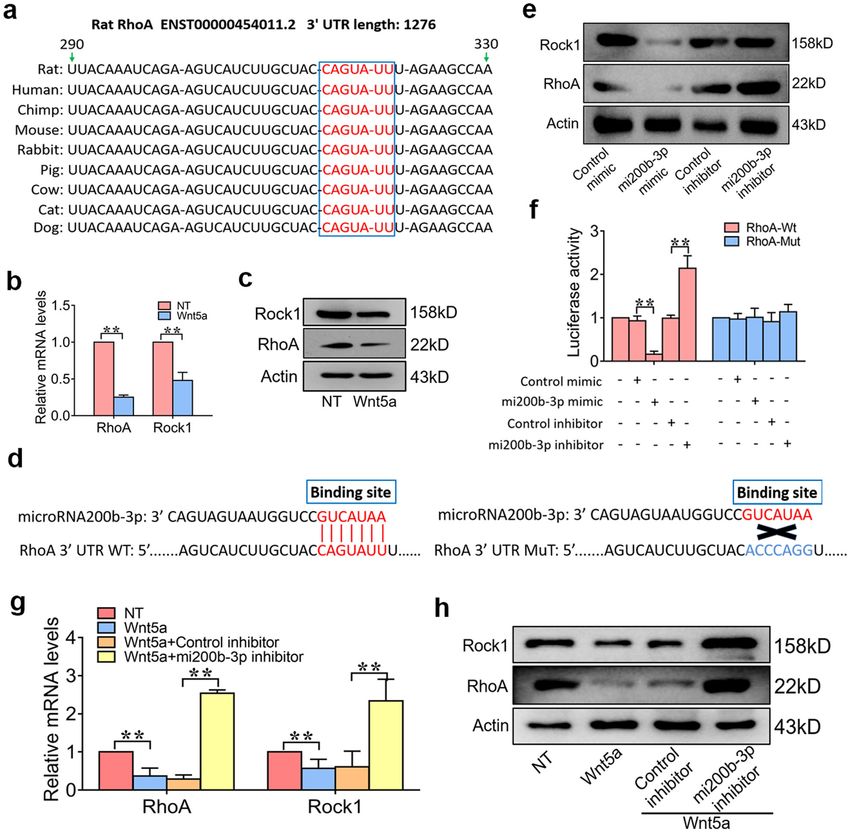
Li et al. Experimental & Molecular Medicine (2020) 52:2020–2033                                                                                 2024

  Fig. 2 Wnt5a suppresses RhoA/Rock signaling by upregulating mi200b-3p. a A schematic diagram illustrating that the RhoA gene is widely
  conserved in many species. b and c RT-qPCR and western blot analyses of RhoA and Rock1 expression in NSCs stimulated with Wnt5a. d A schematic
  diagram illustrating the binding site of mi200b-3p in the RhoA sequence. e Western blot analyses of RhoA and Rock1 expression in NSCs treated with
  an mi200b-3p mimic and inhibitor. f mi200b-3p binding site-directed mutagenesis analysis of the RhoA promoter. g and h RT-qPCR and western blot
  analyses of RhoA and Rock1 expression in NSCs transfected with an mi200b-3p inhibitor and then stimulated with Wnt5a (the data are presented as
  the mean ± SD from one representative experiment of three independent experiments performed in triplicate. **P < 0.01 compared between
  groups.).

transplanted NSCs around the injured site of the spinal                        increased in the LV-Wnt5a group (Fig. 5e–g). In addition,
cord. In the LV-vector and LV-Wnt5a/LV-shmi200b-3p                             our results showed that some GFP-positive NSCs expressed
groups, only a few GFP-positive NSCs showed early                              an oligodendrocyte marker (MBP) in the LV-Wnt5a group,
neuronal marker (β3-tubulin) and mature neuronal mar-                          and only a few GFP-positive NSCs expressed an astrocyte
ker (NeuN) expression. In contrast, the number of GFP+                         marker (GFAP) in the LV-Wnt5a group. In the LV-vector
β3-tubulin+ cells and GFP+ NeuN+ cells was significantly                        and LV-Wnt5a/LV-shmi200b-3p groups, the number of

Official journal of the Korean Society for Biochemistry and Molecular Biology
Li et al. Experimental & Molecular Medicine (2020) 52:2020–2033                                                                          2025

  Fig. 3 Overexpression of RhoA suppresses the neuroinductive effects of Wnt5a/mi200b-3p. a and b Immunofluorescence staining of NSCs
  transfected with LV-RhoA and then treated with Wnt5a. c and d RT-qPCR and western blot analyses of neurogenic marker expression in NSCs
  transfected with LV-RhoA and then treated with Wnt5a. e and f Immunofluorescence staining of NSCs transfected with the RhoA gene plasmid and
  treated with an mi200b-3p mimic. g and h RT-qPCR and western blot analyses of neurogenic marker expression in NSCs transfected with LV-RhoA
  and then treated with an mi200b-3p mimic. (The data are presented as the mean ± SD from one representative experiment of three independent
  experiments performed in triplicate. **P < 0.01 compared between groups.).

Official journal of the Korean Society for Biochemistry and Molecular Biology
Li et al. Experimental & Molecular Medicine (2020) 52:2020–2033 2026
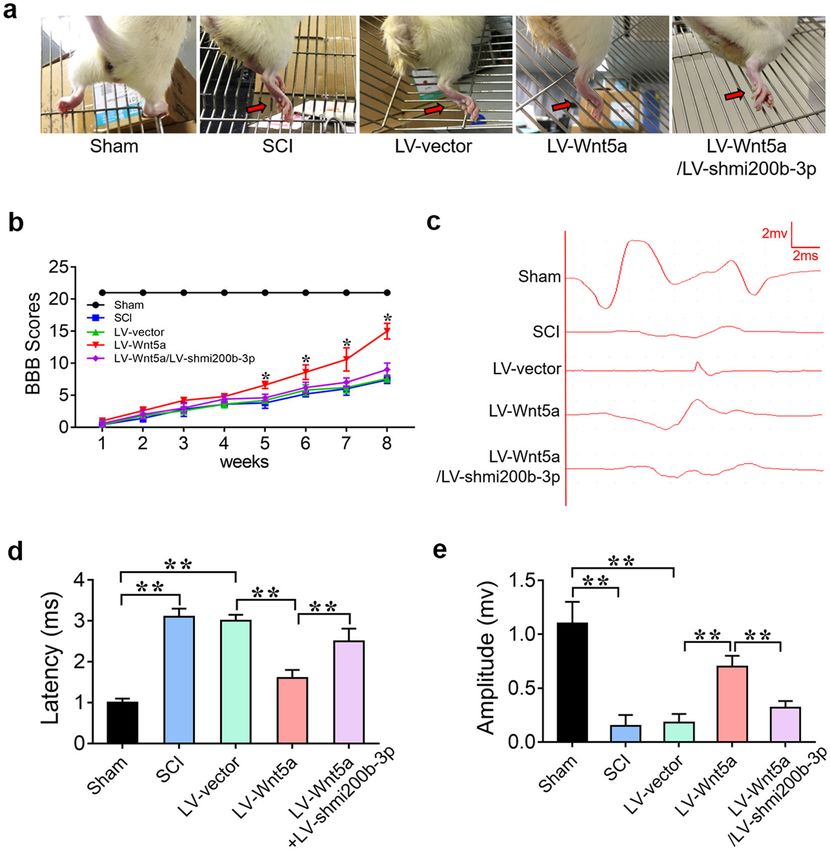
Fig. 4 Wnt5a-modified NSCs promote locomotor functional recovery in SCI. a Behavioral character images showing hindlimb movement in
different groups. b BBB scores in different groups. c Electrophysiological outcomes of the SCEP in different groups. d and e Quantification of the SCEP
latency and amplitude. (**P < 0.01 compared between groups.).
GFP+ MBP+ cells was significantly decreased, and the could grab mildly and step slowly using their hindlimb,
number of GFP+ GFAP+ cells was significantly increased whereas the rats in the LV-Wnt5a/LV-RhoA group could
(Supplementary Fig. S3a–c). These results suggest that hardly grab or stand up, like those in the SCI group
Wnt5a induced transplanted NSCs to mainly differentiate (Fig. 6a). BBB and electrophysiological analyses showed
into neurons through upregulated mi200b-3p expression to that rats in the LV-Wnt5a group exhibited better motor
promote spinal cord repair after SCI. functional recovery, while rats in the LV-Wnt5a/LV-
RhoA group exhibited poor motor functional recovery
Wnt5a induces NSC differentiation into neurons by after SCI (Fig. 6b–e). These results suggest that Wnt5a
suppressing RhoA expression to promote spinal cord promotes motor functional recovery in SCI by suppres-
repair in SCI sing RhoA expression.
To further determine whether Wnt5a promotes spinal To further determine the tissue repair effect of Wnt5a
cord recovery by suppressing RhoA expression in vivo, through suppressing RhoA expression in vivo, H&E
LV-Wnt5a- and LV-RhoA-transfected NSCs were injec- staining, MRI and Nissl staining were used to detect tissue
ted into the injury site to assess the effect on functional repair at 8 weeks after the operation. The total size of the
recovery. As expected, the rats in the LV-Wnt5a group lesion cavity in the LV-Wnt5a group was significantly
Official journal of the Korean Society for Biochemistry and Molecular Biology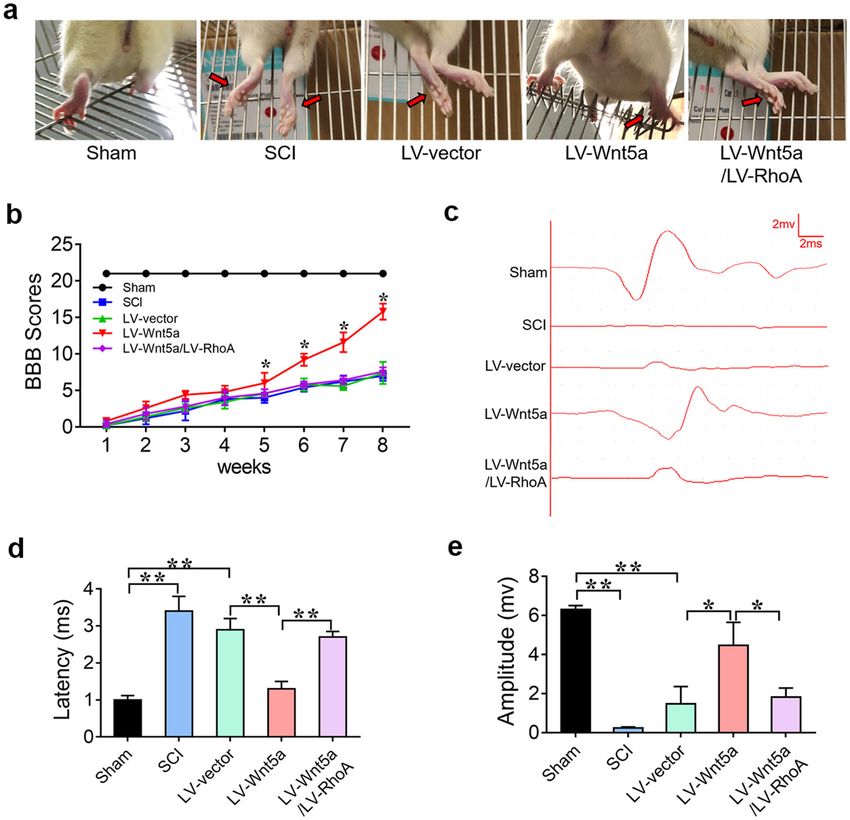
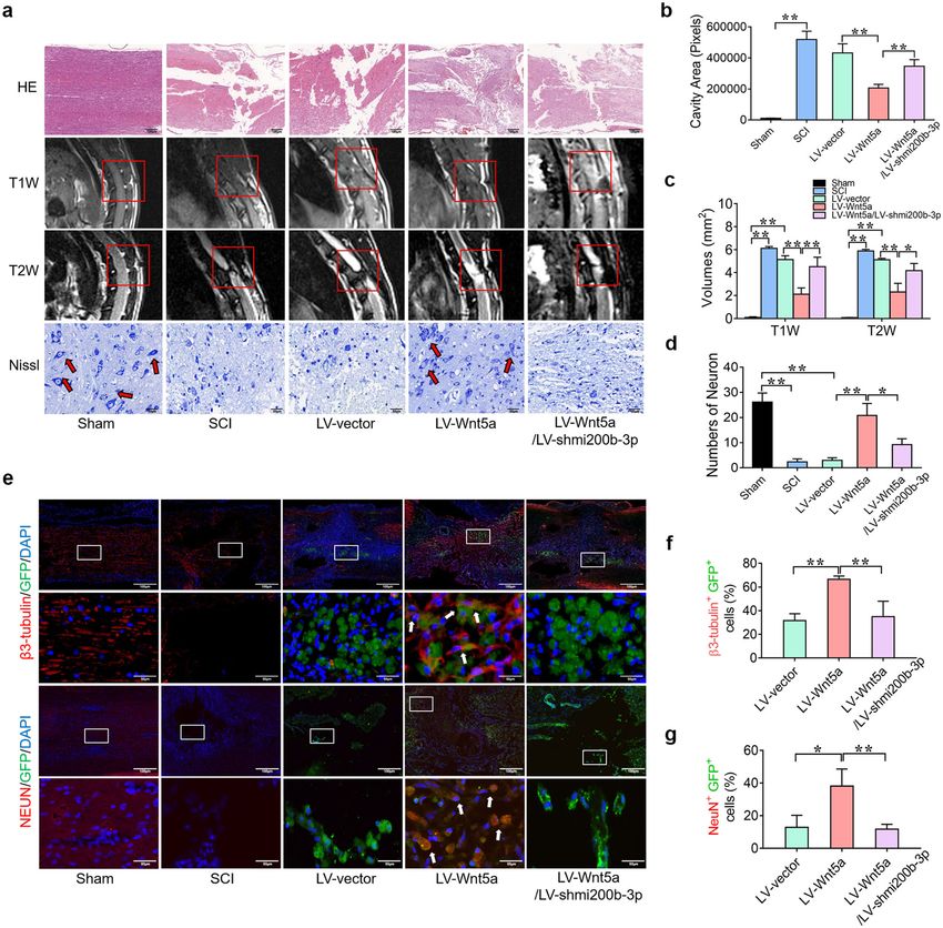
Li et al. Experimental & Molecular Medicine (2020) 52:2020–2033                                                                                    2027

  Fig. 5 Wnt5a-modified NSCs promote histological repair of the damaged spinal cord in SCI. a H&E staining, Nissl staining, and MRI analyses of
  the spinal cord in different groups. b–d Quantification of H&E staining, Nissl staining and MRI results in different groups. e Immunofluorescence
  staining of the spinal cord in different groups. The white boxes indicate images of higher magnification. The white arrow indicates colocalized cells.
  f and g Quantification of immunofluorescence staining. (*P < 0.05 compared between groups; **P < 0.01 compared between groups.).

smaller than that in the SCI group; however, the total size                    that in the LV-Wnt5a group (Fig. 7a, d). Furthermore, we
of the lesion cavity in the LV-Wnt5a/LV-RhoA group was                         investigated the differentiation status of the transplanted
larger than that in the LV-Wnt5a group (Fig. 7a, b).                           NSCs around the injured site of the spinal cord. In the
Findings similar to those of H&E staining were observed                        LV-vector group and the LV-Wnt5a/LV-RhoA group,
on MRI (Fig. 7a, c). Nissl staining showed that the number                     only a few GFP-positive cells showed early neuronal
of surviving neurons in the LV-Wnt5a group was sig-                            marker (β3-tubulin) and mature neuronal marker (NeuN)
nificantly increased compared with that in the SCI group.                       expression. In contrast, the number of GFP+ β3-tubulin+
The number of surviving neurons in the LV-Wnt5a/LV-                            cells and GFP+ NeuN+ cells was significantly increased in
RhoA group was significantly decreased compared with                            the LV-Wnt5a group (Fig. 7e–g). In addition, the number

Official journal of the Korean Society for Biochemistry and Molecular Biology
Li et al. Experimental & Molecular Medicine (2020) 52:2020–2033                                                                                2028

  Fig. 6 Wnt5a induces neuronal differentiation by suppressing RhoA expression to promote locomotor functional recovery in SCI.
  a Behavioral character images showing hindlimb movement in different groups. b BBB scores in different groups. c Electrophysiological outcomes of
  the SCEP in different groups. d and e Quantification of SCEP latency and amplitude. (*P < 0.05 compared between groups; **P < 0.01 compared
  between groups.).

of GFP+ MBP+ cells was significantly decreased and the                          spinal cord injury. Since Wnt signaling has been proven to
number of GFP+ GFAP+ cells was significantly increased                          be neurogenic, we speculated that Wnt5a might have
in the LV-Wnt5a/LV-RhoA groups (Supplementary Fig.                             therapeutic potential in traumatic spinal cord injury.
S3a–c). These results suggest that Wnt5a induced NSCs                            In the current study, we recorded three key observations
to mainly differentiate into neurons by suppressing RhoA                       that provide insights into Wnt5a-induced neurogenesis.
expression to promote spinal cord repair after SCI.                            First, we confirmed that Wnt5a induced neuronal differ-
                                                                               entiation and suppressed RhoA/Rock signaling in NSCs.
Discussion                                                                     Second, we provided the first evidence that mi200b-3p was
  Spinal cord injury due to external trauma is a difficult                      upregulated by Wnt5a and essential for both the neuro-
clinical problem without an ideal solution thus far2,35.                       genesis and suppression of RhoA signaling mediated by
Currently, the current therapeutic strategies for SCI are                      Wnt5a. Third, we provided in vivo evidence that mi200b-3p
not sufficient due to the failure of axonal and neural                          is critical for Wnt5a to induce transplanted NSCs to dif-
regeneration36. Thus, an agent that is able to suppress                        ferentiate into neurons to promote functional and histolo-
negative effects on neuronal differentiation and simulta-                      gical recovery by suppressing RhoA signaling after SCI.
neously promote neurogenesis in the microenvironment                             Previous studies have shown that Wnt5a has a neuro-
would be of great benefit for the treatment of traumatic                        genic effect. Several classic pathways have been found to

Official journal of the Korean Society for Biochemistry and Molecular Biology
Li et al. Experimental & Molecular Medicine (2020) 52:2020–2033 2029 Fig. 7 Wnt5a induces neuronal differentiation by suppressing RhoA to promote histological repair of the damaged spinal cord in SCI. a H&E staining, Nissl staining and MRI analyses of the spinal cord in different groups. b–d Quantification of H&E staining, Nissl staining and MRI results in different groups. e Immunofluorescence staining of the spinal cord in different groups. The white boxes indicate images of higher magnification. The white arrow indicates colocalized cells. f and g Quantification of immunofluorescence staining results. (*P < 0.05 compared between groups; **P < 0.01 compared between groups.). be involved in neuronal differentiation. Varela-Nallar there is substantial evidence that activating noncanonical et al.37 reported that Wnt5a signaling via the Wnt/Ca+ pathways suppresses axonal and neural growth. Wnt-Ryk pathway stimulated dendritic spine morphogenesis in pathways have been reported to suppress axonal and hippocampal neurons and played a trophic role in neu- neurite growth39,40. Elevating the expression of non- ronal differentiation. Wnt5a also activated the JNK and canonical Wnt ligands contributes to the lack of axonal Rac1 pathways to promote ventral midbrain morpho- regeneration in CNS models, while blocking noncanonical genesis and dopaminergic differentiation38. However, Wnt signaling promotes axonal growth and functional Official journal of the Korean Society for Biochemistry and Molecular Biology
Li et al. Experimental & Molecular Medicine (2020) 52:2020–2033                                                                       2030

recovery39,41,42. In addition, miRNAs have been reported                       previous studies have reported that most NSCs transplanted
to play critical roles in regulating gene expression at the                    into SCI lesions differentiate into astrocytes rather than
posttranscriptional level in neural development, tumor                         neurons11. Thus, an agent that increases the neuronal dif-
metastasis, and cell proliferation, apoptosis and differ-                      ferentiation rate of NSCs is necessary for NSC transplan-
entiation24. Previous studies have reported that RhoA,                         tation therapy in SCI. To confirm the role of Wnt5a and
which was recognized to suppress neuronal differentia-                         mi200b-3p in neuronal differentiation and regeneration, we
tion26,43, was the target gene of mi200b-3p and that RhoA                      further conducted an in vivo experiment. Behavioral char-
expression was downregulated by mi200b-3p25.                                   acterization, BBB score evaluation and SCEP analyses
  In this study, we confirmed that Wnt5a, which has been                        revealed that the transplantation of Wnt5a-transfected
controversial in neurogenesis, to the best of our knowl-                       NSCs could lead to better locomotor recovery than naive
edge, and mi200b-3p have strong positive effects on                            NSC transplantation. The silencing of mi200b-3p in
neurogenesis in NSCs (Supplementary Figs. S1 and S2). In                       Wnt5a-transfected NSCs significantly suppressed loco-
addition, we provided evidence that the expression levels                      motor recovery in rodents with SCI (Fig. 4). Moreover, our
of both Wnt5a and mi200b-3p were elevated in the pro-                          histological results revealed that the transplantation of
cess of neuronal differentiation, mi200b-3p was upregu-                        Wnt5a-transfected NSCs could lead to better spinal cord
lated by Wnt5a through the MAPK/JNK pathway in                                 tissue repair than the transplantation of naive NSCs, and
NSCs, and mi200b-3p played a positive role in neuro-                           the silencing of mi200b-3p in Wnt5a-transfected NSCs was
genesis. The inhibition of mi200b-3p partially but sig-                        found to significantly abolish the recovery effect. Most
nificantly      suppressed      Wnt5a-induced      neuronal                     Wnt5a-transfected NSCs could differentiate into neurons
differentiation. These results strongly suggest that Wnt5a                     rather than astrocytes, the latter of which are dis-
upregulated mi200b-3p expression through the MAPK/                             advantageous in spinal cord repair, to reconnect the neural
JNK pathway to promote neuronal differentiation (Fig. 1).                      circuit, and this phenomenon required the involvement of
  The underlying mechanism of Wnt5a-induced neuronal                           mi200b-3p (Fig. 5 and Supplementary Fig. S3).
differentiation is not yet clear. Previous studies have                           Finally, we further confirmed that Wnt5a/mi200b-3p
reported a negative effect of RhoA/Rock signaling on                           improved locomotor functional recovery and promoted
neuronal differentiation. Noncanonical Wnt-planar cell                         tissue repair by suppressing the activation of RhoA sig-
polarity (PCP) signaling controls Rho GTPase activity                          naling. Behavioral characterization, BBB score evaluation
locally by activating or suppressing RhoA and Rac1,                            and SCEP analyses revealed that the transplantation of
resulting in many biological processes44,45. Yang et al.26                     Wnt5a-transfected NSCs could lead to better locomotor
demonstrated that Syx is a gene encoding a RhoA-specific                        recovery than the transplantation of naive NSCs, and the
guanine nucleotide exchange factor. Noggin and RARγ,                           overexpression of RhoA in Wnt5a-transfected NSCs was
which are proteins involved in neural differentiation, were                    found to sabotage the therapeutic effect of Wnt5a-
more abundant in embryonic Syx-/- cells. These phe-                            transfected NSCs in rodents with SCI (Fig. 6).
nomena were blocked by the overexpression of active                            Moreover, our histological results revealed that the
RhoA. This strongly suggested that RhoA/Rock signaling                         transplantation of Wnt5a-transfected NSCs could lead to
prevented neuronal differentiation by limiting the pro-                        better spinal cord tissue repair than the transplantation of
motion of neuronal differentiation protein expression.                         naive NSCs, and the overexpression of RhoA in Wnt5a-
Our results reveal that Wnt5a significantly suppressed                          transfected NSCs was found to significantly abolish the
RhoA and Rock1 expression during neuronal differentia-                         recovery effect. Most Wnt5a-transfected NSCs could
tion. RhoA is the target gene of mi200b-3p according to a                      differentiate into neurons to reconnect the neural circuit,
biological database, and with mi200b-3p silencing, the                         and this phenomenon was inhibited by the overexpression
suppressive effect of Wnt5a was abolished (Fig. 2). These                      of RhoA (Fig. 7 and Supplementary Fig. S3). In addition,
results indicate that mi200b-3p not only mediates the                          some Wnt5a-transfected NSCs were found to differentiate
direct effect of Wnt5a on promoting neurogenesis but                           into oligodendrocytes, which are also beneficial for spinal
also the inhibitory effect of Wnt5a on the RhoA pathway.                       cord repair47,48. The overexpression of RhoA induced
Moreover, our results show that the overexpression of                          NSCs to mainly differentiate into astrocytes. This result
RhoA could resist the neurogenic effect of Wnt5a and                           indicates that Wnt5a have potential as a therapeutic agent
mi200b-3p in NSCs. All the results suggest that Wnt5a/                         to optimize NSC transplantation after SCI.
mi200b-3p promoted neuronal differentiation by sup-                               In conclusion, we showed a novel mechanism of
pressing activation of the RhoA pathway (Fig. 3).                              Wnt5a-induced neuronal differentiation. Wnt5a-induced
  The transplantation of NSCs is considered to be a                            miRNA200b-3p expression is essential for the neu-
potential therapeutic strategy in SCI because NSCs can                         roinductive effect and the inhibitory effect of the RhoA/
differentiate into neurons and oligodendrocytes to                             Rock pathway on neuronal differentiation in spinal cord
reconnect the neural circuit in the lesion1,46. However,                       injury (Fig. 8).

Official journal of the Korean Society for Biochemistry and Molecular Biology
Li et al. Experimental & Molecular Medicine (2020) 52:2020–2033                                                                               2031

  Fig. 8 A schematic diagram illustrating the theory in the current study. Wnt5a-transfected NSCs were injected into the injury site to assess the
  therapeutic effect of Wnt5a-modified NSC transplantation on functional recovery. miRNA200b-3p plays a critical role in Wnt5a promoting
  neurogenesis and suppressing the RhoA/Rock1 signaling pathway.

Official journal of the Korean Society for Biochemistry and Molecular Biology
Li et al. Experimental & Molecular Medicine (2020) 52:2020–2033                                                                                                                 2032

Acknowledgements                                                                             12. Qu, Q. H. et al. Orphan nuclear receptor TLX activates Wnt/beta-catenin sig-
This study was supported by the National Natural Science Foundation of China                     nalling to stimulate neural stem cell proliferation and self-renewal. Nat. Cell
[grant nos.: 81472069, 81971151] and the Guangdong Natural Science                               Biol. 12, 31–U80 (2010).
Foundation, China [grant nos. 2020A1515010265, 2020A1515010306]. X.L., Z.P.,                 13. Rodriguez, J. P. et al. Abrogation of beta-catenin signaling in oligodendrocyte
and L.L. contributed equally to this work. Designed the study: Y.W. and X.L.                     precursor cells reduces glial scarring and promotes axon regeneration after
Conducted the study: X.L., Z.P., X.L., and Y.T. Collected data: X.L., Z.P., L.L., X.L., K.       CNS injury. J. Neurosci. 34, 10285–10297 (2014).
Z., Y.T., and X.Z. Analyzed data: X.L., Z.P., L.L., N.C., L.W., and Y.W. Interpretated       14. Patel, M. et al. Prolonged neural stem cell maturation restores motor function
data: X.L., L.L., L.W., and Y.W. Drafted the manuscript: X.L. and L.L. Revised the               in spinal cord-lesioned rats. Nat. Rev. Neurol. 13, https://doi.org/10.1038/
manuscript content: X.L., L.W., and Y.W. Approved the final version of the                        nrneurol.2017.133 (2017).
manuscript: Y.W. All authors take responsibility for the integrity of the data               15. Seitz, R., Hackl, S., Seibuchner, T., Tamm, E. R. & Ohlmann, A. Norrin mediates
analysis.                                                                                        neuroprotective effects on retinal ganglion cells via activation of the Wnt/
                                                                                                 beta-catenin signaling pathway and the induction of neuroprotective growth
Author details                                                                                   factors in muller cells. J. Neurosci. 30, 5998–6010 (2010).
1                                                                                            16. Jung, Y. S. et al. TMEM9 promotes intestinal tumorigenesis through vacuolar-
 Department of Spine Surgery, The First Affiliated Hospital, Sun Yat-Sen
University, 510080 Guangzhou, Guangdong, China. 2Translational Medicine                          ATPase-activated Wnt/beta-catenin signalling. Nat. Cell Biol. 20, 1421–142
Center, The First Affiliated Hospital, Sun Yat-Sen University, 510080 Guangzhou,                  (2018).
Guangdong, China. 3Department of Pathology, The Seventh Affiliated Hospital,                  17. Li, M. W. et al. Transmembrane protein 170B is a novel breast tumorigenesis
Sun Yat-Sen University, 518107 Shenzhen, Guangdong, China. 4Department of                        suppressor gene that inhibits the Wnt/beta-catenin pathway. Cell Death Dis. 9,
Pathology, The First Affiliated Hospital, Sun Yat-Sen University, 510080                          ARTN 91 https://doi.org/10.1038/s41419-017-0128-y (2018).
Guangzhou, Guangdong, China. 5Department of Spine Surgery, The Seventh                       18. Bresson, L. et al. Podoplanin regulates mammary stem cell function and
Affiliated Hospital, Sun Yat-Sen University, 518000 Shenzhen, Guangdong,                          tumorigenesis by potentiating Wnt/beta-catenin signaling. Development 145,
China. 6Guangdong Province Key Laboratory of Orthopaedics, 510080                                doi:UNSP dev160382 https://doi.org/10.1242/dev.160382 (2018).
Guangzhou, Guangdong, China                                                                  19. Hirabayashi, Y. et al. The Wnt/beta-catenin pathway directs neuronal differ-
                                                                                                 entiation of cortical neural precursor cells. Development 131, 2791–2801
Conflict of interest                                                                              (2004).
The authors declare that they have no conflict of interest.                                   20. Kuwabara, T. et al. Wnt-mediated activation of NeuroD1 and retro-elements
                                                                                                 during adult neurogenesis. Nat. Neurosci. 12, 1097–U1096 (2009).
                                                                                             21. Park, S. Y., Kang, M. J. & Han, J. S. Interleukin-1 beta promotes neuronal
Publisher’s note                                                                                 differentiation through the Wnt5a/RhoA/ JNK pathway in cortical neural
Springer Nature remains neutral with regard to jurisdictional claims in                          precursor cells. Mol Brain 11, ARTN 39 https://doi.org/10.1186/s13041-018-
published maps and institutional affiliations.                                                    0383-6 (2018).
                                                                                             22. Jang, S., Park, J. S. & Jeong, H. S. Neural differentiation of human adipose tissue-
Supplementary information accompanies this paper at https://doi.org/                             derived stem cells involves activation of the Wnt5a/JNK signalling. Stem Cells
10.1038/s12276-020-00536-0.                                                                      Int. 2015, Artn 178618 https://doi.org/10.1155/2015/178618 (2015).
                                                                                             23. Blakely, B. D. et al. Ryk, a receptor regulating Wnt5a-mediated neurogenesis
Received: 16 March 2020 Revised: 15 September 2020 Accepted: 14                                  and axon morphogenesis of ventral midbrain dopaminergic neurons. Stem
October 2020.                                                                                    Cells Dev. 22, 2132–2144 (2013).
Published online: 14 December 2020                                                           24. Smirnova, L. et al. Regulation of miRNA expression during neural cell speci-
                                                                                                 fication. Eur. J. Neurosci. 21, 1469–1477 (2005).
                                                                                             25. Ma, T. & Xue, Y. X. MiRNA-200b regulates RMP7-induced increases in blood-
References                                                                                       tumor barrier permeability by targeting RhoA and ROCKII. Front. Mol. Neurosci.
 1. Assinck, P., Duncan, G. J., Hilton, B. J., Plemel, J. R. & Tetzlaff, W. Cell trans-          9, doi:ARTN 9 https://doi.org/10.3389/fnmol.2016.00009 (2016).
     plantation therapy for spinal cord injury. Nat. Neurosci. 20, 637–647 (2017).           26. Yang, J. N. et al. RhoA inhibits neural differentiation in murine stem cells
 2. Ahuja, C. S. et al. Traumatic spinal cord injury-repair and regeneration. Neu-               through multiple mechanisms. Sci. Signal. 9, doi:ARTN ra76 https://doi.org/
     rosurgery 80, S9–S22 (2017).                                                                10.1126/scisignal.aaf0791 (2016).
 3. Lee, B. B., Cripps, R. A., Fitzharris, M. & Wing, P. C. The global map for traumatic     27. Chen, N. N. et al. Targeted inhibition of leucine-rich repeat and immunoglo-
     spinal cord injury epidemiology: update 2011, global incidence rate. Spinal                 bulin domain-containing protein 1 in transplanted neural stem cells promotes
     Cord. 52, 110–116 (2014).                                                                   neuronal differentiation and functional recovery in rats subjected to spinal
 4. Jin, M. C., Medress, Z. A., Azad, T. D., Doulames, V. M. & Veeravagu, A. Stem cell           cord injury. Crit. Care Med. 44, E146–E157 (2016).
     therapies for acute spinal cord injury in humans: a review. Neurosurg. Focus 46,        28. Zhao, X. Y. et al. Lentiviral vector delivery of short hairpin RNA to NgR1
     Artn E10 https://doi.org/10.3171/2018.12.Focus18602 (2019).                                 promotes nerve regeneration and locomotor recovery in injured rat spinal
 5. Gomes, E. D., Silva, N. A. & Salgado, A. J. Combinatorial therapies for spinal               cord. Sci. Rep-Uk 8, doi:Artn 5447 https://doi.org/10.1038/S41598-018-23751-2
     cord injury: strategies to induce regeneration. Neural Regen. Res. 14, 69–71                (2018).
     (2019).                                                                                 29. Li, X. et al. Wnt4-modified NSC transplantation promotes functional recovery
 6. Mackay-Sim, A. & St John, J. A. Olfactory ensheathing cells from the nose:                   after spinal cord injury. Faseb J. 34, 82–94 (2020).
     Clinical application in human spinal cord injuries. Exp. Neurol. 229, 174–180           30. Kanekiyo, K. et al. Effects of multiple injection of bone marrow mononuclear
     (2011).                                                                                     cells on spinal cord injury of rats. J. Neurotraum. 34, 3003–3011 (2017).
 7. Saberi, H. et al. Safety of intramedullary Schwann cell transplantation for              31. Wu, H. F. et al. The promotion of functional recovery and nerve regeneration
     postrehabilitation spinal cord injuries: 2-year follow-up of 33 cases. J. Neuro-            after spinal cord injury by lentiviral vectors encoding Lingo-1 shRNA delivered
     surg.-Spine 15, 515–525 (2011).                                                             by Pluronic F-127. Biomaterials 34, 1686–1700 (2013).
 8. Ogawa, Y. et al. Transplantation of in vitro-expanded fetal neural progenitor            32. Simard, J. M. et al. MRI evidence that glibenclamide reduces acute lesion
     cells results in neurogenesis and functional recovery after spinal cord con-                expansion in a rat model of spinal cord injury. Spinal Cord. 51, 823–827 (2013).
     tusion injury in adult rats. J. Neurosci. Res. 69, 925–933 (2002).                      33. Ohta, K., Fujimura, Y., Nakamura, M., Watanabe, M. & Yato, Y. Experimental
 9. Lee, K. Z. et al. Intraspinal transplantation and modulation of donor neuron                 study on MRI evaluation of the course of cervical spinal cord injury. Spinal
     electrophysiological activity. Exp. Neurol. 251, 47–57 (2014).                              Cord. 37, 580–584 (1999).
10. Zhu, Y. C., Uezono, N., Yasui, T. & Nakashima, K. Neural stem cell therapy aiming        34. Simon, C. M., Vaughan, E. M., Bement, W. M. & Edelstein-Keshet, L. Pattern
    at better functional recovery after spinal cord injury. Dev. Dynam. 247, 75–84               formation of Rho GTPases in single cell wound healing. Mol. Biol. Cell 24,
    (2018).                                                                                      421–432 (2013).
11. Klein, S. & Svendsen, C. N. Stem cells in the injured spinal cord: reducing the          35. Gabel, B. C., Curtis, E. I., Marsala, M. & Ciacci, J. D. A review of stem cell therapy
    pain and increasing the gain. Nat. Neurosci. 8, 259–260 (2005).                              for spinal cord injury: large animal models and the frontier in humans. World
                                                                                                 Neurosurg. 98, 438–443 (2017).

Official journal of the Korean Society for Biochemistry and Molecular Biology
Li et al. Experimental & Molecular Medicine (2020) 52:2020–2033 2033
36. Binan, L., Ajji, A., De Crescenzo, G. & Jolicoeur, M. Approaches for neural tissue 42. Garcia, A. L., Udeh, A., Kalahasty, K. & Hackam, A. S. A growing field: the
regeneration. Stem Cell Rev. Rep. 10, 44–59 (2014). regulation of axonal regeneration by Wnt signaling. Neural Regen. Res. 13,
37. Varela-Nallar, L., Aranguiz, F. C., Abbott, A. C., Slater, P. G. & Inestrosa, N. C. Adult 43–52 (2018).
hippocampal neurogenesis in aging and Alzheimer’s Disease. Birth Defects Res. 43. Matsukawa, T., Morita, K., Omizu, S., Kato, S. & Koriyama, Y. Mechanisms of
C. 90, 284–296 (2010). RhoA inactivation and CDC42 and Rac1 activation during zebrafish optic
38. Andersson, E. R. et al. Wnt5a cooperates with canonical Wnts to generate nerve regeneration. Neurochem. Int. 112, 71–80 (2018).
midbrain dopaminergic neurons in vivo and in stem cells. Proc Natl Acad. Sci. 44. Gao, C. & Chen, Y. G. Dishevelled: the hub of Wnt signaling. Cell Signal 22,
USA 110, E602–E610 (2013). 717–727 (2010).
39. Tury, A., Tolentino, K. & Zou, Y. M. Altered expression of atypical PKC and Ryk in 45. Mayor, R. & Theveneau, E. The role of the non-canonical Wnt-planar cell
the spinal cord of a mouse model of amyotrophic lateral sclerosis. Dev. polarity pathway in neural crest migration. Biochem. J. 457, 19–26 (2014).
Neurobiol. 74, 839–850 (2014). 46. Tang, Y. W., Yu, P. & Cheng, L. Current progress in the derivation and ther-
40. Lanoue, V. et al. The Wnt receptor Ryk is a negative regulator of mammalian apeutic application of neural stem cells. Cell Death Dis. 8, doi:Artn E3108
dendrite morphogenesis. Sci. Rep-Uk 7, doi:Artn 5965 https://doi.org/10.1038/ https://doi.org/10.1038/Cddis.2017.504 (2017).
S41598-017-06140-Z (2017). 47. Almad, A., Sahinkaya, F. R. & McTigue, D. M. Oligodendrocyte fate after spinal
41. Salinas, P. C. Wnt signaling in the vertebrate central nervous system: from axon cord injury. Neurotherapeutics 8, 262–273 (2011).
guidance to synaptic function. Cold Spring Harb. Perspect. Biol. 4, doi:ARTN 48. Alizadeh, A. & Karimi-Abdolrezaee, S. Microenvironmental regulation of oli-
a008003 https://doi.org/10.1101/cshperspect.a008003 (2012). godendrocyte replacement and remyelination in spinal cord injury. J. Physiol.-
Lond. 594, 3539–3552 (2016).
Official journal of the Korean Society for Biochemistry and Molecular BiologyYou can also read