Understanding the pathogenesis of infectious diseases by single-cell RNA sequencing
←
→
Page content transcription
If your browser does not render page correctly, please read the page content below
Review
                                                                                   www.microbialcell.com

Understanding the pathogenesis of infectious diseases by
single-cell RNA sequencing
Wanqiu Huang1, Danni Wang1 and Yu-Feng Yao1,2,*
1 Laboratory of Bacterial Pathogenesis, Department of Microbiology and Immunology, Institutes of Medical Sciences, Shanghai Jiao
Tong University School of Medicine, Shanghai 200025, China.
2 Department of Infectious Diseases, Shanghai Ruijin Hospital, Shanghai 200025, China.

* Corresponding Author:
Yu-Feng Yao, Rm 710, Bldg 5, 280 S. Chongqing Rd, Shanghai, China 200025; Tel: 021-63846590-776523; Mobile: 13817347153;
Fax: 021-64671226; E-mail: yfyao@sjtu.edu.cn

ABSTRACT Infections are highly orchestrated and dynamic                   doi: 10.15698/mic2021.09.759
processes, which involve both pathogen and host. Transcrip-               Received originally: 19.02.2021;
                                                                          in revised form: 13.07.2021,
tional profiling at the single-cell level enables the analysis of
                                                                          Accepted 21.07.2021,
cell diversity, heterogeneity of the immune response, and                 Published 04.08.2021.
detailed molecular mechanisms underlying infectious dis-
eases caused by bacteria, viruses, fungi, and parasites. Here-
in, we highlight recent remarkable advances in single-cell                Keywords: single-cell RNA sequencing, infectious
RNA sequencing (scRNA-seq) technologies and their applica-                diseases, bacteria, viruses, fungi, parasites, immune
                                                                          response.
tions in the investigation of host-pathogen interactions, cur-
rent challenges and potential prospects for disease treat-
ment are discussed as well. We propose that with the aid of               Abbreviations:
scRNA-seq, the mechanism of infectious diseases will be                   COVID-19 – Coronavirus disease 2019; HC – healthy
further revealed thus inspiring the development of novel                  control; IFN – interferon; LTBI – latent TB infection; Mtb
                                                                          – Mycobacterium tuberculosis; NK – natural killer;
interventions and therapies.
                                                                          PBMC – peripheral blood mononuclear cells; pDC –
                                                                          plasmacytoid dendritic cell; scRNA-seq – single-cell RNA
                                                                          sequencing; TB – tuberculosis; Tfh – follicular helper T
                                                                          cell; UMI – unique molecular identifier.

INTRODUCTION                                                         cellular heterogeneity during infections as well as subpopu-
Infectious diseases have always been serious threats to              lation differences that influence outcomes and individual
public health. Excellent examples are the recent global              differences. Recent advances in scRNA-seq analysis of in-
epidemic of Coronavirus disease 2019 (COVID-19), long-               fections have provided for a new understanding of host-
standing influenza, HIV, Salmonella, as well as other bacte-         pathogen interactions. In this review, we briefly introduce
rial infections. To clarify the pathogenesis of these diseases,      technological improvements in scRNA-seq, highlight re-
it is necessary to understand the interactions between the           ports that interrogate microbial pathogenicity and host
host and the pathogen. Although traditional phenotypic               immune responsiveness at the single-cell level, describe
measurements and bulk transcript analysis can provide                the current limitations of scRNA-seq, and discuss exciting
some insights into pathogenesis, neither the heterogeneity           prospects for the study of infections and possible clinical
of individual cell populations nor the pathogenic states can         application.
be appreciated. With the development of single-cell RNA
sequencing (scRNA-seq), a new technique, which measures              THE TECHNOLOGY ADVANCES IN SINGLE-CELL RNA SE-
the transcriptome at the single-cell level, it is possible to        QUENCING
study cell behaviors at a higher resolution. The use of              The first transcriptome analysis at the single-cell level was
scRNA-seq has refined the human cell landscape [1] and               conducted by Tang et al. in 2009 [5]. Since then, this tech-
driven progress in various areas including immunology [2],           nology has been continuously improved to meet different
developmental biology [3], oncology [4], and infectious              needs, leading to the emergence of several novel methods
diseases. Single cell approaches allow for identification of         such as: SMART-seq2 [6], Drop-seq [7], inDrop [8], CEL-

OPEN ACCESS | www.microbialcell.com                         208                     Microbial Cell | SEPTEMBER 2021 | Vol. 8 No. 9
W. Huang et al. (2021)                                                                                       ScRNA-seq in infection

seq2 [9, 10], and MARS-seq [11]. To date, scRNA-seq has           BACTERIAL INFECTION
developed into a mature workflow, including single cell           The outcomes of an infection are complicated interactions
isolation, cell lysis, conversion of RNA into cDNA with am-       of the pathogen and the host involving multiple biological
plification, library construction, sequencing, and analysis of    factors. Pathogen virulence and growth state, host immun-
the high-throughput data. These new technologies were             ity, diverse cell types, and tissue microenvironments all
developed by improving key steps including cell separation,       impact disease progression and antimicrobial treatment.
library construction, sequencing depth, and quality.              ScRNA-seq has become a powerful tool to probe cell-to-cell
     The emergence of the use of barcodes [12] and unique         variability and uncover both host and bacterial factors that
molecular identifiers (UMI) was a huge advance [13]. The          influence the severity of infection. To date, many scRNA-
single-cell tagged reverse transcription (STRT) sequencing,       Seq studies have been performed to investigate the host-
which first introduced cell-specific barcoding at the reverse     pathogen interactions (Table 1).
transcription stage, enabled highly multiplexed analysis              One well-studied example of infectious diseases is Sal-
[12]. After that, the addition of UMIs identified each mole-      monella infection, a common food-borne pathogen that
cule in a population as distinct, as a random DNA sequence        can produce acute or chronic symptoms, either a limited
label or an aliquot of a complex mixture [14]. Multiple           gastroenteritis or a systemic infection [18]. Recent studies
scRNA-seq methods such as CEL-seq, Drop-seq, and MARS-            have shown that the intracellular bacterial heterogeneity is
seq assess the combination of barcodes and UMIs, provid-          influenced by host cell microenvironments which, in turn,
ing for high throughput and sensitivity. However, multi-          can produce differential host cell immune responses. Dur-
plexing cDNA amplification sacrifices full-length coverage.       ing the infection, macrophages are favorable niches for
These methods profile only the 5'- or 3'-terminus of the          Salmonella survival and proliferation. The scRNA-seq ex-
transcripts. In contrast, SMART-seq2 does not use barcodes        pression profiling of infected macrophages revealed that
or UMIs. The cDNA libraries are generated from individual         the induction of a macrophage type I interferon (IFN) re-
cells, providing full-length transcripts [6] that increase        sponse correlated with variable PhoPQ activity of invading
scalability and availability. A newly developed multiple          Salmonella [19]. When macrophages came across a subset
annealing and dC-tailing-based quantitative single cell RNA       of bacteria with highly modified lipopolysaccharides by
sequencing (MATQ-seq) not only captures the full-length           PhoPQ, the macrophages tended to have a high type I IFN
RNA and genuine biological variation between whole tran-          response. Meanwhile, macrophages harboring non-
scriptomes [15] but also adds UMIs reducing bias with             growing Salmonella displayed hallmarks of the proinflam-
higher sensitivity and lower technical noise.                     matory M1 polarization state that differed little from by-
     Another improvement worth mentioning is the applica-         stander cells [20]. The non-growing bacteria did not trigger
tion of maturing sequencing platforms. Previous methods,          additional immune recognition by intracellular receptors.
e.g., CEL-seq [9], which was inefficient and error-prone,         However, intracellular growing bacteria induced an M2-like
were mainly plate-based. CEL-seq2 [10] employs an auto-           anti-inflammatory response in macrophages, indicating
mated microfluidic platform from Fluidigm (C1 platform).          that intracellular Salmonella were capable of escaping from
With MARS-seq, a high-throughput implementation of the            the host defense by reprogramming macrophage polariza-
original CEL-seq method [11], cells are sorted by fluores-        tion [20]. These data suggest that gene expression hetero-
cence-activated cell sorting (FACS). The newly developed          geneity among infected cells creates diverse environments
Drop-seq [7] and inDrop [8] use nanoliter droplets to cap-        for Salmonella to either persist or exploit its host. In addi-
ture single cells. For Microwell-seq, a high-throughput and       tion, the bacterial pathogenicity also plays an important
low-cost platform, individual cells are trapped in an aga-        role in regulating the host cell state and immune response.
rose microarray and mRNAs are captured with magnetic                  Furthermore, different S. Typhimurium strains have
beads [16]. All these innovative platforms have improved          been shown to produce marked differences during infec-
cell sorting accuracy. The availability of commercial plat-       tion [21]. Compared with non-invasive Salmonella, the
forms such as the Chromium system from 10×Genomics                highly invasive and multi-drug resistant S. Typhimurium
improves scRNA-seq efficiency by automation and lowers            strain ST313 could produce a heterogeneous innate im-
cost as well.                                                     mune response that exploited divergent evasion strategies
     Briefly, even though various technologies have been          for dissemination in vivo. MoDCs infected with invasive
developed, it is necessary to carefully consider the most         Salmonella differentially regulated genes associated with
suitable method for analysis based on actual situations and       endosomal trafficking and antigen presentation pathways.
experimental purposes. A comparative analysis of promi-           Invasive Salmonella induced higher expression of IL10 and
nent scRNA-seq methods revealed that Drop-seq is more             MARCH1 but lower expression of CD83, allowing evasion of
cost-efficient when quantifying the transcriptomes of large       adaptive immune supervision.
numbers of cells at low sequencing depth. Single cell RNA             Moreover, investigations have restricted analysis to on-
barcoding and sequencing (SCRB-seq), with massively par-          ly eukaryotic transcripts, thereby losing the ability to deci-
allel single-cell RNA sequencing (MARS-seq), is preferable        pher the heterogeneity of both host and bacteria at the
when quantifying transcriptomes of fewer cells [17].              same time. The development of scDual-Seq enables the
                                                                  capture of host and pathogen transcriptomes simultane-
                                                                  ously at the single cell level [22]. By utilizing this method to

OPEN ACCESS | www.microbialcell.com                         209                  Microbial Cell | SEPTEMBER 2021 | Vol. 8 No. 9
W. Huang et al. (2021) ScRNA-seq in infection
TABLE 1. The applications of scRNA-seq in infection.
Cell scRNA-seq
Pathogen Host/cell Key findings Ref.
isolation method/platform
Mouse/Bone- The induction of macrophage type I
marrow-derived IFN response was correlated with the
FACS SMART-seq [19]
macrophages variable PhoPQ activity of invading
(BMDMs) bacteria.
Macrophages harboring non-growing
Salmonella displayed proinflammatory
M1 polarization state while macro-
Mouse/BMDMs FACS Smart-seq2 [20]
phages containing growing bacteria
turned into an M2-like anti-
inflammatory expression program.
S. Typhimurium Development of scDual-seq, that cap-
Mouse/BMDMs FACS CEL-Seq2 tured host and pathogen transcrip- [22]
tomes simultaneously.
Invasive Salmonella strain ST313 ex-
Human/Monocyte-
ploited discrete evasion strategies
derived dendritic FACS SMART-seq2 [21]
within infected and bystander MoDCs
cells (MoDCs)
to mediate its dissemination in vivo.
Development of a deconvolution algo-
rithm for inferring cell-type specific
Human/PBMCs FACS 10xGenomics [98]
infection responses from bulk meas-
urements.
Revealed a gradual depletion of a NK
Human/PBMCs FACS 10xGenomics [26]
cell subset from HC LTBI and active TB.
Development of the Seq-well method
M. tuberculosis Human/Monocyte-
and revealed distinct heterogeneity
derived macro- Microwell Seq-Well [24]
between macrophages exposed and
phages (MDM)
unexposed to Mtb.
Identified ACE2 and TMPRSS2 co-
Human/M. mulat-
expressing cells within lung type II
ta/ M. fascicularis/ Microwell Seq-Well
pneumocytes, ileal absorptive entero-
Mouse/multiple Droplets Drop-Seq [38]
cytes, and nasal goblet secretory cells.
tissues, e.g., lung, Microfluidic 10xGenomics
Discovered that ACE2 was a human ISG
ileal, nasal
in vitro.
Development of the Viral-Track meth-
Human/ Bron-
od to scan for viral RNA in scRNA-seq
choalveolar FACS MARS-seq
data and revealed the infection land- [40]
lavage (BAL) sam- Microfluidic 10xGenomics
scape of mild and severe COVID-19
ples
patients.
Combined CRISPR screen with scRNA-
Human/A549 cells,
seq, identified new host factors re-
Primary human ECCITE-seq
SARS-CoV-2 Microfluidic quired for SARS-CoV-2 infection, in- [49]
bronchial epithelial 10xGenomics
creased cholesterol biosynthesis were
cells
related to reduced infection.
Hospitalization was associated with
increased cytotoxic Tfh and cytotoxic T
Human/CD4+ T cells Microfluidic 10xGenomics [45]
helper cells and a reduction in regula-
tory T cells.
Aging induced the dysregulation of the
immune system and increased gene
Human/PBMCs Microfluidic 10xGenomics [104]
expression associated with SARS-CoV-2
susceptibility.
Identified potent neutralizing antibod-
Human/B cells Microfluidic 10xGenomics ies from convalescent COVID-19 pa- [50]
tients.
OPEN ACCESS | www.microbialcell.com 210 Microbial Cell | SEPTEMBER 2021 | Vol. 8 No. 9W. Huang et al. (2021) ScRNA-seq in infection
TABLE 1 (continued). The applications of scRNA-seq in infection.
Cell scRNA-seq
Pathogen Host/cell Key findings Ref.
isolation method/platform
Analyzed viral and host transcrip-
tomes in the same single cell and
Mouse/Lung FACS MARS-seq revealed cellular heterogeneity and [105]
novel markers specific for influenza-
infected cells.
Infections performed at high MOIs
resulted in increased viral gene ex-
pression per cell and IFN lambda 1
Human/A549 cells Microfluidic 10xGenomics [106]
Influenza (IFNL1) showed a widespread pat-
tern of expression more reliant on
paracrine signaling.
Demonstrated the intricate effects
Human/A549 cells Microfluidic 10xGenomics of defective viral genomes on host [107]
transcriptional responses.
Demonstrated that two waves of
Mouse/Lung Microfluidic 10xGenomics pro-inflammatory factors were re- [54]
leased during IAV infection.
Cell state driven by T-cell receptor
mediated cell activation was the
Human/CD4+ T cells Microfluidic Fluidigm C1 main factor of transcriptional heter- [108]
ogeneity and was tested as a bi-
omarker of HIV permissiveness.
Expression of HIV proviruses within
the latent reservoir were influenced
Human/CD4+ T cells Microfluidic 10xGenomics [109]
by the host cell transcriptional pro-
gram.
HIV Characterized cell heterogeneity
during HIV latency and reactivation
Human/CD4+ T cells Microfluidic Fluidigm C1 and identified transcriptional pro- [110]
grams leading to successful reactiva-
tion of HIV expression.
Characterized multiple dynamic
cellular responses and gene expres-
Human/PBMCs Microwell Seq-Well sion modules that varied by time [53]
and cell subsets during acute HIV
infection.
Zika virus (ZIKV) infected NES cells
and radial glia cells and induced
Human/Neuroepithelial
Microfluidic Fluidigm C1 mitochondrial sequestration of cen- [111]
Stem Cells(NES cells)
trosomal phospho-TBK1, nucleoside
analogs inhibited ZIKV replication.
AXL was a candidate Zika entry re-
ceptor in neural stem cells and its
Human/Developing
Zika virus Microfluidic Fluidigm C1 expression was conserved in rodents [112]
cortex
and human cerebral organoid model
systems.
Generated a fully immunocompe-
tent mouse model of ZIKV infection
Mouse/Neuronal stem
Microfluidic 10xGenomics and the NS4B G18R mutation in ZIKV [113]
cells(NSCs)
likely acted through its ability to
diminish IFN-β levels.
Identified cells with viral RNA from
human patients and studied the
Dengue virus Human/PBMCs FACS Smart-seq2 molecular signatures preceding the [114]
development of severe dengue in-
fection.
OPEN ACCESS | www.microbialcell.com 211 Microbial Cell | SEPTEMBER 2021 | Vol. 8 No. 9W. Huang et al. (2021) ScRNA-seq in infection
TABLE 1 (continued). The applications of scRNA-seq in infection.
Cell scRNA-seq
Pathogen Host/cell Key findings Ref.
isolation method/platform
Demonstrated that the EBOV tro-
pism, replication dynamics, and
Rhesus monkeys/
Ebola virus Microwell Seq-Well elicited immune responses were [115]
PBMCs
mediated by viral infection related
to cytokine signaling.
Reconstructed the developmental
FACS Micro- SmartSeq-2 trajectories of Th1 and Tfh (T follicu-
Mouse/CD4+ T cells [70]
fluidic Fluidigm C1 lar helper) cells during blood-stage
P. chabaudi Plasmodium infection.
CD4+ T cell-derived MCSF regulated
Mouse/CD4+ T cells FACS Fluidigm C1 expansion and activation on of spe- [71]
cific myeloid subsets.
Discovered undefined sex-specific
genes as well as three distinct clus-
Human/B+ erythrocytes Microfluidic Fluidigm C1 ters of late-stage asexual parasites [116]
largely defined by stage-specific
genes.
P. falciparum Revealed the gene expression signa-
ture of sexual commitment that
Human/RBCs Droplets Drop-seq AP2-G+ mature schizonts specifically [60]
upregulated additional epigenetic
regulators.
Observed sharp transcriptional tran-
sitions at the asexual stage and dis-
P. falciparum Mouse, Human/Red
FACS Smart-seq2 covered a set of sex-specific genes [61]
P. berghei blood cells (RBCs)
involved in sequestration of mature
gametocytes.
Assembled a Malaria Cell Atlas that
P. falciparum presented the transcriptomic pro-
FACS SMART-seq2
P. berghei Human/RBCs files of individual Plasmodium para- [65]
Microfluidic 10xGenomics
P. knowlesi sites across all morphological life
cycle stages.
Described proteins associated with
the different parasite developmen-
Glossina morsitans
Trypanosoma tal stages in salivary glands and
morsitans/salivary Microfluidic 10xGenomics [117]
brucei highlighted a family of nonvariant
glands
surface proteins associated with
metacyclic parasites.
Revealed that CD14+CD16- mono-
cytes were key regulators of human
Toxoplasma Human/Monocytes Microfluidic 10xGenomics [118]
monocyte transcriptional response
to Toxoplasma.
Integrated GWAS with bulk and
scRNA-seq, identified 27 Candida-
C. albicans Human/PBMCs Microfluidic 10xGenomics response QTLs and revealed a role [77]
for LY86 in the anti-Candida host
response.
Abbreviations: P. knowlesi: Plasmodium knowlesi; FACS: Fluorescence-Activated Cell Sorting; GWAS: Genome-Wide Associated Studies;
HC: healthy control; IAV: Influenza A virus; ISG: Interferon-Stimulated Gene; LTBI: latent TB infection; MCSF: Macrophage Stimulating
Factor; M. fascicularis: Macaca fascicularis; M. mulatta: Macaca mulatta; MOI: Multiplicity Of Infection; Mtb: Mycobacterium tuberculo-
sis; NK: natural killer; PBMCs: Peripheral Blood Mononuclear Cells; P. berghei: Plasmodium berghei; P. chabaudi: Plasmodium chabaudi;
P. falciparum: Plasmodium falciparum; P. knowlesi: Plasmodium knowlesi; QTL: Quantitative Trait Loci; SARS-CoV-2: Severe Acute Respir-
atory Syndrome Corona Virus 2; TB: tuberculosis.
OPEN ACCESS | www.microbialcell.com 212 Microbial Cell | SEPTEMBER 2021 | Vol. 8 No. 9W. Huang et al. (2021) ScRNA-seq in infection
study the process of individual macrophages, the authors VIRAL INFECTION
showed the rate of S. Typhimurium infection was non- Viral infections are always of urgent public concern be-
uniform, supportive by the evidence showing the co- cause of their high degree of transmissibility and pathogen-
existence of all three cell subpopulations. These three cell ic severity. Representative examples are influenza, HIV,
states also showed evidence for a linear progression and SARS-CoV. Advances in single cell technology coupled
through consecutive stages of infection. However, the ex- with mathematical modeling and computer science pro-
periments were limited in that a high multiplicity of infec- vide new horizons for understanding virus and host inter-
tion (MOI) could cause a variable number of bacteria in actions.
infected macrophages, thus masking certain infectious COVID-19, a newly emerged severe acute respiratory
stages and phenotypes. syndrome coronavirus 2 (SARS-CoV-2), has become an on-
Another suitable model for studying host-bacterial in- going global health emergency. Upon entry into the host
teraction is Mycobacterium tuberculosis (Mtb), which sick- cell, SARS-CoV-2 can replicate quickly and trigger a strong
ens millions of people with tuberculosis (TB). Lungs infect- immune response leading to an acute respiratory syn-
ed with Mtb contain several coexisting lesion types such as drome, pulmonary tissue damage, and multiple organ fail-
solid cellular granulomas of densely packed macrophages ure [28]. This highly contagious disease progresses rapidly
and necrotic granulomas with an outer ring of T and B lym- and so far has caused tens of millions of cases. Therefore, it
phocytes [23]. Technological advances in scRNA-seq helped is an urgent need to determine the pathogenic basis of this
to reveal the heterogeneity between different cell types disease in order to improve existing prevention and treat-
and subpopulations during Mtb infection [24]. Gene ex- ment strategies. Single cell technology has shown its
pression shifts associated with cell growth and metabolism unique advantages in relevant investigations that uncov-
were found among cell clusters in response to Mtb. The ered sensitive cell types and the heterogeneity of the host
complex dynamic of host and bacteria can result in distinct immune response, each of which provided valuable in-
symptoms from latent TB infection (LTBI), in which patients sights into vaccine development.
remain clinically asymptomatic, to active TB, a contagious To invade cells, SARS-CoV-2 binds host angiotensin-
state in which patients may suffer from cough, fever and converting enzyme 2 (ACE2) with its spike (S) protein and
night sweats [25]. A small proportion of LBTI patients un- utilizes a type II transmembrane serine protease 2
dergo progression into active TB. For better TB control, it is (TMPRSS2) for priming and activation of the protein [29,
necessary to identify these populations by monitoring reli- 30]. Using scRNA-seq expression analysis of ACE2 and
able biomarkers. Since the intensity of immune response TMPRSS2, different cell types, tissues, and organs were
and alterations in immune cell composition are critical in- assessed. The lung, kidney, bladder, and ileum were found
dicators of severity of disease, many efforts have been to have the greatest expression [31]. Within these organs,
made to investigated the transcript profiles of immune oral mucosa [32], nasal epithelial cells [33], type II alveolar
cells in peripheral blood and lesions. A recent scRNA-seq cells [31], and bronchial transient secretory cells [34] ex-
study compared the transcriptomes of peripheral blood pressed the highest levels of ACE2 and TMPRSS2. As such,
mononuclear cells (PBMC) from healthy controls (HC), LTBI these cells have increased sensitivity to SARS-CoV-2 infec-
and active TB patients. The results showed that there was a tion. Based on evidence obtained from a single cell RNA
gradual depletion of the cytotoxic natural killer (NK) cell expression map of human coronavirus entry factors, sper-
subset (CD3-CD7+GZMB+) during these three states [26]. matogonia and prostate endocrine cells were permissive to
The subset frequency also increased after anti-TB treat- SARS-CoV-2 infection [34], suggesting that males were
ment, which confirmed that the frequency change in NK more vulnerable to coronavirus infection [35]. Multiple risk
cells was involved in host disease severity and could be factors like age, gender and cigarette smoking contribute
used as a novel biomarker to discriminate patients with TB to the infection progression. The chronic smoke exposure
from LTBI and HC. increases lung ACE2 expression and triggers the expansion
The immune response to infectious agents is an orches- of ACE2+ secretory cells in the respiratory tract [35]. Aging
trated process that involves numerous cell types and increases the gene expression associated with SARS-CoV-2
pathways. Ronnie et al. comprehensively characterized the susceptibility and COVID-19 can promote age-induced im-
initial 48 h of the innate immune response to diverse path- mune cell polarization [36]. The expression level of
ogens by MARS-seq [27]. They found that most lymph node TMPRSS2 is increased in older adults compare to children,
cell types showed little pathogen specificity and that anti- which could be a reason for the higher risk of severe dis-
gen-specific immunity was driven by antigen-carrying den- ease among the aged [37]. Furthermore, several studies
dritic cells and monocytes. The NK-driven IFN-γ response demonstrated ACE2 to be an IFN-stimulated gene in hu-
initiated a monocyte-specific signaling cascade that pro- mans that is upregulated by viral infection [38]. These data
moted Th1 development. Taken together, these data allow for the construction of a risk map of vulnerable cell
demonstrate an innate immune heterogeneity in response types, revealing underlying virus transmission mechanisms
to a wide range of pathogens. This knowledge may provide (Figure 1).
insight into the development of safe and effective vaccines. COVID-19 patients present a wide spectrum of clinical
manifestations, which range from asymptomatic infection
to severe pneumonia [39]. The relationship between dis-
OPEN ACCESS | www.microbialcell.com 213 Microbial Cell | SEPTEMBER 2021 | Vol. 8 No. 9W. Huang et al. (2021) ScRNA-seq in infection
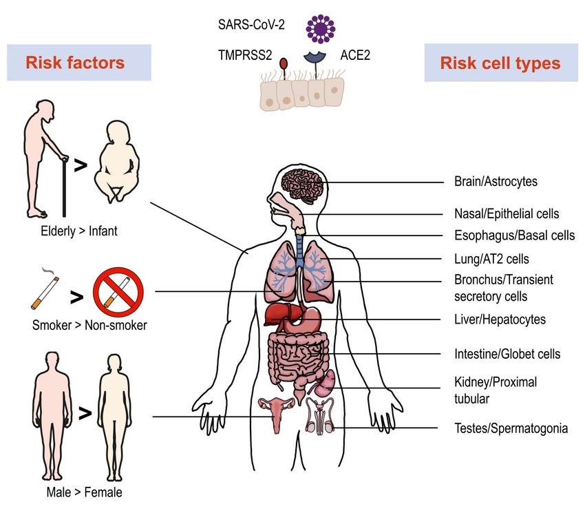
FIGURE 1: Risk factors, vulnerable organs and cell types involved in SARS-Cov-2 infection. Single-cell RNA-seq revealed high expression of
ACE2 and TMPRSS2 in multiple organs and cell types, which are more vulnerable to SARS-Cov-2 infection. Several factors play important roles
in infection and disease process, such as age, smoking and gender.
ease severity and the host immune response is not fully accumulated myeloid cells were neutrophils, FCN+ mono-
understood. Single cell analysis of immune cells and local cytes, and monocyte-derived macrophages, which ex-
landscape signature provide for an understanding of the pressed a viral hallmark IFN type I response [41] and up-
dynamic immune response, which has great heterogeneity regulation of inflammatory chemokine genes, e.g., CCL18.
at the cellular level and differences among individuals. Similarly, severe COVID-19 infections were shown to be
A newly developed computational framework, Viral- associated with developing neutrophils [42, 43] and abun-
Track, enables the global scanning of unmapped viral RNA dant proinflammatory monocyte-derived macrophages
in scRNA-seq data. Integrating these data with the host [44] with an imbalance of the T cell compartment and T cell
transcriptome distinguishes infected cells from bystander function. Proliferating cytotoxic CD8+ T cells and CD4+ fol-
cells and manifests specific virus-induced expression [40]. licular helper T cells (Tfh) were common in COVID-19 pa-
This method was applied to bronchoalveolar-lavage (BAL) tients with reduced proportions of regulatory T cells (Treg)
samples from severe and mild COVID-19 patients and re- [45]. In moderate cases of COVID-19, CD8+ T cells were
vealed the dramatic diversity of immune cell compart- markedly expanded with higher levels of effector mole-
ments and responses. For example, the myeloid compart- cules, e.g., XCL1 [44], and a naïve phenotype for CD4+ T
ment in mild patients was enriched in alveolar macrophage cells in severe cases [40].
and plasmacytoid dendritic cells (pDCs). In severe patients,
OPEN ACCESS | www.microbialcell.com 214 Microbial Cell | SEPTEMBER 2021 | Vol. 8 No. 9W. Huang et al. (2021) ScRNA-seq in infection
In addition to the virus-stimulated alterations in cell at a later stage (day 7 post infection), which may be the
composition, specific host immune response pathways precursor of alveolar macrophages. Enhancing the immune
provide insight into pathogenesis. SARS-CoV-2 infection response against pathogens and reducing the cytokine
induced the activation of the STAT1/IRF3 pathway and storm are the major therapeutic aims of infectious disease.
increased the expression levels of IL6R (interleukin 6 recep- Analysis of these dynamic changes at single cell level could
tor) and IL6ST (interleukin 6 signal transducer), which may give deep insights into a more complete understanding of
have a synergistic effect with elevated IL-6 to induce a immunopathogenesis and offer possible targets for clinical
strong inflammatory response [46]. These results provide treatment.
evidence that cytokine inhibition may be a reliable strategy
to attenuate severe inflammatory responses in COVID-19 PARASITIC INFECTION
patients, e.g., disruption of IL-6 and IL-6R binding [47], Parasitic infection is an important worldwide cause of hu-
CCR1 and/or CCR5 pathway [48]. Additionally, host factors man disease such as Chagas disease, African trypanosomia-
and pathways involved in key elements of the SARS-CoV-2 sis, amebiasis, leishmaniasis, ascariasis, and schistosomia-
viral life cycle have been shown to have a role in regulating sis, leading to millions of deaths [55]. Malaria is a prevalent
the infection [49]. For example, the ATPase proton pump parasitic disease with 229 million victims worldwide in
which interacts with SARS-CoV-2 non-structural protein 6 2019 as reported by the World Health Organization (WHO).
(nsp6) and RAB7A which interacts strongly with non- The disease is caused by a unicellular eukaryotic parasite,
structural protein 7 (nsp7). The loss of RAB7A reduced viral Plasmodium spp., that is transmitted to humans by Anoph-
entry by sequestering ACE2 receptors via altered endoso- eles spp. mosquitoes [56]. Plasmodium falciparum is the
mal trafficking. The upregulation of cholesterol biosynthe- most virulent cause of malaria. During human infection,
sis pathway with the small molecule amlodipine led to re- the parasite attacks the liver and then invades the vascula-
duction of viral infection. It is possible that changes in lipid ture where red blood cells are infected resulting in an in-
composition directly impacted SARS-CoV-2 virion matura- tra-erythrocytic development stage [57]. During this blood
tion and infectivity, indicating potential new therapeutic stage, the fast asexual-replication of parasites produces
targets [49]. the clinical manifestation of the disease. The parasite then
Now that many patients have recovered from the virus converts to a non-replicating sexual gametocyte form,
infection, studying the immune system of these people will which can be transmitted to female mosquito during a
provide valuable information for prevention and treat- blood meal [58]. Understanding the complex life cycle of
ments. Neutralizing antibodies are a powerful weapon to Plasmodium is fundamental for pathogen elimination and
block virus entry. Through analyzing single B cell RNA/VDJ disease treatment. Although these distinct parasitic stages
data of COVID-19 convalescent patients, novel potent neu- have been investigated at the overall population level [57],
tralizing antibodies and S protein-binding antibodies have little is known about the variation between individual para-
been identified, among which BD-368-2 showed a great sites.
therapeutic effect and GD1-69 showed a powerful neutral- ScRNA-seq promises a precise examination of transcrip-
izing activity [50, 51]. Multiple investigations have demon- tional expression heterogeneity during parasitic stage
strated antibody cocktails to largely prevent viral mutation switching and immune response. The transcriptional acti-
escape which produces an optimal antiviral effect [52]. vation of AP2-G is known as the master regulator to initial-
ScRNA-seq technology enables the discovery of new neu- ize the sexual commitment [59]. AP2-G+ mature schizonts
tralizing antibodies, accelerating development of new anti- specifically upregulated epigenetic regulators like histone-
viral drugs and vaccines. modifying enzymes, which facilitated the subsequent ga-
Individual cellular immunity participating in virus-host metocyte development [60]. Individual parasite transcripts
interaction is crucial for controlling virus elimination. A showed distinct stage-specific transcriptional transitions
recent single cell transcriptome analysis revealed a dynam- and revealed differential routes P. falciparum converted
ic cellular program during longitude HIV acute infection, into gametocytes [61-63]. A set of sex-specific genes in-
such as NK cell expansion, naïve CD4+ T cell differentiation, volved in sequestrating gametocytes were found to func-
and a rapid rise in plasma viremia coupled with cell-type tion during immune evasion as well as transmission from
specific interferon-stimulated gene (ISG) upregulation [53]. human to mosquitos. Meanwhile, coupled with the syn-
These cell type frequency shifts at different time points chronized single-cell transcriptome of infected red blood
may shed light on how the immune response were orches- cells, novel gene signature that could be used as an indica-
trated according to the infection progression. In accord- tion to discriminate between sexual and asexual stages
ance with changes in cellular phenotype, the release of were identified [64]. In brief, scRNA-seq has allowed for
pro-inflammatory factors also showed a time-course fluc- high-resolution mapping of the life cycle of Plasmodium
tuation [54]. During the early stage (day 1-3 post infection) spp., providing a fundamental resource for the investiga-
of influenza A virus (IAV) infection, a group of PD-L+ neu- tion of parasite biology and the study of malaria pathogen-
trophils was the major contributor to the first wave of pro- esis [65].
inflammatory factors including Ccl3, Cxcl10, TNF-α, and During the blood-stage of Plasmodium infection, CD4+ T
IL1α. High level of virus HA mRNA and protein were also cells and myeloid cells both play important roles in control-
detected in the cell population. The second wave was ling the progress of disease, in which the intercellular
mainly generated by another subset of Pf4+-macrophages
OPEN ACCESS | www.microbialcell.com 215 Microbial Cell | SEPTEMBER 2021 | Vol. 8 No. 9W. Huang et al. (2021) ScRNA-seq in infection
communication fosters the immune regulation [66]. The ploration. A major limiting factor is the amount of available
CD4+ T-effector cell TCRβ repertoires undergo a polyclonal nucleic acid in that one mammalian cell contains only 10 pg
expansion dominated by TRBV3 gene usage in the acute of total RNA [80]. This limit may result in artificial zeros
phase of P. chabaudi infection [67]. Two types of CD4+ T either systematically or accidently, thus hindering com-
cells are the main sources of protecting against malaria: (1) plete downstream analysis [81]. Using computational sta-
T helper (Th1) cells that secret IFN-γ and stimulate phago- tistical models, it may be possible to circumvent this limita-
cytic cells to capture and kill parasites [68], and (2) Tfh cells tion by modulation of sample variation and noise by use of
that promote development of antigen-specific B cells that VIPER [82] and scSDAEs [83]. With regard to infection,
produce anti-parasitic antibodies [69]. During the activa- most studies have focused on either the host or the patho-
tion and differentiation of CD4+ T cells, myeloid cells were gen, whereas dual RNA-seq allows analysis of the tran-
shown to play a vital regulatory role, not only the dendritic scripts of both host and bacteria simultaneously [84], but
cells presented the antigen stimulation, but inflammatory not at the single-cell bacterial level. However, improved
monocytes could also support a Th1 fate at the Th1/Tfh scDual RNA-seq does enable profiling of both host and
bifurcation [70]. Furthermore, CD4+ T cell-derived macro- bacterial transcripts in one individually infected mammali-
phage stimulating factor (MCSF) facilitated the expansion an cell [22]. It is important to note that the detection of
and activation of specific myeloid subsets such as macro- intracellular bacteria is technically difficult since a single
phages during P. chabaudi infection, which contributed to bacterium contains only 100 fg of total RNA. Further, the
parasite clearance and infection control [71]. Heterogenei- mRNA of bacteria and some virus like hepatitis C virus and
ty revealed by scRNA-seq provides insights into the key dengue virus lack poly (A) tails which are required for li-
molecular and immunological mechanisms of parasite in- brary construction. Enzymatic addition of poly (A) tails,
fection as well as the identification of potential targets for adaptor ligation, and/or random priming for amplification
parasite elimination and vaccine development [72]. [22, 85] address this problem but also require additional
steps to deplete redundant rRNA [86]. Advanced method-
FUNGAL INFECTION ologies for characterization of single infected cells of both
Fungal disease is also a threatening heath problem, causing host and pathogen are needed.
over 1.5 million deaths worldwide each year [73]. With Infection is a complicated process, with outcomes of in-
advances in medical and surgical therapy of many different fection dependent on the heterogeneity of host and path-
diseases, opportunistic fungi have become a major source ogen. Host heterogeneity is at the cell type level, in other
of nosocomial infections especially among immunocom- words, the vulnerability of host cells and their response to
promised patients [74]. Candidiasis and aspergillosis ac- pathogens are attributed to the host cell type [20]. Hetero-
count for a large proportion of these infections [75]. Can- geneity expressed by different pathogens results in various
didiasis is caused by Candida, an opportunistic fungus that host cell responses, resulting in local or systemic infection.
colonizes the skin and mucosa, which can lead to blood- The biological implications of these results are worth ex-
stream infections, known as candidemia [76]. Single cell ploration with spatial and trajectory information of great
transcriptomic analysis showed that Candida stimulation value for analyzing the dynamics of host-pathogen interac-
induced dramatic and differential gene expression within tions. To obtain single cell transcriptomes, traditional isola-
CD4+ T cells, NK cells, and monocytes, while the upregula- tion of single cells usually digests whole tissues or captures
tion of the IFN I pathway was consistent across all cell the entire cell [87], failing to preserve spatial context. Re-
types [77]. Combination with published bulk RNA-seq data cently, pioneering work combining single molecule fluores-
revealed that Candida-response expression quantitative cence in situ hybridization (smFISH) with RNA quantifica-
trait loci (eQTL) was associated with disease susceptibility. tion was used to interrogate spatial gene expression at the
Of note, LY86 was found to be a potential candidemia-risk single cell level [88]. Subsequently, efforts have been made
allele, with reduction of LY86 weakening the migration of to increase the number of transcripts measured within a
monocytes, thus increasing susceptibility for candidemia single cell by use of MERFISH [89] and seqFISH+ [90]. Inte-
[77]. grating microarray-based spatial transcriptomics can assist
Nevertheless, the use of scRNA-seq to determine the in mapping the architecture of scRNA-seq-defined subpop-
transcriptomes of pathogens and hosts during fungal infec- ulations [91]. Related computational analysis strategies
tion are still in their infancy. Most investigations employ such as trendsceek [92], which measures single-cell spatial
single cell transcriptomics that focus on life cycles, cell gene expression have been developed as well.
states, taxonomic diversities, and ecological interactions Both infection and immune response are continuous
[78, 79], rather than fungal infection. Given the increasing processes while most scRNA-seq samples are taken at dis-
morbidity and mortality of fungal disease, deeper and crete time periods. One mean by which to supervise this
more detailed explorations of fungal-host interactions are dynamic process is to implement real-time monitoring,
urgently needed. which is technically challenging and expensive. Another
solution is the use of computer models to simulate poten-
CHALLENGES AND LIMITATIONS tial cell developmental paths based on similarities in ex-
Although single cell technology has revolutionized many pression patterns, which are referred to as trajectory infer-
fields, there are still several challenges impeding deep ex- ence or pseudo-time analysis [93]. Recently, utilizing kinet-
ic scRNA-seq, Abbas et al. characterized pDCs activation
OPEN ACCESS | www.microbialcell.com 216 Microbial Cell | SEPTEMBER 2021 | Vol. 8 No. 9W. Huang et al. (2021) ScRNA-seq in infection
trajectories during mouse cytomegalovirus (MCMV) infec- of TB patients during different stages of disease, they not
tion by pseudo-time analysis [94]. They found pDCs to only distinguished active TB patients from healthy individ-
manifest multiple functions (IFN-I production and T cell uals but also identified individuals with higher risk for de-
activation) in a time and space tightly orchestrated manner. velopment of active TB. Considering the urgent need for
Likewise, Khatun et al. evaluated CD4+ T cell clonal expan- new drugs and vaccines for COVID-19, Alakwaa et al. de-
sion and differentiation trajectories in response to acute veloped a bioinformatic pipeline to prioritize drug candi-
lymphocytic choriomeningitis virus (LCMV) infection [95]. dates based on existing scRNA-seq data and drug perturba-
Although a number of analysis technologies and method- tion databases, which identified four drugs including dida-
ologies are available, method consideration is mainly de- nosine with potential efficacy [99]. Such studies offer ad-
pendent on dataset dimension and trajectory topology [96]. vanced treatment options for infectious disease therapy
These innovative tools will enable the recall analysis of in and intervention.
vivo infections, which will provide for detailed identifica- In addition to the spatial transcriptome, which reveals
tion of pathogenic mechanism, even though limitations still spatial cellular information, emerging multiple-omics single
exist. cell analysis can link chromatin and protein features to
gene expression in a single cell [100], e.g., scDam and T-seq
PERSPECTIVES [101] and SHARE-seq [102]. Combinatorial approaches will
Advances in the application of scRNA-seq for the study of further shed light on the multidimensional features of a
infections have dramatically broadened our understanding single cell, refining characterization of the cell heterogenei-
of the molecular details of host-pathogen interactions ty during infection. Overall, advances in scRNA-seq tech-
(Figure 2). These advances have allowed for the prediction nology have greatly improved our understanding of host-
of infection outcomes and the discovery of new diagnostic pathogen interaction and made fundamental contributions
biomarkers and therapeutic targets [97]. For example, Ben- to the development of new strategies for the control of
Moshe et al. developed a deconvolution algorithm for pre- infectious disease [103]. These advances have enabled
dicting bacterial infection outcomes based on the scRNA- deeper understanding of the infectious progress, although
seq data of human PBMCs infected with Salmonella [98]. analyses of these types have just been initiated. Further
Applying this algorithm to bulk RNA-seq data from cohorts efforts are still required and with the continuous creation
FIGURE 2: Using single-cell
RNA-seq to understand host-
pathogen interactions. Patho-
gens like bacteria, viruses, fungi
and parasites can infect hosts
and cause various infectious
diseases (A). Using scRNA-seq
methods enable the study of
host and pathogen interactions
at a high-resolution level (B-C).
The single cell transcriptomes
reveal the differentially gene
expression map during infec-
tion (D), demonstrate the cell
heterogeneity and immune cell
development (E), uncover the
mechanism behind the inflam-
matory and anti-microbial re-
sponse (F). The application of
scRNA-seq highly contributes to
infection research.
OPEN ACCESS | www.microbialcell.com 217 Microbial Cell | SEPTEMBER 2021 | Vol. 8 No. 9W. Huang et al. (2021) ScRNA-seq in infection
of new approaches, the mechanism of the infectious dis- CONFLICT OF INTEREST
eases will gradually be uncovered, as well as new drugs The authors have declared no conflict of interest.
and effective treatments.
COPYRIGHT
ACKNOWLEDGMENTS © 2021 Huang et al. This is an open-access article released
This work was supported by grants from the National Natu- under the terms of the Creative Commons Attribution (CC
ral Science Foundation of China (No. 81830068, No. BY) license, which allows the unrestricted use, distribution,
81772140, No. 31700120, and No. 81501733), Key Re- and reproduction in any medium, provided the original
search and Development Project of China (No. author and source are acknowledged.
2016YFA0500600), the Program for Professor of Special
Appointment (Eastern Scholar) at Shanghai Institutions of
Higher Learning. Please cite this article as: Wanqiu Huang, Danni Wang and Yu-
Feng Yao (2021). Understanding the pathogenesis of infectious
diseases by single-cell RNA sequencing. Microbial Cell 8(9): 208-
222. doi: 10.15698/mic2021.09.759
REFERENCES
1. Han X, Zhou Z, Fei L, Sun H, Wang R, Chen Y, Chen H, Wang J, Tang A, Yanai I (2016). CEL-Seq2: sensitive highly-multiplexed single-cell
H, Ge W, Zhou Y, Ye F, Jiang M, Wu J, Xiao Y, Jia X, Zhang T, Ma X, RNA-Seq. Genome Biol 17: 77. doi: 10.1186/s13059-016-0938-8
Zhang Q, Bai X, Lai S, Yu C, Zhu L, Lin R, Gao Y, Wang M, Wu Y, Zhang J,
Zhan R, Zhu S, et al. (2020). Construction of a human cell landscape at 11. Jaitin DA, Kenigsberg E, Keren-Shaul H, Elefant N, Paul F, Zaretsky I,
single-cell level. Nature 581(7808): 303-309. doi: 10.1038/s41586- Mildner A, Cohen N, Jung S, Tanay A, Amit I (2014). Massively Parallel
020-2157-4 Single-Cell RNA-Seq for Marker-Free Decomposition of Tissues into
Cell Types. Science 343(6172): 776. doi: 10.1126/science.1247651
2. Stubbington MJT, Rozenblatt-Rosen O, Regev A, Teichmann SA
(2017). Single-cell transcriptomics to explore the immune system in 12. Islam S, Kjällquist U, Moliner A, Zajac P, Fan JB, Lönnerberg P,
health and disease. Science 358(6359): 58-63. doi: Linnarsson S (2011). Characterization of the single-cell transcriptional
10.1126/science.aan6828. landscape by highly multiplex RNA-seq. Genome Res 21(7): 1160-
1167. doi: 10.1101/gr.110882.110
3. Su T, Stanley G, Sinha R, D'Amato G, Das S, Rhee S, Chang AH,
Poduri A, Raftrey B, Dinh TT, Roper WA, Li G, Quinn KE, Caron KM, Wu 13. Kivioja T, Vähärautio A, Karlsson K, Bonke M, Enge M, Linnarsson
S, Miquerol L, Butcher EC, Weissman I, Quake S, Red-Horse K (2018). S, Taipale J (2012). Counting absolute numbers of molecules using
Single-cell analysis of early progenitor cells that build coronary arter- unique molecular identifiers. Nat Methods 9(1): 72-74. doi:
ies. Nature 559(7714): 356-362. doi: 10.1038/s41586-018-0288-7 10.1038/nmeth.1778
4. Jackson HW, Fischer JR, Zanotelli VRT, Ali HR, Mechera R, Soysal SD, 14. Islam S, Zeisel A, Joost S, La Manno G, Zajac P, Kasper M, Lönner-
Moch H, Muenst S, Varga Z, Weber WP, Bodenmiller B (2020). The berg P, Linnarsson S (2014). Quantitative single-cell RNA-seq with
single-cell pathology landscape of breast cancer. Nature 578(7796): unique molecular identifiers. Nat Methods 11(2): 163-166. doi:
615-620. doi: 10.1038/s41586-019-1876-x 10.1038/nmeth.2772
5. Tang F, Barbacioru C, Wang Y, Nordman E, Lee C, Xu N, Wang X, 15. Sheng K, Cao W, Niu Y, Deng Q, Zong C (2017). Effective detection
Bodeau J, Tuch BB, Siddiqui A, Lao K, Surani MA (2009). mRNA-Seq of variation in single-cell transcriptomes using MATQ-seq. Nat Meth-
whole-transcriptome analysis of a single cell. Nat Methods 6(5): 377- ods 14(3): 267-270. doi: 10.1038/nmeth.4145
382. doi: 10.1038/nmeth.1315 16. Han X, Wang R, Zhou Y, Fei L, Sun H, Lai S, Saadatpour A, Zhou Z,
6. Picelli S, Björklund ÅK, Faridani OR, Sagasser S, Winberg G, Sand- Chen H, Ye F, Huang D, Xu Y, Huang W, Jiang M, Jiang X, Mao J, Chen
berg R (2013). Smart-seq2 for sensitive full-length transcriptome pro- Y, Lu C, Xie J, Fang Q, Wang Y, Yue R, Li T, Huang H, Orkin SH, Yuan GC,
filing in single cells. Nat Methods 10(11): 1096-1098. doi: Chen M, Guo G (2018). Mapping the Mouse Cell Atlas by Microwell-
10.1038/nmeth.2639 Seq. Cell 172(5): 1091-1107.e1017. doi: 10.1016/j.cell.2018.02.001
7. Macosko Evan Z, Basu A, Satija R, Nemesh J, Shekhar K, Goldman M, 17. Ziegenhain C, Vieth B, Parekh S, Reinius B, Guillaumet-Adkins A,
Tirosh I, Bialas Allison R, Kamitaki N, Martersteck Emily M, Trombetta Smets M, Leonhardt H, Heyn H, Hellmann I, Enard W (2017). Compara-
John J, Weitz David A, Sanes Joshua R, Shalek Alex K, Regev A, McCar- tive Analysis of Single-Cell RNA Sequencing Methods. Mol Cell 65(4):
roll Steven A (2015). Highly Parallel Genome-wide Expression Profiling 631-643.e634. doi: 10.1016/j.molcel.2017.01.023
of Individual Cells Using Nanoliter Droplets. Cell 161(5): 1202-1214. 18. Kurtz JR, Goggins JA, McLachlan JB (2017). Salmonella infection:
doi: 10.1016/j.cell.2015.05.002 Interplay between the bacteria and host immune system. Immunol
8. Klein Allon M, Mazutis L, Akartuna I, Tallapragada N, Veres A, Li V, Lett 190: 42-50. doi: 10.1016/j.imlet.2017.07.006
Peshkin L, Weitz David A, Kirschner Marc W (2015). Droplet Barcoding 19. Avraham R, Haseley N, Brown D, Penaranda C, Jijon HB, Trombetta
for Single-Cell Transcriptomics Applied to Embryonic Stem Cells. Cell JJ, Satija R, Shalek AK, Xavier RJ, Regev A, Hung DT (2015). Pathogen
161(5): 1187-1201. doi: 10.1016/j.cell.2015.04.044 Cell-to-Cell Variability Drives Heterogeneity in Host Immune Respons-
9. Hashimshony T, Wagner F, Sher N, Yanai I (2012). CEL-Seq: Single- es. Cell 162(6): 1309-1321. doi: 10.1016/j.cell.2015.08.027
Cell RNA-Seq by Multiplexed Linear Amplification. Cell Rep 2(3): 666- 20. Saliba AE, Li L, Westermann AJ, Appenzeller S, Stapels DA, Schulte
673. doi: 10.1016/j.celrep.2012.08.003 LN, Helaine S, Vogel J (2016). Single-cell RNA-seq ties macrophage
10. Hashimshony T, Senderovich N, Avital G, Klochendler A, de Leeuw polarization to growth rate of intracellular Salmonella. Nat Microbiol
Y, Anavy L, Gennert D, Li S, Livak KJ, Rozenblatt-Rosen O, Dor Y, Regev 2: 16206. doi: 10.1038/nmicrobiol.2016.206
OPEN ACCESS | www.microbialcell.com 218 Microbial Cell | SEPTEMBER 2021 | Vol. 8 No. 9W. Huang et al. (2021) ScRNA-seq in infection
21. Aulicino A, Rue-Albrecht KC, Preciado-Llanes L, Napolitani G, Ash- 35. Smith JC, Sausville EL, Girish V, Yuan ML, Vasudevan A, John KM,
ley N, Cribbs A, Koth J, Lagerholm BC, Ambrose T, Gordon MA, Sims D, Sheltzer JM (2020). Cigarette Smoke Exposure and Inflammatory Sig-
Simmons A (2018). Invasive Salmonella exploits divergent immune naling Increase the Expression of the SARS-CoV-2 Receptor ACE2 in
evasion strategies in infected and bystander dendritic cell subsets. Nat the Respiratory Tract. Dev Cell 53(5): 514-529.e513. doi:
Commun 9(1): 4883. doi: 10.1038/s41467-018-07329-0 10.1016/j.devcel.2020.05.012
22. Avital G, Avraham R, Fan A, Hashimshony T, Hung DT, Yanai I 36. Zheng Y, Liu X, Le W, Xie L, Li H, Wen W, Wang S, Ma S, Huang Z,
(2017). scDual-Seq: mapping the gene regulatory program of Salmo- Ye J, Shi W, Ye Y, Liu Z, Song M, Zhang W, Han J-DJ, Belmonte JCI, Xiao
nella infection by host and pathogen single-cell RNA-sequencing. C, Qu J, Wang H, Liu G-H, Su W (2020). A human circulating immune
Genome Biol 18(1): 200. doi: 10.1186/s13059-017-1340-x cell landscape in aging and COVID-19. Protein Cell 11(10):740-770.
doi: 10.1007/s13238-020-00762-2
23. Furin J, Cox H, Pai M (2019). Tuberculosis. Lancet 393(10181):
1642-1656. doi: 10.1016/s0140-6736(19)30308-3 37. Schuler BA, Habermann AC, Plosa EJ, Taylor CJ, Jetter C, Negretti
NM, Kapp ME, Benjamin JT, Gulleman P, Nichols DS, Braunstein LZ,
24. Gierahn TM, Wadsworth MH, 2nd, Hughes TK, Bryson BD, Butler A, Hackett A, Koval M, Guttentag SH, Blackwell TS, Webber SA, Banovich
Satija R, Fortune S, Love JC, Shalek AK (2017). Seq-Well: portable, low- NE, Kropski JA, Sucre JMS (2020). Age-determined expression of prim-
cost RNA sequencing of single cells at high throughput. Nat Methods ing protease TMPRSS2 and localization of SARS-CoV-2 in lung epitheli-
14(4): 395-398. doi: 10.1038/nmeth.4179 um. J Clin Invest 131(1):e140766. doi: 10.1172/jci140766
25. Pai M, Behr MA, Dowdy D, Dheda K, Divangahi M, Boehme CC, 38. Ziegler CGK, Allon SJ, Nyquist SK, Mbano IM, Miao VN, Tzouanas
Ginsberg A, Swaminathan S, Spigelman M, Getahun H, Menzies D, CN, Cao Y, Yousif AS, Bals J, Hauser BM, Feldman J, Muus C,
Raviglione M (2016). Tuberculosis. Nat Rev Dis Primers 2: 16076. doi: Wadsworth MH, 2nd, Kazer SW, Hughes TK, Doran B, Gatter GJ,
10.1038/nrdp.2016.76 Vukovic M, Taliaferro F, Mead BE, Guo Z, Wang JP, Gras D, Plaisant M,
26. Cai Y, Dai Y, Wang Y, Yang Q, Guo J, Wei C, Chen W, Huang H, Zhu Ansari M, Angelidis I, Adler H, Sucre JMS, Taylor CJ, Lin B, et al. (2020).
J, Zhang C, Zheng W, Wen Z, Liu H, Zhang M, Xing S, Jin Q, Feng CG, SARS-CoV-2 Receptor ACE2 Is an Interferon-Stimulated Gene in Hu-
Chen X (2020). Single-cell transcriptomics of blood reveals a natural man Airway Epithelial Cells and Is Detected in Specific Cell Subsets
killer cell subset depletion in tuberculosis. EBioMedicine 53: 102686. across Tissues. Cell 181(5): 1016-1035.e1019. doi:
doi: 10.1016/j.ebiom.2020.102686 10.1016/j.cell.2020.04.035
27. Blecher-Gonen R, Bost P, Hilligan KL, David E, Salame TM, Roussel 39. García LF (2020). Immune Response, Inflammation, and the Clini-
E, Connor LM, Mayer JU, Bahar Halpern K, Tóth B, Itzkovitz S, Schwik- cal Spectrum of COVID-19. Front Immunol 11: 1441. doi:
owski B, Ronchese F, Amit I (2019). Single-Cell Analysis of Diverse 10.3389/fimmu.2020.01441
Pathogen Responses Defines a Molecular Roadmap for Generating 40. Bost P, Giladi A, Liu Y, Bendjelal Y, Xu G, David E, Blecher-Gonen R,
Antigen-Specific Immunity. Cell Syst 8(2): 109-121.e106. doi: Cohen M, Medaglia C, Li H, Deczkowska A, Zhang S, Schwikowski B,
10.1016/j.cels.2019.01.001 Zhang Z, Amit I (2020). Host-Viral Infection Maps Reveal Signatures of
28. Li H, Liu SM, Yu XH, Tang SL, Tang CK (2020). Coronavirus disease Severe COVID-19 Patients. Cell 181(7): 1475-1488.e1412. doi:
2019 (COVID-19): current status and future perspectives. Int J Antimi- 10.1016/j.cell.2020.05.006
crob Agents 55(5): 105951. doi: 10.1016/j.ijantimicag.2020.105951 41. Lee JS, Park S, Jeong HW, Ahn JY, Choi SJ, Lee H, Choi B, Nam SK, Sa
29. Hoffmann M, Kleine-Weber H, Schroeder S, Krüger N, Herrler T, M, Kwon JS, Jeong SJ, Lee HK, Park SH, Park SH, Choi JY, Kim SH, Jung I,
Erichsen S, Schiergens TS, Herrler G, Wu N-H, Nitsche A, Müller MA, Shin EC (2020). Immunophenotyping of COVID-19 and influenza high-
Drosten C, Pöhlmann S (2020). SARS-CoV-2 Cell Entry Depends on lights the role of type I interferons in development of severe COVID-
ACE2 and TMPRSS2 and Is Blocked by a Clinically Proven Protease 19. Sci Immunol 5(49). doi: 10.1126/sciimmunol.abd1554
Inhibitor. Cell 181(2): 271-280.e278. doi: 42. Schulte-Schrepping J, Reusch N, Paclik D, Baßler K, Schlickeiser S,
https://doi.org/10.1016/j.cell.2020.02.052 Zhang B, Krämer B, Krammer T, Brumhard S, Bonaguro L, De Domeni-
30. Walls AC, Park Y-J, Tortorici MA, Wall A, McGuire AT, Veesler D co E, Wendisch D, Grasshoff M, Kapellos TS, Beckstette M, Pecht T,
(2020). Structure, Function, and Antigenicity of the SARS-CoV-2 Spike Saglam A, Dietrich O, Mei HE, Schulz AR, Conrad C, Kunkel D, Vafa-
Glycoprotein. Cell 181(2): 281-292.e286. doi: darnejad E, Xu CJ, Horne A, Herbert M, Drews A, Thibeault C, Pfeiffer
10.1016/j.cell.2020.02.058 M, Hippenstiel S, et al. (2020). Severe COVID-19 Is Marked by a
Dysregulated Myeloid Cell Compartment. Cell 182(6): 1419-
31. Zou X, Chen K, Zou J, Han P, Hao J, Han Z (2020). Single-cell RNA- 1440.e1423. doi: 10.1016/j.cell.2020.08.001
seq data analysis on the receptor ACE2 expression reveals the poten-
tial risk of different human organs vulnerable to 2019-nCoV infection. 43. Wilk AJ, Rustagi A, Zhao NQ, Roque J, Martínez-Colón GJ, McKech-
Front Med 14(2): 185-192. doi: 10.1007/s11684-020-0754-0 nie JL, Ivison GT, Ranganath T, Vergara R, Hollis T, Simpson LJ, Grant P,
Subramanian A, Rogers AJ, Blish CA (2020). A single-cell atlas of the
32. Xu H, Zhong L, Deng J, Peng J, Dan H, Zeng X, Li T, Chen Q (2020). peripheral immune response in patients with severe COVID-19. Nat
High expression of ACE2 receptor of 2019-nCoV on the epithelial cells Med 26(7): 1070-1076. doi: 10.1038/s41591-020-0944-y
of oral mucosa. Int J Oral Sci 12(1): 8. doi: 10.1038/s41368-020-0074-x
44. Liao M, Liu Y, Yuan J, Wen Y, Xu G, Zhao J, Cheng L, Li J, Wang X,
33. Sungnak W, Huang N, Bécavin C, Berg M, Queen R, Litvinukova M, Wang F, Liu L, Amit I, Zhang S, Zhang Z (2020). Single-cell landscape of
Talavera-López C, Maatz H, Reichart D, Sampaziotis F, Worlock KB, bronchoalveolar immune cells in patients with COVID-19. Nat Med
Yoshida M, Barnes JL (2020). SARS-CoV-2 entry factors are highly ex- 26(6): 842-844. doi: 10.1038/s41591-020-0901-9
pressed in nasal epithelial cells together with innate immune genes.
Nat Med 26(5): 681-687. doi: 10.1038/s41591-020-0868-6 45. Meckiff BJ, Ramírez-Suástegui C, Fajardo V, Chee SJ, Kusnadi A,
Simon H, Eschweiler S, Grifoni A, Pelosi E, Weiskopf D, Sette A, Ay F,
34. Lukassen S, Chua RL, Trefzer T, Kahn NC, Schneider MA, Muley T, Seumois G, Ottensmeier CH, Vijayanand P (2020). Imbalance of Regu-
Winter H, Meister M, Veith C, Boots AW, Hennig BP, Kreuter M, Con- latory and Cytotoxic SARS-CoV-2-Reactive CD4(+) T Cells in COVID-19.
rad C, Eils R (2020). SARS-CoV-2 receptor ACE2 and TMPRSS2 are Cell 183(5):1340-1353.e16. doi: 10.1016/j.cell.2020.10.001
primarily expressed in bronchial transient secretory cells. EMBO J
39(10): e105114. doi: 10.15252/embj.20105114 46. Zhu L, Yang P, Zhao Y, Zhuang Z, Wang Z, Song R, Zhang J, Liu C,
Gao Q, Xu Q, Wei X, Sun H-X, Ye B, Wu Y, Zhang N, Lei G, Yu L, Yan J,
Diao G, Meng F, Bai C, Mao P, Yu Y, Wang M, Yuan Y, Deng Q, Li Z,
OPEN ACCESS | www.microbialcell.com 219 Microbial Cell | SEPTEMBER 2021 | Vol. 8 No. 9You can also read