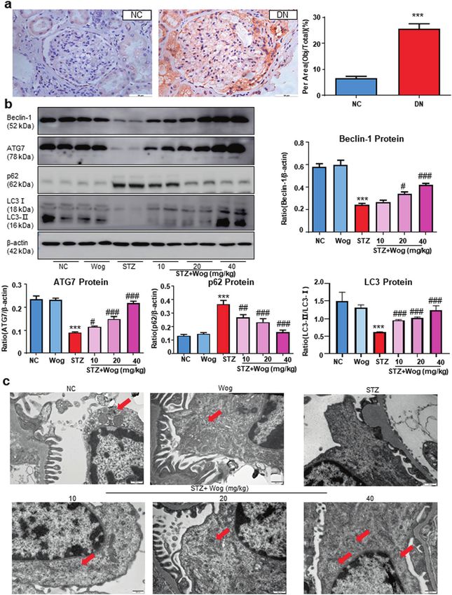
Wogonin protects glomerular podocytes by targeting Bcl-2- mediated autophagy and apoptosis in diabetic kidney disease
←
→
Page content transcription
If your browser does not render page correctly, please read the page content below
www.nature.com/aps

                  ARTICLE                 OPEN

                  Wogonin protects glomerular podocytes by targeting Bcl-2-
                  mediated autophagy and apoptosis in diabetic kidney disease
                  Xue-qi Liu1, Ling Jiang1, Yuan-yuan Li1, Yue-bo Huang1, Xue-ru Hu1, Wei Zhu1, Xian Wang1, Yong-gui Wu1,2, Xiao-ming Meng3 and
                  Xiang-ming Qi1

                     Diabetic kidney disease (DKD) is one of the microvascular complications of diabetes mellitus and a major cause of end-stage renal
                     disease with limited treatment options. Wogonin is a flavonoid derived from the root of Scutellaria baicalensis Georgi, which has
                     shown a potent renoprotective effect. But the mechanisms of action in DKD are not fully elucidated. In this study, we investigated
                     the effects of wogonin on glomerular podocytes in DKD using mouse podocyte clone 5 (MPC5) cells and diabetic mice model.
                     MPC5 cells were treated with high glucose (30 mM). We showed that wogonin (4, 8, 16 μM) dose-dependently alleviated high
                     glucose (HG)-induced MPC5 cell damage, accompanied by increased expression of WT-1, nephrin, and podocin proteins, and
                     decreased expression of TNF-α, MCP-1, IL-1β as well as phosphorylated p65. Furthermore, wogonin treatment significantly inhibited
                     HG-induced apoptosis in MPC5 cells. Wogonin reversed HG-suppressed autophagy in MPC5 cells, evidenced by increased ATG7,
                     LC3-II, and Beclin-1 protein, and decreased p62 protein. We demonstrated that wogonin directly bound to Bcl-2 in MPC5 cells. In
                     HG-treated MPC5 cells, knockdown of Bcl-2 abolished the beneficial effects of wogonin, whereas overexpression of Bcl-2 mimicked
1234567890();,:

                     the protective effects of wogonin. Interestingly, we found that the expression of Bcl-2 was significantly decreased in biopsy renal
                     tissue of diabetic nephropathy patients. In vivo experiments were conducted in STZ-induced diabetic mice, which were
                     administered wogonin (10, 20, 40 mg · kg−1 · d−1, i.g.) every other day for 12 weeks. We showed that wogonin administration
                     significantly alleviated albuminuria, histopathological lesions, and p65 NF-κB-mediated renal inflammatory response. Wogonin
                     administration dose-dependently inhibited podocyte apoptosis and promoted podocyte autophagy in STZ-induced diabetic mice.
                     This study for the first time demonstrates a novel action of wogonin in mitigating glomerulopathy and podocytes injury by
                     regulating Bcl-2-mediated crosstalk between autophagy and apoptosis. Wogonin may be a potential therapeutic drug against DKD.

                     Keywords: diabetic kidney disease; wogonin; podocytes; autophagy; apoptosis; Bcl-2

                     Acta Pharmacologica Sinica (2022) 43:96–110; https://doi.org/10.1038/s41401-021-00721-5

                  INTRODUCTION                                                                                Chinese herbal medicines are widely used for the treatment of
                  Diabetic kidney disease (DKD) remains a growing health concern                           diabetes and its complications [8–10]. Among these, wogonin is a
                  and is characterized by chronic inflammation, hemodynamic                                 flavonoid derived from the root of Scutellaria baicalensis Georgi,
                  changes, and metabolic dysfunction [1, 2]. The early pathological                        which has potent renoprotective effects [11, 12]. It has significant
                  changes in DKD mainly include podocyte injury, shed, and                                 pharmacological actions, such as anti-inflammatory, anti-apopto-
                  apoptosis, while the surviving podocytes show compensatory                               tic, anti-oxidative, and cell cycle regulatory effects, in a variety of
                  hypertrophy and podocyte fusion [3, 4]. Podocyte injury induces                          diseases [13]. We previously reported that wogonin administration
                  proteinuria and leads to the continuous progression of DKD [5]. As                       in rodent models reduced RIPK1-mediated necroptosis following
                  podocytes have limited repair and regeneration ability, the degree                       cisplatin-induced acute kidney injury [8]; however, its underlying
                  of podocyte damage is the main factor determining the prognosis                          mechanism in the early stages of DKD needs to be evaluated.
                  of DKD [6]. At present, prevention and control of DKD in clinical                           Autophagy and apoptosis play important roles in the develop-
                  practice mainly includes early diagnosis, improved control of                            ment and cellular homeostasis. The autophagy level of podocytes
                  blood glucose, and the use of angiotensin-converting enzyme                              is significantly higher than that of other renal intrinsic cells [14].
                  inhibitors or angiotensin receptor blockades [7]. Although these                         Studies have shown that podocyte autophagy substrate protein
                  treatments delay the progression of DKD to a certain extent, they                        p62 is greatly accumulated and LC3-II expression is down-
                  cannot prevent progression to end-stage renal disease. Therefore,                        regulated in the DKD model, indicating the inhibition of
                  the specific molecular mechanism and effective treatment of DKD                           autophagy activity [15]. Apoptosis is an important factor in the
                  need to be further explored.                                                             occurrence of DKD. It has been found that various factors, such as

                  1
                   Department of Nephropathy, The First Affiliated Hospital of Anhui Medical University, Hefei 230022, China; 2Center for Scientific Research of Anhui Medical University, Hefei
                  230022, China and 3The Key Laboratory of Major Autoimmune Diseases, Anhui Institute of Innovative Drugs, School of Pharmacy, Anhui Medical University, The Key Laboratory of
                  Anti inflammatory and Immune Medicines, Ministry of Education, Hefei 230022, China
                  Correspondence: Yong-gui Wu (wuyonggui@medmail.com.cn) or Xiao-ming Meng (mengxiaoming@ahmu.edu.cn) or Xiang-ming Qi (qxm119@126.com)

                  Received: 31 January 2021 Accepted: 18 June 2021
                  Published online: 12 July 2021

                                                                                                                                                                       © The Author(s) 2021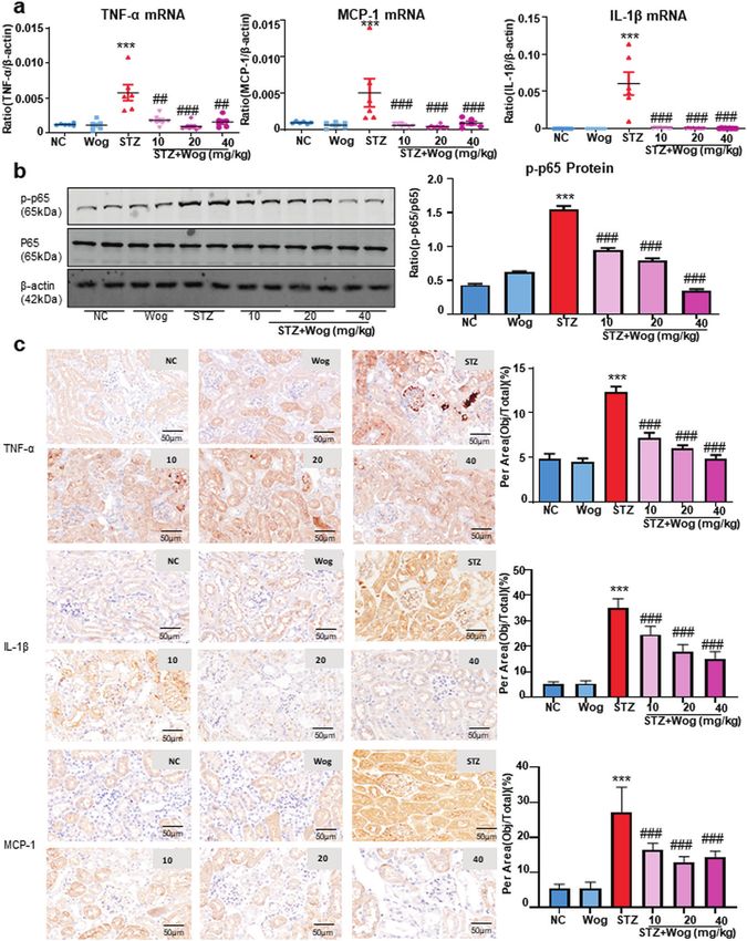
Wogonin protects podocytes in diabetic kidney disease
                                                                       XQ Liu et al.
                                                                                                                                               97
glycolipid toxicity, angiotensin, and glycosylation end products,      MA, USA) and urine protein (UP) was tested by ELISA Kit (Jianglai
induce podocyte apoptosis through different pro-apoptotic path-        Biotechnology Co., Ltd, Shanghai, China) according to the product
ways, leading to the occurrence of DKD [16]. Autophagy and             protocols. We collected mice blood samples in the fasted state by
apoptosis may be triggered by common upstream signals,                 heart punctures. Collected blood was centrifuged for testing of
resulting in combined autophagy and apoptosis or may be                blood urea nitrogen (BUN) and serum creatinine (Cr) (Beyotime,
mutually exclusive [17].                                               Haimen, China).
   The present study aimed to evaluate the effect of wogonin on
glomerular podocytes in DKD in vitro and in vivo using mouse           Kidney histology
podocyte clone 5 (MPC5) cells and diabetic mice models. We             After the kidney was removed, it was fixed with 4% paraformal-
further investigated the mechanism of wogonin. In the present          dehyde for 16 h. The renal tissues were embedded in paraffin and
study, we found that wogonin, used as a single agent, is sufficient     then cut into 4 μm sections. After dewaxing with xylene, the
to significantly reduce proteinuria and decrease podocyte injury.       kidney structure was identified. The sections were stained using
Its mechanism of action was found to be through targeted               PAS and examined under a microscope (Zeiss Spot, Carl Zeiss,
binding to Bcl-2, a well-characterized apoptosis guard that            Gottingen, Germany) at ×400 magnification. Finally, the mesangial
represents a molecular link between autophagy and apoptosis            expansion index and tubular-interstitial injury index of renal tissue
[18]. Our findings clearly showed that the increased activation of      were evaluated from 10 randomly selected fields.
Bcl-2 by wogonin results in the mitigation of Bax-mediated
apoptosis and promotion of Beclin-1-mediated autophagy in              Immunohistochemistry
diabetic kidneys. Therefore, wogonin is expected to become a           The kidney sections were heated in a microwave at 95°C for 20
promising drug for the treatment of DKD.                               min and incubated with 3% hydrogen peroxide. The paraffin
                                                                       tissue sections were treated with anti-p62, anti-TNF-α, anti-IL-1β,
                                                                       anti-MCP-1, anti-Bcl-2, anti-nephrin, and anti-WT-1 antibodies for
MATERIALS AND METHODS                                                  24 h at 4°C, followed with the secondary antibody for 30 min at
Chemicals and reagents                                                 37°C. After staining with DAB for 5 min, the slides were observed
Wogonin was obtained from Aladdin Biology Technology Institute         under a microscope (Zeiss Spot, Carl Zeiss, Gottingen, Germany).
(W101155, CAS 632-85-9, Shanghai, China). STZ, D-glucose, and
D-mannitol were procured from Sigma-Aldrich (Sigma, St. Louis,         Cell culture
MO, USA). Antibodies specific for WT-1, nephrin, podocin, Bax, Bcl-     The MPC5 cells were procured from the Cell Bank of the Chinese
2, cleaved caspase-3, and β-actin were acquired from Abcam             Academic of Sciences (Shanghai, China) and grown in low-glucose
Biotechnology (Abcam, Cambridge, MA, USA). Anti-ATG7, anti-p-          Dulbecco’s Modified Eagle’s Medium (DMEM) (Gibco, San Diego,
p65, anti-p62, anti-p65, and anti-Beclin-1 antibodies were acquired    CA, USA) cultivated with 10% fetal bovine serum (Gibco, San
from Cell Signaling Technology (Danvers, MA, USA). Periodic            Diego, CA, USA). MPC5 cells were propagated at 33°C and treated
acid–Schiff (PAS) kit was obtained from Jiancheng Biotechnology        with interferon (IFN-γ; 10 U/mL). Next, cells were differentiated
Institute (Nanjing, China).                                            without IFN-γ at 37°C for 14 days. For further study, the MPC5 cells
                                                                       were stimulated with high glucose (30 mM glucose) and mannitol
Animals model and experimental design                                  (24.5 mM mannitol + 5.5 mM glucose) containing 1% FBS with or
Male C57BL/6 J mice (6‒8 weeks) were purchased from the                without incubation of wogonin for 24 h.
Experimental Animal Centre of Anhui Medical University (Hefei,
China). The present study protocol was approved by the Ethics          Transmission electron microscopy
Committee of Animal Research of Anhui Medical University (Hefei,       MPC5 cells and renal tissues were treated with 2.5% glutaralde-
China) and conformed to the NIH Guide for the Care and Use of          hyde and 1% osmic acid and fixed at 4°C for 3 days. The samples
Laboratory Animals. The experimental animals were kept in cages        were added with 1% uranyl acetate and embedded in epoxy resin
at 12 h light/dark cycle with free access to food and water in a       (EPON). Polymerized in gelatin capsules at 60°C for 48 h. The
laboratory with controlled temperature (22 ± 2°C) and humidity         sections were observed under a transmission electron microscope
(60%). The mice were randomly divided into six groups: normal          (Hitachi, Japan).
control (NC), NC + wogonin (40 mg/kg), STZ group, STZ +
wogonin groups (10 mg/kg, 20 mg/kg, 40 mg/kg). After adaptable         MTT (thiazole blue colorimetry) assay
feeding for 7 days, 50 mg/kg STZ (dissolved in 0.1 M citrate buffer,   MPC5 cells were cultivated in 96-well plates and incubated with a
pH 4.5) was intraperitoneally administered after 12 h of food          set of concentrations of wogonin, and cells were simultaneously
deprivation daily for 5 consecutive days to generate DM mice [7].      incubated with high glucose for 24 h. Finally, MTT solution (5 mg/
Diabetes mice were defined by fasting blood glucose >250 mg/dL          mL) was added to plates and incubated for 4–6 h. The optical
2 weeks after the first STZ injection. The mice in NC+ wogonin          density (OD) of each well was determined by the microplate
and STZ+ wogonin groups were administered by intragastrical            reader (Multiskan MK3, Thermo Scientific, Waltham, MA, USA) at
gavage with wogonin every other day for 12 weeks. NC and STZ           550 nm wavelength.
groups were given intragastrically with the same amount of saline.
At the end of 12 weeks, 24 h urine was collected from all mice         Bcl-2 overexpression and knockdown in MPC5 cells by shRNA
using the metabolic cage. The mice were anesthetized by                transfections
inhalation of 5% isoflurane and collected blood in the state of         Bcl-2 shRNA (Hanbio, Shanghai, China) was transfected into MPC5
fasting. All experimental subjects were euthanized after               cells mixed with LipofectamineTM 3000 reagent (Invitrogen,
anesthesia.                                                            Carlsbad, CA, USA). The negative scrambled shRNA was used
                                                                       accordingly. First, the mixture of LipofectamineTM 3000 and shRNA
Biochemical analyses                                                   were incubated for 20 min at room temperature in the dark, and
The 12 h fasting blood glucose levels in mice were measured            then applied to the cells. After cultivation for 6 h, the cells were
every 2 weeks according to the Accu-Chek glucose Meters (Roche         cultured in low-glucose DMEM with 10% FBS. Vector and lentivirus
Diagnostic, Inc, Basel, Switzerland) recommended by the Diabetes       Bcl-2 (Hanbio, Shanghai, China) (multiplicity of infection, MOI =
Complications Animal Model Consortium. The kidney weight and           10) were added into the MPC5 cells. Polybrene (10 μg/mL;
bodyweight of each experimental animal were collected. Urine           Solarbio, Shanghai, China) was mixed with the growth medium
albumin was determined using an ELISA kit (Abcam, Cambridge,           to increase transfection efficiency. After 48 h cultivation, 2 μg/mL

Acta Pharmacologica Sinica (2022) 43:96 – 110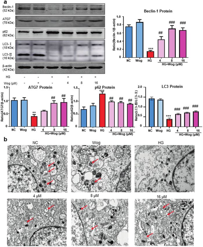
Wogonin protects podocytes in diabetic kidney disease
                                                                XQ Liu et al.
98
      Table 1.   Sequences of primers

     Genes                                           Forward (5′–3′)                                               Reverse (5′–3′)

     Mouse Bcl-2                                     CCTGTGGATGACTGAGTACCTG                                        AGCCAGGAGAAATCAAACAGAGG
     Mouse TNF-α                                     CATCTTCTCAAAATTCGAGTGACAA                                     TGGGAGTAGACAAGGTACAACCC
     Mouse IL-1β                                     CTTTGAAGTTGACGGACCC                                           TGAGTGATACTGCCTGCCTG
     Mouse MCP-1                                     CTTCTGGGCCTGCTGTTCA                                           CCAGCCTACTCATTGGGATCA
     Mouse β-actin                                   GCGTGACATCAAAGAGAAGC                                          GCGTGACATCAAAGAGAAGC

     puromycin (Solarbio, Shanghai, China) was added to the growth              structure of Bcl-2 (PDB code: 6QGH) was downloaded from the
     medium. Stably transfected cells were screened for 2 weeks. The            RCSB Protein Data Bank. Bcl-2 was prepared by preparing a
     surviving cells were considered stable Bcl-2-overexpressing MPC5           protein protocol. CDOCKER module was applied to perform
     cells. Western blot and real-time PCR were used to detect the              molecular docking. Set other parameters as default.
     knockdown and overexpression efficiency of Bcl-2.
                                                                                Flow cytometry
     RNA isolation and real-time PCR                                            Cells were washed with PBS three times and harvested by trypsin
     Renal tissue and cellular RNA were extracted by adding TRIZOL lysis        digestion. After incubation with 10 µL Annexin V-FITC and 5 µL
     buffer (Invitrogen, Carlsbad, CA, USA), and its concentration and          propidium iodide in the dark, the cells were analyzed using a BD
     purity were assessed using NanoDrop2000 spectrophotometer                  FACSVerse flow cytometer (BD Bioscience, San Jose, CA, USA). The
     (Thermo Scientific, Waltham, MA, USA). After the sample quality             rate of apoptosis was expressed by Annexin V+/PI− (early
     was unified, the sample volume was calculated based on the                  apoptosis) and Annexin V+/PI+ (late apoptosis) status.
     concentration. The RNA sample was reverse-transcripted to cDNA
     and the primers for mRNA was designed. CFX96 real-time PCR                 TUNEL assay
     system (Bio-Rad, Hercules, CA, USA) was used to perform on Real-           MPC5 cell apoptosis was detected by TUNEL assay using the
     time PCR assay by SYBR Premix Ex Taq™ II (Takara, Japan). PCR              TUNEL Apoptosis Assay Kit (Beyotime, Haimen, China). The cells
     amplification conditions were as follows: running for over 40 cycles        were incubated in terminal deoxynucleotidyl transferase (TDT)-
     of denaturation at 95°C for 20 s, annealing at 58°C for 20 s, and          mediated dUTP-biotin nick end-labeling (TUNEL). The TUNEL
     elongation at 72°C for 20 s. The primers used in this assay are shown      positive nuclei were observed by optical microscope (Leica DM4P,
     in Table 1.                                                                Shanghai, China).

     Western blot                                                               Cellular thermal shift assay (CETSA)
     Radio-immunoprecipitation assay buffer (Beyotime, Haimen,                  Cellular proteins were extracted from MPC5 cells with or without
     China) was used to extract the fragments of renal tissue and               wogonin for 24 h. Total protein was regulated to the same
     cellular proteins. The extracted protein concentrations were               concentrations using the Protein Assay Kit (Beyotime, Haimen,
     quantified by the bicinchoninic acid reagent (Beyotime, Haimen,             China). The proteins were distributed in PCR tubes and denatured
     China). The samples (40 μg) were separated via 10% or 15%                  at different temperatures (37–65°C) for 8 min on a PCR cycler (Bio-
     sodium dodecyl sulfate-polyacrylamide gel electrophoresis and              Rad, Hercules, CA, USA). After cellular protein was centrifuged, the
     were transferred to nitrocellulose membranes. The membranes                proteins were then frozen and thawed three times with liquid
     were sealed with 5% skimmed milk for 1 h, washed with                      nitrogen, and a Western blot was used to detect the proteins in
     phosphate-buffered saline (PBS) three times, and incubated with            the supernatant.
     applicable primary antibodies overnight. They were then incu-
     bated with the secondary antibody (Rockland Immunochemicals                Immunoprecipitation (IP) assay
     Inc., PA, USA) for 30 min. The band intensities were exposed using         Cells were suspended in standard NP-40 IP buffer (Sigma, St. Louis,
     a chemiluminescence system (Bio-Rad, Hercules, CA, USA). Image J           MO, USA) and the proteins were incubated in NP-40 IP buffer at 4°C.
     software (NIH, Bethesda, MD, USA) was used to quantitatively               The mixture was combined with anti-Bcl-2 antibody 3 h later and
     analyze the protein bands after normalization with β-actin.                then precipitated with Protein G Sepharose beads followed by
                                                                                overnight incubation at 4°C. The beads were washed three times
     Immunofluorescence assay                                                    with 1 mL IP buffer and finally eluted. The immune complexes were
     MPC5 cells were grown on glass coverslips, fixed with acetone at            then subjected to Western blot for determination of protein
     37°C for 10 min, and then blocked with 10% bovine serum                    expression.
     albumin (Beyotime, Haimen, China) at 37°C for 10 min. They were
     incubated with the primary antibody overnight, washed three                Human kidney tissue
     times with PBS, and finally incubated with goat anti-rabbit IgG-            This clinical study has been approved by the Biomedical Ethics
     rhodamine (Bioss, Beijing, China) antibody for 1 h in the dark at          Committee of Anhui Medical University (Hefei, China) (#20170064)
     37°C. The cells were counterstained with DAPI to stain the nuclei          and followed the principles of the Declaration of Helsinki. Normal
     and imaged using an inverted fluorescence microscope (Zeiss                 kidney samples (n = 6) were obtained from renal cancer patients
     Spot, Carl Zeiss, Gottingen, Germany).                                     who underwent routine nephrectomy at the First Affiliated
                                                                                Hospital of Anhui Medical University. Type 1 DM patients (n = 6)
     Molecular docking                                                          were selected. Inclusion criteria: (1) diagnosed with Type 1
     Molecular docking was performed to gain an insight into the                diabetes over 10 years; (2) renal biopsy diagnosed as diabetic
     potential interactions between wogonin and Bcl-2. Discovery                nephropathy; (3) 24 h urine albumin protein >300 mg/24 h; (4) no
     Studio (DS) 2017 R2 software (The Scripps Research Institute, La           obvious symptoms of pyrexia and infection, no cancer, no
     Jolla, CA, USA) was employed in this research. The structure of            autoimmune diseases. Fresh renal tissues were fixed with formalin
     wogonin was optimized by minimizing protocol. The X-ray crystal            for 24 h and embedded in paraffin until use.

                                                                                                         Acta Pharmacologica Sinica (2022) 43:96 – 110
Wogonin protects podocytes in diabetic kidney disease
                                                                           XQ Liu et al.
                                                                                                                                                   99

Fig. 1 Effect of wogonin on HG-treated MPC5 cells’ viability and inhibition on HG-induced inflammation. a The molecular structural
formula of wogonin. b MTT assay to determine the effect of wogonin on viability of MPC5 cells and HG-treated MPC5 cells. c Western blot of WT-1,
nephrin, and podocin in MPC5 cells. d Real-time PCR analysis of TNF-α, MCP-1, and IL-1β expression in MPC5 cells. e Western blot analysis of
phosphorylated p65 (p-p65) in MPC5 cells. Results represent mean ± SEM of three independent experiments. *P < 0.05, **P < 0.01, ***P < 0.001 vs
NC. #P < 0.05, ##P < 0.01, ###P < 0.001 vs HG. HG high glucose, MG 5.5 mM glucose plus 24.5 mM mannitol, NC normal control, Wog wogonin

Statistical analysis                                                       determine the adequate concentration used in the subsequent
All experiments were independently conducted in triplicate. Data           experiments (Fig. 1b). Wogonin showed a minimal effect on MPC5
are expressed as mean ± SD and the difference between groups was           cell viability at concentrations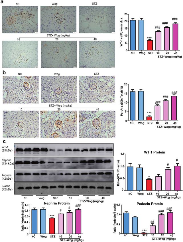
Wogonin protects podocytes in diabetic kidney disease
                                                               XQ Liu et al.
100

      Fig. 2 Wogonin promotes HG-induced autophagy disorders. b Western blot analysis of beclin-1, ATG7, LC3, and p62 in MPC5 cells.
      b Representative transmission electron microscopy images of autophagosomes in MPC5 cells, the arrows indicate autophagosomes. Scale bar =
      500 nm. Results represent mean ± SEM of three independent experiments. **P < 0.01, ***P < 0.001 vs NC. ##P < 0.01, ###P < 0.001 vs HG. HG high
      glucose, NC normal control, Wog wogonin

      upregulated by high glucose (HG) stimulation (Fig. 1d). Moreover,        in MPC5 cells (Fig. 3a). Interestingly, we found that the treatment
      we found that wogonin reduced the HG-induced phosphorylation             of MPC5 cells with wogonin induced the protein, but not mRNA
      of p65 in a dose-dependent manner (Fig. 1e).                             expression of Bcl-2 (Fig. 3b). Therefore, these data suggested that
                                                                               wogonin enhanced the expression of Bcl-2 by inhibiting the
      Wogonin promotes HG-suppressed autophagy in MPC5 cells                   degradation of Bcl-2 in MPC5 cells rather than increasing the
      Western blot analysis showed that wogonin treatment increased            production of Bcl-2. Consistent with the above results, flow
      the ATG7, LC3-II, and Beclin-1 protein levels that were down-            cytometry data of PI/Annexin V-stained MPC5 cells showed that
      regulated due to HG stimulation. Furthermore, the protein level of       wogonin reduced HG-induced cell apoptosis (Fig. 3c). Moreover,
      p62 was decreased with the treatment of wogonin (Fig. 2a). The           TUNEL staining showed that wogonin reduced the level of
      transmission electron microscopy results also showed that                apoptosis (Fig. 3d).
      wogonin significantly promoted autophagy in HG-treated MPC5
      cells. The number of typical autophagosomes with double                  Wogonin target prediction
      membranes was increased in podocytes with wogonin treatment              The target prediction of wogonin was carried out using DS
      (Fig. 2b).                                                               2017 software. As shown in Fig. 4a, the range of binding strength
                                                                               of wogonin to its potential target is shown in red to blue. The fit
      Wogonin alleviates HG-induced apoptosis in MPC5 cells                    values represented the scores of the hypothetical targets, and the
      Wogonin treatment caused a decrease in the protein levels of             top ten disease-related targets were shown in Table 2. Among
      cleaved caspase-3, and Bax and an increase in Bcl-2 protein levels       these targets, Bcl-2, a critical apoptosis-related protein in kidney

                                                                                                         Acta Pharmacologica Sinica (2022) 43:96 – 110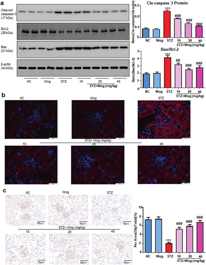
Wogonin protects podocytes in diabetic kidney disease
                                                                         XQ Liu et al.
                                                                                                                                                  101

Fig. 3 Wogonin inhibits HG-induced apoptosis. a Western blot analysis of cleaved caspase-3, Bax, and Bcl-2 in MPC5 cells. b Real-time PCR
analysis of Bcl-2 in MPC5 cells. c Flow cytometry analysis of PI/Annexin V-stained MPC5 cells. d TUNEL assay for MPC5 cells. Scale bar = 50 μm.
Results represent mean ± SEM of three independent experiments. ***P < 0.001 vs NC. ##P < 0.01, ###P < 0.001 vs HG. Abbreviations: HG high
glucose, NC normal control, Wog wogonin

disease, showed a high score of 0.9933 and appeared at a higher          42.1329 kcal/mol (Fig. 4b). Importantly, we found that Bcl-2
frequency, as shown in the mapping.                                      expression levels were decreased in renal tissues of patients with
                                                                         diabetic nephropathy (Fig. 4c). The target engagement between
Wogonin binds directly to Bcl-2 in MPC5 cells                            wogonin and Bcl-2 protein was performed by CETSA. Results
In this article, to study the binding posture between wogonin and        indicated that the denaturation temperature of Bcl-2 differed by
Bcl-2 (PDB ID:6QGH), we used molecular docking. The 2D and 3D            50–65°C with or without wogonin. In MPC5 cells treated with
images showed that wogonin formed three pi-alkyl bonds with              wogonin, the thermal stability of Bcl-2 was significantly improved,
VAL76, ILE62, and ILE72. In addition, wogonin and Bcl-2 formed           showing that wogonin binds to Bcl-2 protein directly (Fig. 4d).
five van der Waals interactions with THR88, ILE89, PHE92, ILE67,            Moreover, bioinformatics prediction pointed out that wogonin
and ARG65, which contributed to the binding affinity. The highest         may target Bcl-2. The results of the IP assay further confirmed the
docking score detected by CDOCKER interaction energy was                 co-precipitation of Bcl-2 with Beclin-1 and Bax in MPC5 cells.

Acta Pharmacologica Sinica (2022) 43:96 – 110
Wogonin protects podocytes in diabetic kidney disease
                                                                XQ Liu et al.
102

      Fig. 4 Prediction of wogonin molecular targets. a Profiling of the predicted protein targets of wogonin using Discovery Studio
      2017 software. b Molecular docking of wogonin binding to Bcl-2 crystal structure. c Immunohistochemistry analysis of Bcl-2 in human kidney.
      Scale bar = 50 μm. Data represent the mean ± SEM for 6 humans. d CETSA analysis in MPC5 cells. e Immunoprecipitation assay. Results
      represent mean ± SEM of three independent experiments. ***P < 0.001 vs NC. HG high glucose, NC normal control, Wog wogonin

                                                                                Wogonin stimulation promoted Bcl-2–Bax-binding power and
       Table 2. Top ten putative protein targets of wogonin predicted using     decreased Bcl-2–Beclin-1-binding ability, indicating that autop-
       Discovery Studio 2017                                                    hagy was increased, and apoptosis was reduced in the cells
                                                                                (Fig. 4e).
      Rank PDB ID Putative target                                  Fit value
                                                                                Wogonin attenuates HG-induced cell apoptosis and promotes
      1       6QGH   B-cell lymphoma-2                             0.9933
                                                                                autophagy through Bcl-2-dependent mechanisms
      2       2BHE   Cell division protein kinase 2                0.9868       First, we knocked down Bcl-2 in MPC5 cells using Bcl-2 shRNA
      3       2POG   Estrogen receptor                             0.9864       (Fig. 5a, b). Inhibition of Bcl-2 expression could significantly
      4       2QC6   Casein kinase II subunit alpha                0.9846       increase cell apoptosis and suppress autophagy. After Bcl-2 was
      5       3BGQ   Proto-oncogene serine/threonine-protein       0.9830       inhibited, wogonin was unable to further increase the expression
                     kinase Pim-1                                               of WT-1 and nephrin (Fig. 5c). This showed that in the absence of
                                                                                Bcl-2, wogonin was unable to exert its cellular protective role,
      6       3A3W   Phosphotriesterase                            0.9799
                                                                                suggesting that its function was through targeting Bcl-2 (Fig. 5d).
      7       3MJW   Phosphatidylinositol-4,5-bisphosphate 3-      0.9762          Next, we overexpressed Bcl-2 using a lentivirus (Fig. 6a, b) and
                     kinase catalytic subunit gamma isoform
                                                                                observed that Bcl-2 overexpression and wogonin treatment
      8       2UZT   cAMP-dependent protein kinase, alpha-         0.9727       played similar cellular protective roles by increasing WT-1 and
                     catalytic subunit                                          nephrin levels, suppressing cell apoptosis, and promoting
      9       2R3P   Cell division protein kinase 2                0.9660       autophagy. Moreover, in Bcl-2 overexpressing cells, the wogonin
      10      3DCV   Proto-oncogene serine/threonine-protein       0.9644       treatment showed a superposition effect to reduce cell injury
                     kinase Pim-1                                               (Fig. 6c, d).

                                                                                                         Acta Pharmacologica Sinica (2022) 43:96 – 110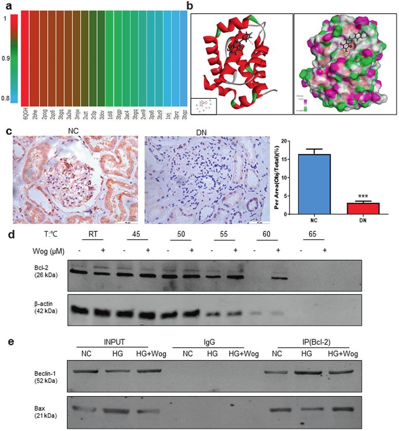
Wogonin protects podocytes in diabetic kidney disease
                                                                             XQ Liu et al.
                                                                                                                                                        103

Fig. 5 Wogonin fails to reduce the HG-induced cell injury, inflammatory response and promotes autophagy in Bcl-2-silenced MPC5 cells.
a Real-time PCR analysis of Bcl-2 in MPC5 cells. b Western blot analysis of Bcl-2 in MPC5 cells. c Western blot analysis of WT-1 and nephrin in
MPC5 cells. d Western blot analysis of cleaved caspase-3, Beclin-1, ATG7, and p62 in MPC5 cells. Results represent means ± SEM for three
independent experiments. *P < 0.05, **P < 0.01, ***P < 0.001 vs NC. #P < 0.05, ##P < 0.01, ###P < 0.001 vs HG. $P < 0.05, $$P < 0.01, $$$P < 0.001 vs
Bcl-2 EV group. HG high glucose, Wog wogonin, EV empty vector, KD Knockdown, NC normal control

Wogonin alleviates physical and biochemical markers and                      remarkably decreased after wogonin treatment (Fig. 7c–f). PAS
pathology in STZ-induced type 1 diabetes mice model                          staining showed that wogonin mitigated the degree of mesangial
For purpose of detecting whether wogonin could protect the                   dilatation and tubulointerstitial injury in the DM group (Fig. 7g).
kidneys in type 1 diabetic mice, diabetic mice were administered             The transmission electron microscopy results also showed that
by intragastrical gavage with wogonin (10, 20, 40 mg/kg body                 wogonin significantly alleviated renal basement membrane
weight). Diabetic mice showed polydipsia, polyuria, and poly-                thickening and foot process fusion in type 1 diabetic mice
phagia accompanied by significant weight loss, hair color messy               (Fig. 7h).
and dirty, and slow movement. But these symptoms
were improved significantly in mice treated with wogonin. At                  Wogonin protects glomerular podocytes in STZ-induced type 1
12 weeks, diabetic mice showed significantly increased albumi-                diabetes mice model
nuria that decreased after wogonin treatment (Fig. 7a). Treatment            Consistent with in vitro findings where wogonin could prevent
with wogonin could not alleviate the blood glucose levels that               HG-induced podocyte injury, WT-1, and nephrin in glomerular
were markedly higher in STZ-induced DM group (Fig. 7b).                      podocytes from diabetic mice were reversed by wogonin
Moreover, the kidney/weight ratio, BUN, Cr and 24-h UP were                  treatment. We observed that the podocyte-specific marker and

Acta Pharmacologica Sinica (2022) 43:96 – 110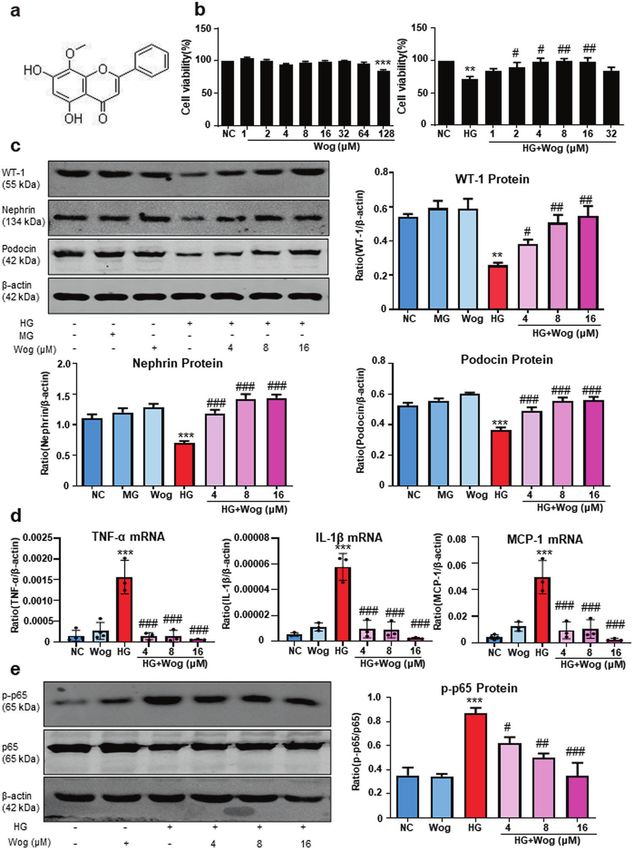
Wogonin protects podocytes in diabetic kidney disease
                                                               XQ Liu et al.
104

      Fig. 6 Bcl-2 overexpression and treatment with wogonin have similar cellular protective roles. a Real-time PCR analysis of Bcl-2 in MPC5
      cells. b Western blot analysis of Bcl-2 in MPC5 cells. c Western blot analysis of WT-1 and nephrin in MPC5 cells. d Western blot analysis of
      cleaved caspase-3, Beclin-1, ATG7, and p62 in MPC5 cells. Results represent means ± SEM for three independent experiments. **P < 0.01, ***P
      < 0.001 vs NC. #P < 0.05, ##P < 0.01, ###P < 0.001 vs HG. $$P < 0.01, $$$P < 0.001 vs Bcl-2-OE group. HG high glucose, Wog wogonin, EV empty
      vector, OE overexpression, NC normal control

      SDs levels significantly decreased as quantified by staining WT-1          of IL-1β, MCP-1, and TNF-α also demonstrated the anti-
      and nephrin in the diabetic group. These changes in diabetic mice        inflammatory effect of wogonin in type 1 diabetic mice (Fig. 9c).
      were attenuated to different degrees by wogonin treatment and
      revealed similar histomorphology to that observed in non-diabetic        Wogonin promotes podocyte autophagy in STZ-induced type 1
      controls (Fig. 8a, b). Further, Western blot results showed that         diabetes mice model
      wogonin increased expressions of WT-1, nephrin, and podocin in           Interestingly, we first found that the expression level of p62 was
      type 1 diabetic mice (Fig. 8c).                                          significantly increased in renal tissues of diabetic nephropathy
                                                                               patients (Fig. 10a). We next tested the effect of wogonin on
      Wogonin protects STZ-induced type 1 diabetes mice model by               podocyte autophagy in diabetic mice. Western blot analysis
      ameliorating inflammatory response                                        showed decreased autophagy-related proteins and concurrently
      Wogonin significantly reduced the levels of TNF-α, MCP-1, and IL-         increased p62 in kidney tissues of diabetic mice (Fig. 10b).
      1β, as determined by real-time PCR (Fig. 9a). Further, results of        Notably, the changes in Beclin-1, ATG7, LC3-II, and p62 levels in
      Western blot revealed that wogonin suppressed phosphorylation            the renal tissue of diabetic mice were partially but significantly
      of p65 in type 1 diabetic mice (Fig. 9b). Immunohistochemical data       reversed by wogonin treatment. In addition, the number of typical

                                                                                                       Acta Pharmacologica Sinica (2022) 43:96 – 110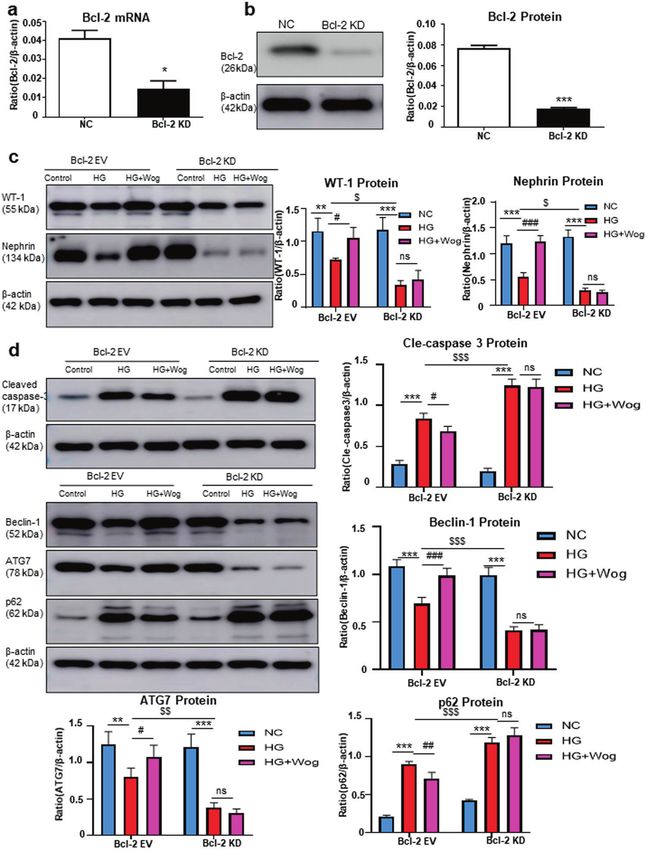
Wogonin protects podocytes in diabetic kidney disease
                                                                        XQ Liu et al.
                                                                                                                                                105

Fig. 7 Physical and biochemical markers and histopathology of type 1 diabetic mice. a Analysis of urine albumin excretion. b Fasting blood
glucose in different groups. c Kidney/body weight. d serum BUN assay. e serum Cr assay. f Twenty-four hours urinary protein quantitation.
g Histological observations of kidney sections stained with periodic acid–Schiff (PAS) from different groups treated with or without wogonin.
Scale bar = 50 μm. h Representative transmission electron microscopy images of podocyte foot processes and glomerular basement
membrane. Scale bar = 1 μm. Results represent mean ± SEM for 6–8 mice. ***P < 0.001 vs NC. #P < 0.05, ##P < 0.01, ###P < 0.001 vs STZ. NC
normal control, Wog wogonin, STZ streptozotocin

autophagosomes with double membranes and the morphology of              proved the beneficial effect of wogonin on the main symptoms
podocytes were observed by transmission electron microscopy.            of DKD.
Reduced autophagosomes were found in the diabetic mice group,
which increased after wogonin treatment (Fig. 10c).
                                                                        DISCUSSION
Wogonin ameliorates apoptosis in STZ-induced type 1 diabetes            The present study explored the renoprotective role of wogonin in
mice model                                                              mice podocyte cells. Our evaluation of clinical symptoms in diabetic
By detecting the expression of apoptosis-related proteins, such as      mice at 12 weeks showed a significant reduction in proteinuria after
cleaved caspase-3, Bax, and Bcl-2, we discovered that podocyte          treatment with wogonin; however, no significant reduction in blood
apoptosis was increased in diabetic mice compared to non-               glucose level was observed. Furthermore, kidney inflammation
diabetic controls and was significantly improved by wogonin              was gradually worsened in diabetic mice, and wogonin showed an
treatment (Fig. 11a). Moreover, TUNEL staining showed that              anti-inflammatory effect in these mice at 12 weeks. We found that
wogonin could reduce cell apoptosis (Fig. 11b). Results of              wogonin played a key role in alleviating cell apoptosis and promoting
immunohistochemistry showed that downregulated Bcl-2 protein            autophagy in DKD by targeting Bcl-2. Our experimental studies
expression was rescued by wogonin (Fig. 11c). In short, our data        highlighted the close link between dysregulated Bcl-2 and

Acta Pharmacologica Sinica (2022) 43:96 – 110
Wogonin protects podocytes in diabetic kidney disease
XQ Liu et al.
106
Fig. 8 Wogonin attenuates glomerular podocytes injury in type 1 diabetic mice. a Immunohistochemistry analysis of WT-1 in mice kidney.
Scale bar = 50 μm. b Immunohistochemistry analysis of nephrin in mice kidney. Scale bar = 50 μm. c Western blot analysis of WT-1, nephrin,
and podocin in mice kidney. Data represent the mean ± SEM for 6–8 mice. *P < 0.05, ***P < 0.001 vs NC. #P < 0.05, ###P < 0.001 vs STZ.
NC normal control, Wog wogonin, STZ streptozotocin
HG-induced MPC5 cells injury. Although previous studies showed TGF-β1/Smad3 signaling pathways [11, 20, 21]. In the current
pharmacological effects of wogonin on inflammation, apoptosis, study, we demonstrated the anti-inflammatory effect of wogonin
oxidative stress, and cell cycle regulation, the underlying mechanisms in HG-treated MPC5 cells. Further study revealed that wogonin
have not been fully elucidated [8]. Thus, our findings indicated that significantly inhibited inflammation in STZ-induced diabetic mice.
wogonin regulates the severity of DKD by alleviating glomerular In particular, wogonin decreased the levels of inflammatory
injury, albuminuria, and cell death. The possible mechanism for cytokines, such as TNF-α, MCP-1, and IL-1β. DKD is considered to
wogonin action on podocytes may be via alleviation of HG-induced be a chronic inflammatory disease, in which the structure of the
apoptosis activation and autophagy disorder through targeting Bcl-2. glomeruli and kidney tubules is altered by chronic microinflam-
Wogonin has a wide range of biological activities and affects mation, leading to proteinuria [22, 23]. Moreover, the study
the inflammatory response, apoptosis, oxidative stress, and other revealed a hyperglycemic environment promotes the activation of
aspects [12, 13, 19]. Wogonin reportedly protected against NF-κB signaling pathway, upregulates the expression of TNF-α,
cisplatin-induced acute kidney injury by targeting RIPK1- MCP-1, and IL-1β, thereby contributing to the renal damage in
mediated necroptosis [8, 20]. It prevented inflammation and type 1 diabetic mice [24]. Thus, specific blocking of inflammatory
fibrosis in diabetic nephropathy by inhibiting the NF-κB and cytokines or the above signaling pathway may reduce renal
Acta Pharmacologica Sinica (2022) 43:96 – 110Wogonin protects podocytes in diabetic kidney disease
XQ Liu et al.
107
Fig. 9 Wogonin attenuates renal inflammation in type 1 diabetic mice. a Real-time PCR analysis of inflammation (TNF-α, MCP-1, IL-1β)
indices in mice kidney samples. b Western blot analysis of phosphorylated p65 (p-p65) in mice kidney tissues. c Immunohistochemistry
analysis of TNF-α, MCP-1, IL-1β in mice kidney tissues. Scale bar = 50 μm. Data represent the mean ± SEM for 6–8 mice. ***P < 0.001 vs NC.
##
P < 0.01, ###P < 0.001 vs STZ. NC normal control, Wog wogonin, STZ streptozotocin
inflammation response and cell damage in DKD mice, thus autophagy [14]. Our results also highlighted that p62, the key
alleviating DKD. protein of autophagy, was significantly increased in renal tissue
Autophagy dysfunction is involved in podocyte injury, and from diabetic nephropathy patients.
autophagy reportedly is inhibited in HG environment [4, 15, 25]. Furthermore, we discovered that wogonin reduces podocyte
Our investigation revealed that wogonin treatment alleviated STZ- apoptosis in DKD. Apoptosis is a strictly controlled process of cell
induced autophagy dysfunction in podocytes. To our knowledge, death, which is necessary for cell growth and body development [29].
this is the first research to demonstrate that wogonin promoted The apoptotic pathway is involved in the development of many
autophagy in STZ-induced diabetic mice. Autophagy is a cellular diseases including diabetic nephropathy [17, 30]. Bax and Bcl-2 are
biological process in which lysosomes degrade cytoplasmic the major mediators of endogenous apoptosis and apoptosis are
components to maintain cell homeostasis [6, 26–28]. Mammalian activated by Bax and inhibited by Bcl-2. Under normal conditions,
target of rapamycin AMP-activated protein kinase and silent autophagy and apoptosis maintain a dynamic balance to ensure the
information regulator 1 (Sirt1) has important regulatory effects in stability of the internal environment. However, the decrease of
Acta Pharmacologica Sinica (2022) 43:96 – 110Wogonin protects podocytes in diabetic kidney disease
XQ Liu et al.
108
Fig. 10 Wogonin attenuates renal autophagy disorder in type 1 diabetic mice. a Immunohistochemistry analysis of p62 in human kidney
tissues. Scale bar = 50 μm. Data represent the mean ± SEM for six humans. b Western blot analysis of Beclin-1, ATG7, p62, and LC3 in mice
kidney tissues. c Representative transmission electron microscopy images of autophagosomes in mice kidney tissues, the arrows indicate
autophagosomes. Scale bar = 500 nm. Data represent the mean ± SEM for 6–8 mice. ***P < 0.001 vs NC. #P < 0.05, ##P < 0.01, ###P < 0.001 vs
STZ. NC normal control, Wog wogonin, STZ streptozotocin
protective autophagy and the increase of apoptosis of podocytes may in assessing the development potential of drugs is target engage-
be one of the important causes of DKD renal injury [31, 32]. ment, which is measured by CETSA at various stages of drug
Interestingly, we found that wogonin significantly increased the development. In the present study, CETSA confirmed that wogonin
protein level of Bcl-2 while did not affect its mRNA expression, which bound to Bcl-2 with high binding affinity. Furthermore, molecular
revealed that wogonin regulated Bcl-2 possibly by reducing the docking was used to elucidate the most stable binding posture on
degradation of Bcl-2 rather than promoting its production. Bcl-2 active sites with wogonin. Importantly, we found that Bcl-2
To better understand the mechanism of wogonin in DKD, we used expression levels were significantly decreased in renal tissues of
DS software to analyze its molecular target. The interaction between diabetic nephropathy patients. Thus, Bcl-2 may have an important
wogonin and Bcl-2 was simulated by computer-aid. A crucial element role in DKD.
Acta Pharmacologica Sinica (2022) 43:96 – 110Wogonin protects podocytes in diabetic kidney disease
XQ Liu et al.
109
Fig. 11 Wogonin attenuates cell death in type 1 diabetic mice. a Western blot analysis of cleaved caspase-3, Bax, and Bcl-2 in mice kidney
tissues. b TUNEL assay in mice kidney tissues. apoptotic cell, red; DAPI, blue. c Immunohistochemistry analysis of Bcl-2 in mice kidney tissues.
Scale bar = 50 μm. Results represent mean ± SEM for 6–8 mice. ***P < 0.001 vs NC. ##P < 0.01, ###P < 0.001 vs STZ. Abbreviations: NC normal
control, Wog wogonin, STZ streptozotocin
To further confirm the role of Bcl-2 in podocyte injury in DKD, we for Bcl-2-mediated autophagy inhibition [33]. Moreover, Bax/Bak
performed an IP assay to verify the role of Bcl-2 in the regulation of translocates to the mitochondrial membrane in response to
autophagy and apoptosis. Interestingly, after wogonin treatment, apoptotic stimuli, promotes the release of cytochrome c into the
the binding of Beclin-1 to Bcl-2 decreased, while that of Bax, a pro- cytosol from the mitochondrial membrane space, and the anti-
apoptotic effector protein, to Bcl-2 increased, indicating inhibition of apoptotic members, Bcl-2 can interact with Bax to inhibit apoptosis
apoptosis and promotion of autophagy [17]. Autophagy and [17]. Previous research reported that phosphorylation of Bcl-2 on
apoptosis play important roles in the development and cellular Ser70 could inhibit Bcl-2 degradation, stabilized Bcl-2–Bax interaction
homeostasis [32, 33]. They may be triggered by common upstream to inhibit apoptosis, and disrupted the Bcl-2–Beclin-1 complex to
signals, resulting in combined autophagy and apoptosis, or may be promote autophagy [18]. Thus, Bcl-2 represents a bridge between
mutually exclusive. Recent studies have shown possible molecular autophagy and apoptosis. Our data further found that Bcl-2 could be
mechanisms of interaction between autophagy and apoptosis [32]. a potential target for wogonin through improving the disorder of
Bcl-2 is a recognized anti-apoptotic factor, which inhibits Beclin-1 autophagy and apoptosis of podocytes.
mediated autophagy by binding to Beclin-1 [31, 34]. In fact, Bcl-2 In conclusion, we demonstrated that the renoprotective effect of
downregulates autophagy by interacting with Beclin-1, the autop- wogonin in STZ-induced diabetic mice was mediated by targeting
hagy effector. Beclin-1 contains a BH3 domain similar to Bcl-2, which Bcl-2-mediated apoptosis and autophagy. We found that wogonin,
is required for binding to anti-apoptotic Bcl-2 and is also necessary used as a single agent, was sufficient to significantly reduce the
Acta Pharmacologica Sinica (2022) 43:96 – 110Wogonin protects podocytes in diabetic kidney disease
XQ Liu et al.
110
symptoms of DKD and attenuate podocyte injury in vivo. Therefore, 15. Gui X, Yang H, Li T, Tan X, Shi P, Li M, et al. Autophagy induction via STING
wogonin might serve as a promising therapeutic agent for treating trafficking is a primordial function of the cGAS pathway. Nature. 2019;567:262–6.
DKD. Moreover, further exploration of Bcl-2 analogs and other drugs 16. Bhatt K, Lanting LL, Jia Y, Yadav S, Reddy MA, Magilnick N, et al. Anti-
based on the interaction between wogonin and Bcl-2 might help inflammatory role of MicroRNA-146a in the pathogenesis of diabetic nephro-
pathy. J Am Soc Nephrol. 2016;27:2277–88.
identify more effective and safer drugs, with potential for translation
17. Levine B, Sinha S, Kroemer G. Bcl-2 family members - Dual regulators of apoptosis
to the clinic, to treat patients with DKD. and autophagy. Autophagy. 2008;4:600–6.
18. Lee EF, Smith NA, Soares da Costa TP, Meftahi N, Yao S, Harris TJ, et al. Structural
insights into BCL2 pro-survival protein interactions with the key autophagy
ACKNOWLEDGEMENTS regulator BECN1 following phosphorylation by STK4/MST1. Autophagy.
The authors thank the Center for Scientific Research of Anhui Medical University for 2019;15:785–95.
valuable help in our experiment. This study was supported by the Scientific Research 19. Enomoto R, Sugahara C, Suzuki C, Nagase I, Takamura Y, Yoshikawa A, et al.
Foundation of the Institute for Translational Medicine of Anhui Province (no. Wogonin prevents glucocorticoid-induced thymocyte apoptosis without dimin-
2017zhyx01); the Promotion Plan of Basic and Clinical Cooperative Research in Anhui ishing its anti-inflammatory action. J Pharmacol Sci. 2007;104:355–65.
Medical University (No. 2019xkjT014); and Natural Science Research Project of 20. Khan NM, Haseeb A, Ansari MY, Devarapalli P, Haynie S, Haqqi TM. Wogonin, a
Universities in Anhui Province (no. KJ2018A0493). plant derived small molecule, exerts potent anti-inflammatory and chon-
droprotective effects through the activation of ROS/ERK/Nrf2 signaling pathways
in human osteoarthritis chondrocytes. Free Radic Biol Med. 2017;106:288–301.
AUTHOR CONTRIBUTIONS 21. Li HD, Chen X, Yang Y, Huang HM, Zhang L, Zhang X, et al. Wogonin attenuates
XQL and LJ conducted the experiments and analyzed the data. XQL and XMM wrote inflammation by activating PPAR-gamma in alcoholic liver disease. Int Immu-
the manuscript. YYL conducted molecular docking experiments. YBH, XW, and WZ nopharmacol. 2017;50:95–106.
contributed to the experimental design. XRH and WZ performed animal experiments. 22. Meng XM, Tang PM, Li J, Lan HY. TGF-beta/Smad signaling in renal fibrosis. Front
YGW. and XMQ conceived, designed the study. XMQ contributed analytical tools. Physiol. 2015;6:82.
23. Wang JN, Yang Q, Yang C, Cai YT, Xing T, Gao L, et al. Smad3 promotes AKI
sensitivity in diabetic mice via interaction with p53 and induction of NOX4-
ADDITIONAL INFORMATION dependent ROS production. Redox Biol. 2020;32:101479.
24. Shao YX, Gong Q, Qi XM, Wang K, Wu YG. Paeoniflorin ameliorates macrophage
Competing interests: The authors declare no competing interests.
infiltration and activation by inhibiting the TLR4 signaling pathway in diabetic
nephropathy. Front Pharmacol. 2019;10:566.
25. Yang D, Livingston MJ, Liu Z, Dong G, Zhang M, Chen JK, et al. Autophagy in
REFERENCES diabetic kidney disease: regulation, pathological role and therapeutic potential.
1. Liu M, Liang K, Zhen J, Zhou M, Wang X, Wang Z, et al. Sirt6 deficiency exacer- Cell Mol Life Sci. 2017;75:669–88.
bates podocyte injury and proteinuria through targeting Notch signaling. Nat 26. Chen K, Dai H, Yuan J, Chen J, Lin L, Zhang W, et al. Optineurin-mediated
Commun. 2017;8:413. mitophagy protects renal tubular epithelial cells against accelerated senescence
2. Xie C, Wu W, Tang A, Luo N, Tan Y. lncRNA GAS5/miR-452-5p reduces oxidative in diabetic nephropathy. Cell Death Dis. 2018;9:105.
stress and pyroptosis of high-glucose-stimulated renal tubular cells. Diabetes 27. Chen K, Feng L, Hu W, Chen J, Wang X, Wang L, et al. Optineurin inhibits NLRP3
Metab Syndr Obes. 2019;12:2609–17. inflammasome activation by enhancing mitophagy of renal tubular cells in dia-
3. Fan Y, Yang Q, Yang Y, Gao Z, Ma Y, Zhang L, et al. Sirt6 suppresses high glucose- betic nephropathy. FASEB J. 2019;33:4571–85.
induced mitochondrial dysfunction and apoptosis in podocytes through AMPK 28. Kong Z, Che K, Hu J, Chen Y, Wang Y, Wang X, et al. Orientin protects podocytes
activation. Int J Biol Sci. 2019;15:701–13. from high glucose induced apoptosis through mitophagy. Chem Biodivers.
4. Kato M, Natarajan R. Diabetic nephropathy–emerging epigenetic mechanisms. 2020;17:e1900647.
Nat Rev Nephrol. 2014;10:517–30. 29. Jiang L, Liu XQ, Ma Q, Yang Q, Gao L, Li HD, et al. hsa-miR-500a-3P alleviates
5. Qin X, Zhao Y, Gong J, Huang W, Su H, Yuan F, et al. Berberine protects glo- kidney injury by targeting MLKL-mediated necroptosis in renal epithelial cells.
merular podocytes via inhibiting drp1-mediated mitochondrial fission and dys- FASEB J. 2019;33:3523–35.
function. Theranostics. 2019;9:1698–713. 30. Shao YX, Xu XX, Wang K, Qi XM, Wu YG. Paeoniflorin attenuates incipient diabetic
6. Stitt-Cavanagh E, MacLeod L, Kennedy C. The podocyte in diabetic kidney dis- nephropathy in streptozotocin-induced mice by the suppression of the Toll-like
ease. ScientificWorldJournal. 2009;9:1127–39. receptor-2 signaling pathway. Drug Des Dev Ther. 2017;11:3221–33.
7. Liu XQ, Jiang L, Lei L, Nie ZY, Zhu W, Wang S, et al. Carnosine alleviates diabetic 31. Salminen A, Kaarniranta K, Kauppinen A. Beclin-1 interactome controls the
nephropathy by targeting GNMT, a key enzyme mediating renal inflammation crosstalk between apoptosis, autophagy and inflammasome activation: impact
and fibrosis. Clin Sci (Lond). 2020;134:3175–93. on the aging process. Ageing Res Rev. 2013;12:520–34.
8. Meng XM, Li HD, Wu WF, Ming-Kuen Tang P, Ren GL, Gao L, et al. Wogonin 32. Zhou F, Yang Y, Xing D. Bcl-2 and Bcl-xL play important roles in the crosstalk
protects against cisplatin-induced acute kidney injury by targeting RIPK1- between autophagy and apoptosis. FEBS J. 2011;278:403–13.
mediated necroptosis. Lab Invest. 2018;98:79–94. 33. Pena-Blanco A, Garcia-Saez AJ. Bax, Bak and beyond–mitochondrial performance
9. Liu XQ, Jin J, Li Z, Jiang L, Dong YH, Cai YT, et al. Rutaecarpine derivative Cpd-6c in apoptosis. FEBS J. 2018;285:416–31.
alleviates acute kidney injury by targeting PDE4B, a key enzyme mediating 34. Yip KW, Reed JC. Bcl-2 family proteins and cancer. Oncogene. 2008;27:6398–406.
inflammation in cisplatin nephropathy. Biochem Pharmacol. 2020;180:114132.
10. Shao YX, Xu XX, Li YY, Qi XM, Wang K, Wu YG, et al. Paeoniflorin inhibits high
glucose-induced macrophage activation through TLR2-dependent signal path- Open Access This article is licensed under a Creative Commons
ways. J Ethnopharmacol. 2016;193:377–86. Attribution 4.0 International License, which permits use, sharing,
11. Zheng ZC, Zhu W, Lei L, Liu XQ, Wu YG. Wogonin ameliorates renal inflammation adaptation, distribution and reproduction in any medium or format, as long as you give
and fibrosis by inhibiting NF-kappaB and TGF-beta1/Smad3 signaling pathways appropriate credit to the original author(s) and the source, provide a link to the Creative
in diabetic nephropathy. Drug Des Devel Ther. 2020;14:4135–48. Commons license, and indicate if changes were made. The images or other third party
12. Du XS, Li HD, Yang XJ, Li JJ, Xu JJ, Chen Y, et al. Wogonin attenuates liver fibrosis material in this article are included in the article’s Creative Commons license, unless
via regulating hepatic stellate cell activation and apoptosis. Int Immuno- indicated otherwise in a credit line to the material. If material is not included in the
pharmacol. 2019;75:105671. article’s Creative Commons license and your intended use is not permitted by statutory
13. Hong M, Almutairi MM, Li S, Li J. Wogonin inhibits cell cycle progression by regulation or exceeds the permitted use, you will need to obtain permission directly
activating the glycogen synthase kinase-3 beta in hepatocellular carcinoma. from the copyright holder. To view a copy of this license, visit http://creativecommons.
Phytomedicine. 2020;68:153174. org/licenses/by/4.0/.
14. Zhang C, Li W, Wen J, Yang Z. Autophagy is involved in mouse kidney devel-
opment and podocyte differentiation regulated by Notch signalling. J Cell Mol
Med. 2017;21:1315–28. © The Author(s) 2021
Acta Pharmacologica Sinica (2022) 43:96 – 110You can also read