In Vitro Biocompatibility and Degradation Analysis of Mass-Produced Collagen Fibers
←
→
Page content transcription
If your browser does not render page correctly, please read the page content below
polymers
Article
In Vitro Biocompatibility and Degradation Analysis of
Mass-Produced Collagen Fibers
Kiran M. Ali, Yihan Huang, Alaowei Y. Amanah, Nasif Mahmood, Taylor C. Suh and Jessica M. Gluck *

                                          Department of Textile Engineering, Chemistry and Science, Wilson College of Textiles, NC State University,
                                          Raleigh, NC 27695, USA; kmmumtaz@ncsu.edu (K.M.A.); yhuang38@ncsu.edu (Y.H.); aya8@duke.edu (A.Y.A.);
                                          nmahmoo3@ncsu.edu (N.M.); tacook3@ncsu.edu (T.C.S.)
                                          * Correspondence: jmgluck@ncsu.edu; Tel.: +1-919-515-6637

                                          Abstract: Automation and mass-production are two of the many limitations in the tissue engineering
                                          industry. Textile fabrication methods such as electrospinning are used extensively in this field because
                                          of the resemblance of the extracellular matrix to the fiber structure. However, electrospinning has
                                          many limitations, including the ability to mass-produce, automate, and reproduce products. For
                                          this reason, this study evaluates the potential use of a traditional textile method such as spinning.
                                          Apart from mass production, these methods are also easy, efficient, and cost-effective. This study
                                          uses bovine-derived collagen fibers to create yarns using the traditional ring spinning method. The
                                          collagen yarns are proven to be biocompatible. Enzymatic biodegradability was also confirmed for
                                          its potential use in vivo. The results of this study prove the safety and efficacy of the material and
                                          the fabrication method. The material encourages higher cell proliferation and migration compared
                                          to tissue culture-treated plastic plates. The process is not only simple but is also streamlined and
                                          replicable, resulting in standardized products that can be reproduced.

Citation: Ali, K.M.; Huang, Y.;
                                          Keywords: collagen; tissue engineering; biomaterials; biocompatibility; scaffolds
Amanah, A.Y.; Mahmood, N.; Suh,
T.C.; Gluck, J.M. In Vitro
Biocompatibility and Degradation
Analysis of Mass-Produced Collagen
                                          1. Introduction
Fibers. Polymers 2022, 14, 2100.
https://doi.org/10.3390/                        Disease, injury, and trauma can damage and degenerate tissues in the human body
polym14102100                             that can be treated via the repair, replacement, or regeneration of the tissue [1]. Often, the
                                          tissues are either taken from the patient’s own body (autograft) or from a donor (allograft).
Academic Editors: Antonia Ressler
                                          An example of an autograft is a bone graft, which is considered the “golden standard”
and Inga Urlic
                                          treatment for spinal surgery [2]. Bone, tendons, cartilage, skin, heart valves, and veins are
Received: 16 April 2022                   some of the tissues that can be treated using these methods [3]. This type of treatment
Accepted: 18 May 2022                     has been revolutionary and has saved numerous lives. However, the treatment has many
Published: 21 May 2022                    drawbacks, including cost and a sometimes difficult recovery, whereas a transplant may
Publisher’s Note: MDPI stays neutral
                                          result in an immune response or even organ rejection [4].
with regard to jurisdictional claims in
                                                For this purpose, tissue engineering is beneficial, as tissues are regenerated instead of
published maps and institutional affil-   being replaced. It involves the expansion of cells from a patient’s biopsy with the help of ex
iations.                                  vivo cell culture that function together to create tissues and organs. Tissue regeneration is
                                          carried out with the help of biological support in the form of engineered scaffolds, which
                                          restore or improve cell function [1]. Scaffolds play an important role in the manipulation of
                                          cell function and guidance toward new tissue development [5].
Copyright: © 2022 by the authors.               Various biomaterials have been used for the fabrication of scaffolds for regenerative
Licensee MDPI, Basel, Switzerland.        medicine. Among them, collagen is one of the most prominent proteins due to its excellent
This article is an open access article    biocompatibility [6]. Its high flexibility, optimal mechanical strength, and ability to absorb
distributed under the terms and           fluids, makes it an ideal biopolymer. Collagen is also one of the most prominent proteins
conditions of the Creative Commons
                                          found in the extracellular matrix (ECM) of humans and is expressed in tissues throughout
Attribution (CC BY) license (https://
                                          the human body such as tendons, ligaments, bones, the dermis, dentins, and blood ves-
creativecommons.org/licenses/by/
                                          sels [7–9]. Additionally, bovine and porcine collagens have shown degradable properties
4.0/).

Polymers 2022, 14, 2100. https://doi.org/10.3390/polym14102100                                            https://www.mdpi.com/journal/polymers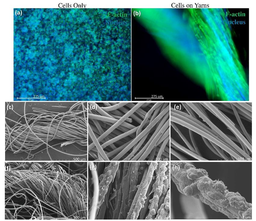
Polymers 2022, 14, 2100                                                                                                            2 of 13

                                   and have been used for the fabrication of biodegradable medical devices/scaffolds [10,11],
                                   such as drug delivery systems [12], and in bone tissue repair [13]. In a clinical study by
                                   Charriere et al. [14], the immunological response to bovine collagen implants was evaluated.
                                   Out of 705 participants, only 2.3% of the patients showed an adverse reaction to the collagen
                                   implant. Another study by Keefe et al. [15] also showed that the adverse effects were only
                                   observed in 1–2% of the treated patients, which were absolved when the material was
                                   resorbed by the hosts. For this reason, collagen polymers, especially porcine and bovine
                                   collagen polymers, are widely used in regenerative medicine.
                                         There are multiple fabrication methods for the creation of scaffolds, such as sol-
                                   vent casting/particle leaching, thermally induced phase separation (TIPS), and three-
                                   dimensional (3D) printing [16]. Although these techniques have been very advantageous
                                   for the creation of scaffolds, they all have disadvantages such as a lack of reproducibility,
                                   low production efficiency, and reduced mechanical strength [16,17]. The most common
                                   problems with these methods are reproducibility, repeatability, and standardization.
                                         Textile fabrication methods, such as ring spinning, knitting, weaving, and braiding,
                                   have been used for centuries. Most of these fabrication techniques, including for yarn and
                                   fabric production, have now been automated and standardized, resulting in efficiently
                                   reproducible products. Using these methods to produce scaffolds will provide us with the
                                   ability to mass-produce products that are identical and that can be created in a standardized
                                   manner [18].
                                         Traditional textile manufacturing methods, including knitting, weaving, and electro-
                                   spinning, have been used for scaffold fabrication. There is a resemblance between the
                                   structure of native ECM and textile fibers [18]. The yarn-like structure of ECM resembles
                                   the fibrous morphology of textile materials, which has been proven to be beneficial for cel-
                                   lular migration and proliferation [19,20]. The fibrous architecture of ECM helps to provide
                                   support to the cell by creating a mesh of collagen, elastin, and other proteins. It is because
                                   of the fibrous structure of ECM that the cells are able to form a communication/signaling
                                   network [20]. Additionally, textile methods also create 3D structures, which are desirable
                                   for cellular scaffolds in terms of cell proliferation and migration [21].
                                         Different methods are available for spinning yarns, including the ring spinning, rotor
                                   spinning, wrap-spinning, and core-spinning methods [22] (Table 1). Among these, ring
                                   spinning is the most popular method for creating yarns using staple fibers (short fibers
                                   ranging from 10–500 mm in length). The spun yarns can be used to create knit, woven, or
                                   braided structures for tissue engineering applications.

                                   Table 1. The advantages and disadvantages of different spinning methods.

        Method                            Advantages                                 Disadvantages                          Ref
                          The yarns that are produced have
                                                                        The method consumes high amounts of
                          high strength.
 Ring Spinning                                                          energy, and therefore, production costs       [22,23]
                          This method is applicable to a wide variety
                                                                        are also high.
                          of fibers.
                          The production costs of using this method
 Rotor Spinning                                                         The resultant strength of the yarns is low.   [22,23]
                          are relatively low.
                          The yarns that are produced through this
 Wrap-Spinning            method are highly absorbent.                  The yarns have low strength.                  [22–24]
                          The method has high production efficiency.
                          This method uses two or more fibers,
                                                                        There are limited applications for
 Core-Spinning            providing excellent properties to the                                                       [22,23,25]
                                                                        this method.
                          resultant composite yarn.

                                       Weaving provides strength to the yarn. A study by Gilmore et al. [26] used round and
                                   grooved cross-sectional fibers to create plain weave and satin weave scaffolds. The results
                                   suggested that the fabric structure has a significant influence on the resultant properties,
                                   especially on the permeability of the scaffold. Similarly, knitting provides elasticity and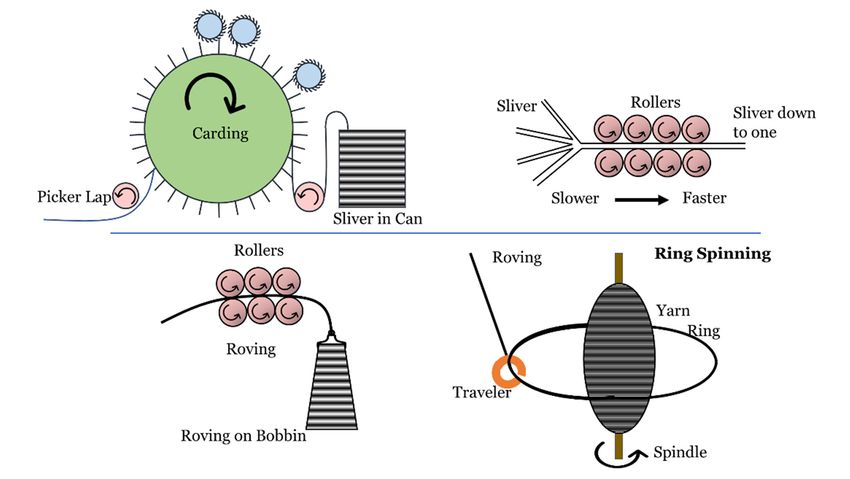
Polymers 2022, 14, 2100                                                                                                          3 of 13

                                     strength to fibrous scaffolds. A study by Lieshout et al. [27] shows a comparison of
                                     knitted and electrospun scaffolds for aortic valves. Human myofibroblasts were cultured
                                     on both scaffolds over the course of 23 days. The results compared tissue formation,
                                     which was evaluated via confocal laser scanning microscopy. The study showed that the
                                     electrospun scaffold tore within 6 h, while the knit scaffold remained intact. Another
                                     study by Zhang et al. [28] explored the idea of using a circular knit machine to produce a
                                     small-caliber vascular graft. The scaffold had excellent mechanical properties (bursting
                                     strength, suture retention strength, and compliance) that were comparable to the coronary
Polymers 2022, 14, x FOR PEER REVIEW artery under normotensive pressure.                                                                     2 of 13
                                           This study evaluates an effort to mass-produce collagen fiber scaffolds that can be
                                     fabricated via traditional textile processes (Figure 1). In this study, collagen yarns are
                                     produced using the ring-spinning method, which is a process of creating yarns by twisting
                                  have   been
                                     fibers     used and
                                            together   for the   fabrication
                                                            winding    them onofa biodegradable
                                                                                    bobbin. The method  medical     devices/scaffolds
                                                                                                              comprises     four processes:[10,11],
                                  such
                                     carding, drawing, roving, and ring spinning (Figure 2). The purpose of carding and by
                                        as  drug   delivery   systems     [12], and   in bone    tissue  repair   [13].  In  a clinical  study
                                  Charriere
                                     drawingetisal.  [14], the
                                                  to clean andimmunological          response
                                                                 align the fibers. After         to bovine
                                                                                           drawing,           collagen
                                                                                                       the fibers         implants
                                                                                                                   are passed          was evalu-
                                                                                                                                 to a roving
                                     machine,
                                  ated. Out ofwhich     fuses the fibers
                                                 705 participants,     onlytogether
                                                                              2.3% ofthrough   twisting,
                                                                                        the patients      creatingan
                                                                                                        showed       a roving.
                                                                                                                       adverseAreaction
                                                                                                                                   roving isto the
                                     a sliverimplant.
                                  collagen    of fibers that is larger
                                                        Another         in diameter
                                                                    study    by Keefethan   yarn.
                                                                                        et al. [15]The
                                                                                                     alsoroving
                                                                                                           showedis then
                                                                                                                      thatpassed    to a ringeffects
                                                                                                                             the adverse
                                  were only observed in 1–2% of the treated patients, which were absolved when thetomate-
                                     spinning   machine,   which   provides    the fibers  with  more   twist, reducing    the  diameter
                                     convert them into a yarn [22]. As a result, yarns are stronger when they are fabricated via
                                  rial was resorbed by the hosts. For this reason, collagen polymers, especially porcine and
                                     ring spinning than other methods, such as rotor spinning. Ring spinning also provides a
                                  bovine   collagen polymers,
                                     high production    rate and canarebewidely   used
                                                                           used for  anyin  regenerative
                                                                                          type  of fiber [23].medicine.

                                        Figure 1. Experimental Design Schematic. Collagen fibers were provided by the Kaneka Corporation
                                    Figure 1. Experimental Design Schematic. Collagen fibers were provided by the Kaneka Corpora-
                                       and spun to yarns using the traditional ring spinning method. The resulting yarns were analyzed for
                                    tion and spun to yarns using the traditional ring spinning method. The resulting yarns were ana-
                                       biodegradability and biocompatibility. The biodegradability of the material was analyzed via weight
                                    lyzed for biodegradability and biocompatibility. The biodegradability of the material was analyzed
                                    viachanges,  tensile strength changes, and morphological analysis. The biocompatibility was checked
                                        weight changes,     tensile strength changes, and morphological analysis. The biocompatibility was
                                       with Alamar blue and live/dead analysis for cell viability, and SEM and phalloidin staining were
                                    checked with Alamar blue and live/dead analysis for cell viability, and SEM and phalloidin staining
                                       used for cytoskeleton analysis.
                                    were  used for cytoskeleton analysis.

                                         There are multiple fabrication methods for the creation of scaffolds, such as solvent
                                    casting/particle leaching, thermally induced phase separation (TIPS), and three-dimen-
                                    sional (3D) printing [16]. Although these techniques have been very advantageous for the
                                    creation of scaffolds, they all have disadvantages such as a lack of reproducibility, low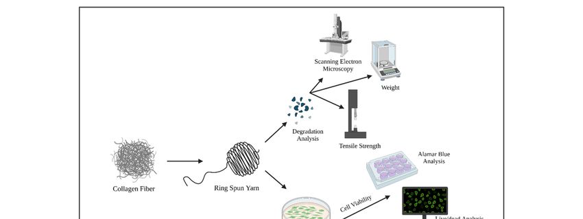
Polymers 2022, 14,
14, 2100
x FOR PEER REVIEW 44of
of 13
Figure 2.
Figure 2. Ring
Ring spinning
spinning process
process for
for collagen
collagen yarns.
yarns. The
The collagen
collagen fibers
fibers were
were provided
provided byby the
the Kaneka
Kaneka
Corporation. The fibers were first run through the card and drawer with multiple sets of rollers. The
Corporation. The fibers were first run through the card and drawer with multiple sets of rollers. The
rollers combed the fibers to clean and aligned them before they were gathered to form a sliver in a
rollers combed the fibers to clean and aligned them before they were gathered to form a sliver in a
can. The sliver then passed through a roving frame, where the sliver reduced in diameter to form a
can. The sliver then passed through a roving frame, where the sliver reduced in diameter to form
roving, which was wound on a bobbin. Finally, the roving bobbins were passed through a ring
a roving, machine,
spinning which was wound
where ondiameters
their a bobbin.were
Finally, the roving
further reducedbobbins were with
via twisting passed
thethrough
help of aa trav-
ring
spinning machine,
eler and a ring. where their diameters were further reduced via twisting with the help of a traveler
and a ring.
After the yarns were fabricated, they were subjected to biocompatibility and biodeg-
Afteranalysis
radation the yarnsto were fabricated,
evaluate they wereuse
the perspective subjected
of this to biocompatibility
technique and biodegra-
in the future to mass-
dation analysis to evaluate the perspective use of this technique in
produce scaffolds. The biocompatibility analysis provides knowledge about thethe future to safety
mass-
produce scaffolds. The biocompatibility analysis provides knowledge about the safety
and efficacy of the fabricated when encountering cells. This is crucial for the application and
efficacy of the fabricated when encountering cells. This is crucial for the application of
of this process in tissue engineering products, especially for dermal [29], cardiovascular
this process in tissue engineering products, especially for dermal [29], cardiovascular [28],
[28], tendon [30], and ligament [18] applications.
tendon [30], and ligament [18] applications.
2. Materials
2. Materials and
and Methods
Methods
Collagen fibers were provided
Collagen fibers were provided by by the
the Kaneka
Kaneka Corporation.
Corporation. TheThe fibers
fibers were
were processed
processed
through a card chute feed system (Rieter Card C4) followed by
through a card chute feed system (Rieter Card C4) followed by two rounds of two rounds of drawing
drawing
(Rieter RSB851). The aligned fibers were then converted to a roving with a roving
(Rieter RSB851). The aligned fibers were then converted to a roving with a roving machine machine
(Rieter Fly
(Rieter Fly F4/1)
F4/1)and
andfinally spun
finally spun into yarn
into on on
yarn a ring spinning
a ring machine
spinning (Rieter
machine G5/2)G5/2)
(Rieter (Fig-
ure 2). The yarns had a fineness of 20 Ne, which is the thickness or coarseness
(Figure 2). The yarns had a fineness of 20 Ne, which is the thickness or coarseness of a yarn, of a yarn,
and were
and were measured according to
measured according to the
the English
English Cotton
Cotton Count
Count (Ne)
(Ne) method,
method, which
which isis defined
defined
as “the number of 840-yard length per pound” [23]. Tensile strength and
as “the number of 840-yard length per pound” [23]. Tensile strength and ultrastructure ultrastructure
characterization were
characterization were carried
carried out
out via
via scanning electron microscopy
scanning electron microscopy (SEM),
(SEM), andand an
an in
in vitro
vitro
biocompatibility analysis
biocompatibility analysis was
was performed
performed on on the
the resulting
resulting yarns
yarns (Figure
(Figure 1).
1).
2.1. Degradation Study
A degradation study was conducted to check the ability of the material to degrade
when implanted
implanted in inthe
thebody
bodyororwhen
when used
used as as
an an in vivo
in vivo application.
application. Collagenase
Collagenase is
is nor-
normally
mally foundfound in almost
in almost all mammalian
all mammalian tissues,
tissues, including
including pig pancreas,
pig pancreas, beef beef pancreas,
pancreas, and
and
humanhuman tissues.
tissues. It in
It helps helps in wound
wound healinghealing
by moving by moving the keratinocytes
the keratinocytes over the
over the collagen-
collagen-rich dermis during re-epithelialization.
rich dermis during re-epithelialization.
A collagenase solution was prepared according to the protocol by Alberti et al. [31].
Collagenase isolated from Clostridium histolyticum cleaves the bonds between neutral
Polymers 2022, 14, 2100                                                                                           5 of 13

                               A collagenase solution was prepared according to the protocol by Alberti et al. [31].
                          Collagenase isolated from Clostridium histolyticum cleaves the bonds between neutral amino
                          acids and glycine, which is found with a high frequency in collagen. This type of degra-
                          dation is a simplistic method to predict the in vivo degradation profile of our ring-spun
                          collagen yarns. First, a collagen yarn sample was dried, weighed, and placed in 12-well
                          plates. In a separate glassware, 0.5 mL of 0.1 M TRIS-HCl and 0.005 M of CaCl2 were mixed
                          with 2 mg/mL of collagenase (Type I, powder, Gibco, >125 units/mg). A 2 mL amount of
                          the prepared solution was added to each well such that each sample was immersed in the
                          solution, and the samples were placed in an incubator at 37 ◦ C for a total of 8 weeks. Their
                          weight, tensile strength, and morphology were monitored biweekly. The ultrastructure and
                          morphology of the yarns were assessed using SEM to evaluate the degradation pattern in
                          the collagen yarns.

                          2.1.1. Weight Change
                               A total of 6 samples were prepared, all of which were 20 cm each in length. It was
                          determined that the length would be longer to increase the accuracy. The samples were
                          weighed and placed in a 12-well plate and immersed in collagenase solution for the entire
                          length of the study. The samples were then placed in the incubator at 37 ◦ C. The solution
                          was maintained at a pH of 7–7.4 throughout the study. The samples were dried every
                          14 days by aspirating the solution and drying in the desiccator for 24 h. After drying, their
                          weight was measured, and the samples were placed in the original well plates again and
                          submerged in collagenase solution at 37 ◦ C. Average mass loss was calculated with the
                          following formula:
                                                                Original Mass − Final Mass
                                                  Mass Loss =
                                                                      Original Mass

                          2.1.2. Tensile Test
                               A total of 24 samples were prepared, each of which were 20 cm in length, and the
                          samples were placed in 12-well plates. Collagenase solution was added to each well, and
                          the samples were stored in the incubator at 37 ◦ C. The pH of the samples was maintained
                          at 7–7.4. Every 14 days, 6 samples were dried and were placed in the desiccator for 24 h.
                          When they had dried completely, the samples were mounted on cardboard (as shown in
                          Figure 3a). An MTS Criterion 43 Tensile tester (MTS Systems, Eden Prairie, MN USA) was
                          used for the tensile test, and a 50 N load cell, a gauge length of 1 cm, and a crosshead speed
                          of 10 mm/min were maintained for the tensile test.

                          2.1.3. Scanning Electron Microscopy
                               The samples were dried and mounted on stubs using double-sided carbon tape. They
                          were sputter-coated with gold/palladium using a SC7620 Mini Sputter Coater (Quorum
                          Technologies, East Sussex, UK) for 45 s, resulting in a 10 nm coating. Samples were imaged
                          with a Phenom G1 desktop SEM (Phenom, ThermoFisher, Eindhoven, The Netherlands).
                          A total of 9 samples were analyzed per time point, and 4 images were taken from each
                          sample. The morphology of the yarns was observed at biweekly intervals. The images
                          were analyzed for surface and bulk degradation.

                          2.2. Biocompatibility
                          2.2.1. Sample Preparation
                              The collagen yarns were sterilized by immersing in 70% ethanol for 20 min. The
                          ethanol was then aspirated followed by washing with phosphate-buffered solution (PBS,
                          Hyclone, Cytiva, Long, UT, USA).
Polymers 2022, 14, x FOR PEER REVIEW 6 of 13
Polymers 2022, 14, 2100 6 of 13
Figure
Figure3. 3. Mechanical
Mechanical analysis over time: Collagen
Collagen yarns
yarnswereweresoaked
soakedin inaacollagenase
collagenasesolution
solutionfor
for8
8weeks
weeks(n(n==33samples
samplesper
perperiod).
period).Samples
Sampleswere
were prepared
prepared andand mounted using a cardboard
cardboard holder
holder
(a) for
(a) for tensile
tensile testing.
testing. The
The samples
samples showed
showed aa significant
significant loss
loss(97%,
(97%,**ppPolymers 2022, 14, 2100 7 of 13
cell indicator (ex/em 488 nm/515 nm); and BOBO-3 Iodide, which is a dead cell indicator
(ex/em 570 nm/602 nm). This test was performed on cell-seeding days 1, 3, 5, and 7.
The alamarBlue assay was also performed to quantify the proliferation of the cells
that had been seeded on collagen fiber scaffolds. This assay provides quantitative data
on cell metabolism (proliferation) by measuring the changes in fluorescence generated
by resazurin, which reduces to resorufin in response to chemical reductions due to cell
growth. The dye changes its color and fluorescence from blue/non-fluorescent (oxidized)
to red/fluorescent (unoxidized).
The cells were seeded on the yarn for 7 days. The alamarBlue assay was carried out
on days 1, 3, 5, and 7 with the positive control cells only and with the negative controls of
the yarn in media only (no cells). The samples were incubated with alamarBlue Reagent
(Fisher Scientific, Waltham, MA, USA) for 1 h. Later, the samples were read with a plate
reader (Synergy HT, BioTek, Santa Clara, CA, USA) set to 540/25 λ excitation, 590/35 λ
emission, and maintained at 37 ◦ C.
2.2.4. Phalloidin Staining
Phalloidin staining was carried out to label and identify F-actin (cytoskeleton) expres-
sion in the cells to observe the cellular morphology. On day 7, the samples were fixed with
4% paraformaldehyde for 20 min and washed with PBS. The samples were then permeabi-
lized with 0.1% TritonX-100 for 30 min and 0.1% Tween-20 for 15 min. Blocking buffer was
prepared with 0.1%Tween-20, 2% bovine serum albumin, and 2% goat serum. Phalloidin
(Invitrogen™ ActinGreen™ 488 ReadyProbes™ Reagent) was used in the concentration
of 2 drops/mL of blocking buffer and incubated for 1 h. After incubation, the samples
were washed with PBS and stained with Hoechst (Invitrogen™ Hoechst 33342, Eugene, OR,
USA) (1:1000 concentration Hoechst: PBS) for 5 min. The samples were then imaged using
fluorescence microscopy (EVOS™ FL Auto 2 imaging system, ThermoFisher, Waltham,
MA, USA).
2.2.5. Ultrastructure Analysis (Scanning Electron Microscopy)
Collagen fiber yarns seeded with NIH 3T3 fibroblasts were fixed in buffered formalin.
The samples were washed 3 × 10 min with 0.15 M sodium phosphate buffer and at a pH of
7.4 followed by post-fixation in 1% osmium tetroxide/0.15 M sodium phosphate buffer at
a pH of 7.4 for 1 h. After washing with deionized water (3 × 10 min), the samples were
treated with 1% tannic acid in water for 30 min. The scaffolds were washed in deionized
water and dehydrated through an increasing ethanol series (30%, 50%, 75%, 90%, 100%,
100%, and 100%—15 min each). The samples were transferred in 100% ethanol to a Samdri-
795 critical point dryer (Tousimis Research Corporation, Rockville, MD, USA) and dried
using liquid carbon dioxide as the transitional solvent. The scaffolds were mounted onto
13 mm diameter aluminum stubs with carbon adhesive tabs and were sputter-coated with
8 nm of gold/palladium alloy (60Au: 40Pd) using a Cressington 208HR Sputter Coater (Ted
Pella, Inc., Redding, CA, USA). Images were taken using a Zeiss Supra 25 FESEM (Carl
Zeiss Microscopy, Jena, Germany) operating at 5 kV or 10 kV using the SE2 detector, 30 µm
aperture, and approximate working distances from 15 to 25 mm. The samples were imaged
for any cell structures that were growing horizontally or vertically over the fiber and yarn
surfaces.
2.3. Statistical Analysis
Data were collected from 3 to 9 samples and expressed as means ± standard devia-
tion. Statistical analysis was performed using the Student’s t test, and significance was
determined at p < 0.0001.
3. Results
This study evaluated the biodegradability and biocompatibility of mass-produced
ring spun collagen fiber yarns (Figure 1). The collagenase (Type I) enzyme was used toData were collected from 3 to 9 samples and expressed as means ± standard devia-
tion. Statistical analysis was performed using the Student’s t test, and significance was
determined at p < 0.0001.
3. Results
Polymers 2022, 14, 2100 8 of 13
This study evaluated the biodegradability and biocompatibility of mass-produced
ring spun collagen fiber yarns (Figure 1). The collagenase (Type I) enzyme was used to
assess yarn degradation. Mass changes, tensile strength, and the morphology of the yarns
assesschecked
were yarn degradation.
at biweeklyMass changes,
intervals. Thetensile strength, and
biocompatibility of the
the morphology of the
yarns was also yarns
checked
were checked at biweekly intervals. The biocompatibility of the yarns was also checked
using mouse fibroblast cells. The cell viability, cell metabolic activity, and the morphology
using
of mouse fibroblast
the cytoskeleton was cells. The cell
observed viability, cell metabolic
for biocompatibility activity, and the morphology
analysis.
of the cytoskeleton was observed for biocompatibility analysis.
3.1. Collagen Yarns Degrade in the Presence of Enzymes in an 8-Week Study
3.1. Collagen Yarns Degrade in the Presence of Enzymes in an 8-Week Study
During the enzymatic degradation with collagenase Type I, the collagen yarns lost
During the enzymatic degradation with collagenase Type I, the collagen yarns lost
most of their mass and mechanical integrity over 8 weeks. On average, the samples lost
most of their mass and mechanical integrity over 8 weeks. On average, the samples
80 ± 6% of their mass over 8 weeks (Figure 3c), which indicates that the material is de-
lost 80 ± 6% of their mass over 8 weeks (Figure 3c), which indicates that the material is
gradable, and the remnants were dispersed in the solution. The pH of the solution re-
degradable, and the remnants were dispersed in the solution. The pH of the solution
mained the
remained the same
samethroughout
throughout thethestudy.
study.The
Thedegradation
degradationofofcollagen
collagenfibers
fiberswas
wasconfirmed
confirmed
via
via the assessment of the tensile strength and morphology of the samples,indicated
the assessment of the tensile strength and morphology of the samples, as by
as indicated
the losses observed in the measured Young’s modulus (Figure
by the losses observed in the measured Young’s modulus (Figure 3b). The modulus loss 3b). The modulus loss in-
dicates
indicates thethe
degradation
degradation ofofthe
thecollagen
collagenfibers
fibersininthe
thepresence
presenceofofcollagenase
collagenasesolution.
solution.TheThe
samples lost 97% of their tensile strength in 6 weeks. Fiber samples
samples lost 97% of their tensile strength in 6 weeks. Fiber samples were mechanically were mechanically
unstable
unstableby byweek
week66and andtherefore
therefore could
could not
not be
be tested
tested for
for tensile
tensile strength
strength inin week
week 8.8.
Gradual degradation was evident in the yarns for eight weeks
Gradual degradation was evident in the yarns for eight weeks (Figure 4) when (Figure 4) whenweekweek0
0and
andweekweek88are arecompared.
compared. A A polymer
polymer undergoes
undergoes bulk bulk degradation
degradation if the diffusion of
if the diffusion of
water/fluids
water/fluidsinto intothe
thepolymer
polymerisisfaster
fasterthan
thanthethebonds
bondsbreak
break[32].
[32].TheTheSEM
SEMimage
imageanalysis
analysis
ofof the
the collagen
collagen yarns
yarns indicated
indicatedthat thatthe
thelength
lengthof of the
the fibers
fibers broken
broken down
down over over 88 weeks
weeks
(Figure
(Figure 4 marked with arrows), suggesting bulk degradation. On the other hand,sur-
4 marked with arrows), suggesting bulk degradation. On the other hand, the the
face of the
surface offiber also changed
the fiber also changedfrom from
smooth to rough
smooth and beady
to rough and (marked with stars),
beady (marked withwhich
stars),
iswhich
a signisof surface
a sign degradation
of surface in the fibers.
degradation in the fibers.
Figure 4.
Figure Bulkand
4. Bulk and surface
surface degradation
degradationisis observed
observedover
over 88 weeks.
weeks. The
Thesamples
sampleswere
were subjected
subjectedto
to
collagenase
collagenasefor
for88weeks.
weeks.The
Themorphology
morphologyof ofthe
thefibers
fiberswas
wasobserved
observedvia
viaSEM.
SEM.The
Thefibers
fibershad
hadbroken
broken
down and degraded significantly at week 8 (images i,j) when compared to week 0 (images a,b). The
surface of the fibers also changed, which shows that there is surface degradation in the fibers (n = 6).
The top images (a,c,e,g,i) show a higher magnification (50 um), whereas the bottom images (b,d,f,h,j)
show a lower magnification (100 um). Arrows indicate broken fiber lengths; stars indicate a surface
transition from smooth to rough and beady.
3.2. The Collagen Material Is Biocompatible with Mouse Fibroblast (NIH 3T3) Cell Line
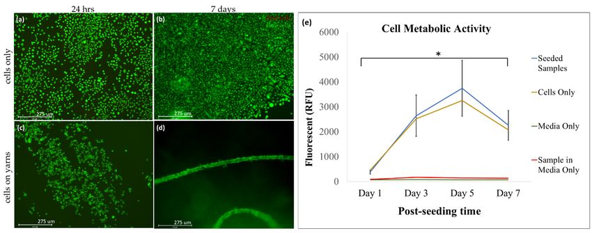
High cell viability on the collagen yarns was observed over 7 days (Figure 5). After
only 24 h of cell culture, the cells were observed to form colonies, which was a strong
indication that the material provides a heterogeneous environment where the cells are more
attracted to some areas, forming clusters and migrating to a suitable area. The cells adhered
to the yarn/fiber surface and proliferated along the length (Figure 5c,d).3.2. The Collagen Material Is Biocompatible with Mouse Fibroblast (NIH 3T3) Cell Line
High cell viability on the collagen yarns was observed over 7 days (Figure 5). After
only 24 h of cell culture, the cells were observed to form colonies, which was a strong
indication that the material provides a heterogeneous environment where the cells are
Polymers 2022, 14, 2100 9 of 13
more attracted to some areas, forming clusters and migrating to a suitable area. The cells
adhered to the yarn/fiber surface and proliferated along the length (Figure 5c,d).
Figure5.5.High
Figure Highbiocompatibility
biocompatibility of of collagen
collagenyarns
yarnsisisobserved.
observed.Viability of of
Viability NIHNIH3T33T3
cells (a) (a)
cells seeded
seeded
on collagen yarns. The green signal indicates live cells, and the red signal indicates
on collagen yarns. The green signal indicates live cells, and the red signal indicates dead cells. dead cells. TheThe
live/dead assay of the seeded samples at 24 hours (c) and after 7 days (d). The cell-only controls at
live/dead assay of the seeded samples at 24 h (c) and after 7 days (d). The cell-only controls at 24 h
24 h and 7 days are also shown (a,b). We see evidence of the cells adhering and proliferating along
and
the7length
days are also
of the shown
fiber for at(a,b).
least We see evidence
7 days, indicatingof the
the cells adhering of
biocompatibility andtheproliferating
collagen yarns along
(n = the
length of results
10). The the fiber forthe
from at least 7 days,
live/dead indicating
assay the biocompatibility
were confirmed and quantifiedof thethe
with collagen yarnsassay
alamarBlue (n = 10).
(e).results
The We observed an increased
from the live/dead in assay
metabolic
wereactivity that was
confirmed andcorrelated
quantifiedto increased
with the proliferation
alamarBlue assayin
theWe
(e). cells that wereanseeded
observed on our
increased in collagen
metabolic yarns (e, blue
activity thatpositive control (cells
was correlated only in yellow)
to increased and
proliferation
ininthe
thecells
negative controls
that were (medium-only
seeded in greenyarns
on our collagen and sample
(e, blueinpositive
mediumcontrol
in red)). A significant
(cells in-
only in yellow)
crease in metabolic activity was observed in the cells seeded on collagen yarns from day 1 to day 5
and in the negative controls (medium-only in green and sample in medium in red)). A significant
(* p < 0.0001). The error bars show the standard deviation (n = 9).
increase in metabolic activity was observed in the cells seeded on collagen yarns from day 1 to day 5
(* p < 0.0001). The error
The collagen barssupport
yarns show the standard
high deviation
cellular viability (n(Figure
= 9). 5), and the corresponding
metabolic activity confirmed the viability (Figure 5e). The metabolic activity results show
an 8xThe collagen
increase in yarns support
metabolic high
activity cellular
from day 1viability
to day 5, (Figure
only to5),reduce
and the corresponding
after that. The
metabolic activity confirmed the viability (Figure 5e). The metabolic activity
reduction in proliferation may be due to limited space for the cells to proliferate any fur- results show
an 8x increase
ther. A study byin Streitchan
metabolicetactivity
al. [33] from day 1that
has shown to day
cells5,slow
only to reduce
down after
or pause that.
their pro-The
reduction in proliferation may be due to limited space for the cells to proliferate
liferation cycle due to spatial constraints in response to contact inhibition. This was high- any further.
Alighted
study by Streitchan et al. [33] has shown that cells slow down or pause
on day 5, where we observe a reduction in metabolic activity. Cells proliferated their proliferation
cycle
moredue to spatial
on the constraints
collagen yarns comparedin response to contact
to standard tissueinhibition. This dishes,
culture plastic was highlighted
indicating on
day 5, where we observe a
their superior biocompatibility. reduction in metabolic activity. Cells proliferated more on the
collagen yarns compared to standard tissue culture plastic dishes, indicating
The morphology of the cells seeded on the collagen yarns is observed to be a bright their superior
biocompatibility.
ellipsoid shape that wraps around the strands of the collagen fibers, as evidenced by f-
actinThe morphology
detection (Figureof the cells
6a,b). seeded on the
The ultrastructure collagen
analysis from yarns is observed
the SEM to be a 6c–
images (Figure bright
ellipsoid shapethat
h) highlights thatcell
wraps
growth around theobserved
can be strands of the the
along collagen fibers,
direction as evidenced
of the by f-actin
collagen fiber.
detection (Figure 6a,b). The ultrastructure analysis from the SEM images (Figure 6c–h)
highlights that cell growth can be observed along the direction of the collagen fiber.Polymers
Polymers2022,
2022,14,
14,x 2100
FOR PEER REVIEW 10 10
ofof1313
Figure
Figure6.6.Cells
Cellsproliferate
proliferatealong
alongthe
thelength
lengthofofthe
thecollagen
collagenyarns.
yarns.TheThesamples
sampleswere
werestained
stainedwith
with
phalloidin
phalloidintotoidentify
identifythe
thecytoskeleton
cytoskeletonofofthe
thecells
cellson
onthe
thecollagen
collagenyarns
yarnsatatday
day77(a,b).
(a,b).F-actin,
F-actin,a a
cytoskeleton
cytoskeletonprotein,
protein,highlights
highlightsthe
thetypical
typicalmorphology
morphologyofofthe thecells
cells(a)
(a)and
andhighlights
highlightsthe
theadhesion
adhesion
ofofthe cells along the length of the collagen yarns (b), scale bar = 275 µm (n = 6). Representative
the cells along the length of the collagen yarns (b), scale bar = 275 µm (n = 6). Representative SEM
images of collagen fibers alone (c–e) at varying magnifications. NIH 3T3 cells were seeded and
SEM images of collagen fibers alone (c–e) at varying magnifications. NIH 3T3 cells were seeded
maintained on collagen fiber yarns for 7 days (f–h). Scale bar (a,b) = 275 µm, (c) = 500 µm, (d–f) =
and maintained on collagen fiber yarns for 7 days (f–h). Scale bar (a,b) = 275 µm, (c) = 500 µm,
100 µm, (g) = 10 µm, (h) = 2 µm (n = 3).
(d–f) = 100 µm, (g) = 10 µm, (h) = 2 µm (n = 3).
4.4.Discussion
Discussion
This
Thisstudy
studydemonstrates
demonstratesthat thattextile-based
textile-basedmass-production
mass-productiontechniques
techniquesmay maybe beused
used
for the fabrication of biocompatible tissue-engineered scaffolds. The
for the fabrication of biocompatible tissue-engineered scaffolds. The yarns in this study yarns in this study
were
wereproduced
producedusingusingaatraditional
traditionalringringspinning
spinningtechnique
techniquefacility
facilitywhere
wheregeneral
generaltextile
textile
yarns are produced. These yarns can be used to produce woven, knit,
yarns are produced. These yarns can be used to produce woven, knit, or braided structures or braided struc-
tures
that that canused
can be be used for tissue
for tissue engineering
engineering applications.
applications. For example,
For example, Learn Learn
et al.et[30]
al. [30]
used
used collagen
collagen yarnsyarns to fabricate
to fabricate a wovena woven scaffold
scaffold for rotator
for rotator cuff tendon
cuff tendon regeneration,
regeneration, whereas
whereas
Ruan etRuan et used
al. [34] al. [34] used a collagen-silk
a collagen-silk blend toblendcreatetoknit
create knit scaffolds
scaffolds for anterior
for anterior cruciate
cruciate
ligament ligament reconstruction.
reconstruction. The dataThe data demonstrate
demonstrate that collagen
that collagen fiber yarnsfiber
areyarns are bio-
biocompatible.
compatible. The biocompatibility
The biocompatibility of a material of isa defined
materialas is the
defined as theof
properties properties
a materialofthat a material
are safe
that are safe and effective for cells to proliferate, migrate, and function
and effective for cells to proliferate, migrate, and function [35]. The results from [35]. The results
our
from our viability
viability and proliferation
and proliferation assays combined
assays combined with our fluorescent
with our fluorescent and electron and electron
microscopy
microscopy
image analysis image analysis
show showthe
that both that both the
material andmaterial and the fabrication
the fabrication method support method sup-
cellular
port cellular proliferation
proliferation and maintain andtheir
maintain their expected
expected cellular morphology
cellular morphology (Figures(Figures
5 and 6). 5 and
It is
6). It is interesting
interesting to observe
to observe that thethat themigrated
cells cells migrated
from thefrom the tissue
tissue culture-treated
culture-treated plate plate
to the
tofiber surface,
the fiber providing
surface, compelling
providing compellingevidence of theofbiocompatibility
evidence the biocompatibility of theofmaterial.
the material.
Similarly,the
Similarly, thefabrication
fabricationmethod
methoddemonstrates
demonstratesthe theability
abilityofofthe
thecollagen
collagenfibers
fiberstoto
degradeover
degrade overtime
timeininthethepresence
presenceofofthe thecollagenase
collagenaseenzyme.
enzyme.Collagenase
Collagenaseisisnormally
normally
presentininmost
present mostmammalian
mammaliantissues,
tissues,including
includingthose
thoseininhumans
humans[36].
[36].Collagenase
Collagenaseprovides
provides
aagood
goodimitation
imitationofofthe theininvivo
vivo environment,
environment,where whereenzymatic
enzymaticactivity
activityisisobserved
observedtotoPolymers 2022, 14, 2100 11 of 13
cleave native fibrillar collagen. In this study, degradation is seen by a reduction in mass
and mechanical integrity over time, which signifies that the fabrication methods provide
additional mechanical strength without changing the degradable nature of the polymer.
Degradation is an important property for implantable biopolymers in situation where the
scaffold is only required for mechanical support until the resulting tissue develops in vivo.
This prevents the need for surgical procedures to remove the scaffold when it is no longer
required [36].
Previously, conventional fabrication techniques such as fiber melts, fiber bonding,
melt molding, and fiber composite foam have been used, which can provide the necessary
morphology. However, these methods have multiple disadvantages, such as a lack of
structural stability or the solvent residue being toxic or harmful [17]. On the other hand,
traditional textile fabrication methods have many advantages, such as good mechanical
stability, a 3D structure, a fibrous nature, and most importantly, the ability to fabricate
products that are identical and that can be produced in bulk [18].
Collagen is one of the most extensively used materials in tissue engineering due to its
abundant presence in the native ECM, where it provides biological and structural integrity
to cells [7]. However, when used as a scaffold, the material does not provide mechanical
strength, which limits its applications [37]. Collagen is normally used as a blended material
or is chemically cross-linked to provide the required strength to the material [37]. However,
the use of ring spinning technology solves this problem due to the twisting of fibers by
providing excellent mechanical support.
Additionally, the fibrous structure of textile yarns replicates the thread-like morphol-
ogy of ECM [18]. Therefore, using collagen in a textile form provides the required support
to the cells whiles also providing a medium for networking and signaling [20]. Moreover,
the 3D structure of textiles is also beneficial for cell proliferation and migration. Biomimetic
fibrous scaffolds are crucial in tissue engineering when the goal is to best recapitulate the
native ECM. Creating fibrous scaffolds from collagen has been repeatedly demonstrated to
be beneficial for a variety of tissue engineering applications [38], including scaffolds for
heart valve repair [39] and cardiac tissue engineering [40], with bone [34,41] and skin tissue
engineering applications being the most common examples.
A study by Xie et al. [42] demonstrated the potential use of wet spun yarn in tissue
engineering applications. However, collagen is sensitive to temperature, and therefore, wet
spinning requires the yarns to be cross-linked to acquire the required thermal and chemical
stability. On the other hand, the ring spinning method is dry and does not require elevated
temperatures. Therefore, it is likely to be a more stable form of spinning for collagen
fibers. Another common method using for processing collagen for tissue engineering
applications includes chemical cross-linking, which is destructive to the natural helical
structure of collagen. Thus, we showed that ring-spun collagen yarns represent an exciting
new opportunity for the scalable production of biomimetic biomaterials that can be used
for a variety of tissue engineering applications.
Regardless of the excellent results of the in vitro analysis of the fabricated textile yarns,
further studies are required to create compact structures that are either woven, knit, or
braided. We are currently working on fabricating knit structures for in vitro studies.
5. Conclusions
Collagen fiber yarns are a highly biocompatible material/technique, as they promote
cell adhesion and proliferation. The structure of the material encourages cell adhesion and
proliferation without requiring any intricate and complex fabrication technique, which not
only saves time but also simplifies and streamlines the fabrication process. The process also
provides a standardized fabrication technique for creating identical scaffolds. Furthermore,
this proof-of-concept study sets the foundation for further analyses of the underlying
textile and biological properties of mass-produced collagen fiber scaffolds that support cell
behavior and that can be used for further tissue engineering and regenerative medicine
applications.Polymers 2022, 14, 2100 12 of 13
Author Contributions: Conceptualization, K.M.A. and J.M.G.; methodology, K.M.A., Y.H. and A.Y.A.;
formal analysis, K.M.A., A.Y.A. and J.M.G.; resources, J.M.G.; data curation, K.M.A., N.M. and T.C.S.;
writing—original draft preparation, K.M.A.; writing—review and editing, K.M.A., N.M., T.C.S. and
J.M.G.; visualization, K.M.A., Y.H., N.M. and T.C.S.; supervision, J.M.G.; project administration,
K.M.A. and J.M.G.; funding acquisition, J.M.G. All authors have read and agreed to the published
version of the manuscript.
Funding: The research was supported by the Kaneka Corporation (K.M.A., J.M.G.), the NCSU
Laboratory Research Equipment Program (LREP) (J.M.G.), the Wilson College of Textiles (J.M.G.), the
Department of Textile Engineering, Chemistry and Science (J.M.G., K.M.A., T.C.S.), and The Provost’s
Fellowship at NC State University (T.C.S.).
Institutional Review Board Statement: Not Applicable.
Informed Consent Statement: Not Applicable.
Data Availability Statement: The data presented in this study are available within this publication.
Acknowledgments: The authors wish to thank Jeff Vercellone and Dennis Mater from the Kaneka
Corporation; Wilson College of Textiles, North Carolina State University; Judy Elson from the
Chemistry and Microscopy Laboratory, Textile Engineering, Chemistry, and Science, Wilson College
of Textiles, North Carolina State University, Physical Testing Laboratory; Timothy Pleasants from
Zeiss Textiles Extension, North Carolina State University; and the Microscopy Services Laboratory,
University of North Carolina at Chapel Hill.
Conflicts of Interest: The authors declare no conflict of interest.
References
1. O’Brien, F.J. Biomaterials & scaffolds for tissue engineering. Mater. Today 2011, 14, 88–95. [CrossRef]
2. Zileli, M.; Benzel, E.C.; Bell, G.R. Chapter 93—Bone graft harvesting. In Spine Surgery, 3rd ed.; Benzel, E.C., Ed.; Churchill
Livingstone: Philadelphia, PA, USA, 2005; pp. 1253–1261. [CrossRef]
3. Transplanting Human Tissue: Ethics, Policy, and Practice; Youngner, S.J.; Youngner, S.J.; Anderson, M.W.; Schapiro, R., Eds.; Oxford
University Press: Oxford, UK, 2004.
4. Atala, A. Tissue engineering and regenerative medicine: Concepts for clinical application. Rejuvenation Res. 2004, 7, 15–31.
[CrossRef] [PubMed]
5. Chen, G.; Ushida, T.; Tateishi, T. Scaffold design for tissue engineering. Macromol. Biosci. 2002, 2, 67–77. [CrossRef]
6. Rahmanian-Schwarz, A.; Held, M.; Knoeller, T.; Stachon, S.; Schmidt, T.; Schaller, H.E.; Just, L. In vivo biocompatibility and
biodegradation of a novel thin and mechanically stable collagen scaffold. J. Biomed. Mater. Res. 2014, 102, 1173–1179. [CrossRef]
7. Gelse, K.; Pöschl, E.; Aigner, T. Collagens—Structure, function, and biosynthesis. Adv. Drug Deliv. Rev. 2003, 55, 1531–1546.
[CrossRef]
8. Glowacki, J.; Mizuno, S. Collagen scaffolds for tissue engineering. Biopolym. Orig. Res. Biomol. 2008, 89, 338–344. [CrossRef]
9. Cen, L.; Liu, W.; Cui, L.; Zhang, W.; Cao, Y. Collagen tissue engineering: Development of novel biomaterials and applications.
Pediatr. Res. 2008, 63, 492–496. [CrossRef]
10. George, J.; Onodera, J.; Miyata, T. Biodegradable honeycomb collagen scaffold for dermal tissue engineering. J. Biomed. Mater. Res.
2008, 87, 1103–1111. [CrossRef]
11. Roßbach, B.P.; Gülecyüz, M.F.; Kempfert, L.; Pietschmann, M.F.; Ullamann, T.; Ficklscherer, A.; Niethammer, T.R.; Zhang, A.;
Klar, R.M.; Müller, P.E. Rotator cuff repair with autologous tenocytes and biodegradable collagen scaffold: A histological and
biomechanical study in sheep. Am. J. Sports Med. 2020, 48, 450–459. [CrossRef]
12. Khan, R.; Khan, M.H. Use of collagen as a biomaterial: An update. J. Indian Soc. Periodontol. 2013, 17, 539–542. [CrossRef]
13. Wei, S.; Ma, J.; Xu, L.; Gu, X.; Ma, X. Biodegradable materials for bone defect repair. Mil. Med. Res. 2020, 7, 54. [CrossRef]
[PubMed]
14. Charriere, G.; Bejot, M.; Schnitzler, L.; Ville, G.; Hartmann, D.J. Reactions to a bovine collagen implant: Clinical and immunologic
study in 705 patients. J. Am. Acad. Dermatol. 1989, 21, 1203–1208. [CrossRef]
15. Keefe, J.; Wauk, L.; Chu, S.; DeLustro, F. Clinical use of injectable bovine collagen: A decade of experience. Clin. Mater. 1992, 9,
155–162. [CrossRef]
16. Dutta, R.C.; Dey, M.; Dutta, A.K.; Basu, B. Competent processing techniques for scaffolds in tissue engineering. Biotechnol. Adv.
2017, 35, 240–250. [CrossRef] [PubMed]
17. Yang, S.; Leong, K.; Du, Z.; Chua, C. The design of scaffolds for use in tissue engineering. Part I. Traditional factors. Tissue Eng.
2001, 7, 679–689. [CrossRef] [PubMed]
18. Jiao, Y.; Li, C.; Liu, L.; Wang, F.; Liu, X.; Mao, J.; Wang, L. Construction and application of textile-based tissue engineering
scaffolds: A review. Biomater. Sci. 2020, 8, 3574–3600. [CrossRef]Polymers 2022, 14, 2100 13 of 13
19. Nivedhitha Sundaram, M.; Deepthi, S.; Mony, U.; Shalumon, K.T.; Chen, J.P.; Jayakumar, R. Chitosan hydrogel scaffold reinforced
with twisted poly(l lactic acid) aligned microfibrous bundle to mimic tendon extracellular matrix. Int. J. Biol. Macromol. 2019, 122,
37–44. [CrossRef]
20. Benjamin, M.; Kaiser, E.; Milz, S. Structure-function relationships in tendons: A review. J. Anat. 2008, 212, 211–228. [CrossRef]
21. Badekila, A.K.; Kini, S.; Jaiswal, A.K. Fabrication techniques of biomimetic scaffolds in three-dimensional cell culture: A review. J.
Cell. Physiol. 2021, 236, 741–762. [CrossRef]
22. Alagirusamy, R.; Das, A. Chapter 8—Conversion of fibre to yarn: An overview. In Textiles and Fashion; Sinclair, R., Ed.; Woodhead
Publishing: Sawston, UK, 2015; pp. 159–189. [CrossRef]
23. Lawrence, C.A. Advances in Yarn Spinning Technology; Elsevier Science: Amsterdam, The Netherlands, 2010.
24. Subramaniam, V.; Mohammad, P. Wrap spinning technology—A critical review of yarn properties. Indian J. Fibre Text. Res. 1992,
17, 252–254.
25. Das, A.; Alagirusamy, R. 3—Fundamental principles of open end yarn spinning. In Advances in Yarn Spinning Technology; Lawrence,
C.A., Ed.; Woodhead Publishing: Sawston, UK, 2010; pp. 79–101. [CrossRef]
26. Gilmore, J.; Yin, F.; Burg, K.J.L. Evaluation of permeability and fluid wicking in woven fiber bone scaffolds. J. Biomed. Mater. Res.
Part B Appl. Biomater. 2019, 107, 306–313. [CrossRef] [PubMed]
27. Van Lieshout, M.I.; Vaz, C.M.; Rutten, M.C.M.; Peters, G.W.M.; Baaijens, F.P.T. Electrospinning versus knitting: Two scaffolds for
tissue engineering of the aortic valve. J. Biomater. Sci. Polym. Ed. 2006, 17, 77–89. [CrossRef] [PubMed]
28. Zhang, F.; Bambharoliya, T.; Xie, Y.; Liu, L.; Celik, H.; Wang, L.; Akkus, O.; King, M.W. A hybrid vascular graft harnessing the
superior mechanical properties of synthetic fibers and the biological performance of collagen filaments. Mater. Sci. Eng. C Mater.
Biol. Appl. 2021, 118, 111418. [CrossRef] [PubMed]
29. Wang, X.; Li, Q.; Hu, X.; Ma, L.; You, C.; Zheng, Y.; Sun, H.; Han, C.; Gao, C. Fabrication and characterization of poly(l-lactide-
co-glycolide) knitted mesh-reinforced collagen–chitosan hybrid scaffolds for dermal tissue engineering. J. Mech. Behav. Biomed.
Mater. 2012, 8, 204–215. [CrossRef] [PubMed]
30. Learn, G.D.; McClellan, P.E.; Knapik, D.M.; Cumsky, J.L.; Webster-Wood, V.; Anderson, J.M.; Gillespie, R.J.; Akkus, O. Woven
collagen biotextiles enable mechanically functional rotator cuff tendon regeneration during repair of segmental tendon defects
in vivo. J. Biomed. Mater. Res. Part B Appl. Biomater. 2019, 107, 1864–1876. [CrossRef]
31. Alberti, K.A.; Xu, Q. Biocompatibility and degradation of tendon-derived scaffolds. Regen. Biomater. 2016, 3, 1–11. [CrossRef]
32. Von Burkersroda, F.; Schedl, L.; Göpferich, A. Why degradable polymers undergo surface erosion or bulk erosion. Biomaterials
2002, 23, 4221–4231. [CrossRef]
33. Streichan, S.J.; Hoerner, C.R.; Schneidt, T.; Holzer, D.; Hufnagel, L. Spatial constraints control cell proliferation in tissues. Proc.
Natl. Acad. Sci. USA 2014, 111, 5586–5591. [CrossRef]
34. Rico-Llanos, G.; Borrego-González, S.; Moncayo-Donoso, M.; Becerra, J.; Visser, R. Collagen type I biomaterials as scaffolds for
bone tissue engineering. Polymers 2021, 13, 599. [CrossRef]
35. Cvrček, L.; Horáková, M. Chapter 14—Plasma modified polymeric materials for implant applications. In Non-Thermal Plasma
Technology for Polymeric Materials; Thomas, S., Mozetič, M., Cvelbar, U., Špatenka, P., Praveen, K.M., Eds.; Elsevier: Amsterdam,
The Netherlands, 2019; pp. 367–407. [CrossRef]
36. Ge, Z.; Jin, Z.; Cao, T. Manufacture of degradable polymeric scaffolds for bone regeneration. Biomed. Mater. 2008, 3, 022001.
[CrossRef]
37. Dong, C.; Lv, Y. Application of collagen scaffold in tissue engineering: Recent advances and new perspectives. Polymers 2016,
8, 42. [CrossRef] [PubMed]
38. Tonndorf, R.; Aibibu, D.; Cherif, C. Isotropic and anisotropic scaffolds for tissue engineering: Collagen, conventional, and textile
fabrication technologies and properties. Int. J. Mol. Sci. 2021, 22, 9561. [CrossRef] [PubMed]
39. Saidy, N.T.; Wolf, F.; Bas, O.; Keijdener, H.; Hutmacher, D.W.; Mela, P.; De-Juan-Pardo, E.M. Biologically inspired scaffolds for
heart valve tissue engineering via melt electrowriting. Small 2019, 15, 1900873. [CrossRef] [PubMed]
40. Roshanbinfar, K.; Vogt, L.; Ruther, F.; Roether, J.A.; Boccaccini, A.R.; Engel, F.B. Nanofibrous composite with tailorable electrical
and mechanical properties for cardiac tissue engineering. Adv. Funct. Mater. 2020, 30, 1908612. [CrossRef]
41. Ma, C.; Wang, H.; Chi, Y.; Wang, Y.; Jiang, L.; Xu, N.; Wu, Q.; Feng, Q.; Sun, X. Preparation of oriented collagen fiber scaffolds and
its application in bone tissue engineering. Appl. Mater. Today 2021, 22, 100902. [CrossRef]
42. Xie, Y.; Chen, J.; Celik, H.; Akkus, O.; King, M.W. Evaluation of an electrochemically aligned collagen yarn for textile scaffold
fabrication. Biomed. Mater. 2021, 16, 025001. [CrossRef]You can also read