Biomarkers of iron metabolism in chronic kidney disease
←
→
Page content transcription
If your browser does not render page correctly, please read the page content below
International Urology and Nephrology
https://doi.org/10.1007/s11255-020-02663-z
NEPHROLOGY - REVIEW
Biomarkers of iron metabolism in chronic kidney disease
Glogowski Tomasz1 · Wojtaszek Ewa1 · Malyszko Jolanta1
Received: 11 May 2020 / Accepted: 21 September 2020
© The Author(s) 2020
Abstract
Iron is the most abundant transition metal in the human body and an essential element required for growth and survival.
Our understanding of the molecular control of iron metabolism has increased dramatically over the past 20 years due to the
discovery of hepcidin, which regulates the uptake of dietary iron and its mobilization from macrophages and hepatic stores.
Anemia and iron deficiency are common in chronic kidney disease. The pathogenesis of anemia of chronic kidney disease
is multifactorial. Correction of anemia requires two main treatment strategies: increased stimulation of erythropoiesis, and
maintenance of an adequate iron supply to the bone marrow. However, there are still many uncertainties in regard to iron
metabolism in patients with chronic kidney disease and in renal replacement therapy. The aim of this review was to summa-
rize the current knowledge on iron metabolism in this population, including new biomarkers of iron status. There is an area
of uncertainty regarding diagnostic utility of both erythroferrone (ERFE) and hepcidin in end-stage renal disease (ESRD)
patients. Higher concentration of hepcidin in oligoanuric patients may reflect decreased renal clearance. Furthermore, the
hepcidin-lowering effect of ERFE in ESRD patients treated with erythropoiesis-stimulating agents (ESAs) may be blunted
by underlying inflammation and concomitant iron treatment. Thus, future studies should validate the use of ERFE as a bio-
marker of erythropoiesis and predictor of response to iron and ESA therapy in dialysis-dependent patients.
Keywords Iron metabolism · Hepcidin · Chronic kidney disease
Introduction (HD) have additional iron loss (up to 3 g per year) [4] as
a consequence of chronic bleeding, repeated phlebotomy
According to World Health Organization (WHO), anemia (venipuncture) and blood lost in the dialyzer and the lines.
is defined as a hemoglobin concentration below 13 g/dl for Furthermore, both HD and peritoneal dialysis (PD) patients
adult males and below 12 g/dl for non-pregnant women [1]. are likely to develop chronic subclinical inflammation as a
The most common cause of anemia worldwide is iron defi- result of exposure to dialyzer membrane and drains and non-
ciency, while anemia of inflammation is the second most biocompatible dialysis fluid, respectively. Anemia in CKD
prevalent type. Prevalence of anemia in patients with chronic patients leads to reduced quality of life and cardiovascular
kidney disease (CKD) increases in more advanced stages performance, cognitive impairment, increased rate of hospi-
of CKD, affecting the majority of stage G4 patients (eGFR talizations and increased mortality [4]. Additionally, anemia
of 15 to 30 ml/min) [2, 3]. There are several underlying may contribute to accelerated progression to end-stage renal
factors contributing to anemia in this population—relative disease (ESRD) [5].
erythropoietin deficiency, iron deficiency (both absolute and
functional), impaired hepcidin clearance, shorter erythrocyte
lifespan, and nutritional deficiencies (folic acid and vitamin Iron metabolism
B12, among others). CKD stage G5 patients on hemodialysis
Iron is one of the essential elements in all living organisms.
Approximately 71% of total body iron is found in hemo-
* Malyszko Jolanta globin and myoglobin in ferrous state (Fe2+). 25% is con-
jolmal@poczta.onet.pl tained in storage proteins, ferritin and hemosiderin, in ferric
1
Department of Nephrology, Dialysis and Internal
state (Fe3+). The unique properties of iron, which can serve
Medicine, Medical University of Warsaw, ul. Banacha 1a, as both acceptor and donor of electrons, are responsible for
02‑097 Warsaw, Poland
13
Vol.:(0123456789)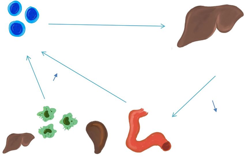
International Urology and Nephrology
its biologic functions but at the same time determine its tox- ferroportin present in the cell membrane of enterocytes and
icity. Excess iron may promote formation of reactive oxygen macrophages and via tyrosine phosphorylation leads to inter-
species (ROS) and lead to oxidative stress via damage to nalization of ferroportin and eventually its degradation in the
DNA, proteins and lipid membranes (Casu C Hepcidin ago- lysosomes. As a result, iron transport from the duodenum to
nists as therapeutic tools). As a heme cofactor, hemoglobin the blood circulation is diminished, iron release from mac-
is responsible for transport of oxygen. Furthermore, iron is rophages and hepatocytes is blocked and, consequently, iron
one of the compounds of catalase, peroxidase, cytochromes recirculation is impaired and serum iron levels decrease [6].
and other enzymes [6, 7]. Daily iron requirements amount Hepcidin production increases in response to iron overload,
to approximately 25–30 mg [8, 9], while iron loss (resulting inflammation or infection, while its synthesis is diminished
from, e.g. menstruation, enterocyte and epidermal desqua- as a result of iron deficiency, increased erythropoiesis and
mation, with traces of iron also found in sweat and urine) hypoxia. Hepcidin expression is regulated by numerous
constitutes 1–2 mg per day. There is no active mechanism proteins—bone morphogenetic protein-6 (BMP-6), hemo-
that enables the elimination of iron. As a result, to main- juvelin (HJV), human hemochromatosis protein (HFE),
tain adequate iron balance, intestinal absorption of the ele- transferrin receptors TfR1 and TfR2, among others. They
ment must cover daily iron loss. Healthy balanced diet pro- are influenced by both liver iron stores and circulatory iron
vides approximately 10–20 mg of iron, of which 1–4 mg is in the form of iron-bound transferrin (holotransferrin) [12,
absorbed [6, 10]. Absorption of ferrous iron (Fe2+) takes 13]. Increased iron levels stimulate production of BMP-6,
place in the apical enterocytes of the duodenum with the aid which binds to its receptor on the surface of hepatocyte and
of the Divalent Metal Transporter 1 (DMT1), while heme forms a complex with HJV (a membrane-bound co-recep-
can also be absorbed via Heme Carrier Protein 1 (HCP1) [4, tor). This process induces SMAD (small mothers of decap-
7]. Ferric iron ( Fe3+) is not easily absorbed; therefore, the entaplegic) phosphorylation pathway, leading to increased
reduction of Fe3+ to Fe2+ is required. This process is ena- expression of hepcidin genes. HFE forms a complex with
bled by duodenal cytochrome b-like ferrireductase enzyme TfR and beta-2-microglobulin and TfR2, which in a not-
(Dcytb). Absorbed iron can be stored in the enterocytes in yet-known fashion induces transcription of hepcidin genes.
ferritin-bound form (and usually lost as a result of entero- Mutations in the abovementioned proteins cause hereditary
cyte desquamation) or transported to the plasma via ferro- hemochromatosis, which manifests itself in hepcidin defi-
portin (FPN1; also found in macrophages, hepatocytes and ciency and iron overload [14]. Erythropoiesis stimulating
the placenta), where it binds to transferrin, which requires factors affect hepcidin synthesis as well—for example, in the
prior iron oxidation by hephaestin, a multicopper ferroxi- event of excessive erythropoiesis, erythropoietin (via bind-
dase present on the basolateral membrane of the enterocyte. ing with EPO receptor on the surface of hepatocytes) and
Transferrin, the essential iron-binding protein produced in growth differentiation factor 15 (GDF-15) decrease expres-
the liver, can reversibly bind two ferric ions, thus changing sion of hepcidin [15]. Furthermore, increased erythropoiesis
its conformation to holotransferrin. Usually approximately is associated with elevated concentration of soluble transfer-
30–40% of transferrin molecules are saturated with iron, rin receptor (sTfR), which is cleaved from transmembrane
which means that the majority of transferrin in the plasma transferrin receptor expressed mainly in cells with high iron
has conformation called apotransferrin and can buffer excess demands [15].
iron, if necessary. Circulating holotransferrin can be taken Recently, a hormone erythroferrone (ERFE) has been
up by cells which have increased iron demands, e.g. erythro- linked with erythropoiesis and iron balance. ERFE is synthe-
cyte precursors, via binding to transferrin receptor 1 (TfR1). sized in erythroblasts in response to increased erythropoiesis
Holotransferrin enters the cytoplasm by means of endocyto- and it suppresses transcription of hepcidin in hepatocytes
sis and as a result of pH-associated change in conformation, and thus increases iron availability in conditions associated
releases iron ions. Ferric iron is once again reduced to F e2+ with greater iron demand [16]. In murine models, ERFE
and crosses endosomal barrier via DMT1 and is ready to deficiency is associated with mild hypochromic anemia and
be incorporated into various enzymes or storage proteins delayed hepcidin suppression following hemorrhage or EPO
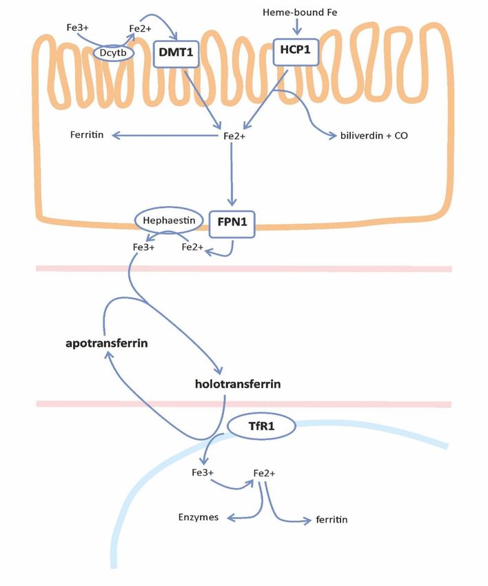
(Fig. 1). injection, while in certain conditions with ineffective eryth-
ropoiesis, such as β-thalassemia, ERFE concentrations were
significantly increased [17] (Fig. 2).
Role of hepcidin Under hypoxic conditions, oxygen deficiency leads to
diminished transcription of hepcidin genes (and conse-
Hepcidin, a 25-amino acid polypeptide discovered in 2000, quently decreased production of hepcidin) via hypoxia-
is one of the key elements of systemic iron metabolism inducible factors (HIF-1α, HIF-2α) [18]. HIFs activate the
[11]. Hepcidin is a hormone produced predominantly in expression of matriptase-2 (also known as the transmem-
the hepatocytes and released into the plasma. It binds to brane protease, serine 6; TMPRSS6), which cleaves HJV
13International Urology and Nephrology
Fig. 1 Iron absorption and
metabolism. Dcytb duodenal
cytochrome b-like ferrireduc-
tase, DMT1 divalent metal
transporter 1, HCP1 heme car-
rier protein 1, FPN1 ferroportin,
TfR1 transferrin receptor 1
from the HFE/TfR2/HJV complex, decreasing hepcidin syn- hepcidin-mediated iron sequestration in the reticuloendothe-
thesis [19]. Recently, it has been suggested that iron affects lial cells and, consequently, decrease iron concentration in
the release of fibroblast growth factor 23 (FGF-23)—a the system. At the same time, iron distribution becomes
marker of increased risk of cardiovascular incidents, espe- impaired; hence, iron availability for the synthesis of hemo-
cially in CKD patients [20–23]. However, outcomes from globin is reduced. As a result, anemia of inflammation (ane-
clinical studies have so far been inconsistent; therefore, the mia of chronic diseases) develops [30]. In addition, in the
impact of iron on FGF23 is unclear [24–26]. Finally, stud- setting of inflammation, patients can have high ferritin lev-
ies in humans revealed that hepcidin is one of the acute- els, low TSAT, and increased iron stores but still experience
phase proteins. It was proved that IL-1α, IL-1β, as well as restricted erythropoiesis resulting from “reticuloendothelial
IL-6 stimulate its expression [11, 27] via activation of the blockade” [5]. Moreover, functional iron deficiency, a state
STAT3 transcription factor [28]—a mechanism responsible of inadequate delivery of iron to the bone marrow in the
for iron restriction in the event of bacterial infection. Fur- setting of adequate iron stores, is caused by impaired iron
thermore, mice with hepcidin antimicrobial peptide (HAMP) mobilization (from the reticuloendothelial system [RES])
gene overexpression were affected by inflammation-related and/or increased bone marrow iron demand (as might be
anemia of chronic diseases [29]. Conditions associated secondary to reduced red cell life span and/or erythropoie-
with inflammation (e.g. chronic kidney disease) lead to sis-stimulating agents [ESA] use) [31]. Proinflammatory
13International Urology and Nephrology
Erythroferron
e
Erythroblast
Liver
Fe3+
Hepcidin
Duodenu
Spleen and m
Under liver hypoxic conditions, oxygen deficiency
Fig. 2 Increased erythroferrone production by erythroblasts suppresses hepcidin synthesis in the liver. Low hepcidin concentration increases iron
availability for erythropoiesis by enhancing iron absorption in the duodenum and iron release from macrophages in the liver and the spleen
cytokines are responsible for various processes typical of evaluation of iron status and the diagnosis of iron defi-
anemia of chronic diseases—they suppress erythropoiesis ciency anemia (IDA) [35]. In general population, decreased
in the bone marrow, impair the production of erythropoietin serum ferritin (< 15 ng/ml) and decreased TSAT (< 16%)
(EPO) [31, 32] and increase the rate of erythrophagocyto- are used for diagnosis of iron deficiency (ID) and iron defi-
sis leading to shorter erythrocyte lifespan [6]. Moreover, in ciency anemia (IDA) in individuals without concomitant
the event of chronic kidney disease the clearance of hepci- inflammation [35]. The international guidelines for the
din is diminished and, as a result, its plasma concentration management of IDA in CKD use the same diagnostic tests;
increases. however, they recommend different cutoff levels of serum
ferritin and TSAT for the diagnosis and initiation of iron
supplementation. Some guidelines recommend higher cutoff
Assessment of iron status levels of TSAT (≤ 30%) and serum ferritin (≤ 200–500 ng/
ml) [36, 37], while others, lower TSAT < 20% and serum
Understanding the dysregulation of iron metabolism is ferritin < 100 ng/ml [38–40]. The reason for the differ-
essential for the precise assessment, predicting treatment ence remains unclear, but at least in part, it may reflect
response as well as effective and safe treatment of anemia the consideration of the influence of inflammation on iron
of chronic kidney disease. A number of biomarkers of iron metabolism disorders in CKD and the distinct prevalence
status in chronic kidney disease have been used in clinical of inflammation severity between patient populations in
settings. However, many of them are influenced by renal fail- different countries. Regardless of the values adopted, the
ure alone and concomitant inflammation. Due to these con- numerous limitations of these diagnostic tools in the assess-
founding effects on the interpretation of most of biomarkers, ment of iron stores in the storage and functional pools, and
the assessment of iron status in chronic kidney disease is still in predicting the response to treatment should be empha-
a challenge [5, 33, 34]. sized. The traditional cutoffs of TSAT at ≤ 20% and serum
Serum iron, transferrin (Tf), total iron binding capac- ferritin ≤ 100 ng/ml have low sensitivity in iron deficiency
ity (TIBC, calculated as Tf × 1389), transferrin saturation detection. In Stancu et al.’s [41] study, these indices identi-
(TSAT, calculated serum iron/total iron binding capac- fied only 17% patients with CKD stage 3–5 as iron defi-
ity × 100) and serum ferritin are traditionally used in the cient whereas 50% prove to be iron deficient based on bone
13International Urology and Nephrology
marrow iron staining. Another limitation of these biomark- status may be predictive of the response to iron supplemen-
ers is scant ability to differentiate between absolute and tation [55, 57], and there is no correction factor that could
functional iron deficiency. It is assumed that low TSAT applied in estimation of iron stores depending on ferritin
combined with normal or elevated serum ferritin level is concentration [48].
diagnostic of functional iron deficiency [42]. However, if The limitations of traditional biomarkers of iron metabo-
functional iron deficiency results of supply/demand mis- lism and the response to treatment created the need to search
match, for example, during treatment with ESA, iron may for alternative diagnostic tools for iron management in CKD
transfer from transferrin faster than it can be mobilized from patients.
the iron stores, resulted in TSAT decrease [43]. Soluble transferrin receptor (sTfR) is produced by prote-
The changes occurring in iron metabolism in CKD olysis of the membrane transferrin receptor (TfR). Its release
patients are different from those observed in iron deficiency into circulation is increased in the setting of iron deficiency;
in general population reflecting the effect of inflamma- hence, sTfR has been evaluated as a potential biomarker
tion being a part of uremic state. With the progression of of iron deficiency. Soluble TfR is not an acute-phase reac-
kidney disease, the production of transferrin in the liver is tant and is less influenced by inflammation than other iron
reduced, and in advanced stages of CKD, transferrin levels metabolism indices [58]. The serum concentration of sTfR
are reduced by 30% [44, 45]. As an acute-phase reactant, is increased in hemodialysis patients with iron deficiency
TIBC progressively decreases with kidney disease progres- and correlate inversely with iron available for erythropoiesis;
sion, and it leads to higher TSAT levels independent of iron however, it is not able to detect occult iron deficiency [42,
status (13) and reduces its credibility as a measure of iron 59, 60]. Unfortunately, the interpretation is confounded by
status and a threshold for initiating iron therapy in CKD the use of ESA [35, 45], and appears to represent erythro-
patients [46]. In the meanwhile, in the majority of patients poietic activity rather than iron deficiency [42, 59, 60]. The
with stage 3–5 CKD, TSAT < 20% may correspond to serum index of sTfR to log10 ferritin has better than TSAT and fer-
iron levels below the lower limit and be indicative of iron ritin predictive value of iron supplementation responsiveness
deficiency [47]. It is postulated that more important parame- in hemodialysis patients [61]. The limitations of widespread
ter to assess iron status and prevent iron-limited erythropoie- measurement of sTfR include not-established standard cut-
sis in CKD patients is iron concentration rather than TSAT. offs, costs and availability in the laboratory.
In recently published review, to avoid iron deficiency, target Other biomarkers of iron status include reticulocyte
serum iron of 60 μg/dl was assumed, which corresponds to hemoglobin content (CHr) and percentage of hypochromic
TSAT of 20% in CKD stage 3, and 22–25% in stage 4 or 5. It red blood cells (%Hypo). CHr provides an expression of iron
is established in clinical practice that serum iron is the more availability for erythropoiesis within 3–4 days [35, 45] and
predictive index of iron sufficiency excluding iron-deficient CHr < 27.2 pg is diagnostic for iron deficiency [35]. %Hypo
erythropoiesis in hemodialysis patients [48]. measures the concentration of hemoglobin in red blood
Ferritin as an acute-phase reactant is frequently ele- cells (RBC), which reflects absolute amount of hemoglobin
vated in CKD patients irrespective of their iron stores [49]. and the RBC size [35, 45] and serves as a sensitive marker
Increased serum ferritin levels are the result of systemic of iron deficiency [35, 45] and iron status changes in the
inflammation and correlate positively with the severity of long-term assessment [35, 45]. Both these biomarkers are
inflammation [50, 51]. Thus, the interpretation of serum fer- influenced by inflammation [62, 63]. Nevertheless, CHr and
ritin is complicated by concomitant inflammation [35, 45]. %Hypo have, compared to TSAT and serum ferritin, better
Under minor inflammation, the specificity of low serum fer- sensitivity and specificity to predict iron deficiency in late
ritin concentration of absolute iron deficiency diagnosis is stages of CKD [48]. CHr and %Hypo are predictive of iron
high [35, 52], but if apparent inflammation is present, nor- responsiveness [64, 65] with at least similar test accuracy
mal or elevated ferritin levels cannot exclude iron deficiency compared with traditional biomarkers in predicting hemo-
in CKD [35, 52]. Inflammation may also reduce the predic- globin increase to intravenous iron administration [66]. It
tive value of serum ferritin for the response to iron supple- needs to be highlighted that during iron supplementation
mentation. The baseline ferritin level may be predictive of the temporal changes of CHr and %Hypo differs—CHr can
the response to oral [53] and intravenous [54] iron treatment normalize within 2–3 days, whereas %Hypo can take even
only in patients with minor inflammation expressed as low months [48]. Unfortunately, both measurements are limited
CRP level. Under concomitant inflammation, ferritin loses by testing requirements. %Hypo must be tested on fresh
its predictive value of the response to iron therapy. Moreo- blood samples (within 6 h) and CHr is time sensitive due to
ver, in highly inflamed patients, not only ferritin but also the maturation of erythrocytes [42].
other biomarkers of iron metabolism (TSAT, CHr, or sTfR) Hepcidin, given its central role in iron metabolism reg-
lose their value in predicting treatment response [55–57]. ulation, has been evaluated as a biomarker of iron status
In these patients, the values of CRP, but not indices of iron and iron responsiveness in CKD patients. Many studies
13International Urology and Nephrology have confirmed increased hepcidin levels in CKD patients available data were very limited due to a high degree of [67–69] Serum hepcidin levels have the strongest correla- heterogeneity. Taking into account the drawbacks and some- tion with serum ferritin, TSAT and sTfR [67, 70–72] and times limited data on the utility of alternative biomarkers are influenced by inflammation [67, 72, 73] iron and ESA of iron status in chronic kidney disease, the traditional bio- administration [67, 71, 72, 74] and dialysis clearance [68, markers still remain the hallmarks of the assessment of iron 69, 75]. Due to a significant intra-individual variability, the metabolism and responsiveness to iron therapy in this patient short-term measurement of serum hepcidin is not useful as population. a biomarker of iron status in CKD patients [76, 77]. And the interpretation of the serum hepcidin level must take into account all confounding factors. Hepcidin is not a good pre- dictor of the response to iron supplementation in dialysis Therapeutic strategies [57] and non-dialysis-dependent patients [78]. Soluble hemojuvelin (sHJV) has been explored as bio- Therapeutic approach should begin with diagnosis and marker of iron status in patients without [79], and with elimination of the underlying condition responsible for iron chronic kidney disease [80, 81]. Opposite to cell surface deficiency. Iron supplementation is the next step. Initiation HJV, soluble HJV may act as an inhibitor of BMP signaling of iron in CKD patients with anemia should be based on and restrain hepcidin expression [82]. Some studies revealed preexisting iron stores and the target Hb level that is desired. that sHJV may be increased in iron deficiency and decreased Even though oral iron is generally considered sufficient in during iron administration [82–84], suggesting that sHJV CKD patients not on dialysis and PD patients, intravenous may be a diagnostic marker of iron status. One important form is the preferred route, especially in hemodialysis concern in soluble HJV assessment is assay validity [81, 82, patients. Oral iron is associated with poor intestinal absorp- 85], and future studies are needed to establish sHJV value as tion and adverse event-related (mainly gastrointestinal) low biomarker of iron status and response to therapy. adherence to therapy [94]. It needs to be emphasized that Growth differentiation factor 15 (GDF15), secreted by the goal of treatment with iron is not to increase Hb levels matured erythroblasts, is involved in hepcidin metabolism to the normal range but to reduce the risk of development of and as such is potentially involved in iron metabolism [86]. severe anemia and associated complications and to minimize However, available data on the role of GDF15 as the marker the need for blood transfusions [4]. ESA therapy is gener- of iron status are scarce. In fact, one study suggested that ally initiated in ESRD patients with replete iron stores (i.e. GDF15 is increased in iron deficiency [87]; the other did TSAT > 30% and/or ferritin > 500 mcg/l) whose Hb levels not confirm it [88]. Moreover, serum GDF15 levels may be are below 10 g/dl [95]. Failure to increase Hb concentration influenced by kidney disease, malnutrition and inflammation after the first month of ESA treatment is defined by KDIGO [89] complicating its usefulness as an iron status biomarker. as ESA hyporesponsiveness—a poor prognostic factor in Plasma neutrophil gelatinase-associated lipocalin terms of patient mortality [36]. There are several factors (NGAL) is known as a predictor of kidney disease pro- responsible for ESA hyporesponsiveness—one of them is gression and marker of inflammation [90, 91]. In addition, inflammation. A new group of oral agents known as HIF NGAL influences iron sequestration; however, the way that prolyl hydroxylase-inhibitors has been developed to improve NGAL influences the iron balance depends on its form. The CKD-associated anemia. The beneficial effect of HIF stabi- bound form of NGAL decreases and free form of NGAL lizers on hemoglobin levels has been observed regardless of increases the level of serum iron. Few studies evaluated the the patient’s iron stores and inflammatory status [17]. How- usefulness of NGAL as a biomarker of iron stores in CKD ever, the long-term safety of these novel agents, especially patients, suggesting its good specificity and sensitivity in the regarding potential risk of tumorigenesis and worsening of detection of decreased iron stores [92, 93]; however, future proliferative diabetic retinopathy, has yet to be established studies are needed to establish the role as a biomarker of [17, 96]. Additionally, the increased usage of intravenous iron status. iron in hemodialysis patients during recent years has led to In summary, we conducted a search in Medline, PubMed, increasing concern over the potential development of iron and Embase using the keywords: iron, biomarkers, kidney overload [97, 98]. Recently, we reported that a substantial failure, CKD, dialysis, hemodialysis, peritoneal dialysis. As number of hemodialysis patients have positive labile plasma described in the Preferred Reporting Items for Systematic iron after intravenous iron administration, which positively reviews and Meta-Analyses (PRISMA) group [94]. We lim- correlated with laboratory parameters that are currently in ited our search to adult patients and publications in English routine clinical use for detecting iron overload and with and Polish till 2020. We found 541 articles, but only 102 higher intravenous iron dose [99]. Thus, we suggest to per- articles were analyzed due to lack of information about full form further studies to establish the clinical implications data, and availability of abstracts only or duplication. The of labile plasma iron, a component of nontransferrin-bound 13
International Urology and Nephrology
iron which may be a more accurate indicator of impending WHO_TRS_405.pdf;jsess i onid = 72382 6 5043 2 7390 B 6E2B
iron overload monitoring in hemodialysis patients. ACE6A264BC23?sequence=1
2. Astor BC, Muntner P, Levin A, Eustace JA, Coresh J (2002)
Association of kidney function with anemia, the Third National
Health and Nutrition Examination Survey (1988–1994). Arch
Conclusion Intern Med 162(12):1401–1408
3. Sofue T, Nakagawa N, Kanda E, Nagasu H, Matsushita K, Nan-
gaku M, Maruyama S, Wada T, Terada Y, Yamagata K, Narita I,
CKD patients tend to have subclinical inflammatory-related Yanagita M, Sugiyama H, Shigematsu T, Ito T, Tamura K, Isaka
immune activation. The pathogenesis of chronic inflamma- Y, Okada H, Tsuruya K, Yokoyama H, Nakashima N, Hiromi
tion in CKD is still not fully understood, yet the proposed K, Ohe K, Okada M, Kashihara N (2020) Prevalence of ane-
underlying factors include oxidative stress, cellular senes- mia in patients with chronic kidney disease in Japan: a nation-
wide, cross-sectional cohort study using data from the Japan
cence, hypoxia, exogenous factors (such as dialyzer mem- Chronic Kidney Disease Database (J-CKD-DB). PLoS ONE
brane or central venous catheter), immune dysfunction, gut 15(7):e0236132. https://doi.org/10.1371/journal.pone.0236132
dysbiosis and retention of uremic toxins [100]. Inflamma- 4. Babitt JL, Lin HY (2012) Mechanisms of anemia in CKD.
tory blockade is associated with resistance to erythropoi- JASN 23(10):1631–1634. https://doi.org/10.1681/ASN.20111
11078
etin despite iron availability, which is more clearly under- 5. Fishbane S, Spinowitz B (2018) Update on anemia in ESRD
stood now that the role of hepcidin in iron metabolism and earlier Stages of CKD: core curriculum. Am J Kidney Dis
has been identified. Studies conducted so far revealed that 71(3):423–435. https://doi.org/10.1053/j.ajkd.2017.09.026
serum ERFE concentration increases in response to ESA 6. Filipczyk L, Król P, Wystrychowski A (2010) Hepcydyna—
hormon wątrobowy kontrolujący gospodarkę żelaza. Forum
treatment in CKD patients, while the correlation between Nefrologiczne 3(4):233–242. https: //journa ls.viamed ica.pl/
ERFE and hepcidin remains unclear [101]. Despite having forum_nefrologiczne/article/view/10359
classical iron biomarkers, we still looking for new ones to 7. Mazur G, Wróbel T, Nosol J (2005) Rola hepcydyny w
improve our diagnostic and predictive tools. There is an area regulacji homeostazy żelaza. Acta Haematologica Polonica
36(2):167–175. https: //pthit. pl/Acta_Haemat ologi ca_Polon
of uncertainty regarding diagnostic utility of both ERFE and ica,Hepcydyna_Hemochromatoza_Niedokrwistosc_w_przeb
hepcidin in ESRD patients [102]. Higher concentration of iegu_chorob_przewleklych,250.html
hepcidin in oligoanuric patients may reflect decreased renal 8. Andrews NC (2008) Forging a field: the golden age iron biol-
clearance. Furthermore, the hepcidin-lowering effect of ogy. Blood 112(2):219–230. https: //doi.org/10.1182/blood
-2007-12-077388
ERFE in ESRD patients treated with ESAs may be blunted 9. Beaumont C, Delaby C (2009) Recycling iron in normal and
by underlying inflammation and concomitant iron treat- pathological states. Semin Hematol 46(4):328–338. https://doi.
ment. Up to date, we have no cost-effective analytical tests org/10.1053/j.seminhematol.2009.06.004
to assess iron metabolism in patients with CKD. Therefore, 10. Lipiński P, Starzyński R (2004) Regulacja ogólnoustrojowej
homeostazy żelaza przez hepcydynę. Postępy Hig Med Dosw
future studies should validate the use of ERFE as a bio- (online) 58:65–73. https://www.phmd.pl/api/files/view/1702.
marker of erythropoiesis and predictor of response to iron pdf
and ESA therapy in dialysis-dependent patients. 11. Park CH, Valore EV, Waring AJ, Ganz T (2001) Hepcidin, a uri-
nary antimicrobial peptide synthesized in the liver. J Biol Chem
Open Access This article is licensed under a Creative Commons Attri- 276(11):7806–7810. https://doi.org/10.1074/jbc.m008922200
bution 4.0 International License, which permits use, sharing, adapta- 12. Camaschella C (2015) Iron-deficiency anemia. N Engl J Med
tion, distribution and reproduction in any medium or format, as long 372:1832–1843. https://doi.org/10.1056/NEJMra1401038
as you give appropriate credit to the original author(s) and the source, 13. Fleming RE, Ponka P (2012) Iron overload in human disease. N
provide a link to the Creative Commons licence, and indicate if changes Engl J Med 366:348–359. https://doi.org/10.1056/NEJMra1004
were made. The images or other third party material in this article are 967
included in the article’s Creative Commons licence, unless indicated 14. Casu C, Nemeth E, Rivella S (2018) Hepcidin agonists as thera-
otherwise in a credit line to the material. If material is not included in peutic tools. Blood 131(16):1790–1794. https://doi.org/10.1182/
the article’s Creative Commons licence and your intended use is not blood-2017-11-737411
permitted by statutory regulation or exceeds the permitted use, you will 15. Zhang A-S, Enns CA (2009) Molecular mechanisms of normal
need to obtain permission directly from the copyright holder. To view a iron homeostasis. Hematology Am Soc Hematol Educ Program
copy of this licence, visit http://creativecommons.org/licenses/by/4.0/. 2009:207–214. https: //doi.org/10.1182/ashedu catio n-2009.1.207
16. Hanudel MR, Rappaport M, Chua K, Gabayan V, Qiao B, Jung
G, Salusky IB, Ganz T, Nemeth E (2018) Levels of the erythro-
poietin-responsive hormone erythroferrone in mice and humans
References with chronic kidney disease. Haematologica 103(4):e141–e142.
https://doi.org/10.3324/haematol.2017.181743
1. Blanc B, Finch CA, Hallberg L, Herbert V, Lawkowicz W, 17. Hasegawa T, Koiwa F, Akizawa T (2018) Anemia in conventional
Layrisse M, Mollin DL, Rachmilewitz M, Ramalingaswami V, hemodialysis: finding the optimal treatment balance. Semin Dial
Sanchez-Medal L, Vintrobe MM (1968) Nutritional anemias. 31(6):599–606. https://doi.org/10.1111/sdi.12719
Report of a WHO Scientific Group. WHO Tech Rep Ser 405:1– 18. Peyssonnaux C, Zinkernagel AS, Schuepbach RA, Rankin E,
40. https://apps.who.int/iris/bitstream/handle/10665/40707/ Vaulont S, Haase VH, Nizet V, Johnson RS (2007) Regulation of
iron homeostasis by the hypoxia-inducible transcription factors
13International Urology and Nephrology
(HIFs). J Clin Invest 117(7):1926–1932. https: //doi.org/10.1172/ 35. Lopez A, Cacoub P, Macdougall IC, Peyrin-Biroulet L (2016)
jci31370 Iron deficiency anaemia. Lancet 387:907–916. https: //doi.
19. Ueda N, Takasawa K (2018) Impact of inflammation on fer- org/10.1016/S0140-6736(15)60865-0
ritin, hepcidin and the management of iron deficiency anemia 36. The Kidney Disease: Improving Global Outcomes (KDIGO)
in chronic kidney disease. Nutrients 10(9):1173. https://doi. Anemia Work Group (2012) KDIGO clinical practice guideline
org/10.3390/nu10091173 for anemia in chronic kidney disease. Kidney Int Suppl 2:279–
20. Parker BD, Schurgers LJ, Brandenburg VM, Christenson RH, 335. https://doi.org/10.1038/kisup.2012.37
Vermeer C, Ketteler M et al (2010) The associations of fibroblast 37. National Kidney Foundation (2006) KDOQI clinical practice
growth factor 23 and uncarboxylated matrix Gla protein with guidelines and clinical practice recommendations for anemia in
mortality in coronary artery disease: the Heart and Soul Study. chronic kidney disease. Am J Kidney Dis 47(5 Suppl 3):S11–
Ann Intern Med 152(10):640–648. https: //doi.org/10.7326/0003- S145. https://doi.org/10.1053/j.ajkd.2006.03.010
4819-152-10-201005180-00004 38. Locatelli F, Bárány P, Covic A, De Francisco A, Del Vecchio
21. Khan AM, Chirinos JA, Litt H, Yang W, Rosas SE (2012) L, Goldsmith D, Hörl W, London G, Vanholder R, Van Biesen
FGF-23 and the progression of coronary arterial calcification W, ERA-EDTA ERBP Advisory Board (2013) Kidney disease:
in patients new to dialysis. Clin J Am Soc Nephrol 7(12):2017– improving global outcomes guidelines on anaemia management
2022. https://doi.org/10.2215/cjn.02160212 in chronic kidney disease: a European Renal Best Practice posi-
22. Faul C, Amaral AP, Oskouei B, Hu MC, Sloan A, Isakova T et al tion statement. Nephrol Dial Transplant 28:1346–1359. https://
(2011) FGF23 induces left ventricular hypertrophy. J Clin Invest doi.org/10.1093/ndt/gft033
121(11):4393–4408. https://doi.org/10.1172/jci46122 39. Tsubakihara Y, Nishi S, Akiba T, Hirakata H, Iseki K,
23. Mendoza MJ, Isakova T, Ricardo AC, Xie H, Navaneethan Kubota M, Kuriyama S, Komatsu Y, Suzuki M, Nakai S,
SD, Anderson AH et al (2012) Fibroblast growth factor 23 and Hattori M, Babazono T, Hiramatsu M, Yamamoto H, Bessho
inflammation in CKD. Clin J Am Soc Nephrol 7(7):1155–1162. M, Akizawa T (2010) 2008 Japanese Society for Dialysis
https://doi.org/10.2215/cjn.13281211 Therapy: guidelines for renal anemia in chronic kidney dis-
24. Wolf M, White KE (2014) Coupling fibroblast growth factor 23 ease. Ther Apher Dial 14:240–275. https: //doi.org/10.111
production and cleavage: iron deficiency, rickets, and kidney dis- 1/j.1744-9987.2010.00836.x
ease. Curr Opin Nephrol Hypertens 23(4):411–419. https://doi. 40. Macginley R, Walker R, Irving M (2013) KHA-CARI guideline:
org/10.1097/01.mnh.0000447020.74593.6f use of iron in chronic kidney disease patients. Nephrology (Carl-
25. Braithwaite V, Prentice AM, Doherty C, Prentice A (2012) ton) 18:747–749. https://doi.org/10.1111/nep.12139
FGF23 is correlated with iron status but not with inflamma- 41. Stancu S, Stanciu A, Zugravu A, Bârsan L, Dumitru D, Lipan
tion and decreases after iron supplementation: a supplemen- M, Mircescu G (2010) Bone marrow iron, iron indices, and
tation study. Int J Pediatr Endocrinol 2012(1):27. https://doi. the response to intravenous iron in patients with non-dialysis-
org/10.1186/1687-9856-2012-27 dependent CKD. Am J Kidney Dis 55:639–647. https://doi.
26. Deger SM, Erten Y, Pasaoglu OT, Derici UB, Reis KA, Onec K, org/10.1053/j.ajkd.2009.10.043
Pasaoglu H (2013) The effects of iron on FGF23-mediated Ca-P 42. Gaweda AE (2017) Markers of iron status in chronic kid-
metabolism in CKD patients. Clin Exp Nephrol 17(3):416–423. ney disease. Hemodial Int 21(Suppl 1):S21–S27. https://doi.
https://doi.org/10.1007/s10157-012-0725-0 org/10.1111/hdi.12556
27. Nemeth E, Valore E, Territo M, Schiller G, Lichtenstein A, Ganz 43. Wish JB (2006) Assessing iron status: beyond serum ferritin and
T (2003) Hepcidin, a putative mediator of anemia of inflamma- transferrin saturation. Clin J Am Soc Nephrol 1(Suppl 1):S4–S8.
tion, is a type II acute phase protein. Blood 101(7):2461–2463. https://doi.org/10.2215/CJN.01490506
https://doi.org/10.1182/blood-2002-10-3235 44. Besarab A, Coyne DW (2010) Iron supplementation to treat ane-
28. Wrighting DM, Andrews NC (2006) Interleukin-6 induces hepci- mia in patients with chronic kidney disease. Nat Rev Nephrol
din expression through STAT3. Blood 108(9):3204–3209. https 6:699–771. https://doi.org/10.1038/nrneph.2010.139
://doi.org/10.1182/blood-2006-06-027631 45. Hayes M (2019) Measurement of iron status in chronic kidney
29. Nicolas G, Bennoun M, Porteu A, Mativet S, Beaumont C, disease. Pediatr Nephrol 34:605–613. https://doi.org/10.1007/
Grandchamp B, Sirito M, Sawadogo M, Kahn A, Vaulont S s00467-018-3955-x
(2002) Severe iron deficiency anemia in transgenic mice express- 46. Wong MMY, Tu C, Li Y, Pecoits-Filho R, Lopes AA, Narita I,
ing liver hepcidin. Proc Natl Acad Sci USA 99(7):4596–4601. Reichel H, Port FK, Sukul N, Stengel B, Robinson BM, Massy
https://doi.org/10.1073/pnas.072632499 ZA, Pisoni RL, the CKDopps Investigators (2019) Anemia and
30. Donderski R, Kardymowicz A, Manitius J (2009) Niedokrwistość iron deficiency among chronic kidney disease Stages 3–5ND
nerkopochodna. Wybrane aspekty diagnostyki i terapii. Choroby patients in the Chronic Kidney Disease Outcomes and Practice
Serca i Naczyń 6(2):82–93. https://journals.viamedica.pl/choro Patterns Study: often unmeasured, variably treated. Clin Kidney
by_serca_i_naczyn/article/view/12052/9930 J 13:613–624. https://doi.org/10.1093/ckj/sfz091
31. Ganz T, Nemeth E (2016) Iron balance and the role of hepcidin 47. Fishbane S, Pollack S, Feldman HI, Joffe MM (2009) Iron indi-
in chronic kidney disease. Semin Nephrol 36:87–93. https: //doi. ces in chronic kidney disease in the National Health and Nutri-
org/10.1016/j.semnephrol.2016.02.001 tional Examination Survey 1988–2004. Clin J Am Soc Nephrol
32. Fasano A (2012) Leaky gut and autoimmune diseases. Clin Rev 4:57–61. https://doi.org/10.2215/CJN.01670408
Allergy Immunol 42(1):71–78. https://doi.org/10.1007/s1201 48. Besarab A, Drueke TB (2020) The problem with trasferrin satu-
6-011-8291-x ration as an indicator of iron “sufficiency” in chronic kidney dis-
33. Babitt JL, Lin HY (2012) Mechanism of anemia in CKD. J ease. Nephrol Dial Transplant. https://doi.org/10.1093/ndt/gfaa0
Am Soc Nephrol 23(10):1631–1634. https://doi.org/10.1681/ 48
ASN.2011111078 49. Kim T, Rhee CM, Streja E, Obi Y, Brunelli SM, Kovesdy CP,
34. Wish JB (2006) Assessing iron status: beyond serum ferritin and Kalantar-Zadeh K (2017) Longitudinal trends in serum ferritin
transferrin saturation. Clin J Am Soc Nephrol 1(suppl 1):S4–S8. levels and associated factors in a national incident hemodialy-
https://doi.org/10.1159/000486968 sis cohort. Nephrol Dial Transplant 32:370–377. https://doi.
org/10.1093/ndt/gfw012
13International Urology and Nephrology
50. Kalantar-Zadeh K, Rodriguez RA, Humphreys MH (2004) Asso- 63. Bovy C, Tsobo C, Crapanzano L, Rorive G, Beguin Y, Albert
ciation between serum ferritin and measures of inflammation, A, Paulus JM (1999) Factors determining the percentage of
nutrition and iron in haemodialysis patients. Naphrol Dial Trans- hypochromic red blood cells in hemodialysis patients. Kidney Int
plant 19:141–149. https://doi.org/10.1093/ndt/gfg493 56:1113–1119. https://doi.org/10.1046/j.1523-1755.1999.00627
51. Ruiz-Jaramillo Mde L, Guizar-Mendoza JM, Amador-Licona .x
N, de Jesus G-N, Hernández-González MA, Dubey-Ortega LA, 64. Tessitore N, Solero GP, Lippi G, Bassi A, Faccini GB, Bedogna
Solorio-Meza SE (2011) Iron overload as cardiovascular risk fac- V, Gammaro L, Brocco G, Restivo G, Bernich P, Lupo A,
tor in children and adolescents with renal disease. Nephrol Dial Maschio G (2001) The role of iron status markers in predicting
Transplant 26:3268–3273. https://doi.org/10.1093/ndt/gfr044 response to intravenous iron in haemodialysis patients on main-
52. Macdougall IC, Bock AH, Carrera F, Eckardt KU, Gaillard C, tenance erythropoietin. Nephrol Dial Transplant 16:1416–1423.
Van Wyck D, Roubert B, Cushway T, Roger SD, FIND-CKD https://doi.org/10.1093/ndt/16.7.1416
Study Investigators (2014) FIND-CKD: a randomized trial of 65. Ratcliffe LE, Thomas W, Glen J, Padhi S, Pordes BAJ, Wonder-
intravenous ferric carboxymaltose versus oral iron in patients ling D, Connell R, Stephens S, Mikhail AI, Fogarty DG, Cooper
with chronic kidney disease and iron deficiency anaemia. Neph- JK, Dring B, Devonald MAJ, Brown C, Thomas ME (2016)
rol Dial Transplant 29:2075–2084. https://doi.org/10.1093/ndt/ Diagnosis and management of iron deficiency in CKD: a sum-
gfu201 mary of the NICE guideline recommendations and their ration-
53. Macdougall IC, Bock AH, Carrera F, Eckardt KU, Gaillard C, ale. Am J Kidney Dis 67:548–558. https://doi.org/10.1053/j.
Van Wyck D, Meier Y, Larroque S, Perrin A, Roger SD (2017) ajkd.2015.11.012
Erythropoietic response to oral iron in patients with non-dialy- 66. Chung M, Moorthy D, Hadar N, Salvi P, Iovin RC, Lau J
sis-dependent chronic kidney disease in FIND-CKD trial. Clin (2012) Biomarkers for assessing and managing iron defi-
Nephrol 88:301–310. https://doi.org/10.5414/cn109198 ciency anemia in late-stage chronic kidney disease. Agency
54. Takasawa K, Takeda C, Wada T, Ueda N (2018) Optimal serum for Healthcare Research and Quality, Rockville. Report No.:
ferritin levels for iron deficiency anemia during oral iron therapy 12(13)-EHC140-EF
(OIT) in Japanese hemodialysis patients with minor inflamma- 67. Zaritsky J, Young B, Wang HJ, Westerman M, Olbina G, Nemeth
tion and benefit of intravenous iron therapy for OIT-nonrespond- E, Ganz T, Rivera S, Nissenson AR, Salusky IB (2009) Hepci-
ers. Nutrients 10:428. https://doi.org/10.3390/nu10040428 din—a potential novel biomarker for iron status in chronic kid-
55. Singh AK, Coyne DW, Shapiro W, Rizkala AR, the DRIVE ney disease. Clin J Am Soc Nephrol 4:1051–1056. https://doi.
Study Group (2007) Predictors of the response to treatment in org/10.2215/CJN.05931108
anemic hemodialysis patients with high serum ferritin and low 68. Malyszko J, Malyszko JS, Kozminski P, Mysliwiec M (2009)
transferrin saturation. Kidney Int 71:1163–1171. https://doi. Type of renal replacement therapy and residual renal function
org/10.1038/sj.ki.5002223 may affect prohepcidin and hepcidin. Ren Fail 31:876–883. https
56. Musanovic A, Trnacevic S, Mekic M, Musanovic A (2013) The ://doi.org/10.3109/08860220903216071
influence of inflammatory markers and CRP predictive value in 69. Zaritsky J, Young B, Gales B, Wang HJ, Rastogi A, Wester-
relation to target hemoglobin level in patients on chronic hemo- man M, Nemeth E, Ganz T, Salusky IB (2010) Reduction of
dialysis. Med Arch 67:361–364. https://doi.org/10.5455/medar serum hepcidin by hemodialysis in pediatric and adult patients.
h.2013.67.361-364 Clin J Am Soc Nephrol 5:1010–1014. https://doi.org/10.2215/
57. Tessitore N, Girelli D, Campostrini N, Bedogna V, Solero GP, CJN.08161109
Castagna A, Melilli E, Mantovani W, De Matteis G, Olivieri O, 70. van der Weerd NC, Grooteman MP, Bots ML, van den Dor-
Poli A, Lupo A (2010) Hepcidin is not useful as a biomarker for pel MA, Den Hoedt CH, Mazairac Albert HA, Nubé MJ, Lars
iron needs in haemodialysis patients on maintenance erythropoie- Penne E, Gaillard CA, Wetzels JFM, Wiegerinck ET, Swinkels
sis-stimulating agents. Nephrol Dial Transplant 25:3996–4002. DW, Blankestijn PJ, ter Wee PM, CONTRAST investigator
https://doi.org/10.1093/ndt/gfq321 (2012) Hepcidin-25 in chronic hemodialysis patients is related
58. Suchdev PS, Williams AM, Mei Z, Flores-Ayala R, Pasricha SR, to residual kidney function and not to treatment with erythro-
Rogers LM, Ml NS (1633S) Assessment of iron status in set- poiesis stimulating agents. PLoS ONE 7:e39783. https://doi.
tings of inflammation: challenges and potential approaches. Am org/10.1371/journal.pone.0039783
J Clin Nutr 106(Suppl 6):1626S–1633S. https://doi.org/10.3945/ 71. Ashby DR, Gale DP, Busbridge M, Murphy KG, Duncan ND,
ajcn.117.155937 Cairns TD, Taube DH, Bloom SR, Tam FW, Chapman RS, Max-
59. Tarng DC, Huang TP (2002) Determinants of circulating solu- well PH, Choi P (2009) Plasma hepcidin levels are elevated but
ble transferrin receptor level in chronic haemodialysis patients. responsive to erythropoietin therapy in renal disease. Kidney Int
Nephrol Dial Transplant 17:1063–1069. https://doi.org/10.1093/ 75:976–981. https://doi.org/10.1038/ki.2009.21
ndt/17.6.1063 72. Mercadal L, Metzger M, Haymann JP, Thervet E, Boffa JJ, Fla-
60. Chiang WC, Tsai TJ, Chen YM, Lin SL, Hsieh BS (2002) Serum mant M, Vrtovsnik F, Houillier P, Froissart M, Stengel B (2014)
soluble transferrin receptor reflects erythropoiesis but not iron The relation of hepcidin to iron disorders, inflammation and
availability in erythropoietin-treated chronic hemodialysis hemoglobin in chronic kidney disease. PLoS ONE 9:e99781.
patients. Clin Nephrol 58:363–369. https://doi.org/10.5414/ https://doi.org/10.1371/journal.pone.0099781
cnp58363 73. Łukaszyk E, Łukaszyk M, Koc-Żórawska E, Tobolczyk J,
61. Chen YC, Hung SC, Tarng DC (2006) Association between Bodzenta-Łukaszyk A, Małyszko J (2015) Iron status and inflam-
transferrin receptor—ferritin index and conventional measures mation in early stages of chronic kidney disease. Kidney Blood
of iron responsiveness in hemodialysis patients. Am Kidney Dis Press Res 40:366–373. https://doi.org/10.1159/000368512
47:1036–1044. https://doi.org/10.1053/j.ajkd.2006.02.180 74. Kato A, Tsuji T, Luo J, Sakao Y, Yasuda H, Hishida A (2008)
62. Hackeng CM, Beerenhout CM, Hermans M, Van der Kuy PHM, Association of prohepcidin and hepcidin-25 with erythropoietin
Van der Dussen H, Van Dieijen-Visser MP, Hamulyák K, Van der response and ferritin in hemodialysis patients. Am J Nephrol
Sande FM, Leunissen KM, Koomanal JP (2004) The relationship 28:115–121. https://doi.org/10.1159/000109968
between reticulocyte hemoglobin content with C-reactive protein 75. Stefánsson BV, Abramson M, Nilsson U, Haraldsson B (2012)
and conventional iron parameters in dialysis patients. J Nephrol Hemodiafiltration improve plasma 25-hepcidin levels: a pro-
17:107–111 spective, randomized, blinded, cross-over study comparing
13International Urology and Nephrology
hemodialysis and hemodiafiltration. Nephron Extra 2:55–65. Herzog H, Law M, Stenvinkel P, Brown DA (2012) Macrophage
https://doi.org/10.1159/000336482 inhibitory cytokine-1 (MIC-1/GDF15) and mortality in end-
76. Ford BA, Eby CS, Scott MG, Coyne Daniel W (2010) Intra-indi- stage renal disease. Nephrol Dial Transplant 27:70–75. https://
vidual variability of serum hepcidin precludes its use as a marker doi.org/10.1093/ndt/gfr575
of iron status in hemodialysis patients. Kidney Int 78:769–773. 90. Bolignano D, Lacquaniti A, Coppolino G, Donato V, Campo
https://doi.org/10.1038/ki.2010.254 S, Fazio MR, Nicocia G, Buemi M (2009) Neutrophil gelati-
77. Peters HP, Rumjon A, Bansal SS, Laarakkers Coby MM, van den nase-associated lipocalin (NGAL) and progression of chronic
Brand JAJG, Sarafidis P, Musto R, Malyszko J, Swinkels DW, kidney disease. Clin J Am Soc Nephrol 4:337–344. https://doi.
Wetzels JFM, Macdougall IC (2012) Intra-individual variability org/10.2215/CJN.03530708
of serum hepcidin-25 in haemodialysis patients using mass spec- 91. Castillo-Rodriguez E, Fernandez-Prado R, Martin-Cleary C,
trometry and ELISA. Nephrol Dial Transplant 27:3923–3929. Pizarro-Sánchez MS, Sanchez-Niño MD, Sanz AB, Fernandez-
https://doi.org/10.1093/ndt/gfs164 Fernandez B, Ortiz A (2017) Kidney injury marker 1 and neu-
78. Gaillard CA, Bock AH, Carrera F, Eckardt KU, Van Wyck DB, trophil gelatinase-associated lipocalin in chronic kidney disease.
Bansal SS, Cronin M, Meier Y, Larroque S, Roger SD, Mac- Nephron 136:263–267. https://doi.org/10.1159/000447649
dougall IC (2016) Hepcidin response to iron therapy in patients 92. Kim IY, Kim JH, Lee DW, Lee SB, Rhee H, Song SH, Seong
with non-dialysis dependent CKD: an analysis of the FIND-CKD EY, Kwak IS (2018) Plasma neutrophil gelatinase-associated
trial. PLoS ONE 11:e0157063. https://doi.org/10.1371/journ lipocalin is associated with iron status in anemic patients with
al.pone.0157063 predialysis chronic kidney disease. Clin Exp Nephrol 22:28–34.
79. Finkenstedt A, Widschwendter A, Brasse-Lagnel CG, Theurl https://doi.org/10.1007/s10157-017-1409-6
I, Hubalek M, Dieplinger H, Tselepis C, Ward DG, Vogel W, 93. Avci Cicek E, Rota S, Dursun B, Cavalci E (2016) Evaluation
Zoller H (2012) Hepcidin is correlated to soluble hemojuvelin of serum NGAL and hepcidin levels in chronic kidney disease
but not increased GDF15 during pregnancy. Blood Cells Mol Dis patients. Ren Fail 38:35–39. https://doi.org/10.3109/08860
48:233–237. https://doi.org/10.1016/j.bcmd.2012.02.001 22X.2015.1107823
80. Malyszko J, Malyszko JS, Levin-Iaina N, Koc-Zorawska E, 94. Moher D, Liberati A, Tetzlaff J, Altman DG (2009) Preferred
Kozminski P, Mysliwiec M (2011) Is hemojuvelin a possible reporting items for systematic reviews and meta-analyses:
new player in iron metabolism in hemodialysis patients? Int the PRISMA statement. PLoS Med 6:e1000097. https://doi.
Urol Nephrol 44:1805–1811. https://doi.org/10.1007/s1125 org/10.1371/journal.pmed.1000097
5-011-0084-x 95. Cappellini MD, Comin-Colet J, De Francisco A, Dignass A,
81. Rumjon A, Sarafidis P, Brincat S, Musto R, Malyszko J, Bansal Doehner W, Lam CS, Macdougall IC, Rogler G, Camaschella
SS, Macdougall IC (2012) Serum hemojuvelin and hepcidin lev- C, Kadir R, Kassebaum NJ, Spahn DR, Taher AT, Musallam
els in chronic kidney disease. Am J Nephrol 35:295–304. https: // KM, IRON CORE Group (2017) Iron deficiency across chronic
doi.org/10.1159/000336528 inflammatory conditions: international expert opinion o defini-
82. Lin L, Goldberg YP, Ganz T (2005) Competitive regulation tion, diagnosis and management. Am J Hematol 92(10):1068–
of hepcidin mRNA by soluble and cell-associated hemoju- 1078. https://doi.org/10.1002/ajh.24820
velin. Blood 106:2884–2889. https://doi.org/10.1182/blood 96. Malyszko J, Malyszko JS, Matuszkiewicz-Rowinska J (2019)
-2005-05-1845 Hepcidin as a therapeutic target for anemia and inflammation
83. Silvestri L, Pagani A, Camaschella C (2008) Furin-mediated associated with chronic kidney disease. Expert Opin Ther Targets
release of soluble hemojuvelin: a new link between hypoxia and 23(5):407–421. https: //doi.org/10.1080/147282 22.2019.159935 8
iron homeostasis. Blood 111:924–931. https://doi.org/10.1182/ 97. Wish JB, Aronoff GR, Bacon BR, Brugnara C, Eckardt KU,
blood-2007-07-100677 Ganz T, Macdougall IC, Núñez J, Perahia AJ, Wood JC (2018)
84. Zhang AS, Anderson SA, Meyers KR, Hernandez C, Eisenstein Positive iron balance in chronic kidney disease: how much is too
RS, Enns CA (2007) Evidence that inhibition of hemojuvelin much and how to tell? Am J Nephrol 47(2):72–83. https://doi.
shedding in response to iron is mediated through neogenin. J Biol org/10.1159/000486968
Chem 282:12547–12556. https://doi.org/10.1074/jbc.M6087 98. Nakanishi T, Kuragano T (2020) Potential hazards of recent
88200 trends in liberal iron use for renal anemia. Clin Kidney J. https://
85. Gutiѐrrez OM, Sun CC, Chen W, Babitt JL, Lin HY (2012) State- doi.org/10.1093/ckj/sfaa117
ment of concern about a commercial assay used to measure solu- 99. Lenihan CR, Winkelmayer WC (2016) The dawning of a new day
ble hemojuvelin in humans. Am J Nephrol 36:332–333. https:// in CKD anemia care? J Am Soc Nephrol 27(4):968–970. https://
doi.org/10.1159/000342519 doi.org/10.1681/ASN.2015091009
86. Malyszko J, Koc-Żórawska E, Levin-Iaina N, Małyszko J, 100. Bnaya A, Shavit L, Malyszko JS, Malyszko J, Slotki I (2020)
Koźmiński P, Kobus G, Myśliwiec M (2012) New parameters Labile plasma iron levels in chronic hemodialysis patients
in iron metabolism and functional iron deficiency in patients on treated by intravenous iron supplementation. Ther Apher Dial
maintenance hemodialysis. Pol Arch Med Wewn 122:537–542 24(4):416–422. https://doi.org/10.1111/1744-9987.13458
87. Lakhal S, Talbot NP, Crosby A, Stoepker C, Townsend ARM, 101. Cobo G, Lindholm B, Stenvinkel P (2018) Chronic inflammation
Robbins PA, Pugh CW, Ratcliffe PJ, Mole DR (2009) Regulation in end-stage renal disease and dialysis. Nephrol Dial Transplant
of growth differentiation factor 15 by intracellular iron. Blood 33(suppl 3):iii35–iii40
113:1555–1536. https://doi.org/10.1182/blood-2008-07-170431 102. Appleby S, Chew-Harris J, Troughton RW, Richards AM, Pem-
88. Theurl I, Finkelstedt A, Schroll A, Nairz M, Sonnweber T, Bell- berton CJ (2020) Analytical and biological assessment of circu-
mann-Weiler R, Theurl M, Seifert M, Wroblewski VJ, Murphy lating human erythroferrone. Clin Biochem 79:41–47. https://
AT, Witcher D, Zoller H, Weiss G (2010) Growth differentiation doi.org/10.1016/j.clinbiochem.2020.02.001
factor in anaemia of chronic disease, iron deficiency anaemia and
mixed type anaemia. Br J Hematol 148:449–455. https://doi.org/ Publisher’s Note Springer Nature remains neutral with regard to
10.1111/j.1365-2141.2009.07961.x jurisdictional claims in published maps and institutional affiliations.
89. Breit SN, Carrero JJ, Tsai VW, Yagoutifam N, Luo W, Kuffner
T, Bauskin AR, Wu L, Jiang L, Barany P, Heimburger O, Muri-
kami MA, Apple FS, Marquis CP, Macia L, Lin S, Sainsbury A,
13You can also read