Dynamic integration of enteric neural stem cells in ex vivo organotypic colon cultures
←
→
Page content transcription
If your browser does not render page correctly, please read the page content below
www.nature.com/scientificreports
OPEN Dynamic integration of enteric
neural stem cells in ex vivo
organotypic colon cultures
Georgina Navoly & Conor J. McCann*
Enteric neural stem cells (ENSC) have been identified as a possible treatment for enteric neuropathies.
After in vivo transplantation, ENSC and their derivatives have been shown to engraft within colonic
tissue, migrate and populate endogenous ganglia, and functionally integrate with the enteric
nervous system. However, the mechanisms underlying the integration of donor ENSC, in recipient
tissues, remain unclear. Therefore, we aimed to examine ENSC integration using an adapted ex vivo
organotypic culture system. Donor ENSC were obtained from Wnt1cre/+;R26RYFP/YFP mice allowing
specific labelling, selection and fate-mapping of cells. YFP+ neurospheres were transplanted to
C57BL6/J (6–8-week-old) colonic tissue and maintained in organotypic culture for up to 21 days. We
analysed and quantified donor cell integration within recipient tissues at 7, 14 and 21 days, along
with assessing the structural and molecular consequences of ENSC integration. We found that
organotypically cultured tissues were well preserved up to 21-days in ex vivo culture, which allowed
for assessment of donor cell integration after transplantation. Donor ENSC-derived cells integrated
across the colonic wall in a dynamic fashion, across a three-week period. Following transplantation,
donor cells displayed two integrative patterns; longitudinal migration and medial invasion which
allowed donor cells to populate colonic tissue. Moreover, significant remodelling of the intestinal ECM
and musculature occurred upon transplantation, to facilitate donor cell integration within endogenous
enteric ganglia. These results provide critical evidence on the timescale and mechanisms, which
regulate donor ENSC integration, within recipient gut tissue, which are important considerations in
the future clinical translation of stem cell therapies for enteric disease.
Abbreviations
3D Three-dimensional
ANOVA One-way analysis of variance
CAM Chick chorioallantoic membrane
Col1a Collagen 1a2
Col4a Collagen 4a1
D Day (in culture)
DMEM Dulbecco’s modified eagle medium
ECM Extracellular matrix
EGF Epidermal growth factor
Eln Elastin
EMT Epithelial-mesenchymal transition
ENS Enteric nervous system
ENSC Enteric neural stem cells
FACS Fluorescence activated cell sorting
FGF Fibroblast growth factor
Fn1 Fibronectin 1
FPS Frames per second
Gapdh Glyceraldehyde 3-phosphate dehydrogenase
GFAP Glial fibrillary acidic protein
GFP Green fluorescent protein
Lama1 Laminin a1
Stem Cells and Regenerative Medicine, UCL Great Ormond Street Institute of Child Health, 30 Guilford Street,
London WC1N, UK. *email: conor.mccann@ucl.ac.uk
Scientific Reports | (2021) 11:15889 | https://doi.org/10.1038/s41598-021-95434-4 1
Vol.:(0123456789)
www.nature.com/scientificreports/
Lamb1 Laminin b1
Mmp1 Matrix metalloproteinase 1
Mmp2 Matrix metalloproteinase 2
Mmp8 Matrix metalloproteinase 8
Mmp 9 Matrix metalloproteinase 9
Mmp13 Matrix metalloproteinase 13
NSM Neurosphere medium
ODU Optical density units
PBS Phosphate-buffered saline
qRT-PCR Quantitative real-time polymerase chain reaction
ROI Region of interest
RT Room temperature
RT-PCR Real-time polymerase chain reaction
SMA Smooth muscle actin
SM22 Smooth muscle protein 22 alpha
TuJ1 Neuron-specific class III beta-tubulin
YFP Yellow fluorescent protein
Loss of neurons within the enteric nervous system (ENS) can impact nearly every region of the gastrointestinal
tract, resulting in a wide variety of disorders commonly termed enteric n europathies1–6. Such enteric neuropa-
thies arise developmentally via disrupted development of the ENS or postnatally via specific neuronal loss or
the disturbance of neuronal signalling. Current interventions for the treatment of these diseases are mainly
focused on symptom management and are limited to chronic pharmacological treatment, or surgical resection
of the affected regions in the most severe cases2,7. Unfortunately, in a significant proportion of patients such
interventions result in significant morbidity, and poor p rognosis8,9, with patients often requiring further surgi-
cal management through early childhood and adolescence10–13. Given the failure of currently available surgi-
cal techniques and drug regimens to provide adequate treatment for such conditions, alternative therapeutic
approaches are required.
Recently, significant research efforts have been employed to investigate the potential of autologous enteric
neural stem cells (ENSC) as a possible treatment option to replace lost or damaged neurons in a range of mouse
models. Early proof-of-principle studies have established the potential for in vivo transplantation of ENSC-
derived neurons in wild-type14–16 and dysmotile transgenic tissues17,18. Importantly, these studies have shown
the successful long-term engraftment of ENSC and their derivatives within the colonic muscularis. Interestingly,
donor-derived cells have been observed to engraft and extend processes at the site of transplantation, forming
anastomosing networks of donor cells within host tissues, which appear to functionally integrate with the endog-
enous ENS. Moreover, donor-derived neurons have been observed at considerable distances from the presumptive
site of transplantation, and appear able to migrate through the muscularis to reside within endogenous ganglia
structures at the level of the myenteric plexus.
However, how transplanted cells integrate into host tissues is currently unclear. Here we show, using an ex vivo
organotypic culture system, that integration of ENSC-derived cells within myenteric ganglia occurs across a
three-week timeframe. We further demonstrate that such integration requires the dynamic remodelling of col-
lagen components within the extracellular matrix (ECM), and tissue architecture, as donor cells migrate across
the gut wall. Thus, we propose that these processes are rate limiting factors in the successful functional integra-
tion of ENSC-derived neurons within transplanted tissues, and are therefore fundamental considerations in the
successful implementation of ENSC-based therapeutic approaches to treat enteric neuropathies.
Methods
Animals. Animals were obtained from The Jackson Laboratory (Bar Harbor, MN, USA). For experimental
procedures, adult C57BL/6J wild-type (6–8 week old) were used to obtain recipient tissue, and early postna-
tal (day 5–7) Wnt1cre/+;R26RYFP/YFP mice (where neural crest cell derivatives express yellow fluorescent protein
(YFP)) were used as donors. Animals were housed and experiments were performed in accordance with relevant
ARRIVE guidelines, the UK Animals (Scientific Procedures) Act 1986, and approved by the University Col-
lege London Biological Services Ethical Review Process. Animal husbandry at UCL Biological Services was in
accordance with the UK Home Office Certificate of Designation.
Donor cell isolation and enrichment. Typically, the entire gut (small intestine and colon) was obtained
from 2 to 3 Wnt1cre/+;R26RYFP/YFP mice at P5-7 after cervical dislocation. Tissues were removed to sterile phos-
phate-buffered saline (PBS, 0.01 mol L −1, pH 7.2 at 4 °C) for further dissection. Strips of the tunica muscularis
were obtained from the jejunum, ileum and colon following removal of the mucosa, via fine dissection, and
pooled for enzymatic dissociation.
Single intestinal cells were obtained after enzymatic dissociation of the tunica muscularis using a Tumor Dis-
sociation Kit (Miltenyi Biotec, Woking, UK), and YFP+ cells isolated using fluorescence activated cell sorting
(FACS) with a MoFloXDP cell sorter (Beckman Coulter, Wycombe, UK). Y FP+ cells were selected using a 530/40
filter set. The ‘n values’ reported refer to the independent FACS experiments.
Neurosphere culture. YFP+ cells were plated at a minimum seeding density of 1 × 10⁵ cells/well on
−1 PBS, Sigma-Aldrich, Gillingham, UK) 6-well dishes. Plated Y
fibronectin-coated (2% w/v in 0.1 mol L FP+ cells
were maintained in “neurosphere medium” (NSM; DMEM/F12 supplemented with B27 (Gibco, Hemel Hemp-
Scientific Reports | (2021) 11:15889 | https://doi.org/10.1038/s41598-021-95434-4 2
Vol:.(1234567890)
www.nature.com/scientificreports/
stead, UK), N2 (Gibco), 20 ng/ml epidermal growth factor (EGF, PeproTech, London, UK), 20 ng/ml fibroblast
growth factor (FGF, PeproTech), and Primocin (100 μg/ml; InvivoGen, Toulouse, France) antibiotic. Typically,
such cultures from P5-P7 intestine formed “neurospheres” between 1 and 2 weeks and were maintained in cul-
ture for up to 4 weeks.
Organotypic culture. The entire colon from adult (6–8-week-old) C57BL/6J mice was removed to sterile
PBS (0.01 mol L −1, pH 7.2 at 4 °C) after cervical dislocation. Colonic tissues were pinned in a Sylgard-lined
chamber and opened along the length of the colon at the mesenteric border. The mucosa was removed, via fine
dissection, taking special care to avoid any damage to the underlying tunica muscularis. Tissue segments were
then mounted (serosa side down) on sterilized 5 mm diameter metal tissue mounts (custom made in-house;
UCL Workshop) and fixed in place with sterilized “O-rings” (Fisher Scientific, Loughborough, UK). Tissues
were washed thoroughly in sterile PBS (0.01 mol L−1, pH 7.2) supplemented with Primocin (100 mg/ml; Invivo-
Gen) and maintained, at 37 °C, 95% O 2/5% CO2, in Dulbecco’s Modified Eagle Medium (DMEM, Gibco) sup-
plemented with L-Glutamine and Primocin (100 mg/ml; InvivoGen) for 1 h before neurosphere transplantation.
The ‘n values’ reported refer to the number of colonic segments, each from a separate mouse, examined at each
timepoint.
Ex vivo neurosphere transplantation. Colonic tissue scaffolds were inverted and placed, submucosal
FP+ neurosphere was subsequently
side down, into individual wells of an untreated 6-well plate. An individual Y
transplanted to the serosal surface, by mouth pipette, using a pulled glass micropipette. Care was taken to gently
place the neurosphere in a central position, without disruption of the underlying tissue. 15 µl media (DMEM,
L-Glutamine and Primocin) was added inside the scaffold well and transplanted tissues were incubated for
2 h at 37 °C, allowing attachment of the neurosphere to the serosal surface. Subsequently, wells were supple-
mented with 3 ml media (DMEM, L-Glutamine and Primocin) and were maintained in culture for between 1
and 21 days post-transplantation.
Immunohistochemistry. Tissues were fixed in paraformaldehyde (4% w/v in 0.1 mol L−1 PBS) for 45 min
at room temperature (RT). After fixation, tissues were washed thoroughly for 1 h in PBS (0.01 mol L −1, pH 7.2
at RT). Tissues were blocked for 1 h (0.1 mol L−1 PBS containing 1% Triton X-100, 10% sheep serum). Tissues
were incubated in primary antibody (diluted in 0.1 mol L−1 PBS containing 1% Triton X-100, 10% sheep serum,
Supplementary Table 1) for 16 h at 4 °C and immunoreactivity was detected using the secondary antibodies
listed in Supplementary Table 2 (1:500 in 0.1 mol L−1 PBS, 1 h at RT). Before mounting, tissues were washed
thoroughly in PBS (0.1 mol L−1 PBS for 2 h at RT). Control tissues were prepared by omitting primary or second-
ary antibodies. Tissues were examined using as LSM710 Meta confocal microscope (Zeiss, Munich, Germany).
Confocal micrographs were digital composites of Z-series scans (0.5–1 μm optical sections). Each antibody label
was assessed in a minimum of three independent replicates to determine reproducibility and consistency. Final
representative images were constructed using FIJI software19. Depth-coding of confocal metafiles was performed
using Imaris Cell Imaging Software (Oxford Instruments, Zurich, Switzerland).
Quantification of transplanted ENSC spread. For montage experiments of cell spread, tissues were
examined using an Axioplan Observer microscope (Zeiss). Micrographs of wholemounts were digital compos-
ites of individual 20X tiled micrographs, stitched using Zen software (Zeiss). Final images were constructed
using FIJI software. The area of donor cell migration within recipient colonic tissue scaffolds was quantified
(Adobe Photoshop, CA, USA), with individual and mean values plotted using GraphPad Prism software (Graph-
Pad, CA, USA). The ‘n values’ reported refer to the number of colonic segments, each from a separate mouse,
which received cells from an independent donor cell isolation experiment.
Quantification of donor cell z‑axis integration. To examine integration through the gut wall trans-
planted tissues were immunohistochemically labelled, as above, including DAPI as standard. Following confocal
imaging (1 μm optical sections), maximal z-axis integration of donor cell body depth was determined by identi-
fying, and recording, the most medially integrated donor cell body (GFP+/DAPI+), relative to the serosal surface.
The ‘n values’ reported refer to the number of colonic segments, each from a separate mouse, which received cells
from an independent donor cell isolation experiment.
RT‑PCR. RNA was extracted from control and transplanted tissues using TRIzol reagent (ThermoFisher,
Hemel Hempstead, UK) and treated with DNase I (Qiagen, Manchester, UK). First-strand cDNA was amplified
from 100 ng RNA using SuperScript VILO cDNA Synthesis Kit (ThermoFisher). qRT-PCR was performed with
a StepOnePlus Real-Time PCR System (ThermoFisher) using the Quantitect SYBR Green PCR kit (Qiagen),
according to the manufacturer’s instructions. qRT-PCR was performed in triplicate, using region-specific prim-
ers designed against mouse sequences for Gapdh, Col1a, Col4a, Fn, Lama1, Lamb1, Eln, Mmp2, Mmp8, Mmp9
and Mmp13 (Supplementary Table 3). Gene expression data were expressed as a proportion of Gapdh, as a refer-
ence, using ΔCT and ΔΔCT calculations. The ‘n values’ reported refer to the number of control and transplanted
colonic segments, each from a separate mouse. Transplanted segments received cells from independent donor
cell isolation experiments.
Statistical analysis. Data are expressed as mean ± standard error of the mean. Statistical analysis was per-
formed using GraphPad Prism software (GraphPad). The intergroup differences for neural network quantifi-
Scientific Reports | (2021) 11:15889 | https://doi.org/10.1038/s41598-021-95434-4 3
Vol.:(0123456789)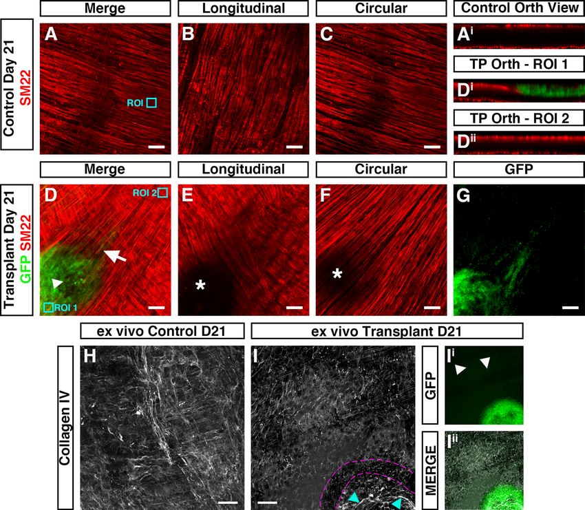
www.nature.com/scientificreports/
cation, and maximal z-axis integration post-transplantation, at day 7, 14, & 21, were evaluated using Welch’s
ANOVA and Welch’s t-tests. For transcriptomic analyses statistical comparison between control (non-trans-
planted) and transplanted samples was performed using ΔCT values by Welch’s t-test. Results were considered
significant at P < 0.05.
Ethical approval and consent to participate. The animals used and the experiments conducted, in this
study, were approved by the University College London Biological Services Ethical Review Process. All of the
procedures performed were in accordance with the UK Animals (Scientific Procedures) Act 1986.
Results
Characterisation of ex vivo organotypic cultured colon. To assess the integration of ENSC, within
intestinal segments, we developed an ex vivo organotypic culture model, based upon methodology previously
used for ENS imaging20,21, in which colonic tissues could be maintained in situ with limited contractile forces
(Fig. 1A–D), which typically lead to tissue damage when pinned. Macroscopic images of tissue segments dem-
onstrate well-preserved, undamaged, tissue structures at day 7, 14 and 21 (Fig. 1E–G). Remarkably, up to 21 days
in culture such preparations did not display evidence of culture contamination, visible structural abnormalities
or any signs of detrimental tissue-scaffold interaction.
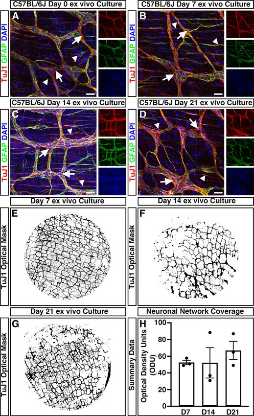
To assess the impact of organotypic culture on the development and maturation of the ENS, immunohisto-
chemistry was performed. Within freshly dispersed (uncultured) colonic tissues, robust neuron-specific class
III beta-tubulin positive ( TuJ1+) neuronal networks could be observed (Fig. 2A). TuJ1+ neuronal networks were
observed, organised in a characteristic mesh-like structure, at the level of the myenteric plexus, in close apposition
to glial fibrillary acidic protein positive ( GFAP+) glial cells (Fig. 2A, arrows). Additional T
uJ1+ intramuscular neu-
rons were observed extending bipolar neuronal fibres within the tunica muscularis (Fig. 2a, arrowheads). Notably,
organotypically cultured tissues demonstrated similar T uJ1+ and G FAP+ expression at day 7 (D7; Fig. 2B), day
14 (D14; Fig. 2C), and day 21 (D21; Fig. 2D) in culture. Again, T uj1+ neuronal populations could be observed
both at the level of the myenteric plexus (arrows) and intramuscularly (arrowheads).
To quantitatively assess the condition of the endogenous enteric neural network, and to define its structure
over time in organotypic culture, the optical density of T uJ1+ positive pixels within wholemount montages of
cultured colonic specimens was assessed. The mean values of the optical density of T uJ1+ neuronal networks
between D7 (52.5 ± 2.7 optical density units (ODU); n = 3, Fig. 2E, H), D14 (52.1 ± 18.2 ODU; n = 3; Fig. 2F,H)
and D21 (66.9 ± 11.0 ODU; n = 3; Fig. 2G,H) cultures where comparable ( W2.0, 2.9 = 0.66, P = 0.582) as determined
by Welch’s one way analysis of variance (ANOVA). These data suggest the preservation of neuronal networks
within ex vivo organotypically cultured specimens up to 21 days in culture.
Donor ENSC migrate extensively within organotypic cultures. ENSC were isolated from donor
Wnt1cre/+;R26RYFP/YFP mice (P5-P7), in which neural crest cells and their derivatives express endogenous yellow
fluorescent protein (YFP). This endogenous YFP expression allowed isolation, by FACS, and fate-labelling of
donor ENSC (Fig. 3A,B). Typically, enzymatic digestion of the tunica muscularis, from the entire small intestine
and colon, and subsequent FACS lead to the enrichment of a Y FP+ neural crest-derived population at 5.6 ± 0.63%
5 4
(3.2 × 10 ± 5 × 10 cells) per sorting experiment (Fig. 3B; n = 5). Upon culture, individually sorted Y FP+ cells
appeared as sparsely distributed spherical shaped cells (Fig. 3C, arrows). By D3, selected YFP+ cells developed
a typical multipolar neural crest cell appearance and began to form cellular connections (Fig. 3D, arrows). At
D7, cell clusters (Fig. 3E, arrows) and elongated interconnected “chains” of cells could be observed forming a
network-like structure (Fig. 3E, arrowheads). Further culture to D9 resulted in the expansion of YFP+ ENSC
cell clusters to form extensive networks which displayed interconnecting filaments (Fig. 3F, arrowheads). Subse-
quently, selected YFP+ ENSC-derived cells were found to form multiple characteristic three dimensional “neu-
rospheres” by approximately 2 weeks in culture (Fig. 3G,H, arrows).
In order to assess integration of ENSC-derived cells, in intestinal specimens, we sought to establish the
ability of cultured neurospheres to colonise and integrate, within organotypically cultured C57BL/6J colonic
specimens ex vivo.
We transplanted a single, individual YFP+ neurosphere (approximately 2 × 10⁴ cells) to the serosal surface
of organotypically cultured C57BL/6J colonic specimens (Supplementary Fig. 1A). At day one (D1) in culture,
neurospheres appeared to be attached to recipient C57BL/6J colonic sections and multiple YFP+ cells were found
to have dissociated from the presumptive site of transplantation, which were observed migrating as individual
cells at the periphery of the donor neurosphere (Supplementary Fig. 1B). At D3, migration of donor cells was
observed to continue in a non-uniform fashion in all directions away from the transplantation site (Supple-
mentary Fig. 2). After three weeks (D21) in culture, neurosphere-derived donor Y FP+ cells were found to have
2 2
migrated extensively to cover an average area of 9.22mm ± 0.85mm (approximately 47% ± 4%) within recipient
colonic tissues (Figs. 3I,J).
Temporal integration of ENSC‑derived cells within organotypic cultures. To assess integration
of YFP+ donor cells, within the tunica muscularis of recipient colonic segments, immunohistochemistry was
performed using the pan-neuronal marker TuJ1 and green fluorescent protein (GFP) antibody.
FP+ cells were observed on the serosal aspect of transplanted colonic segments and appeared to
At D7, G
migrate in multiple directions (Fig. 4A). At this stage, no penetration or integration of donor GFP+ cells, within
the tunica muscularis, was observed. Donor cells were observed on the serosal aspect only (Fig. 4B; arrow),
with no observable migration toward the endogenous T uJ1+ ENS at the level of the myenteric plexus (Fig. 4B;
arrowhead).
Scientific Reports | (2021) 11:15889 | https://doi.org/10.1038/s41598-021-95434-4 4
Vol:.(1234567890)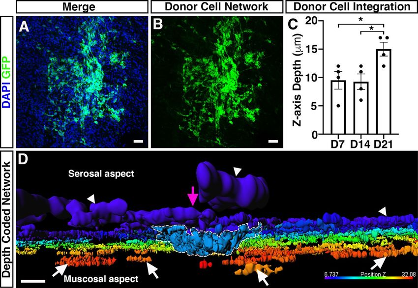
www.nature.com/scientificreports/
Figure 1. Ex vivo organotypic culture of colonic tissue. (A) Schematic representation of ex vivo gut culture
methodology and the strategy used to perform ENSC transplantation. Illustration created by UCL Medical
Illustration. (B) Representative image of colonic tunica muscularis after fine dissection of the mucosal layer.
(C) Representative image of tissue mounting apparatus. (D) Representative image of mounted colonic tissue
segment in organotypic culture. (E–G) Representative images of cultured colonic tissues at Day 7 (E), Day 14
(F), and Day 21 (G) demonstrating the absence of contamination, and lack of gross tissue damage, within long-
term organotypic cultures.
At post-transplantation D14, transplanted Y FP+ neurospheres appeared to have dissociated into single cells
(Fig. 4C) with numerous, spindle shaped, G FP+ cells observed. The majority of G FP+ cells at D14 were again
+
observed at the outer serosal aspect of transplanted tissues. However, donor GFP cells were observed which dis-
played a limited degree of tissue penetration (Fig. 4D; arrow) towards the myenteric plexus (Fig. 4D; arrowhead).
At D21, transplanted GFP+ ENSC-derived cells were visualised at the serosal aspect (Supplementary Fig. 3A).
Though by contrast, GFP+ donor cells were also observed to have migrated through the muscularis to integrate
within, or in close association with, recipient endogenous enteric ganglia at the level of the myenteric plexus
(Fig. 4E–H, Supplementary Fig. 3B). G FP+ cells were observed distributed along the “mesh-like” myenteric neural
network, both as individual cells, and as donor cell clusters, within individual ganglia structures (Fig. 4F–H).
Scientific Reports | (2021) 11:15889 | https://doi.org/10.1038/s41598-021-95434-4 5
Vol.:(0123456789)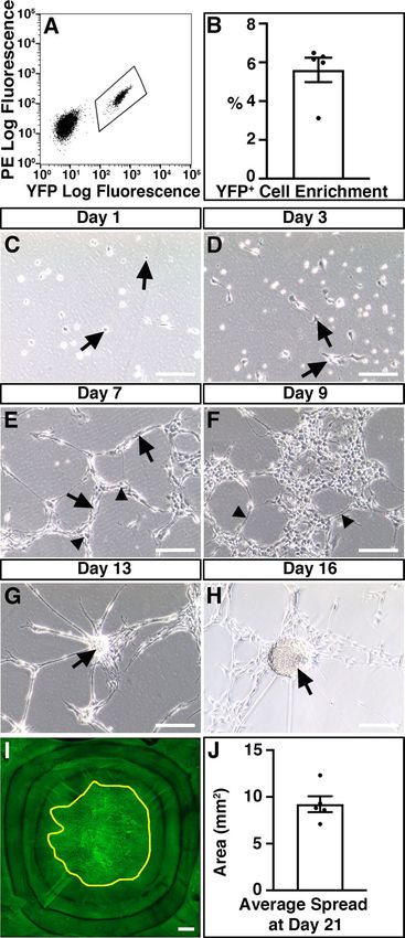
www.nature.com/scientificreports/
Figure 2. Mouse colonic segments cultured ex vivo maintain expression of ENS markers. (A–D) Representative confocal
z-stack images demonstrating the presence of the pan neuronal marker; TuJ1 (red), glia marker; GFAP (green) and DAPI
(blue) in fresh (A), Day 7 (B), Day 14 (C), and Day 21 (D) ex vivo cultured C57BL/6J colonic tissue. TuJ1+ expression was
readily observed across organotypic culture and was found to label the ENS at the level of the myenteric plexus (arrows)
and intramuscular nerves (arrowheads). (E–G) Representative optical density masks of montaged T uJ1+ expression within
cultured colonic segments at Day 7 (E), Day 14 (F), and Day 21 (G). (H) Summary data of neuronal network coverage, as
determined by the optical density of T uJ1+ expression, across organotypic culture (n = 3 for each timepoint). Error bars
represent mean ± s.e.m. Scale bars represent 50 μm.
Scientific Reports | (2021) 11:15889 | https://doi.org/10.1038/s41598-021-95434-4 6
Vol:.(1234567890)www.nature.com/scientificreports/
Figure 3. Isolation and expansion of donor ENSC for ex vivo transplantation. (A) Representative FACS
profile demonstrating the gating scheme for isolation of YFP+ cells (bounded area) from Wnt1cre/+;R26RYFP/YFP
intestine. (B) Summary data showing Y FP+ cell enrichment via FACS (n = 5). (C–H) Representative images
showing the development of FACS-sorted YFP+ cells in culture. At Day 1 (C), the majority of sorted YFP+ cells
displayed a spherical appearance (arrows). By Day 3 (D), YFP+ cells developed a typical multipolar neural crest
cell appearance (arrows). At Day 7 in culture (E), sorted cells appear as multipolar cell clusters (arrows) which
are connected in a network-like structure (arrowheads). At Day 9 (F), YFP+ cell clusters appeared to form
extensive networks which displayed interconnecting filaments (arrowheads). Further culture to Day 13 (G) and
Day 16 (H) led to the development of rounded “neurosphere” structures (arrows). (I) Representative montage
fluorescent image showing the development of YFP+ donor cells within recipient C57BL/6J colonic tissue
21-days after ex vivo transplantation. Yellow line bounds Y FP+ donor cell coverage. (J) Summary data showing
average donor cell spread within recipient tissues at Day 21 (n = 5). Error bars represent mean ± s.e.m (B, J).
Scale bars represent 250 μm (C–H), 50 μm (I). See also Supplementary Fig. 1.
Scientific Reports | (2021) 11:15889 | https://doi.org/10.1038/s41598-021-95434-4 7
Vol.:(0123456789)www.nature.com/scientificreports/
Figure 4. Temporal integration of transplanted ENSC within ex vivo organotypically cultured C57BL/6J colon. (A) Representative
confocal z-stack image of GFP+ (green) ENSC-derived cells and the endogenous TuJ1+ (red) ENS 7-days after ex vivo transplantation.
(B) Orthogonal view of 3D rendered confocal stack showing donor GFP+ cells at the serosal aspect (arrow) and T uJ1+ myenteric
plexus (arrowhead) 7-days after ex vivo transplantation. (C) Representative confocal z-stack image of donor G FP+ (green) cells and
the endogenous TuJ1+ (red) ENS 7-days after ex vivo transplantation. (D) Orthogonal view of 3D rendered confocal stack, 14-days
after ex vivo transplantation, showing donor G FP+ cell penetration (arrow) from the serosal aspect towards the myenteric plexus
(arrowhead). (E, F) Representative low- (E), and high-power (F) confocal z-stacked images of G FP+ (green) donor cells within the
recipient TuJ1+ (red) myenteric plexus, 21-days after ex vivo transplantation. Donor cells were observed as both individual cells, and
donor cell clusters, within ganglia like structures (arrowheads). Donor derived G FP+TuJ1+ neurons (arrows) appeared to extend
GFP+TuJ1+ neuronal fibres which traced the endogenous neural network (cyan arrows). (G) Orthogonal view of 3D rendered
confocal stack showing donor GFP+ cells (arrows) within the endogenous T uJ1+ myenteric plexus (arrowhead) 21-days after ex vivo
transplantation. (H) Zoomed image of inset shown in (F) showing high magnification of donor cell bodies (arrowheads) incorporated
within ganglia as indicated by DAPI (blue), GFP (green) and TuJ1 (red). Scale bars represent 50 μm (A–G), 25 μm (H).
Scientific Reports | (2021) 11:15889 | https://doi.org/10.1038/s41598-021-95434-4 8
Vol:.(1234567890)www.nature.com/scientificreports/
Figure 5. Integration of ENSC-derived donor cells across the gut wall. (A, B) Representative confocal z-stack
image of donor ENSC-derived GFP+ (green), and DAPI (blue), within the gut wall 21-days after ex vivo
transplantation. (C) Summary data showing analysis of donor cell body ( GFP+/DAPI+) integration, measured
as z-axis depth, across the gut wall at day-7 (D7), D14, and D21 after ex vivo transplantation (n = 4 for each
timepoint). (D) Representative depth-coded image of donor cell integration at D21. Note the protrusion of
donor ENSC-derived cells into the gut wall (dashed line) from the site of transplantation on the serosal surface
(magenta arrowhead). Donor GFP+ cells and fibres were observed to have integrated longitudinally along the
gut, both on the serosal surface (arrowheads) and within the tunica muscularis (arrows). Scale bars represent
50 μm (A, B), 30 μm (D). Error bars represent mean ± s.e.m. *P ≤ 0.05, by Welch’s t-test.
At this timepoint, donor derived G FP+ cells could be observed having differentiated to a neuronal phenotype
with co-expression of either TuJ1 (Fig. 4F; arrows) or HuC/D (Supplementary Fig. 3B, Supplementary Movie 1).
Moreover, GFP+TuJ1+ neuronal fibres were also observed at the level of the myenteric plexus, which appeared
to trace the endogenous recipient neural network (Fig. 4F; cyan arrows). Upon three-dimensional (3D) recon-
struction, donor G FP+ cells could clearly be observed throughout the T uJ1+ myenteric neural network (Fig. 4G;
arrowhead), including at both the serosal and mucosal aspects of the myenteric plexus (Fig. 4G; arrows). Here,
individual donor-derived G FP+ cell bodies could be observed within endogenous ganglia structures, via visu-
alisation of DAPI expression within G FP+ donor cells (Fig. 4H, arrowheads).
To establish if donor cells undergo proliferation and/or apoptosis, upon transplantation, we assessed Ki67 &
cleaved Caspase 3 expression in neurospheres and transplanted ex vivo tissues at early (D3) and late (D21) time-
points (Supplementary Figs. 4, 5). Proliferation and apoptosis of Wnt1cre/+;R26RYFP/YFP-derived cells were clearly
observed pre-transplantation, within equivalent neurospheres, as evidenced by robust expression of both Ki67
(Supplementary Fig. 4A) and cleaved Caspase 3 (Supplementary Fig. 5A). However, shortly after transplantation
(D3), neither Ki67 (Supplementary Fig. 4B) nor cleaved Caspase 3 (Supplementary Fig. 5B) were observed within
GFP+ donor cells. Similarly, at D21 post-transplantation, we were unable to observe G FP+-donor derived cells
+
which were undergoing proliferation, or apoptosis, despite evidence of K aspase3+ expression in the
i67 and C
recipient muscularis (Supplementary Figs. 4C, 5C).
We next aimed to quantify the maximal distance ENSC-derived cells had migrated across the gut wall using
confocal z-stacks of GFP-labelled donor cells, in combination with DAPI labelling (Fig. 5A, B). Here, maximal
cell integration was recorded by observing the most medially integrated donor cell body (GFP+/DAPI+), rela-
tive to the serosal surface using Z-series scans (1 μm optical sections), which showed significant differences
in the mean values across the three timepoints as determined by Welch’s ANOVA ( W2.0, 5.9 = 5.58, P = 0.04). At
D7, ENSC-derived donor cells were observed at a z-axis depth of 9.5 ± 1.6 μm (Fig. 5C; n = 4). Similarly, at D14
donor-derived cells were observed at a z-axis depth of 9.3 ± 1.4 μm (P = 0.91; n = 4; Fig. 5C). However, at D21
the most medially integrated donor-derived cell body in ex vivo cultured transplanted colon was observed at a
z-axis depth of 15 ± 1.2 μm (P = 0.03 vs D7, P = 0.02 vs D14; n = 4; Fig. 5C). Importantly, depth-coding of all G
FP+
donor-derived cell bodies and fibres, according to their z-axis depth, revealed integration of donor ENSC along
Scientific Reports | (2021) 11:15889 | https://doi.org/10.1038/s41598-021-95434-4 9
Vol.:(0123456789)www.nature.com/scientificreports/
the serosal aspect of the gut (Fig. 5D; arrowheads, Supplementary Movie 1). Moreover, from the presumptive
site of transplantation (Fig. 5D; magenta arrow), donor cells appear to penetrate the gut wall at the site of neu-
rosphere engraftment (Fig. 5D; dashed white line) and migrate along the longitudinal aspect of the gut within
the tunica muscularis (Fig. 5D; white arrows).
Taken together, these data suggest that ENSC appear to initially migrate from the transplanted neurosphere,
along the serosal aspect of the tissue, in a non-uniform manner. Subsequently, transplanted cells penetrate the
tunica muscularis at the site of neurosphere transplantation and migrate within the tunica muscularis towards the
endogenous neural network at the level of the myenteric plexus, allowing integration within ganglia structures
and extension of donor cell processes, within the myenteric network, by approximately three weeks.
Molecular characterisation of donor ENSC integration after transplantation. Having demon-
strated the integration of donor ENSC-derived cells, within the tunica muscularis, we assessed possible tissue
remodelling within the gut wall; and the molecular basis of ENSC migration and integration within transplanted
colonic segments.
In ex vivo control (non-transplanted) tissues, the smooth muscle architecture appears to be preserved up to
three weeks in organotypic culture (Fig. 6A–C). Here, immunohistochemistry for the smooth muscle marker
SM22 revealed smooth muscle cells in close apposition, both in the longitudinal (Fig. 6B) and circular (Fig. 6C)
orientations. Orthogonal reconstruction of confocal z-stacks also revealed intact S M22+ muscle layers in both the
longitudinal and circular planes (Fig. 6Ai). By contrast, in transplanted tissues, disruption of the smooth muscle
architecture was observed at the site of transplantation 21-days following ENSC transplantation (Fig. 6D–G).
Examination of the longitudinal muscle layers revealed complete loss of S M22+ expression at the site of neu-
rosphere engraftment (Fig. 6E; white star). Similarly, examination of the circular muscle layer revealed partial
reductions in SM22 expression (Fig. 6F; white star). Interestingly, this disruption in SM22 expression appeared
to be limited to the site of transplantation and neurosphere engraftment. Upon orthogonal reconstruction of the
engraftment site (Fig. 6D, ROI 1), GFP+ donor cell expression was observed within the plane of the longitudinal
muscle layer (Fig. 6Di). While this donor cell expression appeared restricted to the serosal aspect, and presump-
tive longitudinal muscle layer, all S M22+ expression in this engraftment region was absent. However, in regions
away from the site of engraftment (Fig. 6D, ROI 2) uninterrupted S M22+ expression was observed in both the
longitudinal and circular muscle layers (Fig. 6Dii). Comparable disruption of the smooth muscle architecture was
also observed following assessment of smooth muscle actin expression in independent replicates (Supplementary
Fig. 6). Immunohistochemistry also revealed apparent alterations in the collagen ECM compartment within
transplanted tissues. As opposed to control non-transplanted colon, where Collagen type IV (Col IV) expression
was observed in near continuity (Fig. 6H), loss of Collagen IV was observed at the boundary of the engraftment
site in transplanted tissues (Fig. 6I; magenta border). Additionally, the presence of Collagen IV expression was
observed, at the site of engraftment, within the remaining neurosphere structure (Fig. 6I; cyan arrow, Figs. 6Ii–Iii).
To further examine the mechanisms involved in donor cell integration, molecular analyses were performed
to examine the expression of candidate genes, known to play critical roles within the intestinal ECM, such as
Collagen 1a2 (Col1a), Collagen 4a1 (Col4a), Fibronectin 1 (Fn1), Elastin (Eln), Laminin a1 (Lama1), Laminin b1
(Lamb1) and in remodelling of the ECM, including Matrix metalloproteinase 1 (Mmp1), Mmp2, Mmp8, Mmp9
and Mmp13. Of note, Mmp1 could not be resolved in control post-natal C57BL/6J wild type colonic tissue nor
in ex vivo cultured control and transplanted tissues whereas all other targets were found to be robustly expressed
(data not shown), in at least one condition, using standard real-time polymerase chain reaction (RT-PCR) analy-
sis. Interestingly, Mmp8 was not observed in control postnatal tissues but could be observed qualitatively at very
low levels in control ex vivo cultures with increasing expression after transplantation (data not shown).
Therefore, all candidate genes, except for Mmp1, were taken forward for quantitative RT-PCR (qRT-PCR)
analysis to assess their expression in transplanted ex vivo tissue segments compared to control non-transplanted
colon.
Interestingly, ENSC integration within transplanted tissues led to increased expression of extracellular matrix
related genes including Col1a (5.8 ± 2.4-fold increase; P = 0.04; n = 4; Fig. 7A) and Col4a (4.0 ± 1.1-fold increase;
P = 0.010; n = 4; Fig. 7B) as determined by comparison of ΔCT values by Welch’s t-test analysis. By contrast, other
ECM related genes were found to be comparable to control ex vivo cultured tissues: including Fn1 (P = 0.38; n = 4;
Fig. 7C), Eln (P = 0.11; n = 4; Fig. 7D), Lama1 (P = 0.12; n = 4, Fig. 7E) and Lamb1 (P = 0.07; n = 4, Fig. 7F). Of the
candidate matrix metalloproteinases, Mmp2 (P = 0.14; n = 4; Fig. 7G) and Mmp8 (P = 0.05; n = 4; Fig. 7H) were
found to be expressed at a similar level to control (non-transplanted) tissues; whereas increases in expression
were observed in Mmp9 (3.4 ± 0.7-fold increase; P = 0.047; n = 4; Fig. 7I) and Mmp13 (5.5 ± 1.9-fold increase;
P = 0.005; n = 4; Fig. 7J) in transplanted tissues when compared to control (non-transplanted) tissues.
Furthermore, assessment of Mmp gene expression of Wnt1cre/+;R26RYFP/YFP neurospheres, by RT-PCR, revealed
the expression of Mmp2 and Mmp9. However, Mmp8 and Mmp13 could not be identified (Supplementary Fig. 7).
Taken together, these data suggest that active remodelling of the ECM collagen compartment and smooth muscle
architecture, by both donor-derived and endogenous matrix metalloproteinases, occurs in response to ENSC
transplantation, which subsequently allows integration of donor ENSC within and along the gut wall.
Discussion
Over the past decade there has been an increasing focus on the development and evaluation of stem cell-based
therapies for treating enteric neuropathies. Recent studies have highlighted the potential of autologous, and
pluripotent-derived ENS progenitor-based therapy, as a means of replacing neurons after in vivo transplantation
to mouse c olon14–17,22–26. However, the precise mechanisms by which donor-derived cells integrate within recipi-
ent tissue remain unclear. Therefore, studies to uncover the timeframe, and mechanisms, underlying donor cell
Scientific Reports | (2021) 11:15889 | https://doi.org/10.1038/s41598-021-95434-4 10
Vol:.(1234567890)www.nature.com/scientificreports/
Figure 6. Donor cell integration induces tissue remodeling after ex vivo transplantation. (A) Representative
confocal z-stack image of SM22+ (red) smooth muscle within control (non-transplanted) C57BL/6J colonic
tissue at day-21. (Ai) Orthogonal view taken from the region of interest (ROI, cyan box) in A showing
SM22+ smooth muscle in two uninterrupted layers. (B, C) Representative images of individual z-stacks at the
longitudinal (B) and circular (C) muscle layers. (D) Representative confocal z-stack image of donor G FP+
(green) and SM22 (red) smooth muscle 21-days after ex vivo transplantation. (D &D ) Orthogonal views
+ i ii
taken from ROIs (cyan boxes) in (D) showing interruption of the longitudinal smooth muscle (Di) at the site of
neurosphere engraftment. At sites away from the area of neurosphere engraftment S M22+ expression appears to
demonstrate two uninterrupted smooth muscle layers (Dii). (E, F) Representative images of individual z-stacks
at the longitudinal (E) and circular (F) muscle layers 21-days after ex vivo transplantation. (G) Representative
image of individual z-stack, at the level of the circular muscle (taken from D), showing migration of GFP+ donor
derived cells from the site of neurosphere engraftment 21-days after ex vivo transplantation. (H) Representative
confocal z-stack image of Collagen IV (grey) in control (non-transplanted) C57BL/6J colonic tissue at day-21.
(I) Representative confocal z-stack image of Collagen IV (grey) 21-days following ex vivo transplantation. Note
the presence of Collagen IV expression at the site of neurosphere transplantation (cyan arrows) and apparent
discontinuity of Collagen IV expression surrounding the presumptive neurosphere boundary (magenta dashed
lines). (Ii, Iii) Representative GFP channel (Ii) and merged (Iii) confocal z-stack images showing GFP+ donor
cells at the site of transplantation and within the tunica muscularis (arrows) having migrated away from the
engrafted neurosphere.
integration are fundamental to progressing towards clinical application of stem cell therapy for gut disorders.
Moreover, mechanistic studies of donor cell integration may uncover useful targets to improve the efficiency of
donor cell integration, and function, within recipient tissues.
Importantly, many previous studies have relied heavily on in vivo surgical transplantation procedures to
rodents14–18,23,27. While this has provided crucial proof-of-principle data that donor cells can integrate within
the gut after transplantation, technical limitations such as tissue opacity, and an inability to perform long-term
in vivo imaging, have limited the mechanistic investigation of how donor cells integrate within recipient gut
tissue. Alternative approaches including explant cultures have also established the feasibility of transplanting
donor enteric precursors into aneural gut28–31. However, these explant methods are often hindered by limitations
Scientific Reports | (2021) 11:15889 | https://doi.org/10.1038/s41598-021-95434-4 11
Vol.:(0123456789)www.nature.com/scientificreports/
Figure 7. Donor cell integration leads to increased expression of collagen and Matrix metalloproteinases. (A–F)
Summary data showing mRNA fold change for ECM-related and matrix metalloproteinase genes: (grey bars):
Collagen 1a (Col1a; A), Collagen 4a (Col4a; B) Fibronectin 1 (Fn1; C), Elastin (Eln; D), Laminin a1 (Lama1:
E), Laminin b1 (Lamb1: F), Matrix metalloproteinase-2 (Mmp2; G), Matrix metalloproteinase-8 (Mmp8; H),
Matrix metalloproteinase-9 (Mmp9; I) and Matrix metalloproteinase-13 (Mmp13; J) 21-days after ex vivo
transplantation compared to non-transplanted control (white bars) tissues. Error bars represent mean ± s.e.m.
n = 4 for each gene examined. *P ≤ 0.05, **P ≤ 0.01 comparing ΔCT values by Welch’s t-test.
in their reproducibility, or in the long-term maintenance of tissues on chick chorioallantoic membrane (CAM)
cultures31–33.
To begin to overcome some of these limitations, here we have employed an ex vivo organotypic culture
method to investigate the temporal integration of ENSC within murine gut segments. We demonstrate that this
ex vivo organotypic culture configuration allows for long-term culture of murine colonic segments, with preserva-
tion of both tissue architecture and ENS structure, providing a robust, reproducible and readily scalable model.
Significantly, this method also appears to limit tissue damage, within culture colonic segments, when compared
to standard ex vivo organotypic culture conditions which typically require pinning of tissue. Hence, this organo-
typic method provides an optimal model to explore the temporal integration of donor cells within gut tissues.
Previous studies have detailed the functional integration of donor neurons, derived from postnatally har-
vested ENSC, in recipient wild-type colonic t issues14,23. These studies, using calcium imaging and optogenetic
approaches, have detailed the functional integration of donor ENSC-derived neurons within the host neuro-
musculature four weeks after in vivo transplantation. By four weeks, engrafted cells were found to have migrated
extensively within the recipient gut wall, including the integration of donor-derived cells within endogenous
ganglia, and the extension of graft-derived axonal fibres which appeared to both trace the endogenous ENS
structure, and project to the circular muscle layer. Moreover, engrafted donor-derived neurons were found to both
Scientific Reports | (2021) 11:15889 | https://doi.org/10.1038/s41598-021-95434-4 12
Vol:.(1234567890)www.nature.com/scientificreports/
functionally integrate with the endogenous ENS c ircuitry14 and provide inhibitory, and excitatory, innervation
to intestinal smooth muscle23. A similar timescale was also observed in the functional integration of ENSC-
derived neurons within dysmotile transgenic tissues17. Here, ENSC transplantation was found to restore neural
responses and rescue gut transit four weeks after transplantation. Again, significant migration of ENSC-derived
cells was observed both within, and along, the gut wall with donor cells appearing to “home” to the myenteric
plexus. More recently, derivation of ENS progenitors from pluripotent sources, such as embryonic stem cells or
induced pluripotent stem cells has been proposed as a potential “off-the-shelf ” alternative to autologous ENSC24.
Similar to ENSC, pluripotent-derived ENS progenitors have been shown to engraft and migrate through the
colonic muscularis to reside within endogenous ganglia structures, across a four week period, following in vivo
transplantation26. Taken together, these findings suggest that the migration of donor ENSC-derived cells into the
gut neuromusculature, and the “homing” of donor neurons to the myenteric plexus region, are likely central pro-
cesses in the establishment of donor-derived functional responses within the host neuromusculature. However,
to date, a more definitive timeframe and mechanistic understanding of donor cell integration remain elusive.
Using our modified ex vivo transplantation approach, we demonstrate that migration of ENSC-derived cells
away from the presumptive site of transplantation, and integration within the colonic musculature, is a dynamic
process which occurs over a three-week period. Initially, ENSC-derived donor cells were observed to migrate
away from the site of neurosphere engraftment, along the serosal aspect of the colon. This migration at the
serosal surface did not appear to show preferential directionality and is reminiscent of the migration pattern of
cultured neural crest cells34–36. A secondary phase of medial migration appears to occur allowing donor cells
to integrate across the gut wall. Crucially, this secondary process appears to be established over a number of
weeks, with penetration of the neuromusculature, and medial migration, appearing to be prominent only after
approximately 14 days.
We hypothesised that donor ENSC utilise molecular processes analogous to that of metastatic cancer cells,
in order to remodel the ECM, infiltrate (i.e., intra/extravasate) and migrate (i.e., invade) within the host gut
wall following transplantation. Indeed, previous studies have highlighted that the matrisome plays multiple
dynamic roles in embryonic neural crest cell migration and differentiation36–39. In tandem, migratory neural
crest cells have been shown to modify the local ECM, suggesting that migration through the ECM is regulated
on multiple levels40,41.
Our initial expectation was that migration through the colonic neuromusculature, and integration in myen-
teric ganglia, would necessitate collagenase-based depletion of the ECM. Our finding of an upregulation in Mmp9
and Mmp13 expression, within transplanted ex vivo cultured tissues, fits with our hypothesis as both have been
shown to be involved in epithelial-mesenchymal transition (EMT) and the progression of colorectal cancer42–44.
Importantly, MMP proteolysis of Collagen IV has previously been shown to promote cell migration, via the
production of specific cleavage fragments with independent biological activity45,46. Therefore, upregulation of
Mmp9 and Mmp13 in transplanted ex vivo colon may provide a possible mechanism of how transplanted ENSC
remodel the ECM, to allow invasion of donor cells. Of note, early emigration of neural crest cells from the neural
tube has been shown to be dependent on M mp947 and is associated with discontinuity of Collagen IV s taining37.
Similarly, here we demonstrate that major remodelling of the musculature and Collagen IV occurs, after ex vivo
transplantation, around the site of neurosphere engraftment which appears to be consistent with neural crest
emigration after transplantation. Yet, our findings of increased Collagen (Col1a and Col4a) gene expression
were unexpected. Previous investigations have, however, demonstrated dynamic regulation of Collagen I and IV
during neural crest cell migration. In particular, Collagen I expression has been shown to be upregulated along
neural crest migratory pathways, whereas Collagen IV has been shown to be increased upon neural crest cell
aggregation and differentiation37. Hence, these processes may account for the observations in the current study,
as transplanted ENSC have been shown to migrate extensively within ex vivo transplanted tissue, and aggregate
within endogenous ganglia structures at the level of the myenteric plexus.
While this study has revealed the timescale and a possible mechanism underlying donor cell integration
within host tissues following ENSC transplantation, future studies will be required to fully elucidate the mecha-
nisms involved. Here, we utilised early post-natal (P5-7) ENSC to investigate donor cell integration within
C57BL/6J wild-type colon. A recent study has shown that enteric neural crest cells progressively lose capacity
to form the ENS with age32. Hence, further studies will be required to determine the integration capacity of
aged donor cells. This caveat may have significant implications in the clinical translation of cellular therapies
for enteric neuropathies, as donor age will need to be considered for both autologous cell transplants, and in
strategies using pluripotent cell sources; given time in culture may alter integrative capacity. Similarly, studies
investigating alternative donor cell forms (i.e., neurospheres vs single cells) may be beneficial in determining
the ideal cell “product” required for integration. Importantly, the outlined ex vivo transplantation methodology
may offer significant benefits in tackling such questions in a medium-throughput, and consistent, manner. Fur-
thermore, we used C57BL/6J wild-type colon as recipient tissue in this proof-of-principle study. We demonstrate
that donor-derived cells infiltrate and “home” to the myenteric ganglia by three weeks post-transplantation. We
assume that trophic factors regulate this “homing” process, though the exact factors involved remain unclear.
Moreover, recent studies have demonstrated that the colonic ECM is altered in diseased s tates48–50, which suggests
the integrative ability of ENSC-derived donor cells is likely to be altered by the recipient microenvironment in
different disease states. Future studies will therefore be required to address how disease-specific parameters alter
ENSC integration, which may be clinically relevant.
Scientific Reports | (2021) 11:15889 | https://doi.org/10.1038/s41598-021-95434-4 13
Vol.:(0123456789)www.nature.com/scientificreports/
Conclusions
We conclude that this study provides critical evidence on the timescale and mechanisms which regulate ENSC
integration within recipient gut tissue. Furthermore, the modified organotypic culture model utilised in the cur-
rent study may be beneficial in the future assessment of donor cell transplantation in neuropathic models, or in
the development of targeted approaches to influence donor cell behaviour as a possible combinatorial strategy
for the treatment of enteric neuropathies.
Data availability
All authors had access to the study data and had reviewed and approved the final manuscript. The data that sup-
port the findings of this study are available from the corresponding author upon reasonable request.
Received: 21 October 2020; Accepted: 26 July 2021
References
1. Goldblum, J. R., Rice, T. W. & Richter, J. E. Histopathologic features in esophagomyotomy specimens from patients with achalasia.
Gastroenterology 111, 648–654 (1996).
2. Knowles, C. H., Lindberg, G., Panza, E. & De Giorgio, R. New perspectives in the diagnosis and management of enteric neuropa-
thies. Nat. Rev. Gastroenterol. Hepatol. 10, 206–218. https://doi.org/10.1038/nrgastro.2013.18 (2013).
3. Harberson, J., Thomas, R. M., Harbison, S. P. & Parkman, H. P. Gastric neuromuscular pathology in gastroparesis: Analysis of
full-thickness antral biopsies. Dig. Dis. Sci. 55, 359–370. https://doi.org/10.1007/s10620-009-1071-2 (2010).
4. Giorgio, V. et al. High-resolution colonic manometry accurately predicts colonic neuromuscular pathological phenotype in pedi-
atric slow transit constipation. Neurogastroenterol. Motil. 25(70–78), e78-79. https://doi.org/10.1111/nmo.12016 (2013).
5. Heanue, T. A. & Pachnis, V. Enteric nervous system development and Hirschsprung’s disease: Advances in genetic and stem cell
studies. Nat. Rev. Neurosci. 8, 466–479. https://doi.org/10.1038/nrn2137 (2007).
6. Amiel, J. et al. Hirschsprung disease, associated syndromes and genetics: A review. J. Med. Genet. 45, 1–14. https://doi.org/10.
1136/jmg.2007.053959 (2008).
7. Westfal, M. L. & Goldstein, A. M. Pediatric enteric neuropathies: Diagnosis and current management. Curr. Opin. Pediatr. 29,
347–353. https://doi.org/10.1097/MOP.0000000000000486 (2017).
8. Catto-Smith, A. G., Trajanovska, M. & Taylor, R. G. Long-term continence after surgery for Hirschsprung’s disease. J. Gastroenterol.
Hepatol. 22, 2273–2282. https://doi.org/10.1111/j.1440-1746.2006.04750.x (2007).
9. Pini Prato, A. et al. Hirschsprung disease: Do risk factors of poor surgical outcome exist?. J. Pediatr. Surg. 43, 612–619. https://doi.
org/10.1016/j.jpedsurg.2007.10.007 (2008).
10. Lu, W. et al. Causes and prognosis of chronic intestinal pseudo-obstruction in 48 subjects: A 10-year retrospective case series.
Medicine (Baltimore) 97, e12150. https://doi.org/10.1097/MD.0000000000012150 (2018).
11. Di Nardo, G. et al. Enteric neuropathology of congenital intestinal obstruction: A case report. World J. Gastroenterol. 12, 5229–5233.
https://doi.org/10.3748/wjg.v12.i32.5229 (2006).
12. Pini-Prato, A. et al. Redo surgery in Hirschsprung disease: What did we learn? Unicentric experience on 70 patients. J. Pediatr.
Surg. 45, 747–754. https://doi.org/10.1016/j.jpedsurg.2009.08.001 (2010).
13. Schweizer, P., Berger, S., Schweizer, M., Holschneider, A. M. & Beck, O. Repeated pull-through surgery for complicated
Hirschsprung’s disease—Principles derived from clinical experience. J. Pediatr. Surg. 42, 536–543. https://doi.org/10.1016/j.jpeds
urg.2006.10.058 (2007).
14. Cooper, J. E. et al. In vivo transplantation of enteric neural crest cells into mouse gut; engraftment, functional integration and
long-term safety. PLoS ONE 11, e0147989. https://doi.org/10.1371/journal.pone.0147989 (2016).
15. Cooper, J. E. et al. In vivo transplantation of fetal human gut-derived enteric neural crest cells. Neurogastroenterol. Motil. https://
doi.org/10.1111/nmo.12900 (2017).
16. Hotta, R. et al. Transplanted progenitors generate functional enteric neurons in the postnatal colon. J. Clin. Invest. 123, 1182–1191.
https://doi.org/10.1172/JCI65963 (2013).
17. McCann, C. J. et al. Transplantation of enteric nervous system stem cells rescues nitric oxide synthase deficient mouse colon. Nat.
Commun. 8, 15937. https://doi.org/10.1038/ncomms15937 (2017).
18. Bhave, S. et al. Enteric neuronal cell therapy reverses architectural changes in a novel diphtheria toxin-mediated model of colonic
aganglionosis. Sci. Rep. 9, 18756. https://doi.org/10.1038/s41598-019-55128-4 (2019).
19. Schindelin, J. et al. Fiji: An open-source platform for biological-image analysis. Nat. Methods 9, 676–682. https://doi.org/10.1038/
nmeth.2019 (2012).
20. VandenBerghe, P., Kenyon, J. L. & Smith, T. K. Mitochondrial C a2+ uptake regulates the excitability of myenteric neurons. J. Neu-
rosci. 22, 6962–6971. https://doi.org/10.1523/jneurosci.22-16-06962.2002 (2002).
21. Boesmans, W. et al. Imaging neuron-glia interactions in the enteric nervous system. Front Cell Neurosci. 7, 183. https://doi.org/
10.3389/fncel.2013.00183 (2013).
22. Cheng, L. S. et al. Endoscopic delivery of enteric neural stem cells to treat Hirschsprung disease. Neurogastroenterol. Motil. 27,
1509–1514. https://doi.org/10.1111/nmo.12635 (2015).
23. Stamp, L. A. et al. Optogenetic demonstration of functional innervation of mouse colon by neurons derived from transplanted
neural cells. Gastroenterology 152, 1407–1418. https://doi.org/10.1053/j.gastro.2017.01.005 (2017).
24. Fattahi, F. et al. Deriving human ENS lineages for cell therapy and drug discovery in Hirschsprung disease. Nature 531, 105–109.
https://doi.org/10.1038/nature16951 (2016).
25. Zhou, Y. & Besner, G. Transplantation of amniotic fluid-derived neural stem cells as a potential novel therapy for Hirschsprung’s
disease. J. Pediatr. Surg. 51, 87–91. https://doi.org/10.1016/j.jpedsurg.2015.10.016 (2016).
26. Frith, T. J. R. et al. Retinoic acid accelerates the specification of enteric neural progenitors from in-vitro-derived neural crest. Stem
Cell Rep. 15, 557–565. https://doi.org/10.1016/j.stemcr.2020.07.024 (2020).
27. Hotta, R. et al. Isogenic enteric neural progenitor cells can replace missing neurons and glia in mice with Hirschsprung disease.
Neurogastroenterol. Motil. 28, 498–512. https://doi.org/10.1111/nmo.12744 (2016).
28. Cheng, L. S. et al. Postnatal human enteric neuronal progenitors can migrate, differentiate, and proliferate in embryonic and
postnatal aganglionic gut environments. Pediatr. Res. 81, 838–846. https://doi.org/10.1038/pr.2017.4 (2017).
29. Li, W. et al. Characterization and transplantation of enteric neural crest cells from human induced pluripotent stem cells. Mol.
Psychiatry https://doi.org/10.1038/mp.2016.191 (2016).
30. Findlay, Q., Yap, K. K., Bergner, A. J., Young, H. M. & Stamp, L. A. Enteric neural progenitors are more efficient than brain-derived
progenitors at generating neurons in the colon. Am. J. Physiol. Gastrointest. Liver Physiol. 307, G741-748. https://doi.org/10.1152/
ajpgi.00225.2014 (2014).
Scientific Reports | (2021) 11:15889 | https://doi.org/10.1038/s41598-021-95434-4 14
Vol:.(1234567890)You can also read