PD-L1 tumor-intrinsic signaling and its therapeutic implication in triple-negative breast cancer
←
→
Page content transcription
If your browser does not render page correctly, please read the page content below
RESEARCH ARTICLE

                                         PD-L1 tumor-intrinsic signaling and its
                                         therapeutic implication in triple-negative
                                         breast cancer
                                         Chunhua Chen,1 Shiheng Li,1 Junli Xue,1 Manlong Qi,1 Xin Liu,2 Yan Huang,1,3 Jinghua Hu,1,3
                                         Haidong Dong,2 and Kun Ling1
                                         Department of Biochemistry and Molecular Biology, 2Departments of Urology and Immunology, and 3Department of
                                         1

                                         Nephrology and Hypertension, Mayo Clinic, Rochester, Minnesota, USA.

                                                 Although the immune checkpoint role of programmed death ligand 1 (PD-L1) has been established
                                                 and targeted in cancer immunotherapy, the tumor-intrinsic role of PD-L1 is less appreciated in
                                                 tumor biology and therapeutics development, partly because of the incomplete mechanistic
                                                 understanding. Here we demonstrate a potentially novel mechanism by which PD-L1 promotes
                                                 the epithelial-mesenchymal transition (EMT) in triple-negative breast cancer (TNBC) cells by
                                                 suppressing the destruction of the EMT transcription factor Snail. PD-L1 directly binds to and
                                                 inhibits the tyrosine phosphatase PTP1B, thus preserving p38-MAPK activity that phosphorylates
                                                 and inhibits glycogen synthase kinase 3β (GSK3β). Via this mechanism, PD-L1 prevents the
                                                 GSK3β-mediated phosphorylation, ubiquitination, and degradation of Snail and consequently
                                                 promotes the EMT and metastatic potential of TNBC. Significantly, PD-L1 antibodies that confine
                                                 the tumor-intrinsic PD-L1/Snail pathway restricted TNBC progression in immunodeficient mice.
                                                 More importantly, targeting both tumor-intrinsic and tumor-extrinsic functions of PD-L1 showed
                                                 strong synergistic tumor suppression effect in an immunocompetent TNBC mouse model. Our
                                                 findings support that PD-L1 intrinsically facilitates TNBC progression by promoting the EMT, and
                                                 this potentially novel PD-L1 signaling pathway could be targeted for better clinical management of
                                                 PD-L1–overexpressing TNBCs.

                                         Introduction
                                         Cancer cells often upregulate programmed death ligand 1 (PD-L1) to exhaust activated T cells by stim-
                                         ulating PD-1 on T cells (1, 2). Thus, antibodies that block PD-1/PD-L1 binding have been applied in
                                         clinical trials to prevent immune evasion in various types of cancers (3). Recent clinical trials confirmed
                                         promising outcomes but also exposed imperfections (low response rate, side effects, and resistance) that
                                         indicate the incomplete understanding of the PD-L1 pathway. Triple-negative breast cancer (TNBC)
                                         remains a major clinical challenge with the worst outcome of all BC subtypes, mostly because of its poor
                                         response to current therapies and high incidence of metastasis. Since TNBC cells express higher levels of
Conflict of interest: The authors have   PD-L1 more often than other BC subtypes (4), PD-L1 antibodies, as monotherapy or a part of combined
declared that no conflict of interest    therapies, have been applied in multiple clinical trials for cancers including TNBCs (5). Current results
exists.                                  from these trials suggest that the combined therapy yields synergistic effects (5), which endorses the FDA
Copyright: © 2021, Chen et al. This is   approval of atezolizumab (a PD-L1 antibody) combined with paclitaxel for PD-L1–positive TNBC (6).
an open access article published under   However, the rational combination and successful application of anti–PD-L1 therapy are dependent
the terms of the Creative Commons        upon the comprehensive understanding of PD-L1 biology in tumor cells.
Attribution 4.0 International License.        In addition to its tumor-extrinsic role of activating PD-1 on immune cells, PD-L1 may influence cancer
Submitted: June 30, 2019                 progression by regulating various tumor-intrinsic events in tumor cells independent of the immune sys-
Accepted: March 18, 2021                 tem (7, 8). For example, an association of PD-L1 and the epithelial-mesenchymal transition (EMT) was
Published: May 10, 2021                  observed in clinical studies of various cancers (9–14) as well as in PD-L1–transgenic mice (15, 16). Studies
                                         in claudin-low breast cancer (17) and cervical cancer (18) also suggested that PD-L1 promotes cancer aggres-
Reference information: JCI Insight.
2021;6(9):e131458.                       siveness by influencing the tumor-intrinsic signaling events in the EMT, metabolism, and metastasis. Consid-
https://doi.org/10.1172/jci.             ering the significant role of the EMT in various aspects of tumor progression, including growth, metastasis,
insight.131458.                          stemness, treatment resistance, dormancy (19), the connection of PD-L1 with the EMT in tumor cells shed

                                                                                                                                                        1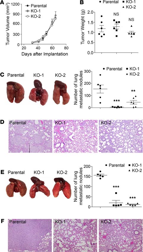
RESEARCH ARTICLE

                                 light on novel avenues for understanding tumor biology. However, these studies are mostly descriptive, and
                                 the molecular mechanism connecting PD-L1 to the EMT in TNBC cells remains unclear. In particular, the
                                 biological significance and clinical indication of PD-L1’s tumor-intrinsic functions are vague.
                                     Here, we demonstrate that PD-L1 facilitates the EMT in TNBC cells by protecting the EMT-promoting
                                 transcription factor Snail from being destructed by the proteosome. This tumor-intrinsic function of PD-L1,
                                 which is achieved via the protein tyrosine phosphatase1B (PTP1B)/p38-MAPK/glycogen synthase kinase
                                 3β (GSK3β) axis and can be activated by PD-1 binding, regulates the aggressiveness of TNBC cells. Intrigu-
                                 ingly, PD-L1 antibodies that constrain its tumor-intrinsic pathway inhibited the growth and metastasis of
                                 TNBC tumors in immunodeficient mice. More importantly, blockade of both tumor-extrinsic and -intrinsic
                                 functions of PD-L1 synergistically suppressed cancer progression in a syngeneic, immunocompetent TNBC
                                 mouse model. These results broaden the current paradigm regarding the role of PD-L1 in cancer progression
                                 and emphasize an underestimated concept that the tumor-intrinsic PD-L1 pathway needs to be considered
                                 when applying the anti–PD-L1 therapy.

                                 Results
                                 Expression of PD-L1 intrinsically promotes the EMT and aggressive malignancy of TNBC cells. To understand
                                 PD-L1 function in TNBC cells, we employed human TNBC cell line MDA-MB-231 that exhibits high
                                 PD-L1 expression (4). Utilizing the CRISPR/Cas9 system, we interrupted PD-L1 expression in MDA-
                                 MB-231 cells (Figure 1A and Supplemental Figure 1, A and B; supplemental material available online
                                 with this article; https://doi.org/10.1172/jci.insight.131458DS1). Unexpectedly, these PD-L1–null cells
                                 exhibited morphological changes and appeared epithelial like (Supplemental Figure 1C). Results from fur-
                                 ther investigation showed that protein levels of epithelial markers E-cadherin and Claudin-1 were nota-
                                 bly increased in PD-L1–null clones compared with their WT counterparts (Figure 1A). Correspondingly,
                                 PD-L1–null cells exhibited a substantial decrease of Snail (Figure 1A), an EMT-promoting transcription
                                 factor (EMT-TF) that suppresses the expression of E-cadherin and Claudin-1 (20). To confirm this obser-
                                 vation, we used RNA interference to achieve transient depletion of PD-L1 in MDA-MB-231 cells using 2
                                 distinct PD-L1–specific siRNAs, which resulted in a substantial increase of E-cadherin and Claudin-1 and
                                 strongly reduced Snail (Figure 1A). Slug, a Snail family EMT-TF, also showed reduction in both PD-L1–
                                 null and PD-L1–knockdown cells (Supplemental Figure 2A), whereas another EMT-TF, zinc finger E-box
                                 binding homeobox 1 (ZEB1), was not affected by PD-L1 deficiency (Supplemental Figure 2A). Notably,
                                 suppressing PD-L1 expression in MDA-MB-231 cells led to a downregulation of matrix protein fibronectin,
                                 which is often upregulated in mesenchymal and metastatic tumor cells (21). Yet, additional mesenchymal
                                 markers, like vimentin and β-catenin, were not markedly or consistently altered, and N-cadherin remained
                                 unexpressed when PD-L1 expression was blocked in MDA-MB-231 cells (Supplemental Figure 2A). These
                                 results suggest that loss of PD-L1 partially reversed the EMT status in MDA-MB-231 cells. Important-
                                 ly, reexpression of PD-L1 (Supplemental Figure 2B) in both PD-L1–null and PD-L1–knockdown MDA-
                                 MB-231 cells overwhelmed the E-cadherin expression and restored Snail (Figure 1B). Thus, we reason that
                                 PD-L1 promotes the EMT in MDA-MB-231 cells, which is likely caused by upregulating Snail family TFs.
                                      To determine if this phenomenon is cell line specific, we examined another PD-L1–expressing human TNBC
                                 cell line, Hs578T. As in MDA-MB-231 cells, depletion of PD-L1 in Hs578T cells caused a marked decrease of
                                 Snail but appeared to have no effect on Slug and ZEB1 (Supplemental Figure 3, A and B). Correspondingly, typi-
                                 cal mesenchymal markers, including N-cadherin, fibronectin, and β-catenin, were decreased in PD-L1–depleted
                                 Hs578T cells (Supplemental Figure 3C). Although we did not detect significant changes on expression of other
                                 examined epithelial proteins, such as E-cadherin, Claudin-1, and ZO-1 (Supplemental Figure 3D), decreases
                                 of Snail and mesenchymal markers in PD-L1–depleted Hs578T cells also suggest a partial reverse of the EMT.
                                 Thus, our results suggest that PD-L1 expression intrinsically promotes the EMT in TNBC cells.
                                      The EMT is a transdifferentiation program that plays an important role in promoting all aspects of
                                 cancer aggressiveness and progression, including tumorigenesis, metastasis formation, resistance to apop-
                                 totic stimuli, as well as the entrance into cancer stem cell states (19, 22). To determine whether the intrinsic
                                 function of PD-L1 affects the aggressiveness of PD-L1–expressing TNBC tumors, we first determined in
                                 vitro behaviors of parental and PD-L1–deficient MDA-MB-231 cells. Compared with parental cells, MDA-
                                 MB-231 cells with stable/complete or transient/partial loss of PD-L1 showed a moderate yet significant
                                 decrease in cell growth/survival in vitro (Figure 1C). Notably, a severely weakened ability of forming
                                 tumor spheroid in soft agar was observed in PD-L1–deficient cells (Figure 1D), which could hardly survive

JCI Insight 2021;6(9):e131458 https://doi.org/10.1172/jci.insight.131458                                                                       2
RESEARCH ARTICLE

                                  Figure 1. Expression of PD-L1 promotes the EMT and aggressive behaviors in MDA-MB-231 cells. (A) Loss of
                                  PD-L1 induced epithelial characteristics in TNBC cells. Left panels, cell lysates from the parental or 2 clones of
                                  PD-L1–null (KO-1 and KO-2) MDA-MB-231 cells. Right panels, MDA-MB-231 cells transiently transfected with
                                  nonspecific control siRNA (siNC) or 2 distinct PD-L1 specific (siPD-L1) siRNAs (si-1 and si-2) for 48 hours. (B)
                                  Reexpression of PD-L1 in PD-L1–deficient MDA-MB-231 cells restored the expression of E-cadherin and Snail to
                                  levels comparable to the parental cells. Cells were transfected with PD-L1 for 24 hours followed by siPD-L1 (si-1)
                                  for another 48 hours. (C) PD-L1 deficiency decreased cell proliferation. Cell proliferation of the parental or PD-L1–
                                  deficient cells was determined at 48 hours or 72 hours. (D) Loss of PD-L1 inhibited the anchorage-independent
                                  growth. Tumorigenesis potential of control or PD-L1–deficient MDA-MB-231 cells was determined using soft agar
                                  colony formation assay. Colony numbers were counted using GelCount. (E) Cells lacking PD-L1 were less migratory.
                                  The in vitro cell migration assay was performed using Boyden chamber with 20,000 cells/chamber. Directional
                                  cell migration was induced by a 4-hour treatment of 10% FBS in cells serum-starved overnight. (C–E) Results (n
                                  = 3 independent experiments) were statistically analyzed and plotted as mean ± SEM using unpaired 2-tailed
                                  Student’s t test with the P value adjusted by Bonferroni’s method. *, P < 0.05; **, P < 0.01; ***, P < 0.001.

JCI Insight 2021;6(9):e131458 https://doi.org/10.1172/jci.insight.131458                                                                              3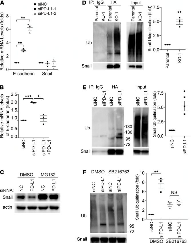
RESEARCH ARTICLE
in soft agar. These results suggest that PD-L1 plays a critical role in cell growth and MDA-MB-231 cells
require PD-L1 for anchorage-independent proliferation and/or survival.
Consistent with the long-standing role of the EMT in promoting cell migration, PD-L1 deficiency
inhibited the in vitro migration of MDA-MB-231 cells. The relative migration rate of the 2 PD-L1–null
clones in response to FBS decreased to 65% and 52% of the parental cells, respectively (Figure 1E, left).
RNA interference–mediated (RNAi-mediated) PD-L1 knockdown also substantially suppressed cell migra-
tion of MDA-MB-231 cells (Figure 1E, right). We hereby conclude that protein-level changes of epithelial
markers and EMT-TFs in PD-L1–deficient tumor cells truly decreased their aggressive behaviors.
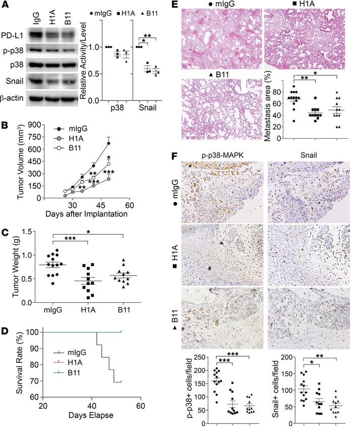
PD-L1 depletion attenuates the lung metastasis of TNBC cells in an immunodeficient host. Our results from in
vitro studies suggested that the tumor-intrinsic function of PD-L1 could contribute to the aggressiveness
of TNBC tumors. To test this possibility, we examined the effect of PD-L1 depletion on tumor growth
and metastasis in vivo. To eliminate the effect of immune response, we employed immunodeficient NOD/
SCID mice as host for the orthotopic transplantation of MDA-MB-231 cells. After inoculating parental
or PD-L1–null MDA-MB-231 cells into the mammary fat pad of NOD/SCID mice, we monitored the
primary tumor growth by measuring tumor size weekly and scaling tumor weight at the experimental end-
point. Both PD-L1–null clones showed similar tumor growth kinetics (Figure 2A) and final tumor weight
(Figure 2B) as parental MDA-MB-231 tumors, suggesting that PD-L1 deficiency did not impact the in situ
growth of primary MDA-MB-231 tumors. However, the number of lung surface metastatic nodules in mice
bearing PD-L1–null tumors was dramatically reduced compared with that in mice bearing parental MDA-
MB-231 tumors (Figure 2C). Histological study on lung tissue sections revealed many fewer micrometa-
static lesions in animals receiving PD-L1–null cells than those receiving parental cells (Figure 2D). Because
the primary tumor size was comparable between the control and PD-L1–null groups, these results suggest
a true suppression on metastasis that resulted from PD-L1 deficiency. This is likely caused by the loss of
tumor-intrinsic functions of PD-L1 and is independent of immune checkpoint blockade, as the tumor-host-
ing animals lack T cells and the systemic immune response.
Metastasis is a multistep process including local spreading/invasion, intravasation, survival in the cir-
culation, extravasation, and colonization and proliferation in the distal organ (23). To check how PD-L1
may participate in the development of metastases, we inoculated tumor cells into the tail vein of NOD/
SCID mice to skip in situ early steps of metastasis, such as local invasion and intravasation. As shown in
Figure 2E, visible metastatic nodules on the lung surface of control mice were evidently greater in number
and size than those bearing PD-L1–null tumors. Histological investigation of lung sections confirmed that
micrometastatic lesions in lung were significantly fewer when PD-L1 expression was depleted (Figure 2F).
Considering our earlier finding that PD-L1 deficiency strongly inhibited the anchorage-independent growth
of MDA-MB-231 cells (Figure 1D), current results suggest that PD-L1 depletion may impair the survival
of circulating tumor cells in bloodstream, in addition to blocking cell migration (Figure 1E) that mainly
impairs the local invasion of PD-L1–null tumors. Taken together, these results strongly suggest that PD-L1
mediates a tumor-intrinsic, tumor-promoting function and is important for TNBC metastasis in vivo.
PD-L1 expression protects Snail proteins from being ubiquitinated and destructed. This effect of PD-L1 is
very likely conducted via regulating the EMT, in the context that the Snail family TFs and the EMT play
important roles in stimulating cancer metastasis and progression not only by improving migration and
invasiveness but also by conferring tumor cells with stem cell–like traits that enhance the ability of tumor
cells to survive in foreign microenvironments, such as in circulation and distant organs (20, 24). To gain
insights into the underlying mechanism by which PD-L1 regulates the EMT, we took a closer look at
E-cadherin and Snail, 2 key EMT-related proteins that showed significant changes when PD-L1 expres-
sion was modified (Figure 1, A and B). Levels of EMT-TFs determine the status of the cell on the EMT
spectrum from complete epithelial to complete mesenchymal by regulating the expression of epithelial
proteins (such as E-cadherin, ref. 20) and mesenchymal proteins (such as fibronectin, ref. 24). Results
from quantitative reverse transcription PCR (RT-PCR) analyses revealed an escalation of E-cadherin
mRNAs not only in PD-L1–null cells but also in siRNA-treated PD-L1–knockdown cells (Figure 3A),
supporting a causal relationship between E-cadherin and PD-L1 in MDA-MB-231 cells. Importantly,
restored expression of PD-L1 diminished the increased transcription of E-cadherin in PD-L1–knock-
down cells (Figure 3B). These results align well with the increase of E-cadherin proteins and decrease
of Snail proteins when PD-L1 was depleted (Figure 1, A and B). Thus, we reason that PD-L1 inhibits
E-cadherin transcription by upregulating Snail expression.
JCI Insight 2021;6(9):e131458 https://doi.org/10.1172/jci.insight.131458 4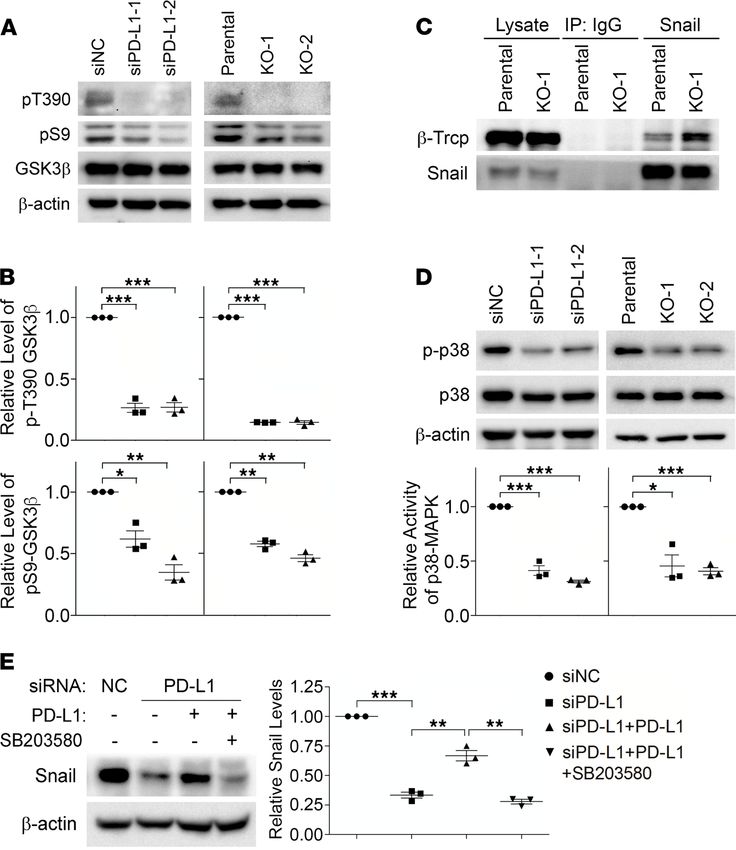
RESEARCH ARTICLE

                                  Figure 2. PD-L1 deficiency reduces the tumor metastasis independent of the antitumor immunity. (A–D) Two mil-
                                  lion parental or PD-L1–null (KO-1 and KO-2) cells were injected into the mammary fat pad of NOD/SCID mouse (6 mice/
                                  group). (A) Tumor volume was measured with calipers weekly and calculated using the standard formula. (B) Tumors were
                                  dissected and weighed at the endpoint (65 days after inoculation). (C and D) Loss of PD-L1 inhibits lung metastasis. (C)
                                  Lungs were dissected at the endpoint. Left panel, images of gross lung showed that parental MDA-MB-231 tumors gen-
                                  erated many more metastatic nodules on lung surface. (D) Representative H&E staining images of lung tissues showing
                                  micrometastatic lesions from mice bearing parental tumors are much more severe than those bearing PD-L1–null tumors.
                                  (E and F) Results from experimental metastasis model suggest that PD-L1 is necessary for the later steps of metastasis
                                  formation. Parental or PD-L1–null MDA-MB-231 cells (8 × 10⁵) were directly injected into the tail vein of NOD/SCID mice (5
                                  mice/group). Animals were terminated 40 days later to examine lung metastasis. (E) Metastatic nodules on lung surface
                                  in each group were analyzed. (F) Representative H&E staining images of lung tissues. PD-L1–null MDA-MB-231 tumors
                                  generated many fewer lung micrometastatic lesions than parental tumors. (D and F) Power of eyepiece: 10×; power of
                                  objective: 10×. (A–C and E) Data were plotted as mean ± SEM and statistically analyzed using 1-way ANOVA analysis with
                                  Dunnett’s test. n = 6 (A–C) and n = 5 (E). N.S., no significant difference; **, P < 0.01; ***, P < 0.001.

                                     However, mRNA levels of Snail were not changed in PD-L1–knockdown cells (Figure 3A),
                                 although Snail transcription appeared impaired in the 2 PD-L1–null clones (Supplemental Figure
                                 4A). This indicates that PD-L1 more likely plays a consistent role in regulating Snail at a posttran-
                                 scriptional level than at the transcriptional level. As a critical TF promoting the EMT program

JCI Insight 2021;6(9):e131458 https://doi.org/10.1172/jci.insight.131458                                                                                   5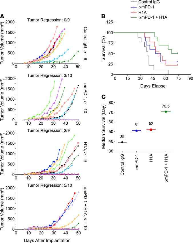
RESEARCH ARTICLE

                                 and regulating cell survival/differentiation (20, 25, 26), Snail is under tight control in cells. The
                                 ubiquitination-dependent, proteasome-mediated destruction limits the protein level of Snail and
                                 makes Snail a short-lived protein (27). Interestingly, treatment with proteasome inhibitor MG132
                                 strongly elevated the protein level of Snail in PD-L1–knockdown cells to a comparable level to that
                                 in parental cells (Figure 3C). Similar results were obtained in PD-L1–null cells (Supplemental Fig-
                                 ure 4B), confirming that PD-L1 has more consistent influence on Snail stability than transcription.
                                 Because proteasome-mediated protein destruction depends on ubiquitination, we next determined
                                 Snail ubiquitination in parental and PD-L1–deficient cells. To eliminate the possibility that some
                                 of the ubiquitin signal accumulated via immunoprecipitation was not from ubiquitinated Snail but
                                 from other ubiquitinated, Snail-binding proteins, we employed the denatured immunoprecipitation
                                 in which the immunoprecipitation of Snail was performed using denatured cell lysates to limit the
                                 noncovalent binding of other proteins to Snail. Using this approach, we showed that exogenously
                                 expressed HA-tagged Snail was much more heavily ubiquitinated in PD-L1–null (Figure 3D) or
                                 PD-L1–knockdown (Figure 3E) cells. Moreover, endogenous Snail also exhibited higher level of
                                 ubiquitination in PD-L1–depleted cells than in parental cells (Supplemental Figure 4C). Our data
                                 suggest an intriguing possibility that the expression of PD-L1 in TNBC cells mediates a tumor-in-
                                 trinsic signaling that inhibits Snail ubiquitination, thus promoting the EMT program.
                                      PD-L1 inhibits GSK3β activity by activating p38-MAPK. Snail ubiquitination is catalyzed by E3 ubiquitin
                                 ligase complexes, mainly the Skp1-Cullin-F-box (SCF) protein complexes (27). Phosphorylation of Snail
                                 enhances its binding to the substrate-recruiting F-box protein of these SCF E3 ligases and facilitates the
                                 subsequent ubiquitination (27). GSK3β-mediated phosphorylation of Snail enhances the binding of Snail
                                 with F-box protein β-transducin repeat-containing protein (β-Trcp) and Snail ubiquitination by SCFβ-Trcp
                                 (28). Indeed, levels of ubiquitinated Snail (Figure 3F), as well as the global ubiquitination (Supplemental
                                 Figure 4D), were comparable in parental and PD-L1–depleted MDA-MB-231 cells after treatment with
                                 GSK3β-specific inhibitor, suggesting that GSK3β is required for the increase of Snail ubiquitination caused
                                 by PD-L1 depletion. Additionally, we found that in PD-L1–null and PD-L1–knockdown cells, GSK3β
                                 phosphorylation at threonine 390 (T390) was almost completely suppressed compared with that in con-
                                 trol cells (Figure 4, A and B). Moreover, the phosphorylation of serine 9 (S9) in GSK3β was also slight-
                                 ly weakened when PD-L1 expression was suppressed (Figure 4, A and B). Because phosphorylations at
                                 T390 and S9 are both inhibitory to GSK3β activity (29), our results suggest that loss of PD-L1 enhances
                                 GSK3β activity, which subsequently promotes the phosphorylation and ubiquitination of Snail. Although
                                 GSK3β-mediated phosphorylation on Snail cannot be directly determined due to the lack of specific anti-
                                 bodies, we indeed found that the association of β-Trcp with Snail was significantly increased when PD-L1
                                 was depleted (Figure 4C). Protein kinase D (PKD) (30) was also reported to facilitate Snail ubiquitina-
                                 tion by phosphorylating Snail at S11 and enhancing its interaction with another F-box protein, FBXO11.
                                 However, we did not see obvious changes of PKD activity in PD-L1–depleted cells by determining the
                                 phosphorylation at T95 of PKDs (Supplemental Figure 5A); and correspondingly, no change of Snail
                                 phosphorylation at S11 was observed in parental and PD-L1–deficient cells using specific antibody (30)
                                 (Supplemental Figure 5B). These data suggest that PD-L1 in TNBC cells suppresses the interaction of Snail
                                 with the E3 ubiquitin ligase SCFβ-Trcp by inhibiting GSK3β activity.
                                      GSK3β phosphorylation at T390 is mediated by p38-MAPK (31), whereas S9 can be phosphorylat-
                                 ed by the PI3K/AKT pathway (32) as well as by other kinases, including the ERK-induced activation
                                 of p90 ribosomal protein S6 kinase (33) and p70 ribosomal protein S6 kinase (p70S6K) (34–36). To
                                 understand how GSK3β is regulated by PD-L1, we compared the activity of these kinases in normal
                                 or PD-L1–depleted MDA-MB-231 cells. Compared with parental cells, ERK1/2 activation in PD-L1–
                                 knockdown or PD-L1–null cells was unchanged, and AKT activity in PD-L1–deficient cells was slight-
                                 ly increased (Supplemental Figure 5C). The change of p70S6K activity was inconsistent in PD-L1–
                                 knockdown cells and PD-L1–knockout cells (Supplemental Figure 5D), indicating that p70S6K might
                                 not be a constant contributor to PD-L1–dependent inhibitory phosphorylation of GSK3β. On the
                                 other hand, we observed a significant, consistent decrease of p38-MAPK activity in both PD-L1–
                                 knockdown and PD-L1–null cells compared with parental cells (Figure 4D and Supplemental Figure
                                 3E), suggesting that PD-L1 expression promotes GSK3β phosphorylation at T390 by regulating the
                                 activity of p38-MAPK. In Figure 1B, we showed that reexpression of PD-L1 recovered the suppressed
                                 Snail expression in PD-L1–depleted cells. Notably, this PD-L1–induced upregulation of Snail could

JCI Insight 2021;6(9):e131458 https://doi.org/10.1172/jci.insight.131458                                                                   6
RESEARCH ARTICLE
Figure 3. PD-L1 promotes the EMT by protecting Snail from being ubiquitinated and destructed. (A) Depletion of
PD-L1 increased E-cadherin transcription but had no effect on Snail transcription. MDA-MB-231 cells were treated with
control or each of 2 distinct PD-L1 siRNAs for 48 hours. mRNA levels of E-cadherin and Snail in these cells were exam-
ined by real-time quantitative PCR. (B) Reexpression of PD-L1 suppressed E-cadherin transcription. MDA-MB-231 cells
were transfected with mock or PD-L1–expressing construct for 24 hours, then transfected with control (siNC) or PD-L1
siRNA (siPD-L1–2) for another 48 hours. The mRNA levels of E-cadherin in each group were examined by RT-qPCR. (A
and B) Data (n = 3 independent experiments) were normalized against the siNC group, plotted as mean ± SEM, and
statistically analyzed using unpaired 2-tailed Student’s t test with the P value adjusted by Bonferroni’s method. *,
P < 0.05; **, P < 0.01; ***, P < 0.001. (C) Inhibition of proteasome recovered the loss of Snail in PD-L1–depleted cells.
Control (siNC) or PD-L1–depleted (siPD-L1–2) MDA-MB-231 cells were treated with 10 μM MG132 for 6 hours. Cells were
then lysed and subjected to immunoblotting with indicated antibodies. (D–F) Snail ubiquitination was enhanced
in PD-L1–depleted cells in a GSK3β-dependent manner. (D) Parental and PD-L1–null (KO-1) MDA-MB-231 cells were
transfected with HA-tagged Snail and Myc-tagged ubiquitin for 48 hours. (E) MDA-MB-231 cells were transfected
with HA-Snail and Myc-ubiquitin for 24 hours, then transfected with control (siNC) or PD-L1 (siPD-L1–2) siRNA for
48 hours. (F) Cells described in E were treated with DMSO or 10 μM SB216763 for 6 hours. (D–F) After being treated
with MG132 (10 μM, 6 hours), cells were lysed with denatured IP buffer, then subjected to immunoprecipitation (IP)
with indicated antibodies (HA antibody for F). The precipitates were analyzed by immunoblotting using indicated
antibodies. Intensity of ubiquitinated Snail was quantified by ImageJ (NIH) and normalized against control. (D–F)
Data (n = 3 independent experiments) were normalized against the parental (D), siNC (E), or siNC/DMSO (F) group;
plotted as mean ± SEM; and statistically analyzed using unpaired 2-tailed Student’s t test. *, P < 0.05; **, P < 0.01.
N.S., no significant difference.
JCI Insight 2021;6(9):e131458 https://doi.org/10.1172/jci.insight.131458 7RESEARCH ARTICLE
Figure 4. PD-L1 prevents Snail ubiquitination via p38-MAPK–mediated inhibition of GSK3β. (A and B) The inhibito-
ry phosphorylations of GSK3β (pT390 and pS9) were decreased in PD-L1–deficient cells. (A) Control, PD-L1–knockdown,
and PD-L1–knockout MDA-MB-231 cells were analyzed by immunoblotting using indicated antibodies. (B) The intensity
of pT390-GSK3β and pS9-GSK3β were measured and normalized against total GSK3β in each group. Results was then
normalized against the control group. (C) Snail exhibited stronger association with β-Trcp in PD-L1–deficient MDA-
MB-231 cells. Endogenous Snail was immunoprecipitated from parental or KO-1 MDA-MB-231 cells. β-Trcp associated
with Snail was determined by immunoblotting. (D) PD-L1–deficient cells exhibited significantly less p38-MAPK activity.
The activating phosphorylation of p38-MAPK (p-p38) was determined by immunoblotting. The relative activity of
p38-MAPK was represented by the ratio of p-p38 to total p38. (E) Selective inhibition of p38-MAPK suppressed the
PD-L1–induced expression of Snail. MDA-MB-231 cells stably expressing PD-L1 were established by lentivirus-mediated
infection. These cells were pretreated with SB203580 (10 μM) for 2 hours before transfection with siNC or siPD-L1 for
48 hours with SB203580, then subjected to immunoblotting analysis using indicated antibodies. After normalizing
against β-actin levels, the expression level of Snail in each group was quantified. (B and D) Results (n = 3 3 indepen-
dent experiments) were plotted as mean ± SEM and statistically analyzed using unpaired 2-tailed Student’s t test with
the P value adjusted by Bonferroni’s method. *, P < 0.05; **, P < 0.01; ***, P < 0.001. (E) Results (n = 3 independent
experiments) were plotted as mean ± SEM and statistically analyzed using 1-way ANOVA with the P value adjusted
by Tukey’s honestly significant differences (HSD) using R function “aov” and “TukeyHSD” from package “stats” in R
version 3.6.3. **, P < 0.01; ***, P < 0.001.
be completely blocked by p38-MAPK inhibitor (Figure 4E). Together, our data strongly suggest that
PD-L1 when upregulated in TNBC cells can inhibit GSK3β by activating p38-MAPK. This prevents
Snail from being phosphorylated by GSK3β and caught by SCFβ-Trcp for ubiquitination and destruction.
We reason that this PD-L1–dependent protection of Snail leads to Snail accumulation and then pro-
motes the EMT and aggressiveness of PD-L1–expressing TNBC cells. Consistent with this conclusion,
we observed that levels of Snail and activated p38 are positively associated with PD-L1 levels in TNBC
patient tissues (Supplemental Figure 5E).
JCI Insight 2021;6(9):e131458 https://doi.org/10.1172/jci.insight.131458 8RESEARCH ARTICLE
PD-L1 directly interacts with and inhibits PTP1B. PD-L1 is a single transmembrane protein with a short
cytoplasmic tail. To investigate how PD-L1 activates p38-MAPK, we employed a proximity-dependent bio-
tin identification (BioID) approach to label the proteins interacting with PD-L1. For this purpose, the car-
boxyl end of PD-L1 was fused to a mutated bacterial biotin ligase BirA* with a labeling radius of approx-
imately 10 nm (37), and this fusion protein was stably expressed in PD-L1–null MDA-MB-231 cells. After
being labeled with biotin, the biotinylated proteins were pulled down from cells by streptavidin-conjugated
beads and then analyzed by mass spectrometry. Compared with cells expressing BirA* alone, we identified
proteins that potentially associate with PD-L1, and one of them was the protein tyrosine phosphatase
PTP1B (also called PTPN1), which is highly correlated with tumorigenesis and progression of various can-
cers (38). By coexpressing exogenous PTP1B and PD-L1 in human embryonic kidney 293T (HEK293T)
cells, we confirmed that PTP1B and PD-L1 associated with each other (Figure 5A). Further, overexpressed
PD-L1 and endogenous PTP1B formed a protein complex, within which endogenous p38-MAPK was also
detected (Figure 5B). More importantly, endogenous PD-L1 successfully pulled down endogenous PTP1B
as well as p38-MAPK in MDA-MB-231 cells (Figure 5C), supporting the presence of p38-MAPK in the
PD-L1–PTP1B complex. To determine whether PD-L1 directly binds to PTP1B, we constructed GST-
tagged PTP1B (GST-PTP1B) and MBP-tagged PD-L1 cytoplasmic domain (MBP-PDL1-CT). These pro-
teins were expressed and purified from E. coli (Figure 5D), then used for in vitro protein pull-down assay.
Results shown in Figure 5D confirmed the direct interaction of PTP1B with the cytoplasmic domain of
PD-L1. Furthermore, truncated PTP1B proteins lacking the C-terminal ER-targeting domain and the intact
proline-rich domain cannot interact with MBP-PDL1-CT (Supplemental Figure 6, A and B), suggesting
that one or both of these domains mediates the interaction with PD-L1.
PTP1B could inhibit p38-MAPK by dephosphorylating p38 at tyrosine 182 (T182) (39). T182 is one
of the 2 sites that when phosphorylated activate p38-MAPK (40, 41), and this phosphorylation can be rec-
ognized by the phosphorylated p38 antibody used in our experiments. The direct interaction of PD-L1
cytoplasmic domain with PTP1B raises an intriguing possibility that PTP1B might play a role in the PD-L1–
dependent regulation of p38-MAPK. Indeed, results from the in vitro PTP1B phosphatase assay showed
that MBP-PDL1-CT, but not MBP alone, inhibited PTP1B activity in a dose-dependent manner (Figure 5E).
This suggests that PD-L1 by binding to PTP1B can inhibit PTP1B-mediated dephosphorylation and inacti-
vation of p38-MAPK. Although the PD-L1–associated PTP1B was a small portion of total PTP1B in resting
MDA-MB-231 cells (Supplemental Figure 6C), we indeed observed a moderate, yet significant, increase of
PTP1B activity when PD-L1 was depleted (Figure 5F). Moreover, PTP1B inhibitor recovered the decreased
p38-MAPK phosphorylation in PD-L1–depleted cells to a level comparable to that in parental cells (Figure
5G), further confirming the participation of PTP1B in the PD-L1–dependent activation of p38-MAPK.
The tumor-intrinsic signaling of PD-L1 can be activated by extracellular stimuli. Our current findings raise an
intriguing possibility that the single transmembrane PD-L1 can function as a receptor to mediate tumor-in-
trinsic signaling. The PD-L1–mediated signaling could be activated by conformational changes in PD-L1,
which may be triggered by ligands binding to its extracellular domain. Another possibility is that PD-L1
may function as a coreceptor. In MDA-MB-231 cells, we did not detect the expression of CD80 or PD-1
(Supplemental Figure 1D), 2 natural PD-L1 binding partners that may induce PD-L1 conformational
changes. To determine how PD-L1 influences intracellular signaling pathways in cultured cells, we exam-
ined the p38 activation induced by FBS in parental and PD-L1–deficient MDA-MB-231 cells. As shown
in Figure 6A, the FBS-induced p38-MAPK activity in PD-L1–deficient cells was significantly weaker than
that in control cells. These results suggest that PD-L1 not only boosts p38-MAPK activity to a higher level
in resting cells but also synergizes with other extracellular stimuli to further enhance p38-MAPK activation.
The hyperactivated p38-MAPK in PD-L1–overexpressing cells, which could result from PD-L1–dependent
PTP1B inhibition, subsequently constrains GSK3β activity and protects Snail from being destructed.
To test if the PD-L1 tumor-intrinsic pathway can be activated by its binding partner, we treated MDA-
MB-231 cells with purified recombinant PD-1 extracellular domain, then determined p38-MAPK activity.
Our results showed that PD-1 ectodomain activated p38-MAPK in parental MDA-MB-231 cells; howev-
er, PD-L1 depletion inhibited p38 activation induced by PD-1 (Figure 6B). Additionally, PD-1 treatment
also caused an increase of Snail in parental MDA-MB-231 cells (Figure 6C). Together, these results sug-
gest that PD-1 binding to PD-L1 can activate the PD-L1 intrinsic tumor-promoting pathway in TNBC
cells. Thus, PD-L1 and PD-1 may be ligand and receptor to each other mutually. To determine if this is
the case in vivo, we analyzed the primary tumors from our orthotopic xenograft studies described above.
JCI Insight 2021;6(9):e131458 https://doi.org/10.1172/jci.insight.131458 9RESEARCH ARTICLE
Figure 5. PD-L1 directly interacts with PTP1B and inhibits its phosphatase activity. (A) PD-L1 and HA-tagged PTP1B
associate with each other. HEK293T cells were transfected with PD-L1 and HA-tagged PTP1B for 48 hours and then
subjected to immunoprecipitation followed by immunoblotting. (B) Ectopically expressed PD-L1 could pull down
endogenous PTP1B and p38-MAPK. PD-L1 overexpressed in HEK293T was immunoprecipitated by PD-L1 antibody
and the associated PTP1B were visualized by immunoblotting. (C) Endogenous PD-L1, PTP1B, and p38-MAPK form a
protein complex in MDA-MB-231 cells. (D) The cytoplasmic domain of PD-L1 directly interacts with PTP1B. GST-tagged
PTP1B (GST-PTP1B) and MBP-tagged PD-L1 cytoplasmic domain (MBP-PDL1-CT) were purified from E. coli. Purified
proteins were analyzed by SDS-PAGE followed by Coomassie blue staining (lower panel). GST pull-down assays were
performed using 0.5 g of each indicated protein. (E and F) PD-L1 inhibited the phosphatase activity of PTP1B. In vitro
phosphatase assay was performed using 120 ng purified GST-PTP1B with indicated amount of MBP or MBP-PDL1-CT (E)
or using endogenous PTP1B immunoprecipitated by anti-PTP1B antibody from indicated cell lysates (F). PTP1B activity
in each sample was normalized against PTP1B with equal amount of MBP (E) or cells treated with control siRNA (siNC,
F, immunoprecipitants pulled down from control cells by normal mouse IgG were used as negative controls of the
phosphatase activity assay). (G) Inhibition of PTP1B recovered p38-MAPK activity in PD-L1–deficient cells. MDA-MB-231
cells were transfected with control (siNC) or PD-L1 (siPD-L1–1) siRNAs along with PTP1B inhibitor (20 or 40 μM) for 48
hours, then lysed for immunoblotting using indicated antibodies. The relative intensity of phosphorylated p38-MAPK
(p-p38) in each sample was determined by GraphPad and normalized against the total p38-MAPK. (E–G) Results (n = 3
independent experiments) were plotted as mean ± SEM and comparisons between indicated groups were statistically
analyzed using unpaired 2-tailed Student’s t test. *, P < 0.05; **, P < 0.01; ***, P < 0.001.
Immunohistochemistry analysis showed strong p38-MAPK phosphorylation in control tumors, especially
at the peripheral area that represents the interface of tumor mass and the host tissue; however, PD-L1–null
tumors showed notably less phosphorylated p38-MAPK, with the KO-2 group showing significant differ-
ence from control tumors (Figure 6D). When we examined Snail expression in the same tumor areas, the
JCI Insight 2021;6(9):e131458 https://doi.org/10.1172/jci.insight.131458 10RESEARCH ARTICLE
numbers of Snail-positive cells in PD-L1–null tumors were significantly decreased compared with those in
control tumors (Figure 6D). These results suggest that the p38-MAPK activity and Snail expression level
in parental MDA-MB-231 tumors are PD-L1 dependent. Since the activated p38-MAPK signal was only
observed at the peripheral region of tumors, we reason that the tumor-associated microenvironment may
activate PD-L1 on tumor cells. Although the immunodeficient NOD/SCID mice do not have B and T cells,
macrophages accumulated in tissues surrounding tumors where PD-1 expression was positive (Supplemen-
tal Figure 7A). Moreover, we showed that mouse PD-1 can bind to human PD-L1 (Supplemental Figure
7B), and this binding can be blocked by our anti–mouse PD-1 antibody (αmPD-1, Supplemental Figure
7C), as suggested previously (42). Interestingly, αmPD-1 treatment suppressed the progression of parental
MDA-MB-231 tumors in NOD/SCID mice but had no effect on PD-L1–deficient MDA-MB-231 tumors
(Supplemental Figure 7D). In the context that MDA-MB-231 cells do not express PD-1 (Supplemental Fig-
ure 1D), these data suggest that the binding of PD-1 in microenvironment to PD-L1 in tumor cells favors
tumor progression. Thus, we reason that host cells in the tumor-associated microenvironment, such as
macrophages, produce PD-1 to activate the PD-L1 pathway in tumor cells. Moreover, p38-MAPK activity
in tumor cells, which can be stimulated by growth factors and cytokines produced by tumor-associated host
cells, would be sustained at higher level for longer time in PD-L1–expressing tumor cells. Together, these
effects could protect Snail and promote the EMT and tumor aggressiveness.
PD-L1 antibodies inhibit TNBC progression independent of antitumor immunity. That PD-1 can activate PD-L1
in tumor cells raises a possibility that the binding of antibodies to its extracellular domain may also affect the
PD-L1–mediated tumor-intrinsic signaling. Subsequently, the tumor-promoting function of PD-L1 could
be interrupted, which may suppress tumor progression. To test this, we employed 2 homemade monoclonal
antibodies of PD-L1, named H1A and B11. According to epitope analysis, H1A and B11 recognize distinct
regions in the extracellular domain of human PD-L1. Our most recent work (43) showed that H1A binding
promotes the degradation of PD-L1 by eliminating the interaction of PD-L1 with its protective binding part-
ner, CMTM6 (44, 45). As shown in Figure 7A, treatment of H1A and B11 both led to a significant reduc-
tion of PD-L1 protein levels, consistent with our previous report (43). The p38-MAPK activity was slightly
decreased in these cells, which is likely because the level of remaining PD-L1 in these cells was much higher
than in PD-L1–deficient cells. Nevertheless, a robust decrease of Snail was observed in H1A- and B11-treat-
ed cells (Figure 7A). These results suggest that H1A and B11 attenuated the tumor-intrinsic function of
PD-L1, likely by promoting PD-L1 degradation, which may then inhibit tumor progression independent of
immune response. To investigate this possibility, we treated the immunodeficient NOD/SCID mice carrying
orthotopically transplanted MDA-MB-231 tumors with H1A and B11.
To maximize the blockade potential of PD-L1 antibodies, we pretreated parental MDA-MB-231 cells
with 1 mg control IgG, H1A, or B11 for 30 minutes. These cells were subcutaneously inoculated into the
mammary fat pad of NOD/SCID mice. Three days later, animals in each group were treated with control
IgG, H1A, or B11, respectively, via intraperitoneal injection every 3 days until the planned endpoint
(day 52 after inoculation). As shown in Figure 7B, tumors treated with both H1A and B11 grew notably
slower than tumors treated with control IgG, with H1A achieving stronger inhibition of tumor growth. By
day 49, 30% of animals treated with control IgG had met the terminating body conditions; however, mice
treated with H1A or B11 all appeared normal. We terminated the study at day 52 and analyzed the prima-
ry tumors and lung metastasis in all animals. The weight of primary tumors in mice treated with control
IgG was significantly higher than tumors treated with PD-L1 antibodies (Figure 7C). Correspondingly, all
mice treated by H1A or B11 survived until the endpoint, whereas 4 out of 13 mice treated by control IgG
met the terminating body condition (Figure 7D). Because no metastatic nodules on the lung surface were
observed in all animals, including mice in the control IgG group in this particular study, we determined
the micrometastatic lesions using histological analyses. As shown in Figure 7E, animals treated with both
PD-L1 antibodies developed significantly fewer metastatic lesions in lung than those treated with control
IgG, which is consistent with the much better survival curves of these treatment groups.
Because NOD/SCID mice lack B and T cells, the tumor-suppressing effect of H1A and B11 is independent
of the antitumor immune response. Compared with tumors treated by control IgG, tumors treated by H1A or
B11 showed markedly less p38-MAPK activity (Figure 7F, left) and Snail expression (Figure 7F, right). Yet, the
expression of PD-1 or the number of macrophages in the tumor-associated microenvironment remained compa-
rable in mice treated by empty vehicle or H1A/B11 (data not shown), indicating that the intrinsic tumor-promot-
ing function of PD-L1 was impaired by H1A or B11. These data suggest that PD-L1–expressing tumors likely
JCI Insight 2021;6(9):e131458 https://doi.org/10.1172/jci.insight.131458 11RESEARCH ARTICLE
Figure 6. PD-L1 mediates a tumor-intrinsic signaling that can be activated by PD-1. (A–C) Cells were subjected to
immunoblotting with indicated antibodies. (A) PD-L1 is necessary for the activation of p38-MAPK by extracellular stim-
uli. MDA-MB-231 cells were transfected with control or PD-L1 siRNAs for 48 hours, serum-starved overnight, and then
treated with FBS (10%) for indicated amount of time. (B) PD-1 activates p38-MAPK in a PD-L1–dependent manner. Con-
trol or PD-L1–depleted MDA-MB-231 cells were treated with PBS or PD-1 (0.5 μg/mL) for 15 minutes. (C) PD-1 treatment
increased the protein levels of Snail in MDA-MB-231 cells. Cells were treated with PBS or PD-1 for 10 or 30 minutes. (A
and C) The relative activity of p38 and protein level of Snail were quantified. Data (n = 3 independent experiments) were
plotted as mean ± SEM and statistically analyzed using unpaired 2-tailed Student’s t test with (C) or without (A) the P
value adjusted by Bonferroni’s method. N.S., no significant difference; *, P < 0.05; **, P < 0.01; ***, P < 0.001. (D) PD-L1
is required for p38 activation and Snail expression in vivo. MDA-MB-231 tumors grown in NOD/SCID mice as described in
Figure 2 were collected and processed for immunohistochemistry staining to determine p38 activity and Snail expression
in tandem tissue slides. Power of eyepiece: 10×; power of objective: 20×. Positive cells were counted in >10 fields/slide/
mice, averaged, and plotted as mean ± SEM. Data (n = 6 mice/group) were statistically analyzed using unpaired 2-tailed
Student’s t test with the P value adjusted by Bonferroni’s method. N.S., no significant difference; *, P < 0.05; **, P < 0.01.
require the tumor-intrinsic signaling of PD-L1 to develop aggressive behaviors, such as metastasis. Together, our
results indicate that PD-L1 antibodies that trigger PD-L1 internalization and degradation would fit the need to
abolish both the intrinsic and extrinsic functions of PD-L1 in TNBC cells.
Targeting both tumor-intrinsic and -extrinsic functions of PD-L1 synergistically suppress TNBC. When character-
izing our homemade anti–human PD-L1 antibodies, we found that H1A and B11 could not block the bind-
ing of PD-1 to PD-L1 (Supplemental Figure 8A), although both of them bound to PD-L1 with high affini-
ty similar to atezolizumab, the FDA-approved PD-1–blocking PD-L1 antibody (Supplemental Figure 8B).
JCI Insight 2021;6(9):e131458 https://doi.org/10.1172/jci.insight.131458 12RESEARCH ARTICLE
Figure 7. PD-L1 antibodies diminish PD-L1 tumor-intrinsic signaling and inhibit TNBC progression independent of anti-
tumor immunity. (A) PD-L1 antibodies suppressed PD-L1 expression and signaling. MDA-MB-231 cells were treated with
control IgG, H1A, or B11 for 48 hours before being subjected to immunoblotting. Relative p38 activity was calculated as the
ratio of phosphorylated p38 and total p38, then normalized against the control group (IgG). Data were plotted as mean ±
SEM and statistically analyzed using unpaired 2-tailed Student’s t test with the P value adjusted by Bonferroni’s method (n
= 3 independent experiments). *, P < 0.05; **, P < 0.01. (B–F) NOD/SCID mice receiving 2 × 10⁶ MDA-MB-231 cells in mammary
fat pad were separated into 3 treatment groups (IgG, H1A, or B11). Starting from day 4 after inoculation, 200 μg antibodies
were administrated intraperitoneally every 3 days until termination. (B and C) Tumor was measured weekly (B) and weighed
at the endpoint (C). (D) Survival (Kaplan-Meier) curve was summarized in each treatment group. (E) Micrometastatic lesions
in lung tissues from each treatment group were visualized by H&E staining and quantified. Power of eyepiece: 10×; power
of objective: 10×. (F) Treatment of PD-L1 antibodies inhibited PD-L1 signaling. The number of phosphorylated p38-positive
or Snail-positive cells in tumor tissues was determined by immunohistochemistry staining in tandem tissue slides prepared
from tumors treated with control or PD-L1 antibodies. Power of eyepiece: 10×; power of objective: 20×. (E and F) Values from
more than 10 fields in slide from each mouse were quantified and averaged. (B–F) Data for each group were plotted as mean
± SEM, and comparisons with the mouse IgG group were statistically analyzed using 1-way ANOVA analysis with Dunnett’s
test (n = 11–13 mice/group). *, P < 0.05; **, P < 0.01; ***, P < 0.001.
This makes it possible to test whether targeting the tumor-intrinsic function of PD-L1 can provide additional
benefit for TNBC patients when combined with the PD-1 antibody that inhibits PD-L1 binding. For this
purpose, we employed E0771 cells, which are syngeneic TNBC cells from C57BL/6 mice and express mouse
PD-L1. To utilize the human PD-L1 antibody H1A that does not block PD-1 binding, we first humanized
JCI Insight 2021;6(9):e131458 https://doi.org/10.1172/jci.insight.131458 13RESEARCH ARTICLE
E0771 cells (E0771-hPDL1) by knocking out the endogenous mouse PD-L1 using CRISPR/Cas9 (Sup-
plemental Figure 8C) and then stably expressing human PD-L1 in them (Supplemental Figure 8D). These
E0771-hPDL1 cells were injected into the mammary fat pad of female C57BL/6 mice to create an immuno-
competent, syngeneic TNBC mouse model. On day 11 after inoculation, when the tumor volume reached
approximately 40 mm3, we divided mice into 4 groups and treated them with 200 μg control IgG, αmPD-1
that suppresses tumor growth by blocking the binding between mouse PD-1 and human PD-L1 (Supplemen-
tal Figure 7, B–D), H1A, or 1:1 mixed αmPD-1 and H1A, respectively. Five treatments were administrated
with 3-day interval between each. Then, we monitored the tumor growth and body condition score of ani-
mals and terminated the ones with body condition score reaching 1 for tissue collection (primary tumor and
lung). On day 74, when at least half of animals in each experimental group had been terminated, we ended
the study and then collected primary tumors and lungs from the remaining mice.
As summarized in Figure 8A, the growth of E0771 tumors was considerably slower in αmPD-1–treated
or H1A-treated groups, with 3 out of 10 and 2 out of 9 mice showing tumor regression, respectively. Signifi-
cantly, the combined treatment of αmPD-1 and H1A exhibited further enhanced tumor suppression effect
and achieved tumor regression in 5 out of 10 mice (Figure 8A). When comparing the average increase of
tumor size in each group, the αmPD-1/H1A group also exhibited slower growth than the αmPD-1 or H1A
group (Supplemental Figure 8E). All 3 treatments suppressed lung metastasis, with the αmPD-1/H1A
combined treatment showing slightly stronger effect (Supplemental Figure 8F). Notably, mice treated with
αmPD-1 plus H1A accomplished the best survival curve among all treatment groups (Figure 8B). On day
74 when the study was completed, all mice in the control IgG group had died, whereas the surviving ones
in the αmPD-1 group, H1A group, and αmPD-1/H1A group were all tumor-regressed or tumor-free. The
median survival was 70.5 days for αmPD-1/H1A combined group, which was considerably longer than
that for the control IgG group (39 days), αmPD-1 group (51 days), or H1A group (52 days) (Figure 8C).
Thus, the combined treatment of αmPD-1 and H1A achieved a significantly improved outcome compared
with each single agent. In the context that H1A does not interrupt PD-1 binding to PD-L1 (Supplemental
Figure 8A), these results clearly support an exciting conclusion that targeting the tumor-intrinsic function
of PD-L1 could provide extra benefits when combined with immune checkpoint blockade reagents.
Discussion
A tumor-intrinsic function of PD-L1 in tumor metastasis was defined in this study. We reported here
that the intracellular domain of PD-L1 preserves p38-MAPK activity by inhibiting PTP1B and subse-
quently GSK3β. As GSK3β-mediated phosphorylation prevents Snail from being ubiquitinated and
degraded, PD-L1 facilitates TNBC metastasis via the p38-MAPK/Snail pathway that promotes the
EMT of TNBC cells. Also, we found that PD-L1 antibodies (H1A and B11) that cannot block PD-1
binding but induce PD-L1 degradation can phenocopy PD-L1 deficiency to make TNBC less aggressive
in growth and metastasis in vivo. Importantly, we showed that H1A synergistically suppressed TNBC
progression when combined with PD-1 blockade antibody in immunocompetent mice. Thus, our study
reveals an immune-independent way of PD-L1 to facilitate TNBC progression and supports a new ther-
apeutic strategy for TNBC treatment.
A bidirectional crosstalk between the EMT status and the PD-L1 expression has been observed in multi-
ple types of cancer, including Claudin-low TNBC patients (9, 17, 46). It was shown that the EMT may drive
PD-L1 expression via ZEB1-dependent downregulation of micro RNA-200 (47). Yet, the molecular mecha-
nism underlying the PD-L1–mediated EMT remains vague. In current study, we defined a potentially novel
physical interaction between the cytoplasmic domain of PD-L1 and PTP1B, which inhibits PTP1B. Our
results demonstrated a PTP1B/p38-MAPK/GSK3β/Snail signaling axis that connects PD-L1 to the EMT
status and aggressiveness of TNBC tumors. The role of PTP1B in cancer appears highly context dependent
(48, 49). PTP1B expression was shown necessary for the ErbB2-induced mammary tumorigenesis (50); how-
ever, in p53-null mice, loss of PTP1B accelerates lymphomagenesis (51). In BC patients, loss of functional p53
is most prevalent with TN/basal-like BC than other BC subtypes (52). Most TNBC cell lines, including MDA-
MB-231 and Hs578T used in our study, are p53 mutated (53). In this context, the PD-L1–mediated inhibition
of PTP1B in these TNBC cells is more likely tumor promoting, which may recapitulate the tumor-suppress-
ing role of PTP1B in the immune system that normally expresses high levels of PD-L1. Our data suggest
that PD-L1 protects the activated p38-MAPK from being dephosphorylated by PTP1B in TNBC cells and
preserves p38-MAPK activity stimulated by conventional extracellular signals in tumor environment. It was
JCI Insight 2021;6(9):e131458 https://doi.org/10.1172/jci.insight.131458 14RESEARCH ARTICLE
Figure 8. Targeting the tumor-intrinsic function of PD-L1 synergistically suppresses TNBC progression when combined
with immune checkpoint blockade reagents. Humanized E0771 cells (1 × 10⁶), in which endogenous mouse PD-L1 was
knocked out and human PD-L1 was overexpressed, were injected into the mammary fat pad of female C57BL/6 mice. On
day 11 after inoculation, when the solid tumor could be touched, mice were randomly separated into 4 groups, which were
treated with 200 μg control IgG, hamster anti–mouse PD-1 antibody (αmPD-1), mouse anti–human PD-L1 antibody H1A, or
100 μg αmPD-1 plus 100 μg H1A, respectively. Antibodies were injected intraperitoneally once every 3 days for 5 injections in
total. (A) Combined treatment of αmPD-1 and H1A exhibited strong synergistic effect on suppressing tumor growth. Tumor
volume was measured with calipers weekly until day 50 after inoculation and calculated as V = 0.5 × LW2. Tumor regression
was shown as ratio of number of animals showing tumor regression and total animal number in each treatment group. (B)
Combined treatment of αmPD-1 and H1A synergistically increased the survival of mice carrying E0771 tumor. Animals were
monitored until day 74 after inoculation, when at least half of mice in each experimental group met the terminating body
condition. (C) The median survival days of mice in each treatment group were calculated and plotted, which clearly showed
that combined treatment of αmPD-1 and H1A elongated the survival time of animals carrying E0771 tumor.
shown that p38 activity is necessary for tumor progression by promoting the production of growth factors
and cytokines that are necessary for tumor cell colonization and angiogenesis (54). Thus, the prolonged p38-
MAPK activity in PD-L1–expressing TNBC tumors would facilitate tumor growth and metastasis.
More than increasing cell mobility, the EMT is a dedifferentiation program that enhances cell sur-
vival against environmental stresses and potentiates cancer stem cell generation, which both contrib-
ute to the development of metastasis and treatment resistance. Our study dissected the tumor-intrinsic
PD-L1 pathway comprising PTP1B, p38-MAPK, GSK3β, and Snail that physically links PD-L1 to the
JCI Insight 2021;6(9):e131458 https://doi.org/10.1172/jci.insight.131458 15You can also read