Rare variants in PPFIA3 cause delayed development, intellectual disability, autism, and epilepsy
←
→
Page content transcription
If your browser does not render page correctly, please read the page content below
medRxiv preprint doi: https://doi.org/10.1101/2023.03.27.23287689; this version posted March 29, 2023. The copyright holder for this preprint
   (which was not certified by peer review) is the author/funder, who has granted medRxiv a license to display the preprint in perpetuity.
                                           All rights reserved. No reuse allowed without permission.

            Rare variants in PPFIA3 cause delayed development,
                        intellectual disability, autism, and epilepsy

       Maimuna S. Paul1,2,3, Sydney L. Michener1,2,3, Hongling Pan2,4, Jessica M. Pfliger1,2,5, Jill A.
        Rosenfeld4, Vanesa C. Lerma1,2,6, Alyssa Tran4, Megan A. Longley1,2, Richard A. Lewis4,7,
      Monika Weisz-Hubshman4, Mir Reza Bekheirnia4,8, Nasim Bekheirnia8, Lauren Massingham9,
           Michael Zech10,11, Matias Wagner10,11,12, Hartmut Engels13, Kirsten Cremer13, Elisabeth
     Mangold13, Sophia Peters13, Jessica Trautmann13, Jessica L. Mester14, Maria J. Guillen Sacoto14,
          Richard Person14, Pamela P. McDonnell15,16, Stacey R. Cohen15, Laina Lusk15, Ana S.A.
       Cohen17, Jean-Baptiste Le Pichon18, Tomi Pastinen17,19, Dihong Zhou20, Kendra Engleman20,
        Caroline Racine21,22, Laurence Faivre22,23, Sébastien Moutton22,23, Anne-Sophie Denommé-
    Pichon21,22, Sarah Schuhmann24, Georgia Vasileiou24, Sophie Russ-Hall25, Ingrid E. Scheffer25,26,
      Gemma L. Carvill27, Heather Mefford28, Undiagnosed Diseases Network, Carlos A. Bacino4,29,
                                      Brendan H. Lee4,29, Hsiao-Tuan Chao1,2,3,4,29,30,31

    Abstract
    PPFIA3 encodes the Protein-Tyrosine Phosphatase, Receptor-Type, F Polypeptide-Interacting
    Protein Alpha-3 (PPFIA3), which is a member of the LAR protein-tyrosine phosphatase-
    interacting protein (liprin) family involved in synaptic vesicle transport and presynaptic active
    zone assembly. The protein structure and function are well conserved in both invertebrates and
    vertebrates, but human diseases related to PPFIA3 dysfunction are not yet known. Here, we
    report 14 individuals with rare mono-allelic PPFIA3 variants presenting with features including
    developmental delay, intellectual disability, hypotonia, autism, and epilepsy. To determine the
    pathogenicity of PPFIA3 variants in vivo, we generated transgenic fruit flies expressing either
    human PPFIA3 wildtype (WT) or variant protein using GAL4-UAS targeted gene expression
    systems. Ubiquitous expression with Actin-GAL4 showed that the PPFIA3 variants had variable
    penetrance of pupal lethality, eclosion defects, and anatomical leg defects. Neuronal expression
    with elav-GAL4 showed that the PPFIA3 variants had seizure-like behaviors, motor defects, and
    bouton loss at the 3rd instar larval neuromuscular junction (NMJ). Altogether, in the fly
    overexpression assays, we found that the PPFIA3 variants in the N-terminal coiled coil domain

       NOTE: This preprint reports new research that has not been certified by peer review and should not be used to guide clinical practice.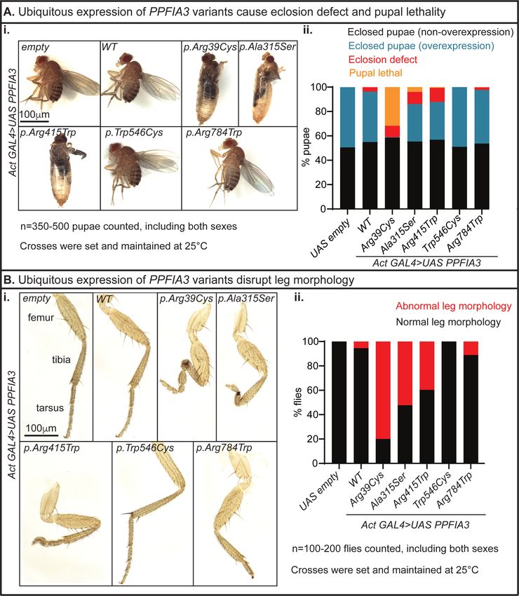
medRxiv preprint doi: https://doi.org/10.1101/2023.03.27.23287689; this version posted March 29, 2023. The copyright holder for this preprint
   (which was not certified by peer review) is the author/funder, who has granted medRxiv a license to display the preprint in perpetuity.
                                           All rights reserved. No reuse allowed without permission.

    exhibited stronger phenotypes compared to those in the C-terminal region. In the loss-of-function
    fly assay, we show that the homozygous loss of fly Liprin-α leads to embryonic lethality. This
    lethality is partially rescued by the expression of human PPFIA3 WT, suggesting human PPFIA3
    protein function is partially conserved in the fly. However, the PPFIA3 variants failed to rescue
    lethality. Altogether, the human and fruit fly data reveal that the rare PPFIA3 variants are
    dominant negative loss-of-function alleles that perturb multiple developmental processes and
    synapse formation.

    Author affiliations:
    1
        Department of Pediatrics, Section of Neurology and Developmental Neuroscience, Baylor
                                                                       2
    College of Medicine, Houston, TX, USA;                              Jan and Dan Duncan Neurological Research
    Institute, Texas Children’s Hospital, Houston, TX, USA; 3Cain Pediatric Neurology Research
    Foundation Laboratories, Jan and Dan Duncan Neurological Research Institute, Houston, TX,
    USA; 4Department of Molecular and Human Genetics, Baylor College of Medicine, Houston,
    TX, USA; 5Graduate Program in Development, Disease Models, and Therapeutics, Baylor
    College of Medicine, Houston, TX, USA; 6Department of Psychology, University of Houston,
    Houston, Texas, USA; 7Department of Ophthalmology, Baylor College of Medicine, Houston,
    TX, USA; 8Renal Genetics Clinic, Baylor College of Medicine, Houston, TX, USA; 9Rhode
                                                                                                                     10
    Island Hospital and Hasbro Children's Hospital Providence, RI, USA;                                                   Institute of
                                                                                                         11
    Neurogenomics, Helmholtz Zentrum München, Munich, Germany;                                                Institute of Human
                                                                                                         12
    Genetics, School of Medicine, Technical University, Munich, Germany;                                      Division of Pediatric
    Neurology, Developmental Neurology and Social Pediatrics, Dr. von Hauner Children's
                  13
    Hospital;          Institute of Human Genetics, School of Medicine, University Hospital Bonn,
                                                                 14                                                        15
    University of Bonn, Bonn, Germany;                                GeneDx Gaithersburg, MD, USA;                             Epilepsy
    NeuroGenetics Initiative (ENGIN), Division of Neurology, Children's Hospital of Philadelphia,
                                      16
    Philadelphia, PA, USA;              Department of Neurology, Perelman School of Medicine, University
                                            17
    of Pennsylvania, PA, USA;                 Children’s Mercy Kansas City, Genomic Medicine Center, The
    University of Missouri-Kansas City (UMKC), School of Medicine, Kansas City, MO, USA;
    18
         Department of Pediatrics, Children's Mercy Kansas City, Kansas City, MO; 19Children's Mercy
                                                           20
    Research Institute, Kansas City, MO;                     Children’s Mercy Hospital, Kansas City, MO, USA;
    21
         University Hospital, Dijon, France INSERM UMR1231 GAD “Génétique des Anomalies du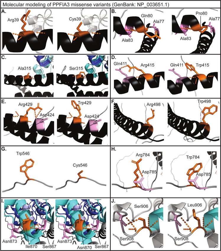
medRxiv preprint doi: https://doi.org/10.1101/2023.03.27.23287689; this version posted March 29, 2023. The copyright holder for this preprint
   (which was not certified by peer review) is the author/funder, who has granted medRxiv a license to display the preprint in perpetuity.
                                           All rights reserved. No reuse allowed without permission.

    Développement,” FHU-TRANSLAD, University of Burgundy, Dijon, France; 22Functional Unit
    for Diagnostic Innovation in Rare Diseases, FHU-TRANSLAD, Dijon Bourgogne; 23Department
    of Genetics and Reference Center for Development Disorders and Intellectual Disabilities, FHU-
    TRANSLAD and GIMI Institute, Dijon Bourgogne University Hospital, Dijon, France;
    24
         Institute of Human Genetics Universitätsklinikum Erlangen, Friedrich Alexander Universität
                                                                          25
    Erlangen Nürnberg, 91054 Erlangen, Germany;                                Epilepsy Research Centre, Department of
                                                                                                                  26
    Medicine, University of Melbourne, Austin Health, Victoria, Australia;                                         Department of
    Pediatrics, University of Melbourne, Royal Children’s Hospital, Florey and Murdoch Children’s
    Research Institutes, Melbourne, Australia; 27Department of Neurology, Northwestern University
                                                                           28
    Feinberg School of Medicine, Chicago, IL, USA;                              Center for Pediatric Neurological Disease
                                                                                                             29
    Research, St. Jude Children's Research Hospital, Memphis, TN, USA;                                          Texas Children’s
                                                 30
    Hospital, Houston, TX, USA;                    Department of Neuroscience, Baylor College of Medicine,
                                  31
    Houston, TX, USA;               McNair Medical Institute, The Robert and Janice McNair Foundation,
    Houston, TX, USA

    Correspondence to Hsiao-Tuan Chao
    Full address: Jan and Dan Duncan Neurological Research Institute, 1250 Moursund St, Suite
    925, Houston, TX, 77030
    E-mail: chao-lab@bcm.edu

    Running title: PPFIA3 variants in neurological disorder

    Keywords: Neurodevelopmental disorder; Synaptic protein; Active zone protein; Disease gene
    discovery; Fruit flies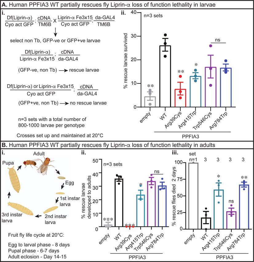
medRxiv preprint doi: https://doi.org/10.1101/2023.03.27.23287689; this version posted March 29, 2023. The copyright holder for this preprint
   (which was not certified by peer review) is the author/funder, who has granted medRxiv a license to display the preprint in perpetuity.
                                           All rights reserved. No reuse allowed without permission.

    Introduction
    Synapses are highly specialized communication junctions between neurons and their target cells
    where neurotransmitter release occurs in an intricately coordinated manner. In the presynaptic
    neuron, a key site for neurotransmitter release is the active zone, which is composed of a
    complex protein matrix.1–4 RIM, ELKS, Munc13, RIM-BP, Piccolo/Bassoon, and Liprin-α are
    the six major protein families comprising the active zone.5 These active zone proteins along with
    other cytoskeletal proteins, Ca2+ channels, and SNAREs (soluble N-ethylmaleimide-sensitive
    fusion protein attachment protein receptors) form a tightly orchestrated unit to mediate synaptic
    vesicle docking, priming, fusion, and neurotransmitter release.5 Disruption of synapse structure
    or function leading to variable defects in neurotransmitter release is widely responsible for
    neurodevelopmental and neuropsychiatric disorders including epilepsy, intellectual disability
    (ID), autism spectrum disorder (ASD), schizophrenia, and bipolar disorder.6–10

    The network of multidomain proteins comprising the active zone falls into different categories
    such as cytoskeletal and scaffolding proteins, adhesion molecules, calcium channels, and
    synaptic vesicle release machinery. Liprins are a critical class of scaffolding proteins found in
    the active zone. In conjunction with the adhesion molecule LAR-PTPs (Leukocyte Antigen
    Receptor-Protein Tyrosine Phosphatases), liprins play a key role in the active zone organization
    and structure. Liprin family members are identified as interactors of LAR-PTPs and are
    subdivided into liprin-α and liprin-β proteins.11,12 Structural studies show that liprins are
    comprised of an N-terminal coiled coil domain and C-terminal sterile-α-motif (SAM)
    domain.11,12        The      N-terminal         coiled      coil     domain        mediates        homodimerization            and
    heterodimerization with other liprin-α members and interactions with other active zone proteins
    such as RIM and ELKS.13–16 In contrast, the C-terminal SAM domain of liprin-α interacts with
    the LAR intracellular domain.17 Additionally, liprins interact with kinesin motor proteins18–20 and
    are involved in the hedgehog signaling-dependent trafficking of Kif7 and Gli to the cilia in the
    context of embryonic development and cortical microtubule organization.18,19

    Liprins are also known as protein-tyrosine phosphatase, receptor-type, f polypeptide (PTPRF)-
    interacting protein α (PPFIA) or β (PPFIB). Vertebrates have four Ppfia (1-4) and two Ppfib (1-
    2) proteins that are encoded by Ppfia1-4 or Ppfib1-2 respectively.12 Expression studies in mice
    show that all four mouse Ppfia1-4 homologs are expressed in the brain, with differences in
medRxiv preprint doi: https://doi.org/10.1101/2023.03.27.23287689; this version posted March 29, 2023. The copyright holder for this preprint
(which was not certified by peer review) is the author/funder, who has granted medRxiv a license to display the preprint in perpetuity.
All rights reserved. No reuse allowed without permission.
distribution and expression levels.21 Ppfia1 is expressed in the brain, lung, heart, liver, muscle,
spleen, and testes.12,22,23 Ppfia2, Ppfia3, and Ppfia4 are predominantly expressed in the
brain,12,22,23 including structures such as the olfactory bulb, striatum, cortex, hippocampus,
thalamus, midbrain, cerebellum, and brainstem.21–23 In contrast, Ppfia1 expression in the brain is
predominantly localized to the cerebellum and olfactory bulb.21–23 A subcellular localization
study showed that Ppfia2 and Ppfia3 are located in both the pre-synaptic and post-synaptic
compartments.23 However, only Ppfia3 specifically colocalizes in the presynaptic compartment
and mediates protein-protein interactions with the active zone proteins Bassoon, RIM, Munc-13,
RIM-BP, and ELKS in hippocampal neurons.24
Ppfia proteins are well conserved in both vertebrates and invertebrates. Studies in C. elegans
identified that the sole Ppfia homolog, syd-2, plays a key role in presynaptic active zone
organization.25,26 Studies showed that syd-2 recruits synaptic components to presynaptic sites and
contributes to the formation of neuromuscular junctions (NMJs), along with active zone
assembly and stabilization.26,27 Mutant syd-2 worms show presynaptic active zone defects due to
disruption of syd-2 oligomerization.13,26,27 A similar role was found for the fruit fly homolog,
Liprin-α, where it is required for synapse formation, synaptic vesicular transport, active zone
assembly, and axonal target selection in the retina.28–30 Consistent with the invertebrate models,
synaptic ultrastructure and electrophysiological studies in Ppfia3 knock-out mice found impaired
presynaptic active zone assembly and synaptic vesicle docking, tethering, and exocytosis.24
Altogether, these studies reveal that Ppfia family members are integral scaffolding proteins for
the assembly of intricate protein complexes involved in synapse formation, synaptic
transmission, and protein trafficking.
Here, we report a cohort of 14 individuals from 12 families with rare mono-allelic variants in
PPFIA3 associated with developmental delay (DD) (11/14), ID (9/14), dysmorphisms (8/14),
hypotonia (6/14), ASD (6/14), and abnormal electroencephalogram (EEG) or epilepsy (5/14). In
these 14 individuals, 11 have PPFIA3 missense variants where ten variants are de novo and one
with unknown inheritance, one individual has a frameshift variant with unknown inheritance, and
two related individuals have a heterozygous splice variant. The phenotypic consequences of rare
variants in PPFIA3 are currently unknown. Molecular modeling of PPFIA3 missense variants
showed that the amino acid changes disrupt the polar contacts and residue interactionsmedRxiv preprint doi: https://doi.org/10.1101/2023.03.27.23287689; this version posted March 29, 2023. The copyright holder for this preprint
(which was not certified by peer review) is the author/funder, who has granted medRxiv a license to display the preprint in perpetuity.
All rights reserved. No reuse allowed without permission.
suggesting these alterations would be deleterious to PPFIA3 protein function. To determine the
pathogenicity of the PPFIA3 variants in vivo, we used fruit fly functional assays where we
examined five of the de novo PPFIA3 missense variants, GenBank NM_003660.4: c.115C>T
[p.(Arg39Cys)], c.943G>T [p.(Ala315Ser)], c.1243C>T [p.(Arg415Trp)], c.1638G>T
[p.(Trp546Cys)], and c.2350C>T [p.(Arg784Trp)]. Overexpression fly assays revealed that the
PPFIA3 variants are associated with behavioral, developmental, and NMJ defects. Loss-of-
function (LOF) fly assays recapitulated an embryonic lethality phenotype induced by loss of fly
Liprin-α, which was partially rescued with human PPFIA3 wildtype (WT) cDNA expression. In
contrast, expression of the PPFIA3 variant cDNAs either failed to rescue or partially rescued the
embryonic lethality compared to PPFIA3 WT cDNA. Altogether, we show that rare PPFIA3
variants are deleterious to protein function with in vivo fruit fly assays and lead to an autosomal
dominant neurodevelopmental disorder characterized by DD/ID, ASD, and epilepsy in humans.
Materials and Methods
Human subjects
Clinical data were acquired after written informed consent to participate in the study and
publication of results was obtained from the participant or their legal representative in
accordance with the ethical standards of the participating institutional review boards (IRB) on
human research at each respective institution. GeneMatcher was used to form an international
collaboration, allowing for comparison of individuals and their variants.31–33 Collection and
analysis of the de-identified clinical cohort was approved by Baylor College of Medicine’s IRB.
PPFIA3 heterozygous variants were identified by exome sequencing (ES) through each
individual’s respective institution. DNA was extracted from peripheral blood mononuclear cells
or buccal sample for ES. Exome or Sanger sequencing of the parental samples were performed
when feasible to confirm de novo or inherited segregation. Paternity was confirmed by the
inheritance of rare single nucleotide polymorphisms from the parents. Sample swap was
excluded. Participant ID’s are not known to anyone outside of the research group.
Molecular modelingmedRxiv preprint doi: https://doi.org/10.1101/2023.03.27.23287689; this version posted March 29, 2023. The copyright holder for this preprint
(which was not certified by peer review) is the author/funder, who has granted medRxiv a license to display the preprint in perpetuity.
All rights reserved. No reuse allowed without permission.
Molecular visualization of the PPFIA3 protein structure was completed with PyMol (The
PyMOL Molecular Graphics System, Version 2.5.2 Schrödinger, LLC.). The crystal structure of
PPFIA3 (GenBank NP_003651.1, Uniprot ID: O75145) was used to build the PPFIA3 protein
structure model in PyMol. Affected residues were altered to the corresponding human variants
and the mutation effects were modeled alongside the native protein. The changes in the PPFIA3
protein structure were assessed by displaying local polar contacts and residue interactions before
and after mutagenesis.
Drosophila melanogaster stocks and maintenance
All the fruit fly stocks used in this study were reared in standard cornmeal and molasses-based
fly food at room temperature (RT, 20-21°C) unless otherwise noted. The fruit fly stocks used in
the study were either obtained from Bloomington Drosophila Stock Center (BDSC) or generated
at the Jan and Dan Duncan Neurological Research Institute. We generated transgenic fly alleles
as previously described34 by utilizing the pUASg-HA-attB vector35 to express the human
PPFIA3 WT and variant cDNAs with a C-terminal hemagglutinin (HA) tag under the control of
upstream activating system (UAS) elements by Gateway LR Cloning (LR Clonase II, Thermo
Fisher Scientific, Cat #11791020). To generate the PPFIA3 variants, we utilized the human full-
length cDNA of PPFIA3 (GenBank: NM_003660.4). PPFIA3 c.115 C>T [p.(Arg39Cys)],
PPFIA3 c.943G>T [p.(Ala315Ser)], PPFIA3 c.1243C>T [p.(Arg415Trp)], PPFIA3 c.1638G>T
[p.(Trp546Cys)], and PPFIA3 c.2350C>T [p.(Arg784Trp)] were generated by Q5 site-directed
mutagenesis (New England Biolabs, Cat #M0491S) in the pDONR221 Gateway compatible
donor vector. The constructs were confirmed by Sanger sequencing. Primer sequences for the
site-directed mutagenesis and Sanger sequencing are listed in Table S1. Human PPFIA3 WT and
variant cDNAs were inserted into the chromosome-3 VK33 (PBac{y[+]-attP}VK00033) docking
site by φC31-mediated recombination for fruit fly transgenesis.35 Transgenic UAS fly alleles
generated in this study include UAS-PPFIA3-WT-HA, UAS-PPFIA3-p.(Arg39Cys)-HA, UAS-
PPFIA3-p.(Ala315Ser)-HA, UAS-PPFIA3-p.(Arg415Trp)-HA, and UAS-PPFIA3-p.(Trp546Cys)-
HA, and UAS-PPFIA3-p.(Arg784Trp)-HA. Fly alleles from the stock centers include: Liprin-
αF3ex15/In(2LR)Gla (BDSC#8563), w[1118]; Df(2L)Exel7027/CyO (BDSC#7801), y[1] w[118];
PBac{y[+]-aatP-3B}-VK00033 (BDSC#9750), and elav-GAL4/CyO (BDSC#8765). UAS-empty-
VK33, Actin-GAL4, and da-GAL4 lines were obtained from Dr. Hugo J. Bellen.medRxiv preprint doi: https://doi.org/10.1101/2023.03.27.23287689; this version posted March 29, 2023. The copyright holder for this preprint
(which was not certified by peer review) is the author/funder, who has granted medRxiv a license to display the preprint in perpetuity.
All rights reserved. No reuse allowed without permission.
Larval brain and NMJ immunostaining and confocal microscopy
Fruit fly larval brains or whole-body wall muscles including the central nervous system were
dissected from wandering third instar larvae reared at 25oC in ice-cold 1X-PBS and fixed in 4%-
paraformaldehyde for 20 minutes at RT. The tissues were washed four times in Tri-PBS (1X-
PBS + 0.2% Triton-X-100) with 1%-Bovine Serum Albumin (BSA) for 15-minutes each
followed by incubation in blocking solution (Tri-PBS with 0.1% BSA and 8% normal donkey
serum) for 30 minutes. Primary antibodies, rat anti-HA (1:50, clone 3F10, Millipore Sigma,
Cat#11867423001), mouse anti-elav (1:100, Developmental Studies Hybridoma Bank,
Cat#9F8A9), mouse anti-Bruchpilot (Brp) (1:50, Developmental Studies Hybridoma Bank,
Cat#nc82), and goat anti-Horseradish Peroxidase (HRP) (1:1000, Jackson ImmunoResearch,
Cat#123-005-021) were diluted in blocking solution, added to the tissues, and incubated
overnight at 4°C. The tissues were rinsed three to four times in Tri-PBS with 1%-BSA for 15
minutes each followed by incubation in blocking solution for 30 minutes at RT. The secondary
antibodies, donkey anti-rat IgG antibody (Cy3) (1:300, Jackson ImmunoResearch, Cat#712-165-
153), Alexa Fluor 488 Affinipure donkey anti-goat IgG (H+L) (1:300, Jackson
ImmunoResearch, Cat#705-545-147) and Alexa Fluor 488 Affinipure donkey anti-mouse IgG
(H+L) (1:300, Jackson ImmunoResearch, Cat#715-545-151) were diluted in blocking solution
and added to the tissues for a 90-minute incubation at RT on a rocker. For NMJ staining,
phalloidin (Phalloidin-iFluor 405 Reagent, Abcam, Cat#ab176752) was added along with the
secondary antibodies to visualize the muscles. After removing the secondary antibody, tissues
were washed three times in Tri-PBS with 1% BSA for 15 minutes each, and then rinsed in 1X-
PBS at RT. For larval brains, this was followed by incubation in 406-diamidino-2-phenylindole
dihydrochloride (DAPI, 1 mg/mL, Cayman Chemical, Cat#14285) for 30 minutes at RT. After
removing DAPI, a final wash was completed with 1X-PBS for 15 minutes at RT. The tissues
were mounted in Prolong Glass anti-fade mountant (Thermo Scientific, Cat#36984). Images
were acquired on a Leica Sp8 laser-scanning confocal microscope. The same settings for laser
power and detector gain were used for all genotypes. Third instar larval brain images were
acquired as a z-stack with a z-step of 1µm and line average of four at 400 Hz with a 20XmedRxiv preprint doi: https://doi.org/10.1101/2023.03.27.23287689; this version posted March 29, 2023. The copyright holder for this preprint
(which was not certified by peer review) is the author/funder, who has granted medRxiv a license to display the preprint in perpetuity.
All rights reserved. No reuse allowed without permission.
objective at 1024 x 1024-pixel resolution. NMJ images were acquired with a 40X objective.
Maximum intensity projections were created from the z-stack in ImageJ. All images were
processed and assembled using ImageJ and Adobe Illustrator.
3rd instar larval NMJ quantifications
The total number of boutons from abdominal segment A3, muscle 6/7 were counted manually
using Imaris. The spot function was used with point style sphere and radius scale 1.0 to count the
number of boutons. The NMJ length was quantified using the HRP staining and measured in
ImageJ. Data was collected and analyzed blinded to genotypes. Statistical analysis between the
control and experimental groups was conducted with one-way ANOVA and Tukey’s post-hoc
analysis in GraphPad Prism 8.
Adult fruit fly behavioral assays
For the climbing assay, 15-day-old flies of both sexes were anesthetized with CO2 24 hours prior
to being tested and two to three flies were housed in food-containing vials at room temperature.
At the time of assay, these flies were transferred without anesthesia to a clear graduated cylinder
with a 20-cm mark. The flies were tapped three times to the bottom of the cylinder to examine
negative geotaxis (climbing upward). The cutoff time to reach the 20-cm mark was 40 seconds.
A total of 40-55 flies of both sexes were tested for each genotype. Crosses for the climbing assay
were set up at 25°C and the assay was performed at 20-21°C.
For the bang sensitivity assay, 15-day-old flies of both sexes were anesthetized with CO2 24
hours prior to being tested and two to three flies were housed in food-containing vials at room
temperature. At the time of assay, these flies were transferred without anesthesia to an empty
food vial and vortexed for 15 seconds. Flies were observed for time to recover from the
vortexing. The cutoff time to recover was 30 seconds. Recovery was defined as being upright
and mobile. Flies were considered bang sensitive if they remained upside down, immobile, or
showing rhythmic involuntary movements suggestive of seizure-like behaviors. A total of 40-55
flies of both sexes were tested for each genotype. Crosses for the bang sensitivity assay were set
up at 25°C and the assay was performed at 20-21°C.medRxiv preprint doi: https://doi.org/10.1101/2023.03.27.23287689; this version posted March 29, 2023. The copyright holder for this preprint
(which was not certified by peer review) is the author/funder, who has granted medRxiv a license to display the preprint in perpetuity.
All rights reserved. No reuse allowed without permission.
Adult fruit fly leg mounting
Adult flies were fixed overnight in ethanol at room temperature and the legs were dissected and
mounted using CMCP-10 Macroinvertebrate High Viscosity Mountant (D/S259) (Electron
Microscopy Sciences, Cat#18004-02). Leg images were taken using the Leica MZ16
stereomicroscope. Images were processed and assembled using Adobe Photoshop CS5.1 and
Adobe illustrator. Crosses were set up at 25°C.
Genomic DNA isolation and PCR
Genomic DNA was extracted by homogenizing three whole flies in 50mM sodium hydroxide
and heating the samples at 95°C for 30 minutes followed by the addition of 1M Tris-HCl (pH
7.5) to stop the lysis. 100 ng of genomic DNA was used to amplify the HA-, PPFIA3-, and
rps17-specific sequences. The experiment was repeated in three independent biological
replicates. HA and PPFIA3 band intensity were quantified by normalizing to the band intensity
of the endogenous reference gene rps17 and plotted as fold-change relative to the control. Flies
were maintained at 20-21°C. Primer sequences are listed in Table S1.
Results
Identification of rare mono-allelic PPFIA3 variants in individuals
with developmental delay, intellectual disability, hypotonia, autism,
and epilepsy
An international collaboration through the Undiagnosed Diseases Network (UDN)36 and
GeneMatcher31–33 led to the identification of 14 individuals with neurodevelopmental phenotypes
and rare missense, frameshift deletion, or consensus splice mono-allelic variants in PPFIA3
(Figure 1A, B). The variants from all affected individuals were identified through exome
sequencing (ES) or Sanger sequencing. Ten of the individuals harbored de novo (I:1, and I:5-13)
missense variants, one individual (I:3) inherited a consensus splice variant from a symptomatic
parent (I:4), and the inheritance pattern for three affected individuals is unknown (I:2, I:4, and
I:14) (Table 1). The individuals with unknown inheritance pattern have either a missense variantmedRxiv preprint doi: https://doi.org/10.1101/2023.03.27.23287689; this version posted March 29, 2023. The copyright holder for this preprint
(which was not certified by peer review) is the author/funder, who has granted medRxiv a license to display the preprint in perpetuity.
All rights reserved. No reuse allowed without permission.
(I:2), a consensus splice variant (I:4), or a frameshift deletion (I:14). The de novo p.(Arg429Trp)
variant was seen in a mosaic state (present in 26% of the reads from ES, suggesting
heterozygosity in ~52% of cells) in I:7. The Combined Annotation Depletion Score (CADD)37
for the variants ranged from 21.1 to 35 (Table S2). Thirteen out of fourteen variants were absent
from the Genome Aggregation Database (gnomAD, v2.1.1).38 The p.(Arg784Trp) variant had a
frequency of 3.19x10⁻⁵ (1/31,386) in gnomAD (v2.1.1) and was identified as a de novo finding in
an individual (I:10) with mild ID and Landau-Kleffner epilepsy syndrome. We also identified a
p.(Arg559Trp) variant in an individual (I:15) with a discordant severe neurodegenerative
phenotype and unknown inheritance of the variant (Table S3). The p.(Arg559Trp) variant was
absent from gnomAD (v2.1.1).
In a statistical model of de novo variants for autism spectrum and intellectual disability disorders
(ASD/ID), the model identified PPFIA3 as one of ~1,000 genes significantly lacking functional
variation in non-ASD/ID individuals but are enriched with de novo variants in ASD/ID
individuals.39 Furthermore, gnomAD v2.1.1. analysis showed that PPFIA3 has a high probability
of loss-of-function (LOF) intolerance (LOEUF = 0.12, pLI = 1.0), as 64.1 LOF variants were
expected given the gene size and GC content but only three LOF variants were observed.38
PPFIA3 is also a highly constrained gene with a missense z-score of 5.49, suggesting intolerance
to missense variation, as 727.5 missense variants were expected but only 311 were observed.38
Together, these findings support that the rare mono-allelic PPFIA3 variants may cause a
neurodevelopmental phenotype.
Twelve individuals in the cohort had DD/ID (I:1-I:4, I:6, I:8-14) (Tables S4, S5), while the
remaining two individuals (I:5 and I:7) could not be assessed for this feature due to early death.
Individual I:5 had renal failure, severe anorectal malformation with complete anal atresia, absent
bladder, and dysmorphisms, and death in infancy. Individual I:7 had a prenatal diagnosis of
abnormal gyration and ventriculomegaly, which led to elective pregnancy termination. Abnormal
EEG (5/14; I:1, I:6, I:10, I:13-14) and epilepsy (3/14; I:1, I:6, I:10) findings were common
(Table 1). The affected individuals had multiple seizure semiologies including focal clonic
seizures, atonic seizures, absence seizures, and focal tonic-clonic seizures with secondary
generalization (Tables 1, S4, S5). Three probands had neuroanatomical changes detected bymedRxiv preprint doi: https://doi.org/10.1101/2023.03.27.23287689; this version posted March 29, 2023. The copyright holder for this preprint
(which was not certified by peer review) is the author/funder, who has granted medRxiv a license to display the preprint in perpetuity.
All rights reserved. No reuse allowed without permission.
MRI (I:6-7, I:12), which included flattening of the posterior globes at the level of optic nerve
insertion, abnormal gyration with ventriculomegaly, and mild periventricular leukomalacia with
mild white matter volume loss (Table S6). Delayed speech development was present in ten
individuals (I:1-3, I:6, I:8, I:10-14) with absent expressive language in one individual (I:6)
(Tables S4, S5). Hypotonia was present in six individuals (I:1-2, I:6, I:12-14) (Table 1). Co-
morbid ASD diagnosis was reported in six individuals (I:2, I:6, I:8-9, I:11, I:13) (Tables S4, S5).
Gastrointestinal dysmotility characterized by constipation, difficulty feeding, and dysphagia was
present in seven individuals (I:1-2, I:5-6, I:10-12) (Tables S4, S5). Dysmorphic facial features
were present in six individuals, and included prominent forehead, strabismus, and bilateral
epicanthal folds (I:1-3, I:6, I:8 I:14) (Tables S4, S5). Macrocephaly or microcephaly were
present in seven individuals (I:3-6, I:11-13) (Tables 1, S4, S5).
Conservation analysis and molecular modeling of PPFIA3 variants
The predicted pathogenicity of PPFIA3 variants was validated in vivo using D. melanogaster
(fruit fly). The fly homolog of PPFIA3 is Liprin-α, and the fly protein shows an overall 48%
identity and 62% similarity with the human protein (Figure 1B). Similar to the human PPFIA3
protein, the fruit fly Liprin-α contains N-terminal coiled coil domains and three C-terminal SAM
domains (Figure 1B). Six of the variants, p.(Arg39Cys), p.(Gln80Pro), p.(Ala315Ser),
p.(Arg415Trp), p.(Arg429Trp), and p.(Arg498Trp) are located in the N-terminal coiled coil
domain (Figure 1B). The variant p.(Ile870Asn) is located in the SAM1 domain (Figure 1B).
The variants p.(Trp546Cys), p.(Arg784Trp), and p.(Ser906Leu) are in the disordered region of
the protein, but p.(Ser906Leu) is located near the SAM1 domain (Figure 1B). Conservation
analysis of the affected residues reveals that the residues in the coiled coil domain, p.Arg39,
p.Gln80, p.Ala315, p.Arg415, and p.Arg429 are well-conserved in invertebrates and vertebrates
(Figure 1C). The affected residue in the SAM1 domain, p.Ile870, and the affected residue near
the SAM1 domain, p.Ser906, are also conserved across species. In the disordered region,
p.Trp546 is conserved in the mice, but not in the fruit flies and worms. In contrast, p.Arg784 and
p.Arg498Trp are conserved in mice and worms, but not in fruit flies. (Figure 1C).
Molecular modeling was completed for the missense variants using PyMol to determine if the
amino acid changes affect the protein function in silico (Figure 2). Regarding the coiled coilmedRxiv preprint doi: https://doi.org/10.1101/2023.03.27.23287689; this version posted March 29, 2023. The copyright holder for this preprint
(which was not certified by peer review) is the author/funder, who has granted medRxiv a license to display the preprint in perpetuity.
All rights reserved. No reuse allowed without permission.
variants, p.(Arg39Cys) removes the positively charged amino acid Arg and introduces an
uncharged amino acid Cys (Figure 2A). Variant p.(Gln80Pro) introduces a ring structure
(Figure 2B), and the variant p.(Ala315Ser) removes the hydrophobic amino acid and introduces
a hydrophilic uncharged amino acid Ser (Figure 2C). The variant p.(Arg415Trp) introduces a
bulky side chain predicted to disrupt the interaction with p.Gln411 (Figure 2D). The variant
p.(Arg429Trp) introduces a bulky side chain that may disrupt the interaction with p.Asp424
(Figure 2E). The variant p.(Arg498Trp) introduces a bulky side chain (Figure 2F). In the
disordered region, variant p.(Trp546Cys) results in the loss of the hydrophobic side chain
(Figure 2G). The variant p.(Arg784Trp) introduces a bulky side chain predicted to disrupt the
polar interaction with p.Asp785 (Figure 2H). Variant p.(Ile870Asn) is located in the SAM1
domain, and it replaces the hydrophobic amino acid Ile with the hydrophilic uncharged Asn
(Figure 2I). The variant p.(Ser906Leu) is near the SAM1 domain and disrupts the interaction
with the neighboring residue p.Ser908 (Figure 2J). Together, the molecular modeling suggests
that these rare variants may hinder PPFIA3 protein function by disrupting the polar interactions.
In vivo functional analysis of PPFIA3 missense variants in fruit flies
To study the functional consequences of PPFIA3 variants in vivo, we selected five of the
missense variants to generate transgenic fruit flies using human cDNAs. We generated UAS-
PPFIA3-WT-HA, UAS-PPFIA3-p.(Arg39Cys)-HA, UAS-PPFIA3-p.(Ala315Ser)-HA, UAS-
PPFIA3-p.(Arg415Trp)-HA, UAS-PPFIA3-p.(Trp546Cys)-HA, and UAS-PPFIA3-
p.(Arg784Trp)-HA fly alleles with C-terminal HA epitope tags. The GAL4-UAS expression
system was used to express PPFIA3 WT and variant cDNAs under the spatiotemporal regulation
of the transactivator protein GAL4 (Figure S1A). A pan-neuronal driver on the second
chromosome, elav-GAL4, was used to express PPFIA3 cDNAs in neurons and a ubiquitous
driver on the second chromosome, Actin-GAL4, was used to express PPFIA3 cDNAs in the
whole fly (Figure S1A). We verified elav-GAL4 mediated expression of PPFIA3 WT and
variant proteins in 3rd instar larval brains (Figure S1B). To confirm the cDNA copy number
insertions, we isolated genomic DNA from adult flies from the following fly lines: UAS-
PPFIA3-WT-HA, UAS-PPFIA3-p.(Arg39Cys)-HA, UAS-PPFIA3-p.(Ala315Ser)-HA, UAS-
PPFIA3-p.(Arg415Trp)-HA, UAS-PPFIA3-p.(Trp546Cys)-HA, and UAS-PPFIA3-
p.(Arg784Trp)-HA. Genomic DNA regions for HA epitope tag, PPFIA3, and rps17 weremedRxiv preprint doi: https://doi.org/10.1101/2023.03.27.23287689; this version posted March 29, 2023. The copyright holder for this preprint
(which was not certified by peer review) is the author/funder, who has granted medRxiv a license to display the preprint in perpetuity.
All rights reserved. No reuse allowed without permission.
amplified, and the PCR product band intensity of either HA or PPFIA3 was quantified using
rps17 as the internal control (Figures S1C, S2A-B). No significance difference in the band
intensity was observed, indicating the cDNA copy number is similar across all the PPFIA3 WT
and variant fly lines.
To determine if expression of PPFIA3 WT and missense variants are deleterious to
developmental processes, we ubiquitously expressed PPFIA3 cDNAs using Actin-GAL4 and
analyzed the fly developmental stages. PPFIA3 cDNAs were expressed in the presence of
endogenous fly Liprin-α. We found that expression of PPFIA3 p.(Arg39Cys), p.(Ala315Ser),
and p.(Arg415Trp) variants in the N-terminal coiled coil domain caused pupal lethality and
eclosion defects, whereas this was not observed for the PPFIA3 p.(Trp546Cys) and
p.(Arg784Trp) variants in the disordered region (Figure 3Ai-ii). Although the expression of the
coiled coil domain variants resulted in pupal lethality and eclosion defects, we obtained rare flies
that eclosed (escapers). In these escapers, we observed a variable penetrance of leg
dysmorphology. The wild-type morphology is comprised of three pairs of legs with each leg
containing three segments: femur, tibia, and tarsus (Figure 3Bi). We found morphological
defects in these segments in either the first, second, third, or all leg pairs with expression of the
PPFIA3 missense variants (Figure 3Bi). Leg dysmorphology was observed in 80% of PPFIA3
p.(Arg39Cys) flies, 50% of PPFIA3 p.(Ala315Ser) flies, 40% of PPFIA3 p.(Arg415Trp) flies,
and 10% of PPFIA3 p.(Arg784Trp) flies (Figure 3Bii). In contrast, the leg dysmorphology
phenotype was absent in the PPFIA3 p.(Trp546Cys) flies (Figure 3Bi-ii).
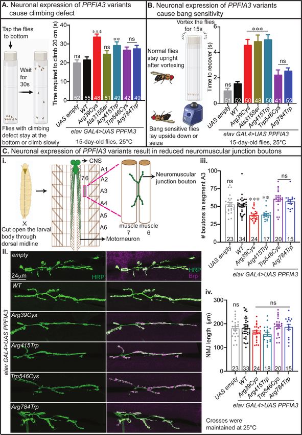
Next, to determine if the neuronal expression of PPFIA3 variants by elav-GAL4 impaired
nervous system development and function we conducted climbing behavior, bang sensitivity
behavior, and NMJ morphology assays. First, we performed a climbing assay to assess for motor
defects in negative geotaxis. The standard behavior of the flies is to climb upward, and any
increase in time to climb represents a motor coordination defect. We found that elav-
GAL4>UAS-PPFIA3 WT flies had motor function similar to control flies that do not express
human PPFIA3 protein (elav-GAL4 > UAS-empty) (Figure 4A). However, climbing was
impaired in both elav-GAL4>UAS-PPFIA3 p.(Arg39Cys) and elav-GAL4>UAS-PPFIA3
p.(Arg415Trp) expressing flies (Figure 4A). Second, to determine if the PPFIA3 variantsmedRxiv preprint doi: https://doi.org/10.1101/2023.03.27.23287689; this version posted March 29, 2023. The copyright holder for this preprint
(which was not certified by peer review) is the author/funder, who has granted medRxiv a license to display the preprint in perpetuity.
All rights reserved. No reuse allowed without permission.
increase seizure susceptibility, we tested bang sensitivity in the flies. We found that elav-
GAL4>UAS-PPFIA3 WT expressing flies were not bang sensitive and recovered similarly to the
elav-GAL4>UAS-empty control. However, elav-GAL4>UAS-PPFIA3 p.(Arg39Cys), elav-
GAL4>UAS-PPFIA3 p.(Ala315Ser), and elav-GAL4>UAS-PPFIA3 p.(Arg415Trp) flies
exhibited bang sensitivity with an increased recovery time (Figure 4B). Third, to explore the
consequence of PPFIA3 variants at the synapse, we examined the fruit fly third instar larval NMJ
morphology in muscle 6/7 of abdominal segment 3 (A3) (Figure 4Ci-ii). The fly NMJ is a
glutamatergic synapse and a well-established model for excitatory glutamatergic synapse
development and function.40,41 We found reduced number of boutons (presynaptic contacts) with
elav-GAL4 mediated expression of the PPFIA3 p.(Arg39Cys) and p.(Arg415Trp) variants
(Figure 4Ciii), indicating these variants perturb synapse formation. Total NMJ length associated
with the PPFIA3 variants remained similar to the PPFIA3 WT and UAS-empty controls (Figure
4Civ).
To determine the functional nature of the human PPFIA3 variants in the absence of wild-type fly
Liprin-α, we performed in vivo rescue experiments with a previously established Liprin-a LOF
allele28, Liprin-αF3ex15, and a Liprin-a deficiency allele, Df(2L)Exel7027/CyO (Figure 5Ai). First,
we observed that complete loss of Liprin-α function (Liprin-αF3ex15 /Df(2L)Exel7027) is
embryonic lethal in control da-GAL4>UAS-empty expressing flies, with a few escapers reaching
larval stage (Figure 5Aii). We expressed the human PPFIA3 WT or variant cDNAs in the
background of Liprin-α LOF using a ubiquitously expressed da-GAL4 at 20°C and assessed if
expression of human PPFIA3 WT or variant proteins are able to rescue the embryonic lethality.
We found a ~25% larval rescue of embryonic lethality with PPFIA3 WT, indicating functional
conservation in fruit flies (Figure 5Aii). Expression of PPFIA3 p.(Arg39Cys) and
p.(Arg415Trp) resulted in significantly reduced larval rescue compared to WT (8% and 13%,
respectively) (Figure 5Aii). However, expression of the PPFIA3 p.(Trp546Cys) and
p.(Arg784Trp) variants resulted in ~17% rescue efficiency of the embryonic lethality, which was
insignificant compared to PPFIA3 WT. Second, we assessed the survival of the rescued larvae to
the adult stage (Figure 5Bi). We found that ~35% of PPFIA3 WT expressing larvae reached the
adult stage (Figure 5Bii), but none of the PPFIA3 p.(Arg39Cys) expressing larvae reached the
adult stage. In contrast, we found that 23% of PPFIA3 p.(Arg415Trp) expressing larvae reachedmedRxiv preprint doi: https://doi.org/10.1101/2023.03.27.23287689; this version posted March 29, 2023. The copyright holder for this preprint
(which was not certified by peer review) is the author/funder, who has granted medRxiv a license to display the preprint in perpetuity.
All rights reserved. No reuse allowed without permission.
the adult stage, which is significantly reduced compared to PPFIA3 WT (Figure 5Bii). However,
the frequency of PPFIA3 p.(Trp546Cys) and PPFIA3 p.(Arg784Trp) expressing larvae reaching
the adult stage was insignificant compared to PPFIA3 WT (33% and 30%, respectively) (Figure
5Bii). Third, we assessed the survival of these rescue adults in the 48 hours post-eclosion. We
found that UAS
empty controls, there is no significant difference in the number of boutons between PPFIA3 WT
and variants (Figure S3Bi). We quantified the total length of the NMJ and found no significant
difference between genotypes (Figure S3Bii). Interestingly, we observed a significantly reduced
ratio of bouton numbers per muscle 6/7 NMJ (segment A3) length in both PPFIA3 WT and
variants compared to the da-GAL4>UAS empty control (Figure S3Bii). However, the bouton to
NMJ length ratio remained unchanged between PPFIA3 WT and variants (Figure S3Bii). This
indicates that there is a significant loss of bouton density in the background of complete Liprin-a
LOF compared to the da-GAL4>UAS empty control. However, expression of neither PPFIA3
WT nor variants were able to rescue the loss of NMJ boutons in the Liprin-a LOF background
(Figure S3Bi, iii). It is possible that due to the severity of the complete Liprin-a LOF, the
PPFIA3 WT or variants expressing larvae examined for NMJ morphology represent a healthier
subset of larvae capable of developing to the 3rd instar stage. Therefore, we may not be capturing
PPFIA3 WT or variants expressing larvae with more severe NMJ phenotypes. This would limit
our ability to identify a morphological difference between PPFIA3 WT and variants in the
background of complete Liprin-a LOF. Together, the in vivo fly functional experiments
demonstrate that rare PPFIA3 variants p.(Arg39Cys), p.(Arg415Trp), and p.(Arg784Trp) result
in loss of PPFIA3 function and are deleterious to multiple developmental processes. ThesemedRxiv preprint doi: https://doi.org/10.1101/2023.03.27.23287689; this version posted March 29, 2023. The copyright holder for this preprint
(which was not certified by peer review) is the author/funder, who has granted medRxiv a license to display the preprint in perpetuity.
All rights reserved. No reuse allowed without permission.
findings and our clinical characterizations show that rare PPFIA3 mono-allelic variants in key
functional domains lead to a syndromic neurodevelopmental disorder.
Discussion
We describe 14 individuals with rare variants in PPFIA3, who have neurodevelopmental
phenotypes including DD/ID, hypotonia, ASD, and epilepsy. The results of our clinical analysis,
in silico molecular modeling, and in vivo functional studies in fruit flies show that rare PPFIA3
variants lead to a previously unrecognized syndromic neurodevelopmental disorder. PPFIA3
protein domain analysis and molecular modeling revealed that six of the PPFIA3 missense
variants, p.(Arg39Cys), p.(Gln80Pro), p.(Ala315Ser), p.(Arg415Trp), p.(Arg429Trp), and
p.(Arg498Trp) are located in the N-terminal coiled coil domain. The coiled coil domain is
critical for PPFIA3’s homodimerization and interaction with active zone proteins, such as RIM
and ELKS, to regulate active zone organization and synaptic vesicle release.13–16 Three PPFIA3
missense variants, p.(Trp546Cys), p.(Arg784Trp), and p.(Ser906Leu), are located in the
disordered region of the protein. One PPFIA3 missense variant, p.(Ile870Asn), is located in the
SAM1 domain. The SAM domains are known to bind to RNA and lipid membranes and the
SAM domains of PPFIA proteins is shown to bind to the adhesion molecule LAR-RPTP.17,42,43
The PPFIA3 frameshift deletion variant, p.(Glu1103Asnfs*8), may result in nonsense mediated
decay followed by reduced protein expression.
Although interpretation is limited by the small sample size, we assessed the occurrence of six
commonly reported features in the 14 individuals with rare PPFIA3 mono-allelic variants and
neurodevelopmental phenotypes. These six features include epilepsy or abnormal EEG, autism,
DD/ID, hypotonia, dysmorphisms, and micro/macrocephaly (Table 2). We found that out of the
six individuals with PPFIA3 missense variants in the coiled coil domain (I:1, p.(Arg39Cys); I:2,
p.(Gln80Pro); I:5, p.(Ala315Ser); I:6, p.(Arg415Trp); I:7, p.(Arg429Trp), and I:8
p.(Arg498Trp)), two of them had premature mortality (I:5 and I:7). I:1 and I:2 had 4/6 clinical
features reported in which I:1 had epilepsy, DD/ID, hypotonia, and dysmorphisms and I:2 had
autism, DD/ID, hypotonia, and dysmorphisms (Table 2). I:6 had 6/6 key clinical features
reported with abnormal EEG, autism, DD/ID, hypotonia, dysmorphisms, and
micro/macrocephaly (Table 2). Individual I:8 had 3/6 clinical features reported (autism, DD/ID,medRxiv preprint doi: https://doi.org/10.1101/2023.03.27.23287689; this version posted March 29, 2023. The copyright holder for this preprint
(which was not certified by peer review) is the author/funder, who has granted medRxiv a license to display the preprint in perpetuity.
All rights reserved. No reuse allowed without permission.
and dysmorphisms) (Table 2). Our amino acid conservation analysis showed that all affected
residues in the coiled coil domain of PPFIA3 are highly conserved across mice, fruit flies, and
worm, suggesting that these residues are critical for the protein’s function across different
species. Molecular modeling of these variants suggested that changes in the affected residues
could hinder the PPFIA3 protein function.
Three individuals have de novo PPFIA3 missense variants (I:9, p.(Trp546Cys); I:10,
p.(Arg784Trp); and I:13, p.(Ser906Leu)) in the disordered region of the protein. The amino acid
conservation analysis suggests that residues in the disordered region of PPFIA3 are not well
conserved across species except for p.Ser906 that is near the SAM1 domain. Individuals I:9
(p.(Trp546Cys)) and I:10 (p.(Arg784Trp)) has 2/6 clinical features reported (Table 2). I:9 had
autism and DD/ID, whereas I:10 had epilepsy and DD/ID. The small number of phenotypic
findings in I:9 and I:10 may be due to the location of the variants in a disordered region of the
PPFIA3 protein. Individual I:13 (p.(Ser906Leu)) had 6/6 clinical features reported with abnormal
EEG, autism, DD/ID, hypotonia, dysmorphisms, and micro/macrocephaly. Together, these
findings suggest that variants in the disordered regions are associated with variable disease
severity, which may be due to the lack of association in this region with functional domains for
PPFIA3. Two individuals had PPFIA3 missense variants in the SAM1 or SAM3 domains (I:11,
p.(Ile870Asn); I:12 p.(Ile870Asn)). Individuals I:11 and I:12 are monozygotic twins and both
had 3/6 clinical features reported. I:11 had autism, DD/ID, and microcephaly. I:12 had DD/ID,
hypotonia, and microcephaly. One individual has a PPFIA3 frameshift variant in the SAM3
domain, I:14, p.(Glu1103Asnfs*8), with 4/6 clinical features reported that include abnormal
EEG, DD/ID, hypotonia, and dysmorphisms. There were two individuals (I:3, I:4) with the same
PPFIA3 intronic splice variant (c.240+1G>A) and the number of key clinical features reported
were 3/5 (I:3) and 2/5 (I:4). Individual I:3 inherited the PPFIA3 variant from the affected parent,
I:4. The inheritance of the PPFIA3 variant in I:4 is unknown. Both I:3 and I:4 have DD, ID, and
microcephaly. Finally, we also identified a missense PPFIA3 p.(Arg559Trp) variant of unknown
inheritance in I:15. The severe neurodegeneration phenotype was not observed in the 14
individuals in our cohort, suggesting that other genetic alterations or etiologies may contribute to
I:15’s clinical findings.medRxiv preprint doi: https://doi.org/10.1101/2023.03.27.23287689; this version posted March 29, 2023. The copyright holder for this preprint
(which was not certified by peer review) is the author/funder, who has granted medRxiv a license to display the preprint in perpetuity.
All rights reserved. No reuse allowed without permission.
Our conservation analysis reveal that the PPFIA3 protein domains are well conserved in the fruit
fly homolog, Liprin-α, which is primarily expressed in the fruit fly embryonic and larval nervous
system.28 In the fruit fly larvae, it is found in the NMJ synapses.28 Several previous fruit fly
studies show that Liprin-α is critical for synapse morphogenesis and axon guidance with Liprin-α
LOF resulting in a reduced number of boutons, axon branching, and active zone dimensions.28
Another important function of Liprin-α is the regulation of synaptic vesicle trafficking. Liprin-α
LOF causes an accumulation of synaptic markers in the axon with a decrease in anterograde
transport and an increase in retrograde transport.44 Moreover, Liprin-α is also essential for retinal
axon targeting in fruit flies.30 Interestingly, a previous study with Ppfia3 knockout mice revealed
that complete loss of Ppfia3 impairs synaptic vesicle exocytosis, reduced the number of synapses
that were responsive to action potentials, and altered the active zone structure.24 Double
knockout of mouse Ppfia2 and Ppfia3 in hippocampal neurons impaired neurotransmitter release
due to perturbations in the number of docked vesicles per synapse.45 Together, these findings
reveal that PPFIA3 homologs in fruit flies and mice are crucial for multiple neurodevelopmental
processes.
We found that the ubiquitous expression of PPFIA3 missense variants in the coiled coil domain
(p.(Arg39Cys), p.(Ala315Ser), and p.(Arg415Trp)) resulted in pupal lethality, eclosion defects,
and abnormal leg morphology. Neuron-specific expression revealed seizure-like behaviors and
climbing defects with PPFIA3 p.(Arg39Cys) and p.(Arg415Trp) variants. As Liprin-α is known
to regulate NMJ development in fruit flies, we examined the effect of human PPFIA3 WT and
variant cDNA expression in the larval NMJ. We found that overexpression of PPFIA3
p.(Arg39Cys) and p.(Arg415Trp) caused a reduction in bouton number compared to PPFIA3
WT, which would potentially impair neurotransmission. Our fly overexpression findings reveal
the PPFIA3 missense variants in the coiled coil domains cause lethality, suggesting these
variants are dominant negative alleles. Interestingly, ubiquitous, or neuronal overexpression of
the PPFIA3 variants in the disordered region (p.(Arg784Trp) and p.(Trp546Cys)) had either mild
or no phenotypes in the fruit flies, which may be related to the fact that the variants are either not
conserved in the flies or cause mild protein dysfunction that is tolerated in the fly model.medRxiv preprint doi: https://doi.org/10.1101/2023.03.27.23287689; this version posted March 29, 2023. The copyright holder for this preprint
(which was not certified by peer review) is the author/funder, who has granted medRxiv a license to display the preprint in perpetuity.
All rights reserved. No reuse allowed without permission.
To further determine whether the variants are LOF or gain-of-function (GOF) in nature and the
functional conservation between human PPFIA3 and fly Liprin-α, we performed a LOF lethality
rescue assay using Liprin-α mutants and PPFIA3 WT and variants. Our LOF rescue assay
showed that human PPFIA3 WT expression partially rescued the Liprin-α LOF embryonic
lethality and development to the adult stage, suggesting that human PPFIA3 WT protein function
is partially conserved in fruit flies. Interestingly, expression of the coiled coil variants, PPFIA3
p.(Arg39Cys) and p.(Arg415Trp) showed reduced rescue efficiency of the Liprin-α LOF
embryonic lethality, indicating these variants are strong LOF variants. In contrast, expression of
the disordered region variants, PPFIA3 p.(Trp546Cys) and p.(Arg784Trp) showed a similar
rescue efficiency of the embryonic lethality as compared to PPFIA3 WT expression. However,
we found in the adult stage that PPFIA3 p.(Arg784Trp) expression significantly reduced the
lifespan of rescue flies compared to PPFIA3 WT, suggesting that p.(Arg784Trp) is a
hypomorphic LOF variant. These findings are consistent with the clinical findings in individual
I:9, p.(Trp546Cys) and I:10, p.(Arg784Trp). Both individuals had only 2/6 commonly reported
features (Table 2). In contrast, individuals I:1, p.(Arg39Cys) and I:6, p.(Arg415Trp) had 4/6 and
6/6 commonly reported features, respectively (Table 2). Together, our overexpression and LOF
rescue assays in fruit flies reveal that rare mono-allelic PPFIA3 variants cause a
neurodevelopmental disorder through a LOF mechanism and that disease severity correlates with
the degree of LOF. The clinical phenotypes and functional assays in fruit flies (Table 3) point
towards a possible domain-specific disease severity mechanism where the variants in the coiled
coil domains might lead to relatively more severe phenotypes in both affected individuals and
fruit flies.
In summary, our study provides clinical and functional evidence that rare mono-allelic PPFIA3
variants cause a syndromic neurodevelopmental disorder characterized by DD/ID, hypotonia,
autism, and epilepsy. Our in vivo functional modeling in fruit flies reveal that PPFIA3 variants
may contribute to disease pathogenesis through LOF mechanisms related to the location of the
affected residues in the PPFIA3 functional domains. Analysis of potential genotype-phenotype
correlations using the clinical and in vivo fruit fly findings suggest that PPFIA3 variants in the
coiled coil domains may have relatively more deleterious effects. A longitudinal assessment in a
larger sample size of affected individuals and functional studies in model organisms wouldmedRxiv preprint doi: https://doi.org/10.1101/2023.03.27.23287689; this version posted March 29, 2023. The copyright holder for this preprint
(which was not certified by peer review) is the author/funder, who has granted medRxiv a license to display the preprint in perpetuity.
All rights reserved. No reuse allowed without permission.
advance our understanding of disease pathogenesis, improve prognostication based on variant
type and location, and identify potential therapeutic avenues.
Acknowledgements
We thank the families and clinical staff at each location for participation in this study. We thank
the CCuB for technical support and management of the computing platform for the individuals
I:3 and I:4. In addition, we thank Mingshan Xue, Dongwon Lee, Kailin Mao, Wu (Charles)
Chen, Brooke Horist, and Cole Deisseroth for critical feedback on the manuscript.
Funding
H.T.C.’s research work is supported by the McNair Medical Institute at The Robert and Janice
McNair Foundation, the Burroughs Wellcome Fund, Child Neurology Foundation and Society,
The Gordan and Mary Cain Foundation, Annie and Bob Graham, The Elkins Foundation, and the
Mark A. Wallace Endowment Award. M.S.P’s research effort is supported in part by the
National Ataxia Foundation and the Burroughs Wellcome Fund. J.M.P.’s research effort was
supported in part by the Burroughs Wellcome Fund. V.C.L and S.L.M. were supported in part by
The Gordan and Mary Cain Foundation and Annie and Bob Graham. S.L.M. was also supported
in part by the Mark A. Wallace Endowment Award. This work was also supported by Texas
Children’s Hospital, the Jan and Dan Duncan Neurological Research Institute, and the Eunice
Kennedy Shriver National Institute of Child Health & Human Development of the National
Institutes of Health under Award Number P50HD103555 for use of the Clinical Translational
Core and Microscopy Core facilities. Research reported in this manuscript was supported by the
NIH Common Fund, through the Office of Strategic Coordination/Office of the NIH Director
under Award Number - U01HG007709. The content is solely the responsibility of the authors
and does not necessarily represent the official views of the National Institutes of Health.
Sequencing and analysis of one individual in this study was made possible by the generous giftsYou can also read