RBBP6 activates the pre-mRNA 3 end processing machinery in humans - Genes & Development
←
→
Page content transcription
If your browser does not render page correctly, please read the page content below

Downloaded from genesdev.cshlp.org on June 10, 2022 - Published by Cold Spring Harbor Laboratory Press

RBBP6 activates the pre-mRNA 3′ end
processing machinery in humans
Vytaute Boreikaite, Thomas S. Elliott, Jason W. Chin, and Lori A. Passmore
Medical Research Council Laboratory of Molecular Biology, Cambridge CB2 0QH, United Kingdom

3′ end processing of most human mRNAs is carried out by the cleavage and polyadenylation specificity factor (CPSF;
CPF in yeast). Endonucleolytic cleavage of the nascent pre-mRNA defines the 3′ end of the mature transcript, which
is important for mRNA localization, translation, and stability. Cleavage must therefore be tightly regulated. Here,
we reconstituted specific and efficient 3′ endonuclease activity of human CPSF with purified proteins. This required
the seven-subunit CPSF as well as three additional protein factors: cleavage stimulatory factor (CStF), cleavage factor
IIm (CFIIm), and, importantly, the multidomain protein RBBP6. Unlike its yeast homolog Mpe1, which is a stable
subunit of CPF, RBBP6 does not copurify with CPSF and is recruited in an RNA-dependent manner. Sequence and
mutational analyses suggest that RBBP6 interacts with the WDR33 and CPSF73 subunits of CPSF. Thus, it is likely
that the role of RBBP6 is conserved from yeast to humans. Overall, our data are consistent with CPSF endonuclease
activation and site-specific pre-mRNA cleavage being highly controlled to maintain fidelity in mRNA processing.
[Keywords: RNA; endonuclease; gene expression; polyadenylation]
Supplemental material is available for this article.
Received November 18, 2021; revised version accepted February 1, 2022.

Eukaryotic protein-coding pre-mRNAs undergo multiple                        2018; Sun et al. 2018). These showed how the CPSF30 and
processing steps during transcription by RNA polymerase                     WDR33 subunits recognize the hexameric polyadenyla-
II. These include 5′ capping, splicing, and 3′ end process-                 tion signal (PAS) sequence, most commonly AAUAAA,
ing (Hocine et al. 2010). During this latter process, a cleav-              thereby recruiting CPSF to cleavage sites on pre-mRNAs.
age event defines the 3′ end of the mature mRNA and is                      The poly(A) polymerase enzyme (PAP) is not a stable sub-
linked to transcription termination (Buratowski 2005;                       unit of human CPSF but is instead recruited to cleaved
Liu and Moore 2021). A poly(A) tail is added to the resul-                  transcripts by hFip1 (Kaufmann et al. 2004; Chan et al.
tant free 3′ end, marking the mRNA for nuclear export                       2014). mCF consists of three subunits: CPSF73,
and controlling mRNA stability and translational effi-                      CPSF100, and symplekin (Zhang et al. 2020). CPSF73 is
ciency in the cytoplasm (Passmore and Coller 2022).                         a zinc-dependent RNA endonuclease that belongs to the
Thus, 3′ cleavage and polyadenylation are critical to the                   metallo-β-lactamase family. CPSF100 is a pseudonuclease
production of functional protein-coding transcripts.                        that is structurally homologous to CPSF73 (Mandel et al.
   In humans, cleavage and polyadenylation are carried                      2006). mCF is tethered to mPSF through a conserved inter-
out by a seven-subunit protein complex known as cleav-                      action between CPSF160 and a peptide within CPSF100,
age and polyadenylation specificity factor (CPSF) (Kumar                    known as the mPSF interaction motif (PIM) (Zhang et al.
et al. 2019; Zhang et al. 2020). CPSF is comprised of two                   2020; Rodríguez-Molina et al. 2021).
stable subcomplexes: mammalian polyadenylation specif-                         To ensure that mature transcripts of a correct length are
icity factor (mPSF) and mammalian cleavage factor (mCF).                    produced, pre-mRNAs must be cleaved at specific sites.
These are equivalent to the polymerase module and nu-                       Deregulation of this process can result in transcriptional
clease module, respectively, of the yeast cleavage and pol-                 defects and nonfunctional transcripts, and can lead to hu-
yadenylation factor (CPF) (Casañal et al. 2017). mPSF                       man disease (Curinha et al. 2014). In vitro, purified CPSF/
contains four subunits: CPSF160, WDR33, CPSF30, and                         CPF is an inherently inactive endonuclease, which pre-
hFip1 (Schönemann et al. 2014). Structures of mPSF/poly-                    sumably must be activated by accessory factors to enable
merase module in apo and RNA-bound states have recent-                      strict regulation of 3′ cleavage (Mandel et al. 2006; Hill
ly been elucidated (Casañal et al. 2017; Clerici et al. 2017,               et al. 2019; Zhang et al. 2020). For example, cleavage stim-
                                                                            ulatory factor (CStF) and cleavage factor IIm (CFIIm) are
                                                                            both multisubunit protein complexes implicated in
Corresponding author: passmore@mrc-lmb.cam.ac.uk
Article published online ahead of print. Article and publication date are
online at http://www.genesdev.org/cgi/doi/10.1101/gad.349223.121. Free-     © 2022 Boreikaite et al. This article, published in Genes & Development,
ly available online through the Genes & Development Open Access             is available under a Creative Commons License (Attribution 4.0 Interna-
option.                                                                     tional), as described at http://creativecommons.org/licenses/by/4.0/.

                  GENES & DEVELOPMENT 36:1–15 Published by Cold Spring Harbor Laboratory Press; ISSN 0890-9369/22; www.genesdev.org               1
Downloaded from genesdev.cshlp.org on June 10, 2022 - Published by Cold Spring Harbor Laboratory Press

Boreikaite et al.

cleavage (Takagaki et al. 1990; De Vries et al. 2000). CStF            ing. This included CPSF (assembled from individually pu-
has been shown to bind a G/U-rich region downstream                    rified mPSF and mCF subcomplexes) as well as the
from the cleavage site on pre-mRNAs and to provide spe-                accessory factors CStF and CFIIm (Fig. 1A). We used short
cificity for poly(A) site selection (Takagaki and Manley               isoforms of CPSF30 and hFip1, and also removed unstruc-
1997). Another accessory factor, cleavage factor Im                    tured regions from WDR33 and the CFIIm subunit Pcf11
(CFIm), is not essential for 3′ cleavage but recruits CPSF             to facilitate purification (Schäfer et al. 2018; Sun et al.
to pre-mRNAs containing an upstream UGUA motif                         2018). We hypothesized that the conserved region of the
and contributes to the use of alternative polyadenylation              multidomain protein RBBP6 (residues 1–335) might also
sites in human cells (Zhu et al. 2018).                                be required for endonuclease activation. RBBP6 did not
   The cleavage activity of human CPSF has been studied                copurify with CPSF and was therefore expressed and puri-
by functional genomics and by in vitro experiments in                  fied separately. As a model pre-mRNA substrate, we used
fractionated nuclear extracts prepared from cultured hu-               a 218-nt fragment of the SV40 pre-mRNA, which has been
man cells (for recent examples, see Eaton et al. 2018; Schä-           shown to be cleaved efficiently in vivo (Ryner and Manley
fer et al. 2018). However, the full protein composition of             1987; Kwon et al. 2021). We omitted the PAP enzyme and
partially purified 3′ end processing machinery from nucle-             ATP from the reactions to focus on the cleavage step of
ar extract is not known, making it difficult to infer molec-           pre-mRNA 3′ end processing (Fig. 1B).
ular mechanisms. Moreover, generating mutants of                          We tested various combinations of 3′ end processing
endogenous proteins to test hypotheses is cumbersome.                  factors in cleavage assays and analyzed the results by
   To enable detailed mechanistic studies of CPSF endo-                denaturing gel electrophoresis of RNA (Fig. 1C). No cleav-
nuclease activation, an in vitro assay containing a well-              age activity was observed when the SV40 pre-mRNA was
defined set of highly pure proteins is required. Recently,             incubated with CPSF alone. Addition of CStF and CFIIm,
this has been achieved for the human histone pre-                      either individually or together, failed to activate CPSF.
mRNA 3′ end processing complex, which shares the endo-                 However, addition of RBBP6 activated CPSF in the pres-
nuclease subunit CPSF73 but differs from CPSF in most of               ence of CStF and CFIIm, promoting efficient cleavage of
its other subunits and its mechanism of RNA recognition                the pre-mRNA substrate. Previous assays in nuclear ex-
(Sun et al. 2020; Gutierrez et al. 2021). The endonuclease             tract used molecular crowding agents such as polyvinyl al-
activity of the budding yeast CPF complex has also been                cohol (Adamson et al. 2005), but these were not required
reconstituted from purified recombinant proteins (Hill                 here. Omitting CPSF from the reaction did not lead to sub-
et al. 2019). The minimal active subcomplex in yeast,                  strate cleavage, showing that the observed endonuclease
called core CPF, contains orthologs of CPSF subunits as                activity cannot be attributed to potential contaminants
well as an additional protein: Mpe1. We recently showed                that copurify with the accessory proteins. Overall, we de-
that Mpe1 is an essential activator of the CPF endonucle-              termined that activation of the CPSF endonuclease re-
ase (Rodríguez-Molina et al. 2021). However, while many                quires three additional protein factors: CStF, CFIIm, and
aspects of 3′ end processing are conserved, there appear to            RBBP6.
be some differences between the yeast and human ma-                       To identify the precise CPSF cleavage site on the SV40
chineries, including in RNA specificity and recognition                pre-mRNA substrate, we sequenced several 5′ cleavage
(Tian and Graber 2012; Rodríguez-Molina et al. 2021).                  products. This revealed that the majority (13 out of 15)
The human ortholog of Mpe1, RBBP6, has been implicat-                  of cleaved RNAs were cut 13 nt downstream from the
ed in pre-mRNA 3′ end processing in humans (Shi et al.                 PAS within a CA|A motif, where | indicates the cleavage
2009; Di Giammartino et al. 2014). Whether it plays a di-              site (Fig. 1D; Sheets et al. 1987). This is consistent with
rect role in the cleavage reaction remains unclear.                    the known sequence preference of 3′ endonucleases and
   Here, we reconstituted CPSF from purified proteins that             with previous observations that pre-mRNAs in cells are
is active in both cleavage and polyadenylation. We dem-                cleaved 10–30 nt downstream from the PAS (Beaudoing
onstrate that human RBBP6 is required for the activation               et al. 2000; Hill et al. 2019).
of 3′ end cleavage even though it is not a stable subunit of              Since the SV40 pre-mRNA contains an upstream
CPSF. Our results show that the mechanism of endonu-                   UGUA motif, we tested whether CFIm affected cleavage
clease activation by Mpe1/RBBP6 is likely to be highly                 of the SV40 substrate with purified CPSF. CFIm is known
conserved.                                                             to bind the RE/D domain of hFip1, which is lacking in iso-
                                                                       form 4 of hFip1 in our CPSF complex (Zhu et al. 2018).
                                                                       Therefore, we also purified CPSF containing the full-
Results                                                                length hFip1 subunit (Fig. 2A). Addition of CFIm into
                                                                       the cleavage assay did not provide any further stimulation
CStF, CFIIm, and RBBP6 are required for activation
                                                                       of CPSF endonuclease activity in our reconstituted sys-
of CPSF endonuclease
                                                                       tem (Fig. 2B; Supplemental Fig. S1A). Nevertheless,
To gain insight into how the human CPSF endonuclease is                CFIm may affect cleavage in other conditions (e.g., when
activated, we attempted to reconstitute pre-mRNA cleav-                the concentrations of CPSF and RNA are lower) or on sub-
age activity from purified recombinant proteins. We used               strates with multiple UGUA motifs and/or multiple po-
baculovirus-mediated expression in insect cells to pro-                tential PAS sequences.
duce highly pure protein complexes predicted to be                        PAP is dispensable for CPSF cleavage activity, high-
directly involved in canonical pre-mRNA 3′ end process-                lighting the fact that cleavage and polyadenylation can

2       GENES & DEVELOPMENT
Downloaded from genesdev.cshlp.org on June 10, 2022 - Published by Cold Spring Harbor Laboratory Press

                                                                                                Reconstitution of human pre-mRNA cleavage

             A

             B                                             C

                                                           D

Figure 1. CStF, CFIIm, and RBBP6 are required for activation of the CPSF endonuclease. (A) Schematic representations and SDS-PAGE
analyses of the purified proteins used in the in vitro endonuclease assays. Residue boundaries and alternative isoforms are indicated for
truncated proteins. An asterisk denotes degradation products. (SII) StrepII tag. (B) Schematic representation of the in vitro pre-mRNA
3′ end processing assay. The cleavage reaction is boxed out. The polyadenylation step was not assayed here. (C) Denaturing gel electro-
phoresis of the SV40 pre-mRNA substrate after incubation with various combinations of human 3′ end processing factors. The full-length
and cleaved RNAs are shown schematically at the right. (D) Part of the sequence of the SV40 pre-mRNA substrate with the experimentally
determined CPSF cleavage sites indicated (scissors). The frequency of a particular cleavage site identified by sequencing of 15 cleavage
products is shown below. The polyadenylation signal (PAS) sequence is marked in green.

be uncoupled in vitro (Moore and Sharp 1985; Ryner and                 Pre-mRNA cleavage by purified, recombinant CPSF
Manley 1987). Addition of PAP and ATP into a cleavage                  is dependent on CPSF73 and a PAS
assay resulted in the polyadenylation of the 5′ cleavage
product with heterogeneous poly(A) tail lengths, but                   Next, we aimed to understand the specificity of the recon-
PAP did not substantially change the cleavage efficiency               stituted 3′ cleavage reaction. First, we generated a CPSF
(Fig. 2C,D; Supplemental Fig. S1B). Thus, our recon-                   complex containing an active site mutant of CPSF73
stituted CPSF complex is active in both cleavage and                   (D75N H76A) in which the coordination of catalytic
polyadenylation.                                                       zinc ions was disrupted (Sun et al. 2020). The complex
   We also tested whether recombinant CPSF could cleave                with a mutant endonuclease subunit was inactive in a
a different pre-mRNA substrate. Under the same reaction                cleavage assay, suggesting that the observed endonuclease
conditions, the adenoviral L3 pre-mRNA was cut with ef-                activity is attributable to CPSF73 (Fig. 3A; Supplemental
ficiency similar to that of the SV40 pre-mRNA (Supple-                 Fig. S2B).
mental Fig. S2A), suggesting that the same complement                     We tested the activity of the mCF subcomplex alone
of accessory proteins (CStF, CFIIm, and RBBP6) is required             and found that it is inactive in the absence of mPSF (Sup-
for activation of the CPSF endonuclease on multiple dif-               plemental Fig. S3). The CPSF30 and WDR33 subunits
ferent pre-mRNA substrates.                                            within mPSF recognize the PAS sequence and contribute

                                                                                                         GENES & DEVELOPMENT           3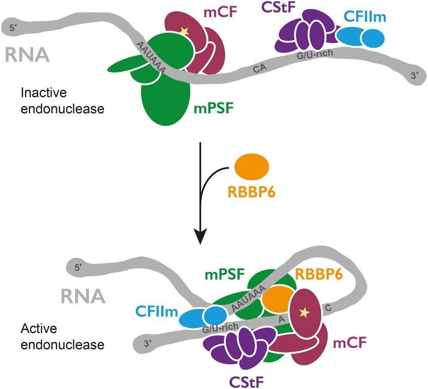
Downloaded from genesdev.cshlp.org on June 10, 2022 - Published by Cold Spring Harbor Laboratory Press

Boreikaite et al.

                    A                                                                         C

                    B                                          D

Figure 2. CFIm is not required for CPSF cleavage activity, and RNA is cleaved and polyadenylated in the presence of CPSF, RBBP6,
CStF, CFIIm, and PAP. (A) SDS-PAGE analyses of purified CPSF containing full-length hFip1 (hFip1FL) and of purified CFIm complex.
Asterisks denote degradation products. (SII) StrepII tag. (B) Cleavage assays of the SV40 pre-mRNA substrate with CPSF-hFip1FL in the
presence of increasing concentrations of CFIm. CFIm does not substantially affect CPSF cleavage activity. (C ) SDS-PAGE analysis of
purified PAP. An asterisk denotes degradation products. (D) Coupled cleavage and polyadenylation assays of the SV40 pre-mRNA sub-
strate at two different concentrations of PAP in the presence of either ATP or ATP and 3′ -dATP together. 3′ -dATP is also called cor-
dycepin and is known to inhibit polyadenylation. The heterogeneous products that appear in the presence of ATP are largely absent
when 3′ -dATP is also added. This demonstrates that polyadenylation is responsible for the diffuse band. Some substrate RNAs may
also get polyadenylated by free PAP.

to specific recruitment of CPSF73 to pre-mRNAs (Clerici                cessing complex in vitro (Gutierrez et al. 2021). We there-
et al. 2018; Sun et al. 2018). Replacement of the canonical            fore tested whether the JTE-607 acid analog was also
AAUAAA polyadenylation signal in the SV40 pre-mRNA                     inhibitory to the reconstituted human canonical 3′ end
with an AACAAA hexamer resulted in a substantial re-                   processing complex. Titrating the compound into the
duction in cleavage by CPSF (reduced by ∼80%), demon-                  cleavage reaction showed dose-dependent inhibition of
strating that CPSF has specificity for PAS-containing                  the endonuclease activity with an IC50 of ∼350 nM (Fig.
RNAs (Fig. 3B). It is likely that mPSF is not only involved            3C,D), which is very similar to the Kd of the acid form of
in RNA binding but is also required for conformational re-             JTE-607 for isolated CPSF73 (∼370 nM) (Ross et al.
arrangements that allow endonuclease activation (Rodrí-                2020). Together, these data confirm that the observed in
guez-Molina et al. 2021).                                              vitro endonuclease activity is specific to CPSF73.
   Recently, CPSF73 was identified as the direct target of
JTE-607, a prodrug with anti-inflammatory and antican-
                                                                       Canonical and histone pre-mRNA 3′ end processing
cer properties (Kakegawa et al. 2019; Ross et al. 2020).
                                                                       complexes are activated by different mechanisms
The active acid form of JTE-607 inhibits both the purified,
recombinant yeast 3′ endonuclease (Ross et al. 2020) and               The human histone pre-mRNA 3′ processing reaction was
CPSF73 within the human histone pre-mRNA 3′ end pro-                   recently reconstituted with purified proteins, and the

4       GENES & DEVELOPMENT
Downloaded from genesdev.cshlp.org on June 10, 2022 - Published by Cold Spring Harbor Laboratory Press

                                                                                               Reconstitution of human pre-mRNA cleavage

                  A                                                    B

                  C                                                    D

Figure 3. Cleavage activity of purified recombinant CPSF is dependent on CPSF73 and a PAS. (A) Time-course cleavage assays of the
SV40 pre-mRNA substrate comparing the activities of wild-type (CPSFWT) and nuclease-dead (CPSFCPSF73 D75N H76A) CPSF complexes.
(B) Time-course cleavage assays of SV40 pre-mRNA substrates containing either a canonical PAS (RNAAAUAAA) or a mutant PAS
(RNAAACAAA) sequence. (C ) Cleavage assays in the presence of increasing concentrations of the JTE-607 acid compound. (D) Dose re-
sponse curve of the CPSF cleavage activity as a function of the concentration of JTE-607 acid. Each dot represents a single measurement.
At least three measurements were performed for each concentration of the drug, but some points overlap.

structure of the substrate-bound complex was determined                the mechanism of CPSF73 activation is fundamentally
in an active state (Sun et al. 2020; Gutierrez et al. 2021).           different in each complex.
The histone processing complex shares three subunits
with CPSF: symplekin, CPSF100, and CPSF73 (termed
                                                                       RBBP6 is not a stable subunit of human CPSF
the histone cleavage complex, equivalent to mCF in
CPSF). Although some aspects of endonuclease activation                The yeast ortholog of RBBP6, Mpe1, is a constitutive sub-
are carried out by proteins exclusive to the histone com-              unit of the native yeast CPF complex (Vo et al. 2001; Casa-
plex, the N-terminal domain (NTD) of symplekin (which                  ñal et al. 2017), but recombinant RBBP6 did not copurify
is also found in CPSF) was shown to be essential for acti-             with CPSF. To determine whether RBBP6 is stably associ-
vating CPSF73. We tested whether the symplekin NTD                     ated with endogenous CPSF from human cells, we used
plays a similar role in CPSF. To this end, we prepared                 CRISPR–Cas9 to generate a stable HEK293T cell line in
a CPSF complex in which the NTD of symplekin was de-                   which endogenous WDR33 carries a C-terminal HTBH
leted. The CPSF complex lacking the symplekin NTD                      tag (His6-TEV protease cleavage site–biotin acceptor pep-
retained activity similar to that of wild-type CPSF, sug-              tide-His6) (Wang et al. 2007). The biotin acceptor peptide
gesting that the mechanism of endonuclease activation                  becomes biotinylated by endogenous enzymes in the
is different between the two CPSF73-containing complex-                cell, which allows the purification of CPSF on Strep-Tac-
es (Fig. 4A).                                                          tin beads. We purified endogenous CPSF from the
   In addition, the phosphatase SSU72 was shown to inhib-              WDR33-HTBH cell line and analyzed its protein content
it the histone processing complex by binding to and                    by SDS-PAGE and mass spectrometry. The complex was
sequestering the symplekin NTD (Sun et al. 2020).                      relatively pure, and the enriched bands of CPSF subunits
Ssu72 is a subunit of yeast CPF (Casañal et al. 2017), and             could be detected by SDS-PAGE (Fig. 5A). Mass spectrom-
hence we tested whether human SSU72 also interacts                     etry analysis revealed that all seven CPSF subunits copuri-
with CPSF. We found that SSU72 interacts with mCF                      fied from human cells across multiple replicates (Fig. 5B;
and CPSF but not with mCF lacking the symplekin                        Supplemental Material). Among the accessory factors re-
NTD (Fig. 4B,C). However, titrating SSU72 into the                     quired for CPSF cleavage activity, only CStF subunits
CPSF cleavage reaction did not affect the in vitro endonu-             were pulled down by the endogenous CPSF complex. In
clease activity (Fig. 4D). Together, these results suggest             particular, the CStF64 subunit copurified with CPSF con-
that, similar to the histone 3′ processing complex,                    sistently. CStF64 is associated with the native histone
SSU72 interacts with the symplekin NTD in CPSF, but                    pre-mRNA 3′ end processing complex (Skrajna et al.

                                                                                                        GENES & DEVELOPMENT           5
Downloaded from genesdev.cshlp.org on June 10, 2022 - Published by Cold Spring Harbor Laboratory Press

Boreikaite et al.

          A                                                               B

          C

                                                                          D

Figure 4. Canonical and histone pre-mRNA 3′ end processing complexes are activated by different mechanisms. (A) Time-course cleav-
age assays of the SV40 pre-mRNA substrate comparing wild-type CPSF (CPSFWT) and CPSF lacking the symplekin NTD (CPSFsymplekin
ΔNTD). (B) Gel filtration chromatograms (top) and SDS-PAGE analyses (bottom) of CPSF in the presence (red) or absence (black) of
SSU72. (C ) Gel filtration chromatograms (top) and SDS-PAGE analyses (bottom) of wild-type mCF (mCFWT; green) and mCF lacking
the NTD of symplekin (mCFsymplekin ΔNTD; blue) mixed with SSU72. (D) Cleavage assays in the presence of increasing concentrations
of SSU72.

2018), and our data suggest that CStF64 may also be part of            processing (Fig. 6A; Supplemental Fig. S4; Sakai et al.
endogenous CPSF. Importantly, RBBP6 did not copurify                   1995; Simons et al. 1997; Li et al. 2007; Batra et al.
with CPSF, consistent with a previous study (Chan et al.               2018). A construct encompassing only the N-terminal do-
2014). Therefore, despite its critical role in activating              mains of RBBP6 was sufficient to stimulate CPSF (Fig.
the CPSF endonuclease, RBBP6 is not a stable component                 1C), suggesting that the C-terminal region is dispensable
of the CPSF complex.                                                   for pre-mRNA cleavage in vitro.
                                                                         To further investigate the interaction of RBBP6 and
                                                                       CPSF, we used size exclusion chromatography. When
RBBP6 is a conserved activator of canonical pre-mRNA
                                                                       mixed together, RBBP6 and CPSF eluted from the column
3′ end cleavage
                                                                       in two separate peaks, indicating that the affinity of any
Since RBBP6 is not a constitutive subunit of CPSF, we                  potential interaction is not sufficiently high for them to
were particularly intrigued by the role of RBBP6 in activat-           coelute (Fig. 6B). However, when a 41-nt fragment of L3
ing the 3′ endonuclease. Human RBBP6 is an ∼200-kDa                    pre-mRNA containing a canonical PAS (AAUAAA) was
protein with a conserved N-terminal region containing                  included, a substoichiometric amount of RBBP6 comi-
several ordered domains, and a long, disordered, noncon-               grated with RNA-bound CPSF (Fig. 6B). We also per-
served C-terminal tail, which interacts with various bind-             formed pull-downs using MS2-tagged L3 pre-mRNA and
ing partners that are not directly related to mRNA 3′ end              found that RBBP6 was pulled down by RNA only in the

6       GENES & DEVELOPMENT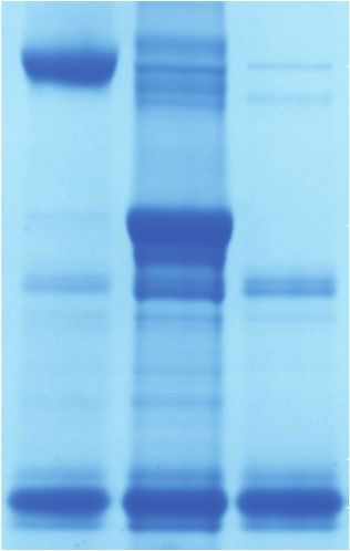
Downloaded from genesdev.cshlp.org on June 10, 2022 - Published by Cold Spring Harbor Laboratory Press

                                                                                         Reconstitution of human pre-mRNA cleavage

A                             B                                               Figure 5. RBBP6 is not a stable subunit of CPSF pu-
                                                                              rified from human cells. (A) SDS-PAGE analysis of
                                                                              the endogenous CPSF complex. The bands represent-
                                                                              ing CPSF subunits are indicated. TEV protease was
                                                                              used to elute the complex from Strep-Tactin beads
                                                                              and remains present in the sample. The gel was
                                                                              stained with SYPRO Ruby. (B) Heat map representing
                                                                              the sequence coverage of each protein required for
                                                                              CPSF endonuclease activity in vitro in the endoge-
                                                                              nous CPSF preparations as detected by mass spec-
                                                                              trometry. No RBBP6 peptides were detected across
                                                                              three independent experiments.

presence of CPSF (Supplemental Fig. S5A). This suggests           pre-mRNA-sensing region (PSR). The PSR sequence is con-
that RBBP6 is recruited to CPSF in an RNA-dependent               served in RBBP6 and, using AlphaFold2 (Jumper et al.
manner, which is reminiscent of RNA-mediated stabiliza-           2021), we predict that this region is likely to adopt an over-
tion of Mpe1 on the yeast polymerase module (Rodríguez-           all structure similar to that of the Mpe1 PSR (Fig. 6E; Sup-
Molina et al. 2021). CStF and CFIIm must also be recruited        plemental Fig. S4). In the predicted structure, the C-
to then activate cleavage.                                        terminal helix of the RBBP6 PSR is in an alternative bind-
   Yeast Mpe1 contacts two subunits of CPF (Hill et al.           ing position on WDR33. Interestingly, the site of Mpe1 in-
2019; Rodríguez-Molina et al. 2021). First, the ubiquitin-        teraction on Pfs2 is occupied by a loop of CPSF30 in the
like domain (UBL) of Mpe1 stably interacts with the               human complex, suggesting that the C-terminal helix of
N-terminal nuclease domain (NTD) of the endonuclease              the RBBP6 PSR may bind to a different site on human
subunit (Hill et al. 2019). The interacting residues are          mPSF.
highly conserved, and a structure of the complex could               To test the functional relevance of the RBBP6 PSR, we
be confidently modeled (Hill et al. 2019). An isoform of          mutated a conserved aromatic residue in RBBP6, Y228.
RBBP6 that contains only the UBL domain inhibits cleav-           This residue is equivalent to W257 in Mpe1, which forms
age in nuclear extract by competing with full-length              critical contacts with the yeast polymerase module. We
RBBP6 (Di Giammartino et al. 2014).                               also mutated P195, which contacts RNA in the yeast com-
   To test whether human RBBP6 and CPSF73 interact in a           plex (Rodríguez-Molina et al. 2021). Both RBBP6-Y228G
manner similar to the yeast proteins, we coexpressed Stre-        and RBBP6-P195G mutants were almost completely inef-
pII-tagged RBBP6-UBL with various constructs of CPSF73            fective at activating the CPSF endonuclease (Fig. 6D). In ad-
in insect cells and performed pull-down studies. This             dition, neither RBBP6 mutant comigrated with CPSF
showed that tagged RBBP6-UBL pulled down stoichiomet-             during gel filtration chromatography, even in the presence
ric amounts of both full-length CPSF73 and the CPSF73             of RNA (Fig. 6B). Since RBBP6 and Mpe1 have been impli-
nuclease domain (NTD) (Fig. 6C). Thus, the interaction            cated in RNA binding on their own (Baejen et al. 2014; Lee
of RBBP6-UBL and the CPSF73 nuclease is conserved in              and Moore 2014), we compared the relative affinities of the
humans. However, the complex between RBBP6-UBL                    RBBP6 mutants for RNA using electrophoretic mobility
and the CPSF73 nuclease domain dissociated during fur-            shift assays (EMSAs). None of the mutations affected the
ther purification, demonstrating that the affinity between        ability of RBBP6 to bind the RNA used in gel filtration as-
the human proteins is relatively low. We introduced muta-         says (Supplemental Fig. S5B), which suggests that the mu-
tions in RBBP6-UBL (D43K and R74E) at the putative                tated residues are involved in RNA-dependent binding to
RBBP6–CPSF73 interaction interface (Hill et al. 2019).            CPSF, not in binding RNA directly. These observations
The RBBP6-D43K-R74E mutant failed to activate CPSF                demonstrate that the PSR of RBBP6 plays a crucial role in
in a cleavage assay, likely due to a weakened association         stimulating the endonuclease.
with the CPSF–RNA complex (Fig. 6B,D). These results                 Together, these data suggest that RBBP6 interacts with
highlight that, like in yeast, the RBBP6-UBL contacts the         CPSF in an RNA-dependent manner to act as an essential
CPSF73 nuclease domain. Interestingly, the interaction            activator of the canonical pre-mRNA 3′ endonuclease.
between RBBP6-UBL and CPSF73-NTD is stoichiometric,               RBBP6 interactions with CPSF, and therefore the mecha-
whereas the RBBP61–335 interaction with the CPSF–RNA              nism of endonuclease activation by RBBP6, are likely to
complex is substoichiometric. It is therefore possible            be conserved from yeast to humans.
that the RBBP6-UBL–CPSF73-NTD interaction is partially
blocked in the context of the full CPSF complex.
   We recently showed that yeast Mpe1 also binds to the           Discussion
Pfs2 subunit of the yeast polymerase module and directly
contacts the pre-mRNA substrate (Rodríguez-Molina                 3′ cleavage of nascent protein-coding transcripts is essen-
et al. 2021). We therefore named this region of Mpe1 the          tial for both mRNA maturation and transcription

                                                                                                  GENES & DEVELOPMENT           7
Downloaded from genesdev.cshlp.org on June 10, 2022 - Published by Cold Spring Harbor Laboratory Press

Boreikaite et al.

    A                                                                            B

    C                                   E

    D

Figure 6. RBBP6 is a conserved activator of canonical pre-mRNA 3′ end cleavage. (A) Domain diagram of full-length human RBBP6 (1792
residues). The construct used in this study (residues 1–335) is indicated. (UBL) Ubiquitin-like domain, (ZnK) zinc knuckle, (PSR) pre-
mRNA-sensing region, (Pro) proline-rich domain, (RS) arginine, serine-rich domain, (Rb) retinoblastoma protein-interacting region,
(p53) p53-interacting region. (B) Gel filtration chromatograms of CPSF and RBBP6 in the presence or absence of a 5′ -FAM fluorescently
labeled 41-nt L3 RNA (top), and denaturing PAGE analysis of proteins and RNA from the indicated fractions (bottom). The gels are cropped
and outlined in color to correspond with the colors of the chromatogram traces. (C ) Pull-down of the SII-tagged UBL domain of RBBP6 in
the presence of various constructs of CPSF73 from Sf9 insect cells. RBBP6 pulls down full-length CPSF73 and CPSF73-NTD. (FL) Full
length, (NTD) N-terminal domain (residues 1–460), (CTD) C-terminal domain (residues 461–684). (D) Cleavage assays in the presence
of various concentrations of either wild-type (RBBP6WT) or mutant (RBBP6Y228G, RBBP6P195G, and RBBP6D43K R74E) RBBP6. (E) Overlay
of the experimental structure of the yeast Mpe1 PSR (orange) (Rodríguez-Molina et al. 2021) and an AlphaFold2 prediction of the structure
of the equivalent region in human RBBP6 (magenta) overlaid on human mPSF (PDB 6BLL) (Sun et al. 2018). Residues of functional signifi-
cance are indicated. A loop of CPSF30 would clash with the C-terminal helix of the Mpe1 PSR. (Yellow) WDR33, (pink) CPSF30, (green)
CPSF160, (gray) PAS RNA.

termination. Here, we reconstituted the canonical pre-                 However, yeast CF IB is also needed to enforce the specif-
mRNA 3′ endonuclease activity of human CPSF with pu-                   icity of cleavage. There is no clear ortholog of CF IB in
rified proteins and determined that CStF, CFIIm, and                   humans.
RBBP6 are all required for its activation (Fig. 7). Together,             Purified CPSF73 in isolation only weakly and nonspecif-
these four factors likely represent the minimal and uni-               ically cleaves RNA (Mandel et al. 2006). Thus, its incorpo-
versal machinery that cleaves pre-mRNAs at their 3′                    ration into a seven-subunit protein complex may ensure
ends. In agreement with this, orthologous factors (core                that the endonuclease is inhibited until it is specifically ac-
CPF and CF IA) are required in yeast (Hill et al. 2019).               tivated on PAS-containing transcripts. The additional

8       GENES & DEVELOPMENT
Downloaded from genesdev.cshlp.org on June 10, 2022 - Published by Cold Spring Harbor Laboratory Press
Reconstitution of human pre-mRNA cleavage
requirement for three RNA-binding accessory factors taining the same fundamental mechanism of endonucleo-
would further restrict activation, precisely positioning lytic cleavage.
the endonuclease on RNA and preventing premature RBBP6 interacts with CPSF in an RNA-dependent man-
cleavage. In vivo, variations in nuclear concentrations of ner. This RNA dependence explains why RBBP6 was de-
basal cleavage factors (as has been shown for CStF) (Taka- tected in an RNA-bound postcleavage 3′ end processing
gaki and Manley 1998) as well as other accessory proteins complex but not in endogenous apo CPSF (Shi et al.
(for example, CFIm) (Tseng et al. 2021) additionally regu- 2009; Chan et al. 2014). The C-terminal domain of
late cleavage site selection in a transcript- and context-spe- RBBP6 is absent from our construct, and it is not required
cific manner (Gruber and Zavolan 2019). It has been for cleavage in vitro. Interestingly, this domain contains
proposed that human CPSF100 may also be able to cata- linear peptide motifs that bind transcription factors (Rb
lyze endonucleolytic cleavage (Kolev et al. 2008). Howev- and p53) (Saijo et al. 1995; Sakai et al. 1995; Simons
er, under the conditions used here, CPSF73 is the only et al. 1997) and also has an RS domain, which in other pro-
active endonuclease within CPSF. teins is known to bind the spliceosome and SR proteins
Previously, RBBP6 was suggested to regulate alternative that regulate alternative splicing (Graveley and Maniatis
polyadenylation site selection (Di Giammartino et al. 1998). Therefore, RBBP6 may help coordinate 3′ end pro-
2014), but its role has been largely underestimated, pri- cessing with transcription and splicing in vivo.
marily because RBBP6 is not a constitutive subunit of hu- The in vitro endonuclease activity of human CPSF is
man CPSF. In contrast, yeast Mpe1 is a core subunit of substantially slower than that of yeast CPF under similar
CPF (Vo et al. 2001; Casañal et al. 2017; Hill et al. 2019). conditions (Hill et al. 2019; Rodríguez-Molina et al. 2021).
Despite differences in affinity, the molecular nature of This could be due to the RNA-dependent nature of
the interaction of RBBP6/Mpe1 with CPSF/CPF is likely RBBP6 binding to CPSF or because additional, unknown
largely conserved, as demonstrated in our mutational protein factors are involved in vivo. However, it is also
analyses. The affinities of other components of the 3′ possible that human CPSF is an inherently inefficient
end processing machineries also differ between humans and potentially more accurate endonuclease that allows
and yeast. For example, the poly(A) polymerase enzyme more extensive regulation, for example, to enable correct
is a constitutive subunit of the yeast but not the human cleavage site selection even on very long 3′ UTRs with
complex (Kaufmann et al. 2004; Chan et al. 2014). Human multiple potential PAS sequences (Martin et al. 2012).
CPSF has a nanomolar affinity for PAS-containing RNA On the other hand, CPF cleavage must be very efficient
(Hamilton et al. 2019), while the interaction of CPF with to prevent transcriptional readthrough into downstream
RNA is orders of magnitude weaker (Hill et al. 2019). In open reading frames in yeast, where genes are closely
addition, human CStF and CFIIm are separate complexes, spaced (David et al. 2006; Rodríguez-Molina et al. 2021).
whereas in yeast they form a constitutive complex called The structure of the active histone pre-mRNA 3′ end
CF IA (Gordon et al. 2011; Schäfer et al. 2018). These dif- processing machinery demonstrated how the propagation
ferences may enable alternative types of regulation of pre- of conformational rearrangements across many protein
mRNA 3′ end processing in different organisms while re- factors can lead to the opening of the CPSF73 active site
(Sun et al. 2020). Although we show that the precise na-
ture of endonuclease activation differs between CPSF
and the histone complex, we envision that a coordinated
assembly of CPSF, CStF, CFIIm, and RBBP6 on a pre-
mRNA substrate (Fig. 7) leads to a similar conformational
change in CPSF73.
In this issue, Schmidt et al. (2022) also report in vitro re-
constitution of the human pre-mRNA 3′ end cleavage re-
action that is dependent on RBBP6. Efficient cleavage in
the reconstituted system of Schmidt et al. (2022) requires
the addition of ATP and PAP. While further investigation
will be required to understand this, subtle difference in
the assay conditions (including buffer composition, pro-
tein and RNA concentrations, and the exact protein se-
quences used) may account for the differences.
Inhibitors of 3′ endonucleases have been demonstrated
to have anticancer (Kakegawa et al. 2019; Ross et al.
2020) and antiprotozoan (Jacobs et al. 2011; Palencia
et al. 2017; Sonoiki et al. 2017; Swale et al. 2019) proper-
ties. Thus, understanding how CPSF73 is activated may
aid in the development of new therapeutics for a variety
of diseases. The reconstitution of human canonical pre-
Figure 7. Model for activation of CPSF cleavage. Coassembly of mRNA 3′ end processing with purified proteins provides
CPSF, CStF, CFIIm, and RBBP6 activates the endonuclease new opportunities for studying the molecular mecha-
CPSF73 (star). Remodeling of protein and RNA may occur. nisms of cleavage and polyadenylation in detail.
GENES & DEVELOPMENT 9Downloaded from genesdev.cshlp.org on June 10, 2022 - Published by Cold Spring Harbor Laboratory Press
Boreikaite et al.
Materials and methods were harvested by centrifugation when the cell viability fell be-
low ∼90% (after 3–4 d). The cell pellets were flash-frozen in liquid
Cloning N2 and stored at −80°C. All of these procedures were described in
detail previously (Hill et al. 2019; Kumar et al. 2021).
CPSF, CStF, CFIIm, CFIm, RBBP6, PAP, and SSU72 E. coli codon-op-
timized genes encoding each full-length protein and isoform 2 of E. coli E. coli BL21(DE3) Star cells transformed with the His6-
CPSF30 (UniProt O95639-2) in pACEBac vectors were synthe- SSU72-encoding pET-28a vector were grown at 37°C and then in-
sized by Epoch Life Science. All cloning was validated by se- duced with 0.5 mM IPTG at OD600 ∼0.6 and grown overnight at
quencing (Source Bioscience). All primers and plasmids used 20°C. The cells were harvested by centrifugation, flash-frozen
and generated in this study are listed in Supplemental Tables S1 in liquid N2, and stored at −80°C.
and S2.
To generate isoform 4 of hFip1 (UniProt Q6UN15-4), fragments
containing residues 1–28 and 44–393 were amplified by PCR. Protein purification
Substitution F393K was also introduced during the PCR of frag-
ment 44–393. Both fragments were assembled into an empty mPSF-hFip1iso4 A frozen cell pellet of Hi5 cells was thawed in ly-
pACEBac vector using Gibson assembly. sis buffer [50 mM HEPES-NaOH at pH 8.0, 300 mM NaCl, 1 mM
To express SSU72 in E. coli, the coding region of SSU72 was TCEP, 2 mM Mg(OAc)2] supplemented with 50 µg/mL DNase I,
amplified by PCR from its pACEBac vector. The forward primer three protease inhibitor tablets (Roche 11836153001), and 1 mL
contained an NdeI cleavage site, and the reverse primer had a of BioLock (IBA 2-0205-050) per 1 L of cell culture. The cells
BamHI cleavage site. After digestion with NdeI (NEB R0111) were lysed by sonication, and the lysate was cleared by centrifu-
and BamHI–HF (NEB R3136) enzymes, the SSU72 coding region gation. The cleared lysate was filtered through a 0.65-µm filter
was ligated into an empty pET-28a vector that had been cleaved and incubated with Strep-Tactin beads (IBA 2-1201-025) for
with the same enzymes. The vector contained an in-frame His6 2–3 h. The beads were washed with lysis buffer, and the complex
tag followed by a 3C protease cleavage site on its 5′ end. was eluted with 2.5 mg/mL desthiobiotin (IBA 2-1000-005) in ly-
The full coding regions of CStF77, symplekin, CFIm25, and sis buffer. The eluate was diluted to reduce the NaCl concentra-
PAP were amplified by PCR from their original pACEBac vectors tion to 75 mM, filtered through a 0.45-µm filter, and applied to a
and cloned using Gibson assembly into pACEBac vectors con- 1-mL Resource Q column (Cytiva 17117701) equilibrated in buff-
taining an in-frame TEV cleavage site followed by an SII tag on er A [20 mM HEPES-NaOH at pH 8.0, 75 mM NaCl, 0.5 mM
their 3′ ends. For the following genes, only the sequences encod- TCEP, 2 mM Mg(OAc)2]. The complex was eluted using a linear
ing the indicated residues were amplified by PCR: 1–576 of gradient of buffer B [20 mM HEPES-NaOH at pH 8.0, 1 M NaCl,
WDR33, 769–1555 of Pcf11, 1–335 of RBBP6, 1–142 of RBBP6 0.5 mM TCEP, 2 mM Mg(OAc)2] over 50 column volumes. The
(RBBP6UBL), and 341–1274 of symplekin (symplekinΔNTD). These peak fractions were pooled, concentrated, and injected onto a
fragments were also cloned into pACEBac-TEV-SII vectors as de- Superose 6 XK 17/600-pg column (Cytiva 71501695) equilibrated
scribed above. in size exclusion buffer [20 mM HEPES-NaOH at pH 8.0, 150 M
To produce catalytically inactive CPSF73 D75N H76A, the NaCl, 0.5 mM TCEP, 2 mM Mg(OAc)2]. Selected fractions were
CPSF73 pACEBac plasmid was divided into three overlapping pooled and concentrated. The concentrated protein was ali-
fragments, and these fragments were amplified by PCR. The mu- quoted, flash-frozen in liquid N2, and stored at −80°C.
tations were located in the overlapping region between two of the
three fragments. All three fragments were ligated together using mPSF-hFip1FL mPSF-hFip1FL was purified from Hi5 cells by Strep-
Gibson assembly. To produce CPSF73NTD and CPSF73CTD con- Tactin affinity chromatography and anion exchange chromatog-
structs, CPSF73 residues 1–460 and 461–684, respectively, were raphy as described for mPSF-hFip1iso4. The peak fractions of
amplified by PCR and inserted into empty pACEBac vectors using mPSF-hFip1FL from a 1-mL Resource Q column were pooled, ali-
Gibson assembly. quoted, flash-frozen in liquid N2, and stored at −80°C.
Assembly into pBig1 vectors A modified biGBac protocol was used mCF, mCFCPSF73 D75N H76A, and mCFsymplekin ΔNTD mCF and its vari-
to generate pBig1 vectors encoding all subunits of each complex ants were purified from Sf9 cells using the same protocol as
(mPSF, mCF, CStF, CFIIm, CFIm, and their variants) as described mPSF except that (1) 50 µg/mL RNase A was added to lysis buffer,
previously (Weissmann et al. 2016; Hill et al. 2019). (2) buffers were supplemented with 5% (v/v) glycerol before each
concentration step, and (3) size exclusion buffer contained 20 mM
HEPES-NaOH (pH 8.0), 150 M NaCl, and 1 mM TCEP.
CRISPR–Cas9 gene targeting in mammalian cells Plasmids to target
the 3′ end of the endogenous WDR33 gene were a kind gift from
CStF CStF was purified from Sf9 cells using the same protocol as
Steven West (University of Exeter). The sequence of the HTBH
mCF, except that the size exclusion buffer contained 20 mM
tag (Wang et al. 2007) was purchased as a gBlock from IDT and in-
HEPES-NaOH (pH 8.0), 200 mM NaCl, and 1 mM TCEP.
serted into a homology-directed repair plasmid by Gibson
assembly.
CFIIm CFIIm was purified from Sf9 cells using the same protocol
as mCF with a few modifications. In the lysis buffer, DNase I and
Protein expression RNase A were replaced by 50 U/mL benzonase (Merck E1014),
and 100 µM PMSF (Merck 93482) was also added. The size exclu-
Baculovirus pBig1 (mPSF, mCF, CStF, CFIIm, and CFIm) or sion buffer of CFIIm contained 20 mM Tris-HCl (pH 8.5), 150 mM
pACEBac (RBBP6 and PAP) vectors were transformed into NaCl, 0.5 mM TCEP, and 5% (v/v) glycerol.
EMBacY cells. Extracted bacmids were transfected into Sf9 insect
cells to generate the P1 virus. To produce the P2 virus, Sf9 cells RBBP6, RBBP6Y228G, RBBP6P195G, and RBBP6D43K R74E RBBP6 was pu-
were infected with the P1 virus. Proteins were overexpressed by rified from Sf9 cells using the same protocol as mPSF but with dif-
infecting large-scale cultures of Sf9 cells (except for mPSF, which ferent buffers: lysis buffer (50 mM HEPES-NaOH at pH 8.0, 400
was overexpressed in Hi5 insect cells) with the P2 virus. The cells mM NaCl, 1 mM TCEP), buffer A (20 mM HEPES-NaOH at pH
10 GENES & DEVELOPMENTDownloaded from genesdev.cshlp.org on June 10, 2022 - Published by Cold Spring Harbor Laboratory Press
Reconstitution of human pre-mRNA cleavage
8.0, 40 mM NaCl, 0.5 mM TCEP), buffer B (20 mM HEPES-NaOH 5′ end and a BamHI site on its 3′ end. After restriction digest with
at pH 8.0, 1 M NaCl, 0.5 mM TCEP), and size exclusion buffer [20 both enzymes, the L3 fragment was ligated into a linearized
mM HEPES-NaOH at pH 8.0, 200 mM NaCl, 0.5 mM TCEP, 2 pUCIDT plasmid encoding the T7 RNA polymerase promoter fol-
mM Mg(OAc)2]. Also, a HiLoad 16/600 Superdex 200-pg column lowed by three MS2 loops upstream of the insert.
(Cytiva 28989335) was used for the size exclusion step.
All unlabeled pre-mRNA substrates were
In vitro transcription
CFIm CFIm was purified from Sf9 cells by Strep-Tactin affinity transcribed using HiScribe T7 high-yield RNA synthesis kit
chromatography and anion exchange chromatography as de- (NEB E2040) and subsequently purified with Monarch RNA
scribed for mPSF-hFip1iso4 but using different buffers: lysis buffer cleanup kit (NEB T2040).
[50 mM bicine-NaOH at pH 9.0, 400 mM NaCl, 0.5 mM TCEP,
2 mM Mg(OAc)2, 10% (v/v) glycerol], buffer A [20 mM bicine-
NaOH at pH 9.0, 150 mM NaCl, 0.5 mM TCEP, 2 mM Mg Cleavage assays with purified proteins
(OAc)2, 10% (v/v) glycerol], and buffer B [20 mM bicine-NaOH Each protein factor was first diluted in protein dilution buffer [20
at pH 9.0, 1 M NaCl, 0.5 mM TCEP, 2 mM Mg(OAc)2, 10% mM HEPES-NaOH at pH 7.25 (measured at room temperature),
(v/v) glycerol]. The peak fractions of CFIm from a 1-mL Resource 150 mM NaCl, 0.5 mM TCEP, 2 Mg(OAc)2]. Individually purified
Q column were pooled, aliquoted, flash-frozen in liquid N2, and mPSF and mCF complexes at 2.5 µM each were mixed in protein
stored at −80°C. Before running assays, ∼ 100 µL of CFIm was dilution buffer and incubated for 30 min on ice. All protein com-
thawed and dialyzed overnight against 500 mL of dialysis buffer ponents were then mixed on ice in 19 µL per condition and/or
[20 mM bicine-NaOH at pH 9.0, 400 mM NaCl, 0.5 mM TCEP, time point at the final concentrations of 50 nM CPSF, 100 nM
2 mM Mg(OAc)2, 10% (v/v) glycerol]. CStF, 100 nM CFIIm, and 300 nM RBBP6. The final buffer com-
position of complete reactions was 20 mM HEPES-NaOH (pH
PAP PAP was purified from Sf9 cells by Strep-Tactin affinity chro- 7.25; measured at room temperature), 50 mM NaCl, 0.5 mM
matography as described for mPSF-hFip1iso4. The eluate was incu- TCEP, 2 Mg(OAc)2, and 1 U/µL RiboLock (Thermo EO0381).
bated overnight at 4°C with 20 µg/mL TEV protease to remove the The tubes were transferred to 37°C, and the reaction was initiat-
StrepII tag. The protein was further purified using a 1-mL HiTrap Q ed by addition of the RNA substrate to a final concentration of
column (Cytiva 29051325) equilibrated in buffer A (50 mM 100 nM. Unless indicated otherwise, the reactions were stopped
HEPES-NaOH at pH 8.0, 100 mM NaCl, 1 mM TCEP) and eluted after 150 min by adding 5 µL of stop buffer (130 mM EDTA, 5%
with a linear gradient of buffer B (50 mM HEPES-NaOH at pH 8.0, 1 [v/v] SDS, 12 mg/mL proteinase K in protein dilution buffer) and
M NaCl, 1 mM TCEP). The peak fractions were concentrated and incubating them for a further 15 min at 37°C. The samples were
loaded onto a HiLoad 26/600 Superdex 200-pg column (Cytiva mixed with RNA gel loading dye (Thermo Scientific R0641) and
28989336) equilibrated in buffer containing 50 mM HEPES- loaded onto a prerun (30 W for 30 min) denaturing 10% (SV40) or
NaOH (pH 8.0), 150 mM NaCl, and 1 mM TCEP. The peak frac- 6% (L3) polyacrylamide gel containing 7 M urea in TBE buffer.
tions were pooled, concentrated, and aliquoted. The aliquots The gels were run for 25 min at 400 V, stained with SYBR Green
were flash-frozen in liquid N2 and stored at −80°C. (Invitrogen S7564), and imaged using a ChemiDoc XRS+ (Bio-
Rad).
SSU72 E. coli cells were lysed by sonication in buffer A (50 mM The relative activity of CPSF under condition x was calculated
HEPES-NaOH at pH 8.0, 500 mM NaCl, 1 mM TCEP, 20 mM im- as the relative intensity of the cleavage product bands in each lane
idazole) supplemented with two protease inhibitor tablets and relative to this ratio in control conditions (no PAP, no CFIm, or no
50 µg/mL DNase I. The lysate was cleared by centrifugation and JTE-607):
loaded onto a HisTrap HP 5-mL column (Cytiva 17524701) equil- 5′ productx + 3′ productx total RNA0
ibrated in buffer A. The protein was eluted with a linear gradient of relative activity = × ′ .
total RNAx 5 product0 + 3′ product0
buffer B (50 mM HEPES-NaOH at pH 8.0, 500 mM NaCl, 1 mM
TCEP, 500 mM imidazole) over 20 column volumes. 3C protease The intensity values were measured in Fiji.
(43 µg/mL) was added to the pooled peak fractions to remove the
His6 tag, and the protein was dialyzed overnight using a 7-kDa
Coupled cleavage and polyadenylation assay with purified proteins
cutoff membrane against dialysis buffer (50 mM HEPES-NaOH
at pH 8.0, 500 mM NaCl, 1 mM DTT). The dialyzed sample was Cleavage reactions were set up as described above. To test polya-
concentrated in the presence of 5% (v/v) glycerol and loaded denylation, PAP was added to the cleavage reaction at a final con-
onto a HiLoad Superdex 75 26/600 column (Cytiva 28989334) centration of either 12.5 nM or 25 nM. 3′ -dATP (Merck C9137)
equilibrated in size exclusion buffer (20 mM HEPES-NaOH at and/or ATP (Thermo Scientific R0441) were also included. The
pH 8.0, 200 mM NaCl, 1 mM TCEP). The peak fractions were con- assays were run and analyzed as described above for cleavage-
centrated in the presence of 5% (v/v) glycerol, aliquoted, and only assays.
flash-frozen in liquid nitrogen. The protein was stored at −80°C.
Sequencing of 5′ cleavage products
Preparation of RNA substrates
A standard cleavage reaction of the SV40 substrate was analyzed on
Sequences of all RNAs used in this study are listed in Supplemen- a denaturing gel as described above. The band corresponding to the
tal Table S3. 5′ -FAM fluorescently labeled 41-nt L3 RNA was syn- 5′ cleavage product was excised and submerged in 50 µL of crush
thesized by IDT. The DNA sequences encoding fragments of and soak buffer [3 M Na(OAc) at pH 5.2, 0.1 M EDTA at pH 7.4,
SV40 pre-mRNA with either wild-type (AAUAAA) or mutant 20% (v/v) SDS]. The gel band was crushed with a sterile pipette
(AACAAA) PAS were purchased as gBlocks from IDT. The se- tip and incubated overnight at 37°C. After taking off the superna-
quence of the T7 RNA polymerase promoter was added to the tant, the same steps were repeated with 50 µL of fresh crush and
5′ end of the gBlock by PCR amplification. soak buffer for 2 h. The two supernatants were combined, and the
The template of the L3 pre-mRNA was purchased from IDT as a extracted RNA was precipitated for 2 h at −20°C in 300 µL of abso-
gBlock. The fragment had a KpnI (NEB R0142) cleavage site on its lute ethanol with 1 µL of Glycoblue (Invitrogen AM9516). The RNA
GENES & DEVELOPMENT 11Downloaded from genesdev.cshlp.org on June 10, 2022 - Published by Cold Spring Harbor Laboratory Press
Boreikaite et al.
was pelleted in a chilled microcentrifuge at maximum speed for 10 The extract was diluted to the final KCl concentration of 300 mM
min and washed with 500 µL of 70% ethanol. The RNA pellet was before applying the sample to Strep-Tactin beads. CPSF was purified
resuspended in 20 µL of DEPC water. An adenylated adaptor of a as described in experiment 1.
known sequence was ligated to the 3′ end of the extracted 5′ cleav- In experiment 3, nuclear extract of the HEK293T-WDR33-
age product using T4 RNA ligase 2, truncated (NEB M0242). The HTBH cell line was prepared using detergent lysis. The cell pellet
RNA was purified from the ligation reaction components using was resuspended in lysis buffer [10 mM HEPES-KOH at pH 8.0,
Monarch RNA cleanup kit. The 5′ cleavage products that contained 100 mM KCl, 2 mM Mg(OAc)2, 0.3 M sucrose, 0.2% (v/v) Igepal
the adaptor were converted into cDNA using SuperScript IV first (Merck I3021), 1 mM TCEP]. The cells were incubated on ice,
strand synthesis system (Invitrogen 18091050) with a forward prim- and the intact nuclei were isolated by centrifugation. The pellet
er specific to a 5′ region of the SV40 RNA and a reverse primer that containing the nuclei was resuspended in extraction buffer
anneals to the adaptor. The cDNA was further amplified by PCR [20 mM HEPES-KOH at pH 8.0, 300 mM KCl, 2 mM Mg(OAc)2,
and ligated into a bacterial vector using Zero Blunt PCR cloning 10% (v/v) glycerol, 0.2% (v/v) Igepal, 1 mM TCEP]. The break-
kit (Invitrogen K270040). After transformation into TOP10 E. coli down of the nuclei was checked by Trypan blue staining. The nu-
cells, 15 colonies were picked, and the isolated plasmids were se- clear extract was clarified by centrifugation, and the sample was
quenced using the M13R primer (Source Bioscience) to determine applied to Strep-Tactin beads. CPSF was purified as described in
the 3′ end of the 5′ cleavage product. experiment 1.
The eluate from each experiment was analyzed by SDS-PAGE.
The gels were stained with SYPRO Ruby (Invitrogen S12000). The
Assays with JTE-607 acid compound gel in Figure 5A came from experiment 1. The samples were also
subjected to protein identification by tandem mass spectrometry.
The prodrug of JTE-607 was purchased from Tocris and hydro-
Mass spectrometry data were analyzed using Scaffold4 software.
lyzed to JTE-607 acid analog as previously described (Ross et al.
2020; Gutierrez et al. 2021). Standard cleavage assays were set
up in the presence of various concentrations of the acid form of Pull-downs from insect cells
JTE-607, and the samples were analyzed by denaturing polyacryl-
amide gel electrophoresis as described above. The quantitation A P2 virus encoding RBBP6UBL-SII and a P2 virus carrying a gene
data were plotted in Prism 9 and fitted to the equation of “[inhib- of one of the CPSF73 variants (CPSF73FL, CPSF73NTD, or
itor] versus response − variable slope (four parameters)” with an CPSF73CTD) were used to coinfect Sf9 cells at ∼2 million cells/
R2 value of 0.9656. mL. The cultures were harvested after 3 d by centrifugation and
washed in ice-cold PBS. The cell pellets were lysed using glass
beads (Merck G8772) in lysis buffer [50 mM HEPES-NaOH at
Endogenous pull-downs from mammalian cells pH 8.0, 300 mM NaCl, 1 mM TCEP, 2 mM Mg(OAc)2] supple-
mented with two protease inhibitor tablets per 50 mL of buffer.
A stable HEK293T cell line in which the endogenous WDR33 The lysates were cleared by centrifugation and applied to Strep-
subunit carried a C-terminal HTBH tag was generated using an Tactin beads. After a 2-h incubation, the beads were washed in ly-
established protocol for CRISPR–Cas9-based gene targeting (Ea- sis buffer, and the bound proteins were eluted by incubating the
ton et al. 2018). The correct clones were identified by sequencing samples in NuPAGE LDS sample buffer (Invitrogen NP0007) for
and Western blotting. 2 min at 98°C. The eluted proteins were analyzed on a NuPAGE
HEK293T cells were grown on 150-mm dishes in high-glucose 4%–12% Bis-Tris 1.0-mm mini protein gel (Invitrogen NP0321),
GlutaMAX DMEM medium (Gibco 10566016) supplemented and the gel was stained with Instant Blue (Abcam 119211).
with 10% fetal bovine serum and penicillin–streptamycin. Na-
tive CPSF was purified from either total cell extract (replicate 1)
or nuclear extract (replicates 2 and 3). Gel filtration chromatography
In experiment 1, the HEK293T-WDR33-HTBH cells were har-
vested using a cell scraper, washed in PBS, and resuspended in hy- All samples were incubated for 30 min on ice before analysis. To
potonic lysis buffer [20 mM HEPES-NaOH at pH 8.0, 2 mM Mg investigate RBBP6 binding to CPSF, 2.5 µM CPSF and 7.5 µM
(OAc)2, 2 mM EDTA, 1 mM EGTA, 1 mM DTT, 10% glycerol] RBBP6 or its point mutants were mixed with or without 5 µM
supplemented with protease inhibitor tablets and 100 µM 5′ -FAM 41-nt L3 RNA. The CPSF-RBBP6 samples were loaded
PMSF. Total cell extract was prepared by freeze–thaw lysis before onto a Superose 6 Increase 3.2/300 column (Cytiva 29091598)
adjusting the NaCl concentration to 300 mM. The lysate was equilibrated in HEPES-NaOH (pH 8.0), 50 mM NaCl, and 0.5
clarified by centrifugation and incubated with Strep-Tactin mM TCEP. To test SSU72 binding to CPSF and mCF variants,
beads. The beads were washed in buffer containing 50 mM 2.5 µM CPSF/mCF/mCFsymplekin ΔNTD was incubated with
HEPES-NaOH (pH 8.0), 300 mM NaCl, 1 mM DTT, 2 mM Mg 10 µM SSU72. The samples were loaded onto the same column
(OAc)2, and 10% (v/v) glycerol, and the complex was eluted but in a buffer containing HEPES-NaOH (pH 8.0), 150 mM
from the beads in the same buffer by cleavage with TEV protease. NaCl, and 0.5 mM TCEP. The protein content of the peak frac-
TEV protease remained in the eluted sample. tions was analyzed by SDS-PAGE as described above. To detect
In experiment 2, nuclear extract of the HEK293T-WDR33-HTBH the RNA, stop buffer was added to an aliquot of each fraction. Af-
cell line was prepared using homogenization. The cell pellet was re- ter incubation for 10 min at 37°C, RNA loading dye was added,
suspended in hypotonic lysis buffer (10 mM HEPES-KOH at pH 7.9, and the samples were loaded onto 15% Novex TBE-urea gels
10 mM KCl, 1 m DTT, 1.5 mM MgCl2) supplemented with protease (300 V, 50 min). The gels were scanned using a FAM channel on
inhibitor tablets and 100 µM PMSF. The cells were incubated on a Typhoon FLA 7000 instrument (GE Healthcare).
ice, and the intact nuclei were isolated by centrifugation. The pellet
containing the nuclei was resuspended in extraction buffer (20 mM
In vitro pull-downs on M2-L3 pre-mRNA
HEPES-KOH at pH 7.9, 420 mM KCl, 1 m DTT, 1.5 mM MgCl2, 0.2
mM EDTA, 25% [v/v] glycerol). The nuclei were lysed by homoge- The pull-downs were performed in pull-down buffer containing
nization, and the nuclear extract was clarified by centrifugation. 20 mM HEPES-NaOH (pH 8.0), 50 mM NaCl, 0.5 mM TCEP,
The breakdown of the nuclei was checked by Trypan blue staining. and 2 mM Mg(OAc)2. First, 520-nt MS-L3 pre-mRNA was
12 GENES & DEVELOPMENTYou can also read