A versatile, compartmentalised gut on a chip system for pharmacological and toxicological analyses - Nature
←
→
Page content transcription
If your browser does not render page correctly, please read the page content below
www.nature.com/scientificreports
OPEN A versatile, compartmentalised
gut‑on‑a‑chip system
for pharmacological
and toxicological analyses
Pim de Haan1,2,6, Milou J. C. Santbergen2,3,6, Meike van der Zande4, Hans Bouwmeester5,
Michel W. F. Nielen3,4 & Elisabeth Verpoorte1*
A novel, integrated, in vitro gastrointestinal (GI) system is presented to study oral bioavailability
parameters of small molecules. Three compartments were combined into one hyphenated, flow-
through set-up. In the first compartment, a compound was exposed dynamically to enzymatic
digestion in three consecutive microreactors, mimicking the processes of the mouth, stomach, and
intestine. The resulting solution (chyme) continued to the second compartment, a flow-through
barrier model of the intestinal epithelium allowing absorption of the compound and metabolites
thereof. The composition of the effluents from the barrier model were analysed either offline by
electrospray-ionisation-mass spectrometry (ESI–MS), or online in the final compartment using
chip-based ESI–MS. Two model drugs, omeprazole and verapamil, were used to test the integrated
model. Omeprazole was shown to be broken down upon treatment with gastric acid, but reached
the cell barrier unharmed when introduced to the system in a manner emulating an enteric-coated
formulation. In contrast, verapamil was unaffected by digestion. Finally, a reduced uptake of
verapamil was observed when verapamil was introduced to the system dissolved in apple juice, a
simple food matrix. It is envisaged that this integrated, compartmentalised GI system has potential
for enabling future research in the fields of pharmacology, toxicology, and nutrition.
A main entry route of compounds required by the body for its well-being is via the mouth, with these compounds
contained in foods and medicines. A material that is ingested undergoes a series of processes which result in
the liberation of the compounds of interest from the material matrix and their entry into the circulatory sys-
tem. Once in the circulatory system, they find their way to other locations in the body where they will be most
effective. The fraction of orally ingested compounds that makes it into the circulatory system is defined as the
oral bioavailability1. The oral bioavailability of ingested compounds is thus of nutritional, pharmacological and
toxicological interest. It is a crucial factor, for example, for drug dosing in pharmacotherapy, in the occurrence of
oral intoxications, and in nutrition studies. Oral bioavailability is determined by three processes, namely bioac-
cessibility, intestinal absorption and metabolism by enzymes in gut or liver cells. A chemical is considered to be
bioaccessible if a fraction of it has been released from the ingested matrix (e.g. food, drug, etc.) in a form that can
be absorbed by the intestinal w all2. Parameters in the gastrointestinal (GI) tract, such as pH, enzymatic content,
and residence time, are of great influence on the bioaccessibility of a chemical, and there are several in vitro
digestion models available to study this. These models mimic the chemical and enzymatic reactions that take
place in the mouth, stomach and small i ntestine3,4. In the mouth phase, the main process involved in digestion
can be ascribed to the enzyme amylase, which digests s tarch5. In the stomach phase, a low pH causes denatura-
tion of proteins (as well as damage to other acid-labile chemical bonds), which are then hydrolysed into smaller
peptides by the enzyme, pepsin6. In the small intestine, the pH is neutralised by the addition of bicarbonate.
Enzymes from the pancreas, including proteases and lipases, are also i ntroduced7, as is bile from the gallbladder
1
Pharmaceutical Analysis, Groningen Research Institute of Pharmacy, University of Groningen,
P.O. Box 196, XB20, 9700 AD Groningen, The Netherlands. 2TI-COAST, Science Park 904, 1098 XH Amsterdam,
The Netherlands. 3Laboratory of Organic Chemistry, Wageningen University, Stippeneng 4, 6708
WE Wageningen, The Netherlands. 4Wageningen Food Safety Research, Wageningen University & Research,
P.O. Box 230, 6700 AE Wageningen, The Netherlands. 5Division of Toxicology, Wageningen University, Stippeneng
4, 6708 WE Wageningen, The Netherlands. 6These authors contributed equally: Pim de Haan and Milou
J. C. Santbergen. *email: e.m.j.verpoorte@rug.nl
Scientific Reports | (2021) 11:4920 | https://doi.org/10.1038/s41598-021-84187-9 1
Vol.:(0123456789)
www.nature.com/scientificreports/
to emulsify fats8. These latter additions complete the formation of a digestive mixture known as chyme. It is this
mixture that enters the intestine, and from which compounds are absorbed through the intestinal wall.
The next process involved in determining oral bioavailability is absorption by the intestinal wall. First-pass
metabolism by the intestine and liver is sometimes also included in the definition of oral b ioavailability1. How-
ever, in this study on bioavailability we have focused only on absorption of a compound through the intestinal
wall, which may occur via mechanisms classified as either active or passive. Currently, static in vitro cell cul-
ture models are used to predict oral bioavailability9. Static in vitro cell culture models of the intestine, based
on immortalized human cell lines such as Caco-2 cells, have been used in the past to provide data for human
intestinal uptake10,11. Caco-2 cell layers have been found to provide good predictability for absorption of small
lipophilic drugs that are commonly absorbed through transcellular diffusion when cultured in static transwell
systems. Transwell systems are based on porous membrane inserts immersed in wells to create apical (above the
membrane) and basolateral (below the membrane) volumes, with cell layers cultured on the porous membrane
in the apical compartment. Test compounds are transported passively by diffusion to the membrane. Transwell
models thus do not capture dynamic features, such as peristaltic motion and flow-induced shear stress present
in the intestine.
The rise of microfluidic technology and organ-on-a-chip devices provides an opportunity to miniaturize
the systems used for bioavailability studies. One example is the recent development of a three-stage, flow-based
digestion-on-a-chip model for determination of bioaccessibility, where chemical and enzymatic breakdown in
the mouth, stomach and intestinal phase is recapitulated12. Each digestive chamber is individually addressable
so that conditions can be tuned with respect to pH, enzyme activity, and other parameters. Moreover, a range
of different dynamic cell culture systems which mimic the intestinal epithelial barrier has been developed for
compound permeability s tudies13–19. These systems share a common design consisting of two chambers separated
by a porous membrane containing cells of human intestinal origin. The cells used are either from cell lines, or
less frequently, primary human cells or cells derived from human stem cells. The cells inside the devices are
subjected to flow (hence the designation “dynamic”), resulting in a better representation of the in vivo intestinal
microenvironment by inducing shear stress on the cells18,20. Absorption of chemical compounds found in drugs
and nutrients has been investigated in some of these dynamic cell culture systems by integrating them with
analytical detection platforms allowing for automated real-time measurements and identification of (un)known
metabolites17,21–25. Two examples of systems that approach what we show in this paper with respect to digestion
coupled to absorption are known. In one case, there were enzymes and bile salts in the digestion medium being
flushed through the absorption module24. Though these examples represent a high level of compartmentalisation,
analysis of processes running within them were not done in real-time or o nline24,25.
In this study, we have taken the development of these in vitro systems one step further to open up a route
to a more complete analytical approach to determine oral bioavailability, before possible first-pass metabolism
by the liver1. We propose that an ideal, automated platform to study oral bioavailability should consist of a
digestion-on-a-chip coupled to an in vitro, flow-through, intestinal epithelial model which in turn is coupled to
mass spectrometric (MS) detection. This is highly challenging for a number of reasons: (1) The relatively high
levels of digestive enzymes and bile salts present in the undiluted chyme coming from the digestion-chip are
toxic to the intestinal epithelial cells in the absorption compartment. (2) Volumetric flow rates in the different
compartments should be matched to ensure that the resulting shear stress is within the physiological range.
(3) The physiological concentrations of proteins and salts in the chyme interfere with MS analysis, and must
be removed by sample pretreatment to minimize background chemical n oise26. As a first demonstration of our
system, we have chosen to determine the bioavailability parameters of two small-molecule drugs, omeprazole
and verapamil. These two drug molecules were chosen to benchmark our system because of their well-known
behaviour in the GI tract. Omeprazole is normally administered as an enteric-coated formulation, as the mol-
ecule is sensitive to acidic degradation in the stomach27,28. The fate of omeprazole was studied using either full
digestion (i.e., mouth, stomach, and intestine), or a simplified, intestine-only digestion. Verapamil, on the other
hand, is unaffected by the digestive processes; this drug was studied in the presence of a simple food matrix to
study possible effects thereof.
Materials and methods
Chemicals. Verapamil hydrochloride, omeprazole, penicillin–streptomycin, formic acid, lucifer yellow,
4-(2-hydroxyethyl)-1-piperazineethanesulfonic acid (HEPES), sodium bicarbonate, Triton-X100 and Hank’s
balanced salt solution (HBSS), with and without phenol red, were all purchased from Sigma-Aldrich/Merck
(Zwijndrecht, the Netherlands). Dulbecco’s Modified Eagle Medium (DMEM) with 4.5 g/L glucose and L-glutamine
with and without phenol red, bovine serum albumin (BSA) and heat-inactivated fetal bovine serum (FBS) were
obtained from Gibco (Bleiswijk, the Netherlands). Rabbit polyclonal antibody ZO-1/TJP1 conjugated to Alexa
fluor 594, Prolong Diamond Antifade Mountant, dimethyl sulfoxide (DMSO), phosphate-buffered saline (PBS)
and non-essential amino acids (NEAA) were bought from Thermo Fisher Scientific (Landsmeer, the Netherlands)23.
All chemicals for digestive juices, including enzymes, came from Sigma-Aldrich/Merck, except sodium dihy-
drogen phosphate monohydrate and hydrochloric acid (Acros, Geel, Belgium), and potassium chloride and
sodium chloride (Duchefa, Haarlem, the Netherlands). Acetonitrile was purchased from Actu-All Chemicals
(Oss, the Netherlands), and Ultrahigh Pressure Liquid Chromatography-Mass Spectrometry (UPLC-MS) grade
water from Biosolve (Valkenswaard, the Netherlands). Paraformaldehyde was obtained from VWR (Amsterdam,
the Netherlands), polydimethylsiloxane (PDMS) from Dow Corning (Sylgard, Midland, Michigan, USA), and
WST-1 reagent from Roche Diagnostics GmbH (Mannheim, Germany). Water was prepared fresh daily using a
Milli-Q Reference Water Purification System from Millipore (Burlington, Massachusetts, USA).
Scientific Reports | (2021) 11:4920 | https://doi.org/10.1038/s41598-021-84187-9 2
Vol:.(1234567890)www.nature.com/scientificreports/
Cell culture. The human colorectal adenocarcinoma cell line, Caco-2, was obtained from the American Type
Culture Collection (ATCC, Manassas, Virginia, USA) and co-cultured with the human colon adenocarcinoma
mucus secreting cell line HT29-MTX-E12 obtained from the European Collection of Authenticated Cell Cul-
tures (ECACC, Salisbury, UK). Cells were used at passage numbers 29–40 (Caco-2) and 52–70 (HT29-MTX-
E12). Cell lines were cultured in separate, 75 cm2, cell culture flasks (Corning Inc., Corning, New York, USA) in
cell culture medium (DMEM containing 10% FBS, penicillin–streptomycin (100 U/mL and 100 µg/mL) and 1%
NEAA). Cells were maintained in a humidified 5% CO2 atmosphere at 37 °C and subcultured every 2 to 3 days.
The cells were seeded at a density of 40,000 cells/cm2 on a polycarbonate 24-well transwell insert (0.4 µm pore
size, 0.6 cm2 surface area, Millipore) in cell culture medium. Caco-2 and HT29-MTX-E12 cells were seeded on
the apical side of the insert at a 3:1 ratio; cell culture medium was replaced every other day. For permeability
experiments, transwell inserts were placed into the QV600 system from Kirkstall (Rotherham, UK), henceforth
referred to as a flow-through transwell, at day 20 of culture. The apical and basolateral compartments of the
flow-through transwell each had internal volumes of 2 mL. Cell culture medium containing 25 mM HEPES was
introduced into the apical compartment (200 µL/min, maximum shear stress 6 × 10⁻³ dyne/cm229) and baso-
lateral compartment (100 µL/min) of the flow-through transwell system, using a separate syringe pump (New
Era Pump Systems, Farmingdale, New York, USA) for each compartment. Medium was maintained at 37 °C
and perfusion continued for 24 h, as described by Giusti et al.29. After 24 h, the apical and basolateral syringes
were both replaced by syringes containing HBSS with 25 mM HEPES and 0.35 g/L N aHCO3 added. To assure a
biologically relevant environment, syringe heaters (New Era Pump Systems) were used to heat the medium and
keep the cells at 37 °C without the need for an additional incubator.
Cell viability. Possible cytotoxic effects of omeprazole and chyme were evaluated using the WST-1 cell via-
bility assay. First, Caco-2 and HT29-MTX-E12 cells (ratio 3:1) were seeded in flat bottom 96-well plates (Greiner
Bio-One, Alphen aan den Rijn, the Netherlands) at a concentration of 1 × 10⁵ cells/mL in cell culture medium
(100 µL/well). Plates were incubated at 37 °C under 5% C O2 for 24 h. Cell culture medium was removed, and
the cells were subsequently exposed for 24 h to 100 µL/well volumes of serial dilutions of omeprazole (0–50 µg/
mL) or chyme (0–100%) in cell culture medium at 37 °C. Then, the exposure media containing the compounds
was discarded and the cells were washed with pre-warmed HBSS. Subsequently, WST-1 reagent (in cell culture
medium without phenol red, as this interferes with absorbance measurements) was added to the cells (1:10, 100
µL/well). After 1.5 h of incubation at 37 °C, the absorbance of each well was measured at 440 nm using a Synergy
HT Multi-Mode microplate reader (Bio-Tek, Winooski, Vermont, USA). The viability of the cells for each con-
centration of chyme was expressed as a percentage of the negative control consisting of only cell culture medium.
For omeprazole, the negative control consisted of cell culture medium with 0.5% DMSO added to match the
concentration of DMSO in the samples. Triton-X100 (0.25%, v/v) was used as a positive control and decreased
the cell viability to 0.0 ± 0.4%.
Evaluation of cell barrier integrity. Barrier integrity was evaluated after cells had been cultured on a
transwell membrane for 21 days and subsequently stained for tight junction protein, ZO-1, as described before23.
Just before staining, the cells were washed with PBS and fixed with 4% paraformaldehyde (w/v) for 15 min, per-
meabilized with 0.25% Triton-X100 (v/v) and blocked with 1% BSA (w/v). The cells were then incubated with
100 µL of solution containing 10 µg/mL of the conjugated antibody ZO-1/TJP1-Alexa Fluor 594 for 45 min.
Between steps, cells were washed with PBS three times. Cells were mounted in a 120-µm-thick spacer (Sigma-
Aldrich) on a microscope slide (Thermo Scientific) with ProLong Diamond Antifade Mountant. Slides were
then examined using a confocal microscope (LSM 510-META, Zeiss, Jena, Germany), with samples excited with
a 543 nm laser at a magnification of 40 X. Cell layer integrity was also evaluated using the transport marker,
lucifer yellow. Following drug permeability experiments, the cells were incubated with lucifer yellow at an apical
concentration of 500 µg/mL in HBSS for 30 min. HBSS was collected from the apical and basolateral side at t = 0
and t = 30 min and analysed for fluorescence at 458/530 nm (excitation/emission) using a microplate reader. Cell
layers that transported more than 5% of lucifer yellow to the basolateral compartment were considered leaky
and discarded.
Artificial digestive juices. Artificial saliva, stomach juice, duodenal juice and bile were prepared as
described by de Haan et al.12; a detailed composition of the juices can be found in Table S1 of the supplemen-
tary information (SI). We refer the reader to de Haan et al.12 for a more detailed explanation about the choices
made underlying this model, with respect to the amounts of the different physiologically relevant components
included in the juices. In short, all chemicals except the enzymes were dissolved in ultrapure water and the pH
was evaluated using a pH meter (Metrohm 713, Barendrecht, the Netherlands) and adjusted as necessary using
HCl or NaOH. Only after setting the right pH (leading to local pH values of 7.0, 3.0 and 7.0 in the mouth, stom-
ach, and intestine compartments, respectively) were the enzymes added to the juices, this to prevent inactivation
or denaturation of enzymes if added to a solution with an aberrant pH.
Compartmentalised system design and operation. Fabrication of the digestion-on-a-chip system
(Compartment 1), shown in Fig. 1a, has been previously described12. In short, identical micromixer devices for
the three phases of digestion were fabricated by micromolding PDMS on molds made by photolithography in
SU-8 photoresist layers deposited on 0.7-mm-thick, polished glass substrates. Mixing channels were 300 µm
wide and 51.5 mm long, and contained 16 sequential arrays of 12 herringbone-shaped grooves each embedded
in the bottom of the channel structure. Channels were 60 µm deep, and 50 µm deeper in the groove regions.
Grooves were 110 µm wide and spaced 60 µm apart. The total internal volume of each micromixer was 1.48 μL
Scientific Reports | (2021) 11:4920 | https://doi.org/10.1038/s41598-021-84187-9 3
Vol.:(0123456789)www.nature.com/scientificreports/
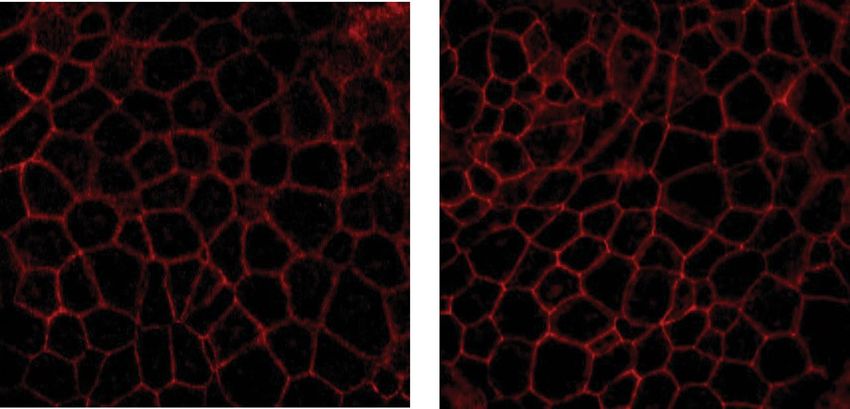
Figure 1. Schematic overview of the different components making up the three compartments used throughout
our experiments. (a) Compartment 1—Digestion-on-a-chip: Three chaotic micromixers representing the
mouth, stomach and intestinal phase of digestion are coupled to one another using PTFE tubing. Conditions
are individually controlled by addition of appropriate artificial juices to each micromixer. (b) Compartment
2—Absorption: A fourth micromixer is used to mix the chyme from the digestion-on-a-chip with cell culture
matrix needed for the cells, in order to dilute the chyme before exposing cells to it. This solution was introduced
to the apical side of the flow-through transwell (FTTW) system containing a co-culture of Caco-2 and HT29-
MTX-E12 cells. For offline analysis, a fraction collector was used to continuously obtain samples from both the
apical and basolateral chambers. For online analysis, the flow-through transwell is coupled to Compartment 3
(c). (c) Compartment 3—On-line analysis with automated sample clean-up: Sample is automatically collected
from the apical and basolateral chambers in the first and second switching valves, respectively. In the third
switching valve, two nanotraps were integrated to retain the analyte of interest and wash away unwanted
sugars and salts. Subsequently, the analyte of interest was eluted to a microfluidic C18 chip-based column and
analysed by QTOF-MS. The state of the valves in this figure indicates the initial configuration at the start of the
experiment, when apical effluent is being collected in the sample loop of Valve 1, while the sample loop in Valve
2 is being flushed with aqueous eluent.
including inlet channels leading to the groove arrays. The grooves perturb the profile of side-by-side laminar
flows entering the mixing channel to generate ‘chaotic’ flow patterns that result in larger contact areas between
solutions. In this way, diffusion distances are shortened substantially and diffusional mixing times dramati-
cally reduced. The different digestive compartments were connected to each other via polytetrafluoroethylene
Scientific Reports | (2021) 11:4920 | https://doi.org/10.1038/s41598-021-84187-9 4
Vol:.(1234567890)www.nature.com/scientificreports/
(PTFE) tubing (0.8/1.6 mm inner/outer diameter, Polyfluor Plastics, Breda, the Netherlands) (Fig. 1a). Flow for
the digestion-on-a-chip was regulated by a pressure-driven flow control system30. Pressurised air was passed
through a microfilter (PTFE, 0.45 µm pore size, Boom B.V., Meppel, the Netherlands) and distributed to the
four glass bottles into which 15 mL tubes (Greiner Bio-One, Frickenhausen, Germany) containing the diges-
tive juices and the sample had been placed. Digestive juices in each of the containers were kept at a constant
pressure of 500 mbar. PTFE tubing was used to connect the liquid-containing glass bottles to Coriolis-based
mass flow controllers (ML120 and BL100, Bronkhorst High-Tech, Ruurlo, The Netherlands), using blunt Fine-
Ject 21G needles (HenkeSassWolf, Tuttlingen, Germany) fitted directly inside the open end of the tubing. The
micro-Coriolis-based mass flow sensors were used to regulate the flow of juices and samples with a far greater
stability and accuracy than would be possible with syringe pumps and flow sensors based on other measurement
principles30. A Bronkhorst software package was used to change flow controller settings and to take measure-
ments of mass flow and density. In Compartment 2, a fourth micromixer (Fig. 1b) was incorporated to dilute the
chyme from the digestion-on-a-chip with the exposure medium (HBSS) required for permeability experiments,
to prevent any cytotoxic effect of chyme on the cells. The effluent from this last micromixer was connected to
the apical side of the flow-through transwell (Fig. 1b). Subsequently, the flow-through transwell was coupled
to a fraction collector, collecting one sample every minute in a 96-well plate. Alternatively, the flow-through
transwell was connected to Compartment 3, which consisted of a series of three switching valves connected to a
microfluidic chip-based UPLC-QTOF-MS (Fig. 1c) as described by Santbergen et al.23 In this latter option, apical
and basolateral effluents from the flow-through transwell were alternately loaded in 5 µL stainless steel sample
loops mounted on the first and second switching valve (Fig. 1c). Each sample loop was loaded for 15 min. After
sample collection, the content of each sample loop was depleted of proteins and bile salts by flushing for 4 min
through an Optimize Technologies (Oregon City, Oregon, USA) C8 nanotrap column (180 µm × 5 mm, 2.7 µm)
using an aqueous solvent (H2O with 1% acetonitrile) at a flow rate of 20 µL/min. Following the clean-up, the trap
column was eluted with a microflow gradient at 3 µL/min towards a microfluidic iKey chip BEH C18 analytical
column for UPLC-QTOF-MS analysis (see below for more details on MS analysis).
Offline and online UPLC‑QTOF‑MS analysis. Offline analysis of omeprazole. For the static cell per-
meability experiments in transwells, 5 µg/mL omeprazole was suspended in HBSS (without phenol red) contain-
ing 25 mM HEPES and 0.35 g/L NaHCO3 as the donor solution. At day 21 of culture, the donor solution was
directly applied to the apical side of the cells (400 µL/insert). The basolateral side was filled with 600 µL of HBSS
per insert. Samples were taken (100 µL) from the basolateral side at 15, 30, 45, 60, 120 and 180 min. The sampled
medium was replenished after each sample acquisition with 100 µL of fresh medium. At t = 0 and t = 180 min,
apical samples were taken.
For dynamic experiments in the flow-through device, including the complete integrated modular GI tract
(Fig. 1a,b), omeprazole was introduced into the digestion-on-a-chip at a concentration of 1 mg/mL in DMSO (1
µL/min). When intestinal digestion only was desired, omeprazole was dissolved in the combined digestive juices
from the mouth, stomach and intestine at a concentration of 40 µg/mL before introduction to the micromixer
in which chyme was diluted with HBSS. In both cases, the final omeprazole concentration on the apical side of
the flow-through transwell was 5 µg/mL. The apical and basolateral effluent flows of the flow-through transwell
were directed to a Gilson 234 autosampler (Villiers-le-Bel, France), which was used as a fraction collector in this
case to collect samples every minute in a 96-well plate. All samples were analysed undiluted by UPLC-QTOF-MS,
using the procedure that is described next. One 2-position/10-port Ultralife switching valve (IDEX Health &
Science, Oak Harbor, Washington, USA) with 1/16″ fittings was used to incorporate online sample preparation
with the microfluidic chip UPLC-QTOF-MS. A nano Acquity autosampler (Waters) set at 10 °C and with a 2 µL
injector was used. The sample loop was flushed for 4 min with mobile phase A (water with 1% acetonitrile and
0.1% formic acid) at a flow rate of 3 µL/min towards a C8 nanotrap column (Optimize Technologies) (180 µm
I.D. × 5 mm, 2.7 µm particles). Following the clean-up, the nanotrap column was eluted towards a microfluidic
chip-based iKey BEH C18 analytical column (150 µm I.D. × 50 mm, particle size 1.7 µm) (Waters) by switching
the valve. The 3 µL/min microflow gradient elution consisted of mobile phase A (cf. above) and mobile phase
B consisting of acetonitrile with 1% water and 0.1% formic acid. The gradient started at 0% B, and after 1 min
was increased to 50% B in 0.1 min. This composition was maintained for 3.9 min, and then increased to 90% in
0.1 min, to be kept constant for 3.9 min. The composition was returned to 0% in 0.1 min and an equilibration
time of 3.9 min was allowed prior to the next injection. MS detection was performed with a Waters Xevo QTOF
MS equipped with an iKey nano electrospray ionisation source operated in the positive ion mode, with a capillary
voltage of 3.9 kV, desolvation temperature of 350 °C, gas flow rate 400 L/h, source temperature of 80 °C and cone
gas flow rate of 10 L/h. Data were acquired and processed using MassLynx v4.1 (Waters) software.
Online analysis of verapamil. Verapamil was introduced into the compartmentalised GI-tract, total-analysis
system at a concentration of 1 mg/mL (Fig. 1a) in either ultrapure water or apple-juice sample matrices (1 µL/
min). The final concentration of verapamil on the apical side of the flow-through transwell was 5 µg/mL. The
process of automated sample clean-up and trapping was described above in the section “Compartmentalised
system design and operation”. In the case of verapamil analysis, the C8 nanotrap column was eluted towards a
microfluidic chip-based iKey BEH C18 analytical column using the following gradient. The 3 µL/min microflow
gradient was based on a published method23 and consisted of mobile phase A (water with 1% acetonitrile) and
mobile phase B (acetonitrile with 1% water), both containing 0.1% formic acid. The gradient started at 10% B
and, after 4 min, was linearly increased to 100% B in 4 min. This composition was kept constant for 3 min, and
then reverted to 10% B in 0.1 min. An equilibration time of 3.9 min was allowed prior to the next injection. MS
detection was performed with a Waters Xevo QTOF mass spectrometer with the same settings as for the offline
Scientific Reports | (2021) 11:4920 | https://doi.org/10.1038/s41598-021-84187-9 5
Vol.:(0123456789)www.nature.com/scientificreports/
analysis of omeprazole. Data were collected using MassLynx, yielding a separate data file for each trap-column
analysis.
Permeability calculations. The apparent permeability coefficient (Papp, cm/s) was calculated as described
ark31, according to the following equation:
by Yeon and P
dQ 1
Papp =
dt AC0
In this equation, dQ/dt is the transport rate into the basolateral compartment (µmol/s), A is the surface
area of the cell layer (0.6 c m2) and C
0 is the initial concentration of the compounds in the apical compartment
(µmol/cm3).
Results and discussion
General considerations. Our aim in this study was to develop a compartmentalised in vitro model of the
GI tract to investigate the bioavailability of orally consumed compounds. Static cell culture systems have gen-
erally been applied for absorption studies, which may not always be optimal for predicting in vivo absorption
behaviour10,32,33. These systems also don’t allow for the inclusion of hands-off digestive sample processing. In our
case, the digestive compartment is based on a well-established batch-based in vitro digestion approach, using
fermenters having volumes of 10–100 mL. The use of batch-based systems implies the intermittent sequential
addition of artificial digestive juices to mimic the different environments that an ingested sample finds itself in in
the GI tract. Transfer of digested material to the absorption model cannot be performed automatically. We have
chosen to implement this digestive approach in a flow-through system to facilitate automation and impart the
freedom to dynamically change conditions in the different stages as desired to achieve enhanced versatility of the
in vitro model12. Our system is unique by virtue of the fact that it combines microfluidic digestion-on-a-chip, an
in vitro intestinal epithelial barrier model and online MS analysis in one automated total analysis system. Besides
automation, the application of flow to transport and process sample from the mouth through to the intestine and
MS analysis also means that the epithelial cell culture in the absorption module is nourished and maintained
under more in vivo-like conditions.
We faced three challenges in the construction and demonstration of our system, namely: (1) the coupling
of compartments having different internal volumes and thus operated at different flow rates, (2) the need for
automated sample clean-up for MS, and (3) cell damage in the absorption module resulting from exposure to
chyme. Above all, maintaining a relevant biological barrier is essential for studying the uptake of compounds.
How we addressed these three challenges to ensure sequential in vitro digestion and absorption is described in
the next three sections.
Assembly of the total analysis system: resolving flow rate incompatibilities. In Fig. 1, a sche-
matic representation is given of our three hyphenated compartments. Figure 1a depicts Compartment 1, the
microfluidic digestion-on-a-chip system, consisting of three ‘chaotic’ micromixers representing the three phases
of digestion in the mouth, stomach and intestine. In the first micromixer, the sample containing the compound
of interest (1 µL/min) was mixed with artificial saliva (4 µL/min), resulting in an effluent flow of 5 µL/min from
the mouth phase. The mouth phase was connected to the stomach phase by PTFE tubing, creating an incubation
time in the oral phase of 2 min dictated by the internal volume of the tubing. In the second micromixer, artificial
gastric juice (8 µL/min) was mixed with the 5 µL/min effluent from the mouth, with a gastric incubation time
of 120 min determined by the volume of the tubing used to connect the stomach micromixer with the intestinal
micromixer. Finally, the 13 µL/min effluent from the gastric phase was mixed with the intestinal juices (12 µL/
min) in the third micromixer, resulting in a final chyme flow rate of 25 µL/min at the outlet of the digestion-
on-a-chip. The incubation time of chyme in the intestinal phase was also 120 min, again dictated by the volume
of the PTFE tubing connecting the intestinal phase with the cell culture barrier compartment. All microfluidic
chips were kept at a constant temperature of 37 °C for optimal enzymatic activity. The selected flow rates and
residence times in the different microreactors were based on average values that are relevant for in vivo human
physiology34–36. Also, the ratios between the respective flow rates represent the in vivo volumetric ratios of the
digestive phases.
The internal volume of the absorption module is 4 mL, 2 mL for the apical chamber and 2 mL for the basolat-
eral chamber. The output flow rate of the digestion-on-a-chip therefore needs to be supplemented substantially
in order to provide a sufficiently high flow rate to operate the absorption module at a physiologically relevant
shear stress for the cells. The chyme from the digestion-on-a-chip (25 µL/min) was therefore mixed with trans-
port buffer HBSS (175 µL/min) using a fourth micromixer, resulting in an eight-fold dilution of the chyme on
the apical side of the cells. Note that at this point in the sample processing, the original sample solution had
been diluted 200 times in total (25 times during passage through the digestion-on-a-chip, 8 times in the fourth
micromixer). An important consideration for the samples analysed is thus that the compounds of interest are
sufficiently soluble in the sample solution to achieve reasonably high initial concentrations to be detectable after
processing-related dilution. Related to this, the detection limit of the final analysis method must be sufficiently
low. The effluent from the fourth micromixer was connected to the apical side of the flow-through transwell
(Fig. 1b) with a total flow rate of 200 µL/min, causing a realistic shear stress on the cultured epithelial cells in
accordance with the in vivo range for the intestine (0.002–0.08 dyne/cm2) 18,29. Finally, the effluent from the
flow-through transwell was connected either to a fraction collector or to the automated online analysis system
(Compartment 3, Fig. 1c).
Scientific Reports | (2021) 11:4920 | https://doi.org/10.1038/s41598-021-84187-9 6
Vol:.(1234567890)www.nature.com/scientificreports/
a 120 b c
100
Cell viability (%)
80
60
40
20
0
0 20 40 60 80 100
% of chyme
Figure 2. (a) Cell viability of Caco-2/HT29-MTX-E12 co-culture after 24 h exposure to increasing
concentrations of chyme, measured using the WST-1 mitochondrial activity assay. Viability is given as a
percentage of the control (% ± standard error of the mean (SEM); triplicates). A nonlinear curve was fitted
through the points for clarity using GraphPad Prism. (b) Confocal image of Caco-2/HT29-MTX-E12 cells
cultured in a transwell for 21 days (control). (c) Confocal image of Caco-2/HT29-MTX-E12 cells cultured in
a transwell for 21 days and exposed to 12.5% chyme for 24 h. All exposures under static conditions. Cells were
stained for tight junction protein ZO-1/TJP1 (red). Scale bar: 20 µm.
Automated sample clean‑up. In our previous work, a flow-through transwell barrier model was also
coupled to MS detection, and sample dissolved in HBSS matrix was added to the apical chamber of the absorp-
tion module23. In this study, however, we have subjected our sample to in vitro digestion first, using the diges-
tion compartment presented e arlier12. The sample thus finds itself in a chyme matrix, which includes not only
physiological concentrations of nutrients and ions, but enzymes and bile salts as well. MS analysis of chyme
will result in increased chemical interference compared to previous studies where the sample was dissolved in
cleaner HBSS buffer. This is an important consideration if UPLC-QTOF-MS is to be used as an online detector.
As mentioned above, the chyme was diluted by a factor of 8 with HBSS buffer before entering the absorp-
tion module. Besides ensuring physiological shear rates for the cell culture, this dilution also served to lower
concentrations of species in the chyme matrix that cause higher background signal in the MS analysis. After the
absorption module, the effluent flows were passed through C8 trap columns to accumulate compounds of interest
and wash away interfering proteins. The compounds of interest were then analysed as described in our previous
study using a set-up described in the Materials and Methods section and shown in Fig. 1c. Despite dilution, a
higher background caused by the increased complexity of the sample matrix was observed. However, it was still
possible to record mass spectra and reconstructed ion chromatograms of characteristic drug ions.
In vitro intestinal barrier integrity upon exposure to chyme. The cell media used conventionally
for absorption studies do not contain digestive compounds (enzymes and bile salts). However, because the
absorption model in our system is preceded by a digestion compartment that produces chyme, it was crucial to
investigate the effects of chyme on the barrier integrity of the cell layer, both before and after the experiments.
Pure chyme is toxic to the Caco-2 and HT29-MTX-E12 cells used in our flow-through transwell m odel24,37. We
therefore undertook a study in which this co-culture was exposed to differing dilutions of chyme in order to
determine a non-toxic chyme composition. This investigation used three different techniques.
First, to determine the toxicity of the mixture of digestive juices coming from the digestion-on-a-chip, we
used a WST-1 viability assay on proliferating cells to assess the mitochondrial activity as a measure of cell viabil-
ity. Intestinal cell cultures were exposed to varying concentrations of chyme for 24 h. As shown in Fig. 2a, cell
viability remained unaffected after 24 h exposure for chyme concentrations up to 62.5% (v/v) in medium. These
results are comparable with a previous in vitro study37, suggesting that the living cells in Compartment 2 can be
exposed to a mixture containing chyme.
For experiments with the digestion-on-a-chip connected to the flow-through transwell, pure chyme was
diluted by a factor of 8 in HBSS to achieve the desired flow rate (200 μL/min) for the apical compartment. This
yielded a 12.5% chyme solution in HBSS, which is well below the upper acceptable concentration of 62.5%
reported above to maintain proper cell viability during experiments. Our choice of a chyme concentration below
62.5% is supported by data from Mahler et al.24 using a microfluidic gut-on-a-chip system. Their study used a
chyme solution, originally described by Glahn et al.38, that exhibited an enzymatic activity that was equivalent to
77% of the enzymatic activity of our chyme (based on pancreatin content in the solution that is presented to the
cells). They observed cell damage (indicated by a drop in both viability and trans-epithelial electrical resistance)
after exposing a Caco-2/HT-29MTX co-culture to the chyme solution for only 2 h. In some cases, damage was
observed after even shorter exposure times. In the in vivo situation, uptake of compounds may start at the same
time as intestinal digestion (i.e., upon entering the duodenum), especially in fasted state and less pronounced
in the fed state of our model. However, undiluted chyme is toxic to the cells in our absorption compartment, so
it must be diluted before entering that compartment. Dilution of chyme causes the concentration of enzymes
contained in the chyme to decrease, with an associated decrease in enzyme activity, i.e. reduced digestive capacity.
Scientific Reports | (2021) 11:4920 | https://doi.org/10.1038/s41598-021-84187-9 7
Vol.:(0123456789)www.nature.com/scientificreports/
Figure 3. Permeability of omeprazole across a monolayer of Caco-2/HT29-MTX-E12 cells in a static
transwell, without digestive juices in the apical matrix. Permeability is given as a percentage of the initial apical
concentration (% ± SEM; n = 3).
This is why digestion and absorption cannot be performed concurrently in our model. This is in agreement with
in vitro digestive systems that are currently used to digest samples before doing uptake s tudies35,36.
For our second series of experiments, we statically exposed a 21-day old co-culture of Caco-2 and HT29-
MTX-E12 cells to 12.5% chyme for 24 h and subsequently stained the tight junction protein ZO-1/TJP1. Co-
cultured cells were exposed to 0% chyme (control, Fig. 2b) and to 12.5% chyme (Fig. 2c), and an interconnect-
ing network of tight junction proteins is shown in red. No differences in the quality of the tight junctions were
observed for the two co-cultures, indicating good barrier integrity. Moreover, cell barrier integrity was also
confirmed after each permeability experiment using the fluorescent marker, lucifer yellow, which is not trans-
ported by the cells. Any translocation of this compound to the basolateral compartment amounting to more
than 5% of the total amount present in the apical compartment thus indicates leakiness of the barrier. Cell layers
were exposed to 500 µg/mL lucifer yellow in HBSS for 30 min after every permeability experiment. Cell layers
allowing more than 5% of lucifer yellow to translocate to the basolateral side were considered leaky, and data
from these cultures were discarded. The cell barriers used for calculating permeability showed 0.9 ± 0.4% lucifer
yellow transport, confirming that the biointegrity of living cells can be fully maintained in the presence of chyme.
In vitro digestion and intestinal permeability of omeprazole. To evaluate digestion-on-a-chip in
combination with our dynamic model of the intestinal barrier, we used the model drug compound, omeprazole
(molecular structure, Fig. S1, SI). Omeprazole is a proton pump inhibitor that irreversibly blocks the last step of
acid production in the stomach wall, thereby increasing the gastric p H39. Omeprazole is preferably administrated
orally via a suspension, tablet or capsule. As omeprazole itself is acid-labile, these drug formulations contain an
enteric coating to protect omeprazole from acid degradation in the stomach27. Omeprazole is then released in
the small intestine and absorbed. Prior to evaluation of the combined set-up comprising digestion and cellular
uptake of omeprazole, we determined a 5 µg/mL concentration of this drug to be non-toxic to the cell co-culture,
using the WST-1 assay (Fig. S2, SI). Static co-cultures of Caco-2 and HT29-MTX-E12 cells were then exposed
to omeprazole at 5 µg/mL for 3 h. In Fig. 3, the cumulative percentage of omeprazole that has crossed the cell
barrier to the basolateral side is given at different time points, reaching 36.1% of the apical concentration after
3 h. The apparent permeability coefficient (Papp) was calculated to be 54.9 ± 12.9 × 10⁻⁶ cm/s, which is in the
same range as in vitro P app data found for omeprazole in the literature (13.4–53.2 × 10⁻⁶ cm/s, for monolayers of
Caco-2 and L-MDR1 cells)40,41. Extrapolation of data obtained in vitro to the in vivo situation remains difficult42,
but the Biopharmaceutics Classification System (BCS)43 that is used by North American and European authori-
ties discriminates drugs based on their in vitro permeability with respect to the drug, metoprolol, of which the
Papp is in the order of 30–50 × 10–6 cm/s44. Both the in vivo and in vitro permeability of drugs in the BCS have
been evaluated extensively, and the correlation between in vitro and in vivo permeabilities has been established.
In other words, if the in vitro permeability of a drug is higher than that of metoprolol, the drug is considered
highly permeable in vitro and likely is highly permeable in vivo as well. This is the case for omeprazole, which
we too find to have a high in vitro permeability. A next step in the extrapolation of data obtained from in vitro
models as described in this work to the in vivo situation would be their use in so-called Physiologically Based
Kinetic Models (PKB models) and Quantitative in vitro to in vivo Extrapolations (QIVIVE)45,46. These models
use in vitro intestinal uptake rates as input in addition to other human physiological parameters, and have been
extensively exploited to support safety assessments of chemicals.
Scientific Reports | (2021) 11:4920 | https://doi.org/10.1038/s41598-021-84187-9 8
Vol:.(1234567890)www.nature.com/scientificreports/
1500
intestine
full
1000
intensity (a.u.)
500
0
8.5 9.0 9.5 10.0 10.5
Time (min)
Figure 4. Reconstructed-ion chromatogram of m/z 346 ([M + H]+) at time point 90 min on the apical side after
only intestinal digestion (black) (Fig. 1b) or full digestion (grey) (Fig. 1a,b). Samples were collected by a fraction
collector followed by offline analysis using chip-based UPLC-QTOF-MS.
Next, we coupled digestion-on-a-chip to the dynamic model of the intestine, using two different set-ups. In
the first set-up, all three chaotic micromixers simulating the mouth, stomach and intestine were implemented,
and connected via a fourth micromixer to the flow-through transwell, using the compartmental set-up depicted
in Fig. 1a,b. Every minute, apical and basolateral samples were collected in two separate 96-well plates using
two fraction collectors. In the second set-up, we emulated the working mechanism of an enteric-coated tablet of
omeprazole, which only releases omeprazole in the intestinal compartment to prevent exposure to gastric acid.
This was done by excluding the mixers for the mouth and stomach compartments to realise a simplified version
of chip-based digestion. Only one micromixer was used to mix the sample (omeprazole) and the pre-mixed
digestive juices (saliva, gastric juice, and intestinal juice). After dilution in the fourth mixer and perfusion of the
dynamic cell coculture, samples were collected from the apical and basolateral side in two separate 96-well plates.
In Fig. 4, the reconstructed ion currents of the [M + H]+ ion of omeprazole at m/z 346 are given for both complete
digestion and exposure to only intestinal digestion after 90 min. The figure clearly shows that the unprotected
omeprazole is fully degraded in the total digestion system, in accordance with expectations; no signal remains
for the m/z 346 ion (in grey). We did not observe any clear degradation products of omeprazole in the MS data47.
In the second experiment mimicking the ingestion of enteric-coated omeprazole, we clearly observed the
omeprazole ion in the apical effluent (Fig. 4, in black), as expected. However, we did not observe any translo-
cation of omeprazole to the basolateral site. This is in contrast to the static permeability data for omeprazole
(Fig. 3), and in vivo data which predict that uptake of omeprazole could be expected in the dynamic flow-through
system40,41. From the literature, it is known that omeprazole heavily binds to plasma proteins48. A control experi-
ment was conducted to examine the effect of digestive juices (chyme) on the translocation of omeprazole in a
static transwell (Fig. S3, SI). It was found that the uptake of omeprazole in the presence of digestive juices was
about three times lower compared to omeprazole dissolved in only HBSS buffer. This may explain why no ome-
prazole was detected in the basolateral compartment of the flow-through transwell after fraction collection and
offline analysis. Translocation appears to have been lowered due to binding with proteins in the chyme matrix.
Moreover, omeprazole will generally be more difficult to detect in the flow-through case than in the static case,
as in a dynamic system no accumulation of the translocated compound occurs due to the collection of samples
every minute.
Compartmentalised in vitro GI tract with online analysis: proof‑of‑principle with and without
co‑exposure to a food matrix. All the compartments of our system (digestion-on-a-chip, flow-through
transwell with co-cultured intestinal cells, and MS analysis) were combined in one hyphenated, online system
(Fig. 1a–c), creating a multi-module GI tract with automated online analysis to monitor in vitro oral bioavail-
ability over time. In a previous study23, the flow-through transwell was combined with online MS analysis. In
this study, we further challenged the system by including microfluidic digestion-on-a-chip (Fig. 1a), allowing
pre-treatment of samples with digestive juices before studying translocation through a model of the gut wall. We
used the model compound, verapamil (molecular structure, Fig. S4, SI), a drug for treatment of high blood pres-
sure and other conditions, for evaluation of the modular in vitro GI tract. Verapamil has the advantage that there
are plenty of data available in the literature for both static and dynamic transwell systems, making it possible
to benchmark our s ystem23,49,50. First, we examined if verapamil is affected by digestion in the different phases
by performing a test tube digestion. As can be seen in Fig. S5 in the supplementary information, verapamil is
not affected by digestion. Therefore, it was hypothesised that verapamil would exhibit similar behaviour in our
modular in vitro GI tract compared to its behaviour in the earlier flow-through set-up reported previously23.
Scientific Reports | (2021) 11:4920 | https://doi.org/10.1038/s41598-021-84187-9 9
Vol.:(0123456789)www.nature.com/scientificreports/
Cumulative permeability verapamil (%)
14
12 apple juice
no matrix
10
8
6
4
2
0
0 15 45 75 105 135 165 195
Time (min)
Figure 5. Permeability of verapamil in apple juice matrix (black) or no food matrix (ultra-pure water, white)
across a monolayer of Caco-2/HT29-MTX-E12 cells, measured in the integrated GI tract with online analysis
set-up combining the three compartments in Fig. 1a–c. Permeability is given as a calculated cumulative
percentage of the starting apical concentration (% ± SEM; n = 1).
Figure 5 shows the cumulative permeability of verapamil over 195 min measured in the entire system shown
in Fig. 1a–c (in white). The results are very similar to the data from Santbergen et al. indicating that including
the additional digestion-on-a-chip functionality affects neither the biointegrity of the co-culture of Caco-2 and
HT29-MTX-E12 cell model, nor the overall analytical p erformance23. In contrast to the reduced absorption of
omeprazole in the presence of digestive juices, the translocation of verapamil is not affected at all.
To emulate the functions of the GI tract even further, a final experiment was performed in which verapamil
was not administered in ultrapure water, but in apple juice as a simplified food matrix. Apple juice was chosen
because it is a reasonably complex, liquid food matrix suitable for the setup (i.e., liquid form), containing sugars
and minerals in water. The absence of mastication in the oral phase of our system does not allow for samples
based on solid foods, and special care must be taken in order to keep the microchannels free of blockages by
precipitates. In Fig. 5, the uptake of verapamil dissolved in apple juice (black) is depicted versus the control in
water (white). Clearly, the absorption of verapamil is much slower in the presence of an apple juice matrix com-
pared to the control. It is well known that food (or certain food ingredients) alters the bioavailability of drugs, for
example by drugs binding to proteins, fats, or calcium ions contained in f ood51,52. Fruit juices have been shown to
inhibit the transport of drugs into cells by organic anion-transporting polypeptides (OATPs) in cell membranes53.
However, since verapamil is mainly transported via passive d iffusion54, this effect was not expected to play a role
in this study. Nevertheless, the uptake of verapamil seems to be affected by apple juice in this proof-of-principle
experiment, and more experiments with different sample matrices are required to ascertain if OATPs are involved
in verapamil transport after all, or if there is another as yet unidentified effect taking place.
Conclusion
The integrated compartmentalised model of the GI tract described in this paper comprises pretreatment of
samples with digestive juices, followed by absorption of sample molecules and their possible metabolites through
an in vitro intestinal epithelial barrier. Online coupling to UPLC-QTOF-MS resulted in an automated online
read-out of oral bioavailability parameters of the molecules. To the best of our knowledge, this system is the
first of its kind developed for assessment of oral bioavailability parameters in pharmacological and toxicological
applications. This system encompasses the two key processes of the human intestinal tract, namely digestion
and absorption. The sequential combination of different compartments having different volumes has been dem-
onstrated, by adjustment of volumetric flow rates between compartments. The physiological relevance of this
system has been improved by the use of medium which has been made to resemble dilute chyme. By applying
an automated sample clean-up system developed in a previous study23, it was possible to analyse the compounds
of interest in dilute chyme using MS detection.
There are few previous studies which have pursued this total-analysis-system route to better mimic the
in vivo situation24,25,37. While development of such a system requires some effort, a good system may lead to a
significant reduction in the need for animal models in these types of studies. This is certainly relevant now, at
a time where many companies are committing to doing far fewer animal experiments. For future applications,
the incorporation of cell types that have higher metabolic capacity than Caco-2 cells is desired. In particular,
3-D culture formats such as organoids could greatly improve the applicability of the s ystem55. In silico models
are being developed, but they rely heavily on data that have been obtained in vivo or in in vitro systems that
extrapolate to the in vivo situation. Systems like ours will be required to optimise these in silico models42,56,57.
Scientific Reports | (2021) 11:4920 | https://doi.org/10.1038/s41598-021-84187-9 10
Vol:.(1234567890)www.nature.com/scientificreports/
In this work, we show maximum versatility for our in vitro model. The complete system as described can
be used to study many physiological processes that involve digestion and absorption of nutritionally as well
as pharmacologically important compounds. There is one caveat associated with the system, namely that it is
unsuited for mimicking in vivo processes in which the digestion of a compound in the intestine actually drives
its absorption through the creation of high local concentrations. (One such process is the supersaturation of
lipid-based drug delivery systems in vivo, the investigation of which would require a different model in vitro58.)
Each compartment can be tailored to specific applications according to the needs of end-users, including resi-
dence times and digestive juice compositions. An example of this in this paper is the simple customisation of
the digestive system for the study of omeprazole. Another route for future research is to include drugs that are
wholly or partially affected by enzymatic action, for example in the conversion of prodrugs into active drugs.
Finally, the flow-through nature of our hyphenated system has potential for the automation of oral bioavailability
testing in drug development (novel drug and drug formulation development, next generation risk assessment)
and chemical toxicity testing.
Received: 28 April 2020; Accepted: 5 February 2021
References
1. Rowland, M. & Tozer, T. N. Clinical Pharmacokinetics and Pharmacodynamics 4th edn, 183–215 (Lippincott Williams & Wilkins,
New York, 2011).
2. Fernandez-Garcia, E., Carvajal-Lerida, I. & Perez-Galvez, A. In vitro bioaccessibility assessment as a prediction tool of nutritional
efficiency. Nutr. Res. 29, 751–760. https://doi.org/10.1016/j.nutres.2009.09.016 (2009).
3. Walczak, A. P. et al. Behaviour of silver nanoparticles and silver ions in an in vitro human gastrointestinal digestion model. Nano-
toxicology 7, 1198–1210. https://doi.org/10.3109/17435390.2012.726382 (2013).
4. Alegria, A., Garcia-Llatas, G. & Cilla, A. in The Impact of Food Bioactives on Health: in vitro and ex vivo models (eds K. Verhoeckx
et al.) 3–12 (2015).
5. DeSesso, J. M. & Jacobson, C. F. Anatomical and physiological parameters affecting gastrointestinal absorption in humans and
rats. Food Chem. Toxicol. 39, 209–228. https://doi.org/10.1016/s0278-6915(00)00136-8 (2001).
6. Trout, G. E. & Fruton, J. S. The side-chain specificity of pepsin. Biochemistry 8, 4183–4190. https://doi.org/10.1021/bi00838a041
(1969).
7. Whitcomb, D. C. & Lowe, M. E. Human pancreatic digestive enzymes. Dig. Dis. Sci. 52, 1–17. https://doi.org/10.1007/s10620-006-
9589-z (2007).
8. Carey, M. C., Small, D. M. & Bliss, C. M. Lipid digestion and absorption. Annu. Rev. Physiol. 45, 651–677. https://doi.org/10.1146/
annurev.ph.45.030183.003251 (1983).
9. Musther, H., Olivares-Morales, A., Hatley, O. J. D., Liu, B. & Hodjegan, A. R. Animal versus human oral drug bioavailability: Do
they correlate? Eur. J. Pharm. Sci. 57, 280–291. https://doi.org/10.1016/j.ejps.2013.08.018 (2014).
10. Lea, T. in The Impact of Food Bioactives on Health: in vitro and ex vivo models (eds K. Verhoeckx et al.) 95–102 (2015).
11. Costa, J. & Ahluwalia, A. Advances and current challenges in intestinal in vitro model engineering: A digest. Front. Bioeng. Bio-
technol. 7, 144. https://doi.org/10.3389/fbioe.2019.00144 (2019).
12. de Haan, P. et al. Digestion-on-a-chip: A continuous-flow modular microsystem recreating enzymatic digestion in the gastroin-
testinal tract. Lab Chip 19, 1599–1609. https://doi.org/10.1039/c8lc01080c (2019).
13. Kulthong, K. et al. Implementation of a dynamic intestinal gut-on-a-chip barrier model for transport studies of lipophilic dioxin
congeners. RSC Adv. 8, 32440–32453. https://doi.org/10.1039/c8ra05430d (2018).
14. Bein, A. et al. Microfluidic organ-on-a-chip models of human intestine. Cell. Mol. Gastroenterol. Hepatol. 5, 659–668. https://doi.
org/10.1016/j.jcmgh.2017.12.010 (2018).
15. Villenave, R. et al. Human gut-on-a-chip supports polarized infection of coxsackie B1 virus in vitro. PLoS ONE 12, e0169412. https
://doi.org/10.1371/journal.pone.0169412 (2017).
16. Imura, Y., Asano, Y., Sato, K. & Yoshimura, E. A microfluidic system to evaluate intestinal absorption. Anal. Sci. 25, 1403–1407.
https://doi.org/10.2116/analsci.25.1403 (2009).
17. Gao, D., Liu, H. X., Lin, J. M., Wang, Y. N. & Jiang, Y. Y. Characterization of drug permeability in Caco-2 monolayers by mass
spectrometry on a membrane-based microfluidic device. Lab Chip 13, 978–985. https://doi.org/10.1039/c2lc41215b (2013).
18. Kim, H. J., Huh, D., Hamilton, G. & Ingber, D. E. Human gut-on-a-chip inhabited by microbial flora that experiences intestinal
peristalsis-like motions and flow. Lab Chip 12, 2165–2174. https://doi.org/10.1039/c2lc40074j (2012).
19. Kulthong, K. et al. Microfluidic chip for culturing intestinal epithelial cell layers: Characterization and comparison of drug transport
between dynamic and static models. Toxicol In Vitro https://doi.org/10.1016/j.tiv.2020.104815 (2020).
20. Kim, H. J. & Ingber, D. E. Gut-on-a-chip microenvironment induces human intestinal cells to undergo villus differentiation. Integr.
Biol. (Camb) 5, 1130–1140. https://doi.org/10.1039/c3ib40126j (2013).
21. Santbergen, M. J. C., van der Zande, M., Bouwmeester, H. & Nielen, M. W. F. Online and in situ analysis of organs-on-a-chip.
Trac-Trends Anal. Chem. 115, 138–146. https://doi.org/10.1016/j.trac.2019.04.006 (2019).
22. Zhang, Y. S. et al. Multisensor-integrated organs-on-chips platform for automated and continual in situ monitoring of organoid
behaviors. Proc. Natl. Acad. Sci. U.S.A. 114, E2293–E2302. https://doi.org/10.1073/pnas.1612906114 (2017).
23. Santbergen, M. J. C., van der Zande, M., Gerssen, A., Bouwmeester, H. & Nielen, M. W. F. Dynamic in vitro intestinal barrier
model coupled to chip-based liquid chromatography mass spectrometry for oral bioavailability studies. Anal. Bioanal. Chem. 412,
1111–1122. https://doi.org/10.1007/s00216-019-02336-6 (2020).
24. Mahler, G. J., Esch, M. B., Glahn, R. P. & Shuler, M. L. Characterization of a gastrointestinal tract microscale cell culture analog
used to predict drug toxicity. Biotechnol. Bioeng. 104, 193–205 (2009).
25. Imura, Y., Yoshimura, E. & Sato, K. Micro total bioassay system for oral drugs: Evaluation of gastrointestinal degradation, intestinal
absorption, hepatic metabolism, and bioactivity. Anal. Sci. 28, 197–199 (2012).
26. Mallet, C. R., Lu, Z. & Mazzeo, J. R. A study of ion suppression effects in electrospray ionization from mobile phase additives and
solid-phase extracts. Rapid Commun. Mass Spectrom. 18, 49–58. https://doi.org/10.1002/rcm.1276 (2004).
27. Riedel, A. & Leopold, C. S. Degradation of omeprazole induced by enteric polymer solutions and aqueous dispersions: HPLC
investigations. Drug Dev. Ind. Pharm. 31, 151–160. https://doi.org/10.1081/Ddc-200047787 (2005).
28. Bouwman-Boer, Y., le Brun, P., Woerdenbag, H., Tel, R. & Oussoren, C. Recepteerkunde. 5th edn, (Bohn Stafleu van Loghum, 2009).
29. Giusti, S. et al. A novel dual-flow bioreactor simulates increased fluorescein permeability in epithelial tissue barriers. Biotechnol.
J. 9, 1175–1184. https://doi.org/10.1002/biot.201400004 (2014).
Scientific Reports | (2021) 11:4920 | https://doi.org/10.1038/s41598-021-84187-9 11
Vol.:(0123456789)You can also read