Reappraisal and new material of the holotype of Draconyx loureiroi (Ornithischia: Iguanodontia) provide insights on the tempo and modo of ...
←
→
Page content transcription
If your browser does not render page correctly, please read the page content below
Zoological Journal of the Linnean Society, 2022, 195, 125–156. With 15 figures.

Reappraisal and new material of the holotype of
Draconyx loureiroi (Ornithischia: Iguanodontia) provide
insights on the tempo and modo of evolution of
thumb-spiked dinosaurs

                                                                                                                              Downloaded from https://academic.oup.com/zoolinnean/article/195/1/125/6535466 by guest on 21 June 2022
FILIPPO MARIA ROTATORI1,2,*, , MIGUEL MORENO-AZANZA1,2,3, and
OCTÁVIO MATEUS1,2,
1
  GEOBIOTEC, Department of Earth Sciences, NOVA School of Science and Technology, Campus de
Caparica, P-2829 516 Caparica, Portugal
2
  Museu da Lourinhã, Rua João Luis de Moura 95, 2530-158, Lourinhã, Portugal
3
  Grupo Aragosaurus-IUCA, Facultad de Ciencias, Universidad de Zaragoza, 50009, Zaragoza, Spain

Received 9 April 2021; revised 9 November 2021; accepted for publication 26 November 2021

The Upper Jurassic Lourinhã Formation is well known for its rich assemblage of fossil vertebrates. In this formation,
ornithopod dinosaurs are represented by two iguanodontian species, Eousdryosaurus nanohallucis and Draconyx
loureiroi. We recently became aware of unreported material belonging to the holotype of Draconyx loureiroi, consisting
of partially articulated manual elements. We here re-describe the holotype specimen ML 357, including the newly
discovered material. The specimen was subjected to CT-scanning and its surface data used to assess anatomical
characters. Linear measurements of metatarsal III were used to estimate the body length of the specimen. The
Draconyx loureiroi holotype was included in two datasets and analysed with maximum parsimony and Bayesian
inference approaches to estimate evolutionary rates among Iguanodontia. We present evidence that Draconyx
loureiroi is a valid taxon nested in Styracosterna and is clearly diagnosable by a unique combination of characters.
Both maximum parsimony and Bayesian inference indicate high evolutionary rates across the Jurassic/Cretaceous
transition for the base of Iguanodontia. Length estimation suggests that Draconyx loureiroi was a relatively small,
bipedal and possibly cursorial animal. Given its basal phyletic position, we interpret this bauplan was the ancestral
condition for Styracosterna, that only later in the Cretaceous evolved into giant quadrupedal forms.

KEYWORDS: Ornithopoda – Phylogenetic analysis – Bayesian inference – Jurassic – Europe.

                   INTRODUCTION                                    2015; McDonald et al., 2012), while Late Jurassic taxa
                                                                   are represented by fewer and, in some cases, incomplete
Iguanodontia (sensu Madzia et al., 2018) is a highly
                                                                   specimens (Galton & Powell, 1980; Mateus & Antunes,
diverse clade of ornithischian dinosaurs with basal
                                                                   2001; Ruiz-Omeñaca et al., 2006, Escaso et al., 2014).
(non-hadrosauroid) forms as a frequent component of
                                                                      The relationships at the base of Iguanodontia
Late Jurassic and Early Cretaceous ecosystems (Galton,
                                                                   (generally within basal Ornithopoda) have
1980, 2006, 2009; McDonald et al., 2010b; Foster,
                                                                   undergone several changes in recent years (Boyd,
2020). Despite their long history of research (Mantell,
                                                                   2015; Dieudonné et al., 2016, 2021; Bell et al., 2018;
1825) and their widespread biogeographic distribution,
                                                                   Madzia et al., 2018; Herne et al., 2019; Rozadilla
most of what is known of the anatomy and phyletic
                                                                   et al., 2019), profiling a more complex evolutionary
relationships of Late Jurassic species is relegated to the
                                                                   history of this clade than previously inferred (e.g.
North American continent and Africa. Europe has also
                                                                   McDonald, 2012; Norman, 2015). After McDonald
produced many relevant specimens and species dated
                                                                   (2012) and derived matrices (Xu et al., 2018; Verdú
to the Early Cretaceous (Norman, 1980, 1986, 2004,
                                                                   et al., 2018, 2019), several studies have considered the
                                                                   global relationships of Iguanodontia, often focusing
*Corresponding author. E-mail: filippo.rotatori.93@gmail.com       on less inclusive clades (Boyd, 2015; Madzia et al.,

© The Author(s) 2022. Published by Oxford University Press on behalf of The Linnean Society of London.                125
All rights reserved. For permissions, please e-mail: journals.permissions@oup.com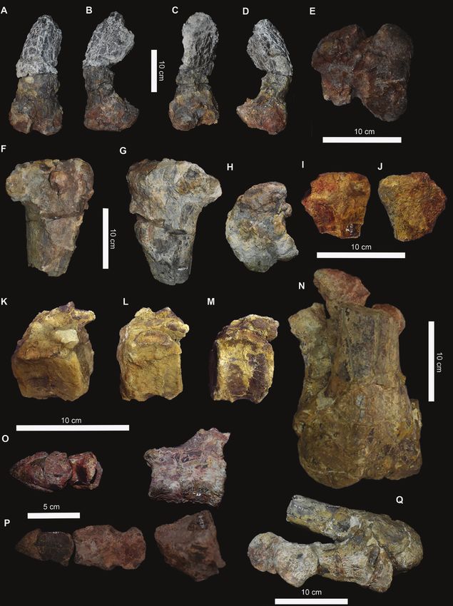
126    F. M. ROTATORI ET AL.

2018; Dieudonné et al., 2021). European taxa, despite                            Geological setting
their paucity and poor preservation, may help to              The Lourinhã Formation (Fig. 1) is a heterogenous
untangle the intricate evolutionary relationships at          siliciclastic succession of continental deposits dated to
the root of Iguanodontia. Recently, two independent           the Kimmeridgian–Tithonian interval and located in the
lines of evidence (ichnological and body fossil               Meso-Cenozoic Lusitanian Basin (Kullberg et al., 2014),
records) have pointed out that the diversity of               in which subdivisions have been debated (Hill, 1989;
iguanodontians in the Late Jurassic of Europe may             Taylor et al., 2014). Generally, it is composed of sandstones
be severely underestimated, suggesting the presence           and mudstones with rare limestone intercalations,
of styracosternans more similar to Early Cretaceous           representing a braided fluvial system and alluvial fans,
forms than to other coeval iguanodontians (Castanera          with occasional marine transgressions (Hill, 1989; Taylor
et al., 2020, 2021; Rotatori et al., 2020).                   et al., 2014). Since a detailed revision and overview of this
   Draconyx loureiroi Mateus & Antunes, 2001 is

                                                                                                                               Downloaded from https://academic.oup.com/zoolinnean/article/195/1/125/6535466 by guest on 21 June 2022
                                                              lithostratigraphic unit is beyond the scope of the current
one of two ornithopod taxa described from the Late            study, for simplicity the subdivisions described by
Jurassic of the Iberian Peninsula. The holotype ML            Mateus et al. (2017) are used. The Lourinhã Fm. outcrops
357, and the only specimen so far, is a medium-sized          in two sub-basins: the Consolação and Turcifal. In the
iguanodontian that was recovered from the Upper               first, three members are distinctly recognizable (from
Jurassic Lourinhã Formation (Portugal) in 1991                bottom to top): Porto Novo/Praia da Amoreira Member,
(Mateus & Antunes, 2001). Histological studies                Praia Azul Member and the Santa Rita Member. In
have shown that ML 357 is a senile individual,                the Turcifal sub-basin, the only member outcropping is
approximately 30 years old (Waskow & Mateus,                  the Assenta Member, which is laterally equivalent to the
2017). Mateus & Antunes (2001) classified the                 Santa Rita Member.
specimen as a basal iguanodontian, attributing the               The Draconyx loureiroi holotype was recovered from
species to the family ‘Camptosauridae’, composed at           the Praia Azul Member, which is composed of three
that time of the species Camptosaurus dispar Marsh,           extensive carbonated shell-layers, cutting a succession
1879, ‘Camptosaurus’ (= Cumnoria) prestwichii                 of fine mudstones (Hill, 1989; Taylor et al., 2014).
Hulke, 1880, Callovosaurus leedsi Ruiz-Omeñaca                Each layer represents a marine transgression episode
et al. 2006 and Draconyx loureiroi. Subsequent                along the second layer in the transition between the
analyses by McDonald (2011) recovered the genus               Kimmeridgian and Tithonian stages (Hill, 1989;
Camptosaurus as paraphyletic, while Ruiz-Omeñaca              Taylor et al., 2014; Mateus et al., 2017). The specimen
et al. (2006) have recovered Callovosaurus leedsi as          ML 357 was recovered just above the second layer and
a member of Dryosauridae. Since then, the clade               is, therefore, considered lowermost Tithonian in age
‘Camptosauridae’ has been recovered from a few                (Mateus & Antunes, 2001).
analyses with weak support (i.e. Verdú et al., 2018).
Other authors treated Draconyx loureiroi as a
wildcard taxon (McDonald, 2012) or they excluded
                                                                           MATERIAL AND METHODS
it a priori in their analyses due to its fragmentary
nature. Recently, Verdú et al. (2018) recovered               ML 357 is a partial skeleton, including appendicular
D. loureiroi in a more derived position than                  and axial elements. It was recovered in 1991 and
Camptosaurus dispar.                                          has been housed in the collections of the Museu da
   In the present study, we contribute to our knowledge       Lourinhã since. The specimen includes an almost
of the anatomy and phylogenetic relationships of Late         complete and articulated foot and several other
Jurassic European ornithopod taxa by re-describing the        disarticulated and isolated postcranial elements.
holotype of Draconyx loureiroi (ML 357) and including         We were recently informed by the discoverer of
undescribed material that was made available by the           this specimen, Carlos Anunciação, of the existence
original collector. We analyse in detail its phylogenetic     of unreported material belonging to the holotype
relationships and its significance for the evolutionary       specimen, stored in his home since its collection in
history of iguanodontians via time-calibrated analyse,        1991. This unreported material has now been donated
employing both maximum parsimony and Bayesian                 to the Museu da Lourinhã and is added to the original
inference. The results indicate a more deeply nested          ML 357 entry number. This newly donated material
position in Ankylopollexia than previously inferred by        includes manual phalanges and further carpal
other authors.                                                elements. It is clearly from the same locality, matches
   Abbreviations: ML, Museu da Lourinhã, Lourinhã,            in size and is consistent in other features of ML 357.
Portugal; SHN, Sociedade de História Natural, Torres          Furthermore, the lack of repeated elements supports
Vedras, Portugal; NMNH, National Museum of Natural            the interpretation that both the old and the new
History, Washington DC, USA.                                  material represent the same individual. In addition,

                         © 2022 The Linnean Society of London, Zoological Journal of the Linnean Society, 2022, 195, 125–156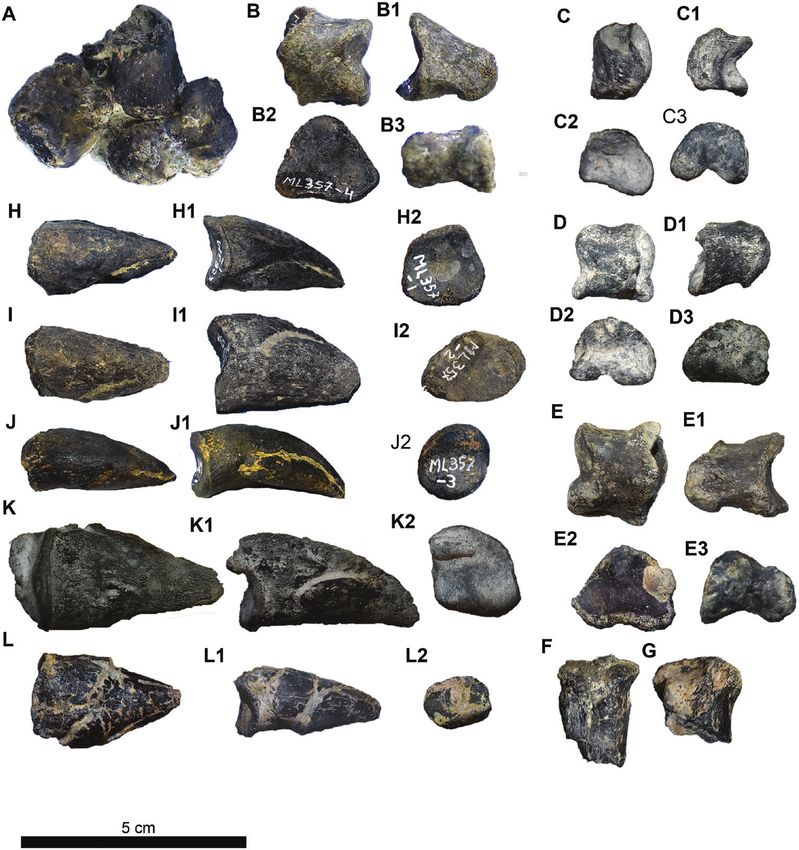
HOLOTYPE OF DRACONYX LOUREIROI                    127

                                                                                                                               Downloaded from https://academic.oup.com/zoolinnean/article/195/1/125/6535466 by guest on 21 June 2022
Figure 1. Geographical and geological settings of ML 357. A, B, Stratigraphic log of the Lourinhã Formation and position of
the specimen. C, Re-adapted from Rotatori et al. (2020). Map of the Iberian Peninsula courtesy of Eduardo Puertolas-Pascual.

the fragments of rock attached to the bones of the new           Therefore, the surface data were exported as 3D
material, a dense grey muddy siltstone, are identical            meshes by AVIZO v.9 in the free software BLENDER
to that found encasing the old material, adding further          (Hess, 2010) and anatomical structures were isolated.
support to the suggestion that it is part of the same            As a third step, the specimen was articulated and
individual. Both the old and new material of ML 357              rendered producing the images present herein.
have been prepared mechanically. Its post-cranium,                 To test its phylogenetic affinities, the old and newly
except for the manual elements, has been subjected to            reported material of ML 357, was first added to the
CT-scanning in order to produce a three-dimensional              dataset of Boyd (2015), followed by the modifications
model to illustrate its anatomy and better assess                of Madzia et al. (2018) and Bell et al. (2018). We
its phylogenetic characters. The specimen was first              performed one global analysis, since the new material
scanned at a veterinary facility, ‘Hospital veterinario          did not provide any new character scores. The analysis
do Oeste’, using a Siemens Somatom Emotion 6 with                was carried out in TNT v.1.5 (Goloboff & Catalano,
voltage set to 130 kV and a current of 200 ųA. To                2016), the tree search was performed in two steps, as
improve resolution of the first scan, the tarsus and             described by Bell et al. (2018), and a ‘New Technology
pedal elements were re-scanned by the company                    Search’ was conducted to find starting trees, setting 30
‘MiconSense’ using a GE VtomeX M 240, with voltage               cycles of Drift and 50 cycles of Ratchet. Default options
of 200 kV and a current of 500 ųA. It was not possible           were used for the other parameters. The search was
to segment the specimen, because of noise caused                 performed using 1000 random additional sequences.
by permineralization due to diagenetic processes.                Subsequently, the most parsimonious trees (MPTs)

© 2022 The Linnean Society of London, Zoological Journal of the Linnean Society, 2022, 195, 125–156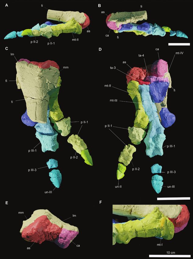
128    F. M. ROTATORI ET AL.

were subjected to an additional round of tree bisection         The analysis sampled 10 000 000 generations per run,
reconnection (TBR) to further explore the tree-space.           sampled with a MCMC method for four runs of six
   After the first phylogenetic analysis confirmed the          chains per run. The initial ‘burn-in’ was set at 25%.
inclusion of ML 357 in Ankylopollexia, the specimen             Convergence and stationarity were again assessed in
was added to the dataset of Xu et al. (2018) in order to        TRACER v.1.7.1, adopting the same criteria described
explore its affinities within the clade. We corrected the       in the non-clock analysis. We provided a soft root upper
scoring for character 121 of Dryosaurus altus Marsh,            prior (220, 205) for the tree, lower than the stratigraphic
1878, Dysalotosaurus lettowvorbecki Virchow, 1919               occurrence of the outgroup taxon (Lesothosaurus
(scored as: 0), Valdosaurus canaliculatus Galton, 1975          diagnosticus Galton, 1978) and being consistent with
and Uteodon aphanoecetes (Carpenter & Wilson, 2008)             the ghost lineage of Ornithischia inferred by other
(scored as ‘?’) based on a literature review (Galton,           works (Baron, 2019). Since MrBayes needs an extant
1981; Carpenter & Wilson, 2008; Barrett et al., 2011;           taxon to perform the calculation of evolutionary rates,

                                                                                                                                     Downloaded from https://academic.oup.com/zoolinnean/article/195/1/125/6535466 by guest on 21 June 2022
Barrett, 2016). As a tree-search strategy, we performed         we set the occurrence interval for Lesothosaurus
a heuristic search with 1000 replicates, keeping ten            diagnosticus as (10, 0 Mya). We opted for this operation
trees per replicate. The MPTs were then subjected to            instead of creating a dummy taxon coded for just 0
the TBR algorithm as in the previous search.                    because the matrix of Xu et al. (2018) is designed in a
   We investigated the variation of evolutionary                way that L. diagnosticus has this coding. The addition of
rates across the topology obtained by the analysis of           a dummy taxon would create unnecessary noise within
the dataset of Xu et al. (2018). We used the script for         the topology and the rooting. Following Simões et al.
TNT developed by Rauhut & Pol (2019) to calculate               (2020a, b), we implemented an informative prior for
the homoplasy concentration (HC) index, which is                the base of the clock rate. This prior value is obtained
linearly related to an increase in evolutionary rates.          by subdividing the median value for tree length in
The ages (FAD/LAD) of terminal taxa were downloaded             substitutions from posterior trees by the age of the tree,
from the Paleobiology Database on 19 January 2021,              based on the median of the distribution for the root
inserting the term Iguanodontia, and in case of multiple        prior (4.0714/205.5 = 0.02) (Simões et al., 2020a, b).
contradictory entries, we chose the age range with the            We m o d e l l e d r a t e s b a s e d o n a l o g - n o r m a l
greatest number of entries.                                     distribution, with the rates sampled from the log-
   As an additional line of evidence, we performed a            normal distribution and the mean of the log-normal
third set of analyses using Bayesian inference. We              distribution given the value based on the non-clock
re-analysed the dataset of Xu et al. (2018) in the program      tree estimate (0.02) in natural log scale = –3.9120. We
MrBayes v.3.2.7 (Ronquist et al., 2012) in two steps:           chose to use the exponent of the mean to provide a
first, we performed a non-clock analysis to calculate           broad standard deviation (e0.02 = 1.0202). The terminal
background evolutionary rate and then we proceeded to           age uncertainty of the previously downloaded FAD/
a clock analysis, time-calibrating the topology recovered       LAD was modelled according to a uniform prior.
from the non-clock analysis. The character evolution              In our attempt to reconstruct the body length
followed the Mk model (Lewis, 2001) and the values              of specimen ML 357, we used the linear equations
were sampled from a gamma distribution. The analysis            introduced by Becerra & Ramirez (2018). We opted for
sampled 10 000 000 generations per run, sampled with            the ones derived for metatarsal III, since it is the most
Markov chain Monte Carlo (MCMC) method for four                 complete and unaltered appendicular element in ML
runs of six chains per run. The initial ‘burn-in’ was set at    357. In addition, Becerra & Ramirez (2018) categorized
25%. Convergence of independent runs and stationarity           locomotory models of ornithischian dinosaurs as
were assessed through the program TRACER v.1.7.1                (1) cursorial, (2) subcursorial and (3) graviportal,
(Rambaut et al., 2018) considering effective sample             based on the ratio of the femur and whole leg length.
size (ESS) for each parameter informative with values           Ornithopods (including hadrosaurs) are considered
equal or > 200. Then we proceeded to the clock-analysis         to range from a cursorial to subcursorial locomotion
according to the fossilized birth–death model (FBD;             stance. Since the hindlimb of ML 357 is not preserved
Heath et al., 2014), under maximum diversity sampling           in its integrity, we estimated the body length of ML 357
tree methods corrected to exclude a sampled-ancestor            assuming a fully cursorial locomotory model according
model. The topology recovered from parsimony and                to the equation (1):
non-clock analyses was implemented as strict prior.
It employed the relaxed uncorrelated clock model                               bl = −147.18 + 17.45 × (m3)
independent gamma rate (IGR) and the background
values for the clock-rates were sampled by a log-normal         Second, a subcursorial locomotory model according to
distribution. As in the previous analysis, character            the equation (2):
evolution followed the Mk model (Lewis, 2001) and
                                                                               bl = −441.11 + 24.54 × (m3)
the values were sampled from a gamma distribution.

                           © 2022 The Linnean Society of London, Zoological Journal of the Linnean Society, 2022, 195, 125–156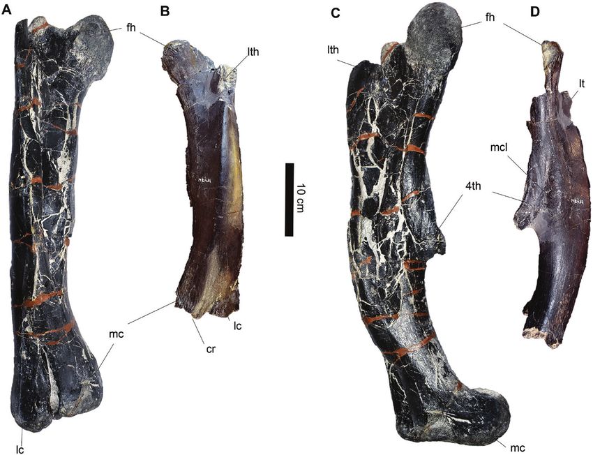
HOLOTYPE OF DRACONYX LOUREIROI                  129

Where bl is the estimated body length and m3 is the              Description and comparisons
linear measurement of the metatarsal III. The ecological         Cranial material: Maxillary teeth (ML 357–31) (Fig.
implications of both models are discussed below in the           2A, B): One of the maxillary teeth preserves part of
Discussion. Selected measurements of ML 357 are                  the root and its crown is complete (Fig. 2A). The other
provided in Table 1. Supporting Information including:           tooth is just an isolated crown (Fig. 2B). The specimens
TNT files of the parsimony analyses (Files S2, S3), the          appear to have suffered some erosion and post-mortem
infiles of the non-clock (File S4) and clock-analysis (File      breakage. The root is slightly labiolingually curved
S5) and a 3D model of the leg of ML 357 (File S6).               and tapers smoothly into the crown (Fig. 2A). There
                                                                 is no cingulum at the junction between the root and
                        RESULTS                                  the crown. Overall, the crown is leaf-shaped and the
                                                                 veneer of enamel is thicker on the labial side (Fig. 2A).

                                                                                                                             Downloaded from https://academic.oup.com/zoolinnean/article/195/1/125/6535466 by guest on 21 June 2022
             Systematic palaeontology                            Labially a thick primary ridge is distally offset and
                Dinosauria Owen, 1842                            five accessory ridges are present on the mesial surface
              Ornithischia Seeley, 1887                          (Fig. 2A, B). Non-mammillated hook-like denticles are
                                                                 coarsely present on the mesial crown margin, while
              Ornithopoda Marsh, 1881                            distally they appear to have been obliterated by either
             Iguanodontia Sereno, 1986                           erosion or occlusal wear. An extensive occlusal surface
       Dryomorpha Milner & Norman, 1984                          develops on the apex of the crown and is inclined
                                                                 labiolingually approximately around 30° (Fig. 2A, B).
            Ankylopollexia Sereno, 1986
            Styracosterna Sereno, 1986
                                                                 Remarks: The tapering root, slightly labiolingually
  Draconyx loureiroi Mateus & Antunes, 2001                      recurved and the leaf-shaped crown are common
                        (Figs 2–7)                               characteristics of Dryomorpha (Galton, 1983, 2006;
                                                                 Norman, 1986, 2004). The maxillary crowns possess a
Type material: The holotype specimen (newly reported             distally offset primary ridge resembling the condition of
material marked with *), ML 357 (subnumbers from 1               ankylopollexians but differing from dryosaurids in which
to 31) includes two maxillary teeth, carpal bones, two           the primary ridge is located towards the centre of the
metacarpal distal ends*, three left carpal phalanges,            crown (Galton, 2006). In the original description, Mateus
one right carpal phalanx*, two left unguals, three               & Antunes (2001) indicated the presence of five accessory
right unguals* and one right leg including proximal              ridges in the distal half of the crown as an autapomorphy
femoral epiphysis, proximal and distal epiphysis of              of Draconyx loureiroi. However, the number of accessory
the tibia, astragalus, calcaneum, four metatarsals, five         ridges is variable throughout the dental series (Galton,
phalanges and two unguals.                                       2006). Therefore, we regard it as a non-informative
                                                                 character if the tooth position is not articulated.
Referred specimen: An isolated left femur, ML 434 from
1 km south of the type locality previously referred to           Axial skeleton: Caudal vertebrae (ML 357–9–11) (Figs
Draconyx loureiroi by Mateus & Antunes (2001), now               3K–M, 4A–F): Three proximal caudal vertebrae are
referred to Ankylopollexia indet.                                preserved and, as for the rest of the skeleton, they have
                                                                 suffered breakage and erosion. The centra are stout
Type locality, horizon and age: Vale de Frades,                  and craniocaudally compressed. The largest centrum
Lourinhã Municipality, Portugal. Praia Azul Member               is ML 357–9, while ML 357–10 and ML 357–11 are
of Lourinhã Formation, lower Tithonian, 151–152 Mya.             slightly smaller. The cranial and caudal surfaces are
                                                                 slightly amphicoelous, having a subelliptical, rounded
Emended diagnosis: Styracosternan iguanodontian                  shape (Fig. 4 A, C, G, I, M, O). Dorsally, the neural
distinguished from other basal iguanodontians by the             arch preserves the ventralmost portion of the neural
following combination of characters: unfused and non-            spine but the neural suture is not visible. Immediately
interlocked carpus; absence of a sharp crest running             caudal to the spine, in the most complete specimens
from the medial condyle of the femur towards of the              ML 357-9 and ML 357 - 10, (Fig. 4C, O) two small,
lesser trochanter, fully open U-shaped extensor groove           rounded and steeply inclined post-zygapophyses
on distal epiphysis of the femur; fully open V-shaped            diverge laterally. On the lateral surface of the neural
flexor groove without overhangs on distal epiphysis              arch, the articulation for the transverse processes
of the femur; concave medial margin of proximal                  is overlain by a broken surface. The presence of this
epiphysis of the tibia; caudally pointing fibular                surface indicates that these vertebrae represent the
condyle of the tibia; and a splint-like metatarsal I.            cranialmost portion of the caudal series. In ventral

© 2022 The Linnean Society of London, Zoological Journal of the Linnean Society, 2022, 195, 125–156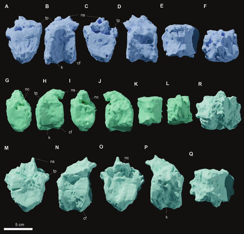
Table 1. Selected measurements of ML 357. All measurements are in mm (fragmented material marked with *)
130
Height Mesiodistal width Labiolingual width
Complete maxillary tooth 16 10 6
ML 357–31
Maxillary crown tooh 11 9 9
ML 357–31
Total Length Centrum cranial Centrum cranial Centrum caudal Centrum caudal Neural spine Neural arch
mediolateral dorsoventral mediolateral dorsoventral height mediolateral
width height width height width
Caudal vertebra n.1 59 58 52 55 56 56 80
ML 357–9
F. M. ROTATORI ET AL.
Caudal vertebra n.2 47 48 53 47 52 45 60
ML 357–10
Caudal vertebra n.3 47 44 48 40 50 45 60
ML 357–11
Articulated carpus Total Length Mediolateral width Craniocaudal height
Metacarpal (fragment) 32
ML 357–5
Carpal n.1 ML 357–5 20 22
Carpal n.2 ML 357–5 13 13
Carpal n.3 ML 357–5 14 18
Mediolateral distal Extensor-palmar
width distal height
Distal metacarpal 1 ML 23 18 6
357–25
Distal metacarpal 2 ML 18 18 16
357–26
Left manual elements Mediolateral distal Mediolateral Dorsovental Dorsovental
width proximal width distal height proximal height
Manual phalanx n.2 ML 8 15 18 10 10
357–22
Manual phalanx n.3 ML 17 16 19 10 13
357–21
Manual phalanx n.4 ML 19 18 20 9 15
357–24
Manual ungual n.1 ML 35 22 15
357–1
Manual ungual n.4 ML 38 15 15
357–23
© 2022 The Linnean Society of London, Zoological Journal of the Linnean Society, 2022, 195, 125–156
Downloaded from https://academic.oup.com/zoolinnean/article/195/1/125/6535466 by guest on 21 June 2022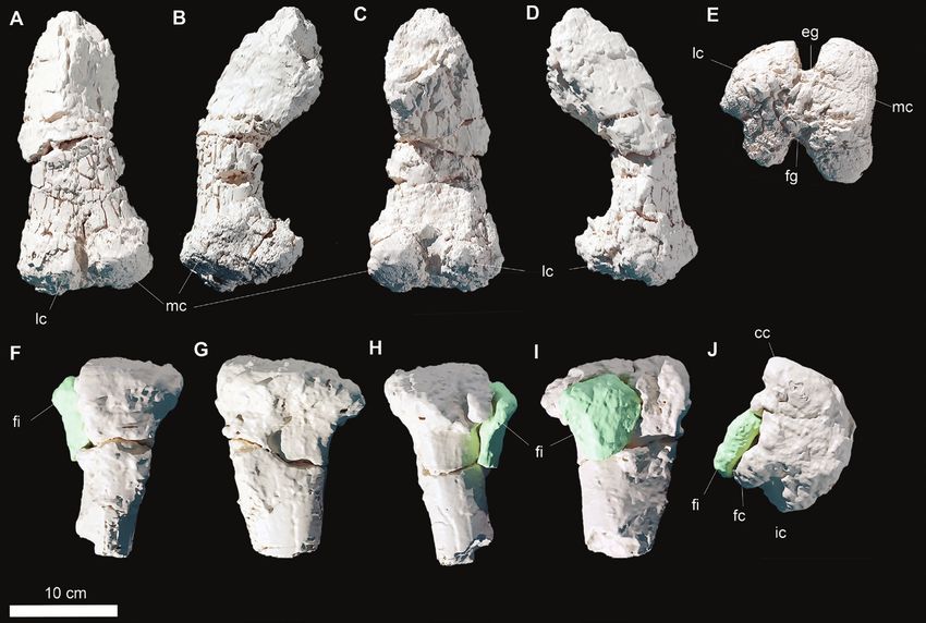
Table 1. Continued
Right manual elements
Manual phalanx n.1 17 17 22 10 17
ML 357–4
Manual ungual n.2 zML 30 17 15
357–2
Manual ungual n.3 33 12 14
ML 357–3
Manual ungual n.5 33 18 13
ML 357–20
Total Length Mediolateral distal Medial condyle Lateral condyle Flexor groove Extensor groove
width length length width width
Femur ML 357–6 280 120 120 110 50 30
Total Length Craniocaudal distal Mediolateral width
width
Tibia proximal epiphysis 210 140 135
ML 357–7
Total Length Mediolateral distal
width
Tibia distal epiphysis ML 210 150
357–12
Craniocaudal Craniocaudal length
length
Fibula (proximal) ML 75 90
357–8
Pes ML 357–12
Mediolateral Dorsoventral height
width
Tarsal 3 ML 357–12 50
Tarsal 4 ML 357–12 70
Astragalus ML 357–12 90 50
Calcaneum ML 357–12 30 70
Total Length Mediolateral distal Mediolateral Dorsoplantar Dorsplantar
© 2022 The Linnean Society of London, Zoological Journal of the Linnean Society, 2022, 195, 125–156
width proximal width distal height proximal height
Metatarsal I ML 357–12 30*
Metatarsal II ML 357–12 140 50 25 45
Metatarsal III ML 357–12 175 70 40 40
Metatarsal IV ML 357–12, 60* 35 70 45
18
HOLOTYPE OF DRACONYX LOUREIROI
131
Downloaded from https://academic.oup.com/zoolinnean/article/195/1/125/6535466 by guest on 21 June 2022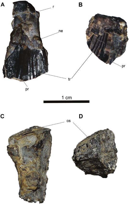
132 F. M. ROTATORI ET AL.
view, the centra have an hourglass-shaped outline (Fig.
4E, K, Q). The ventral surfaces of the centra possess a
narrow keel and the margin is highly concave (Fig. 4B,
P). Caudally, broad facets for chevrons are present.
Remarks: The three caudal vertebrae are stout and
cylindrical, similar in overall shape and proportions
to those of other basal iguanodontians, such as:
Camptosaurus dispar, Cumnoria prestwichii,
Dryosaurus altus, Dysalotosaurus lettowvorbecki,
Mantellisaurus atherfieldensis Mantell, 1825 and
Uteodon aphanoecetes (Galton & Powell, 1980; Galton,
Downloaded from https://academic.oup.com/zoolinnean/article/195/1/125/6535466 by guest on 21 June 2022
1981; Norman, 1986; Carpenter & Wilson, 2008;
Carpenter & Galton, 2018). They do differ remarkably
from those of Iguanodon bernissartensis Van Beneden,
1881 and Iguanacolossus fortis McDonald et al., 2010b
in being less discoidal in shape, and from Barilium
dawsoni Lydekker, 1888a by being less compressed
55
40
45
dorsoventrally (Norman, 1980, 2011; McDonald
et al., 2010b).
Manus (ML 357–1–5, 20–26) (Fig. 5):The partial carpus
(ML 357–5) is composed of the proximal part of a
30
25
20
25
20
metacarpal, two distal carpals and a proximal carpal
(Fig. 5A). Two isolated distal epiphyses of metacarpals
(ML 357–25, 26) are associated with the semi-
articulated carpus, being both consistent in size and
their state of preservation (Fig. 5F, G). Four manual
phalanges and five ungual phalanges are associated
with the carpus (Fig. 5B–E). Three manual phalanges
(phalanx n.2 ML 357–22, phalanx n.3 ML 357–21 and
69
28
48
45
phalanx n.4 ML 357–24) and two unguals (ungual n.1
ML 357–1 and ungual n.4 ML 357–23) are likely to
belong to the left manus (Fig. 4), while the remaining
three unguals (ungual n.2 ML 357–2, ungual n.3 ML
357–3 and ungual n.5 ML 357–20) and manual phalanx
n.1 ML 357–4 are attributed to the right manus. Given
the fragmentary condition and the weathering of the
specimen, it is not possible to identify the bones of each
35
25
30
30
45
individual carpal. The two distal carpals are cuboid in
shape and are stout elements, while the proximal carpal
is more lightly built and slightly arched (Fig. 5A). The
proximal end of the metacarpal preserves a concave
proximal margin. Despite the poor preservation, it is
possible to determine that the carpus does not include
50*
35
60
60
40
63
fused elements, as all of the contacts between all the
identifiable carpals are clearly visible (Fig. 5A).
Pedal Phalanx III-1 ML
Pedal Phalanx III-3 ML
The two isolated distal metacarpals fragments are
Pedal Phalanx II-1 ML
Pedal Phalanx II-2 ML
Pedal Ungual III ML
similar in shape: they are compressed along their
Table 1. Continued
Pedal Ungual II ML
extensor–palmar axis, broadening mediolaterally
towards their distal ends. The medial and lateral
ginglymi are subequal in size and preserved on their
357–19
357–13
357–15
357–16
357–17
357–14
collateral ligament pits. The manual phalanges, ML
357–22 and ML 357–21, articulate with each other.
Furthermore, phalanx ML 357–22 articulates with
© 2022 The Linnean Society of London, Zoological Journal of the Linnean Society, 2022, 195, 125–156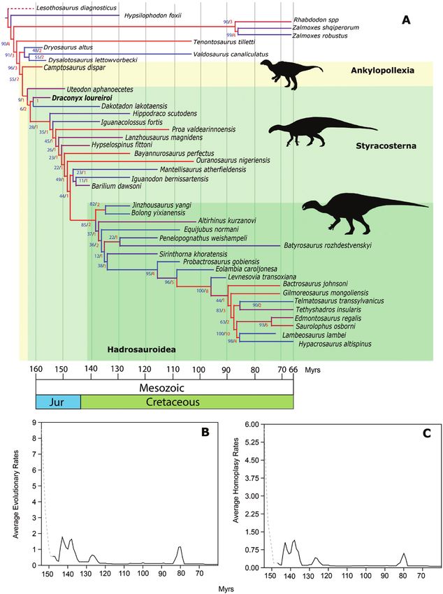
HOLOTYPE OF DRACONYX LOUREIROI                       133

                                                                                                                                  Downloaded from https://academic.oup.com/zoolinnean/article/195/1/125/6535466 by guest on 21 June 2022

Figure 2. Two maxillary teeth of Draconyx loureiroi holotype ML 357 in labial and lingual views. Completely preserved
maxillary in labial (A) and lingual (C) views; isolated maxillary crown in labial (B) and lingual (D) views. Abbreviations: ne:
neck, os: occlusal surface, pr: primary ridge, tr: tertiary ridges.

the ungual ML 357–23. Therefore, these latter manual             exception of phalanx n.2 where they are subequal (Fig.
elements compose a complete finger (Fig. 5C, D, K). In           5B–E). The unguals are generally elongated and claw-
general, all of the phalanges have a distal triangular           like, slightly arched along the extensor–palmar axis
section, moderately arched ventral surface and strongly          and have a subtriangular articular facet (Fig. 5H–L).
inclined and robust ginglymi. The lateral ginglymus              The only exception, ungual n.5 (Fig. 5L), appears
is smaller with respect to the medial one, with the              instead to be compressed along its extensor–palmar

© 2022 The Linnean Society of London, Zoological Journal of the Linnean Society, 2022, 195, 125–156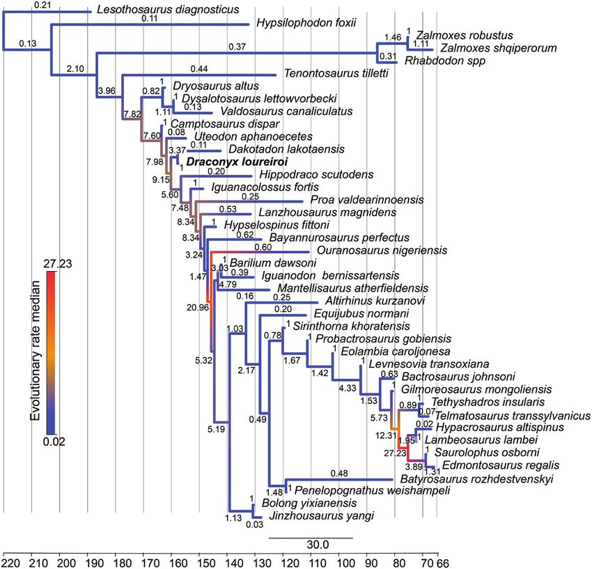
134    F. M. ROTATORI ET AL.

                                                                                                                                  Downloaded from https://academic.oup.com/zoolinnean/article/195/1/125/6535466 by guest on 21 June 2022

Figure 3. General overview of Draconyx loureiroi holotype ML 357 post-cranial skeleton. Caudal vertebrae (K-M) in left
lateral view. Right femur (A–E) in frontal (A), medial (B), caudal (C), lateral (D) and distal (E) views. Right tibia in medial
(F) and lateral (G) views. Proximal end of the fibula in medial (I) and lateral (J) views. Right articulated pes in dorsal (N)
and medial (Q) view. Pedal digit III (O) and pedal digit II (P) in dorsal view.

                           © 2022 The Linnean Society of London, Zoological Journal of the Linnean Society, 2022, 195, 125–156
HOLOTYPE OF DRACONYX LOUREIROI 135
Downloaded from https://academic.oup.com/zoolinnean/article/195/1/125/6535466 by guest on 21 June 2022
Figure 4. Three dimensional models of axial skeleton of Draconyx loureiroi holotype ML 357 (A–F): Caudal n.2 ML 357-10 in
cranial view (A), left lateral view (B), caudal view (C), right lateral view (D), ventral view (E) and dorsal view (F). Caudal n.3
ML 357-11 (G–L) in cranial view (G), left lateral view (H), caudal view (I), right lateral view (J), ventral view (K) and dorsal
view (L). Caudal n.1 ML 357-9 (M-R): cranial view (M), left lateral view (N), caudal view (O), right lateral view (P), ventral
view (Q) and dorsal view (R). Abbreviations: cf: chevron facet, k: keel, nc: neural canal, ns: neural spine, tp: transverse process.
axis. A fracture develops mediolaterally and is slightly iguanodontians (Norman, 2004). However, the carpus
inclined craniocaudally. Both parts are separated is constituted by isolated blocky elements, resembling
by this fracture and have suffered mediolateral the condition in dryosaurids and Tenontosaurus spp.
displacement. (Dodson, 1980; Galton, 1981; Forster, 1990; Winkler
et al., 1997). The distal carpals of ML 357 differ from
R e m a rk s : D e s p i t e t h e p o o r p r e s e r v a t i o n o f the ones of Camptosaurus dispar, which are arranged
the carpus of ML 357, certain features allow in two co-ossified and highly interlocked blocks.
comparisons with other taxa. The small metacarpals Furthermore, the carpus of ML 357 does not exhibit
are not significantly different from those of other the total ossified condition present in Iguanodon
© 2022 The Linnean Society of London, Zoological Journal of the Linnean Society, 2022, 195, 125–156136 F. M. ROTATORI ET AL.
Downloaded from https://academic.oup.com/zoolinnean/article/195/1/125/6535466 by guest on 21 June 2022
Figure 5. Manual elements of Draconyx loureiroi holotype ML 357 (ML 357 1-5, 20-26), including articulated partial
carpus, manual phalanges and unguals (A–L). Articulated partial carpus (ML 357 – 5) in extensor view (A), right phalanx
n.1(ML 357 – 4) in dorsal view (B), lateral view (B1), proximal view (B2) and distal view (B3), left phalanx n.2 (ML 357 –
22) in dorsal view (C), lateral view (C1), proximal view (C2) and distal view (C3), left phalanx n.3 (ML 357 – 21) in dorsal
view (D), lateral view (D1), proximal view (D2) and distal view (D3), left phalanx n.4 (ML 357 – 24) in dorsal view (E), lateral
view (E1), proximal view (E2) and distal view (E3), isolated distal ends of tarsals (ML 357 – 25,26) in extensor view (F, G),
left ungual phalanx n.1 in dorsal view (D), mediolateral view (D1) and proximal view (D2), left ungual phalanx n.1 (ML 357-
1) in dorsal view (H), mediolateral view (H1) and proximal view (H2), right ungual phalanx n.2 (ML 357 – 2) in dorsal view
(I), mediolateral view (I1) and proximal view (I2), right ungual phalanx n.3 (ML 357 – 3) in dorsal view (J), mediolateral
view (J1) and proximal view (J2), left ungual phalanx n.4 in dorsal view (K), mediolateral view (K1) and proximal view (K2),
ungual phalanx n.5 (ML 357 – 20) in dorsal view (L), mediolateral view (L1) and proximal view (L2).
© 2022 The Linnean Society of London, Zoological Journal of the Linnean Society, 2022, 195, 125–156HOLOTYPE OF DRACONYX LOUREIROI 137
Downloaded from https://academic.oup.com/zoolinnean/article/195/1/125/6535466 by guest on 21 June 2022
Figure 6. Three dimensional models of limb bones elements of Draconyx loureiroi holotype ML 357 (ML 357 6-8), femur
(A–E) and tibia and proximal end of fibula (F–J). Femur (ML 357 – 6) in cranial view (A), medial view (B), caudal view (C),
lateral view (D) and distal view (E). Tibia (ML 357 – 7) and proximal end of fibula (ML 357 – 8) in cranial view (F), medial
view (G), caudal view (H), lateral (I) and proximal (J) views.
bernissartensis, Magnamanus soriaensis Vidarte proximalmost part of the preserved shaft is
et al., 2016, Mantellisaurus atherfieldensis and other mediolaterally crushed and compressed, due
styracosternans (Fig. 12A–J) (Dodson, 1980; Norman, to taphonomic processes. The femur is strongly
1980, 1986, 2004, 2011, 2015, Vidarte et al., 2016). bowed craniocaudally; the section of the shaft was
The carpals and the manual unguals are similar to subcircular but is heavily distorted proximally by
the ones present in Camptosaurus dispar, Cumnoria the compression and damaged distally (Fig. 6B, D).
prestwichii and Uteodon aphanoecetes, differing The distal epiphysis is subrectangular in shape in
from the ones of more derived styracosternans (i.e. distal view, extending slightly more mediolaterally
Iguanodon bernissartensis, Magnamanus soriaensis, than craniocaudally. The two distal condyles are
Mantellisaurus atherfieldensis and all other preserved, appearing subequal in size with the
hadrosauriformes) in being, respectively, more arched lateral condyle slightly larger than the medial one
and more claw-like instead of extremely compressed (Fig. 6E). A deep, extensive, fully open and U-shaped
and hoof-like (Fig. 12A–J) (Galton & Powell, 1980; extensor groove separates the two condyles cranially
Norman, 1980, 1986, 2004; Carpenter & Wilson, 2008, (Fig. 6E). The cranial process of the lateral condyle
Vidarte et al., 2016). is rounded and deflected caudally. The caudal finger-
like process of the lateral condyle (condylid, according
Femur ML 357–6 (Figs 3A–E, 6A–E): The preserved to Bertozzo et al., 2017) is not preserved, being broken
right femur consists of the heavily eroded distalmost at its base, but the crest for the muscular insertion
part of the shaft and the distal epiphysis. The is distinguishable. The medial condyle appears stout
© 2022 The Linnean Society of London, Zoological Journal of the Linnean Society, 2022, 195, 125–156138 F. M. ROTATORI ET AL.
and rectangular, although its cranial and caudal 1980; Carpenter & Wilson, 2008). The inflection
processs have been eroded. The flexor groove is fully point of the curvature is located more cranially in
open and its margin, consisting of the caudal process Draconyx loureiroi than in Camptosaurus dispar
of the lateral condyle and the condylid of the medial and Uteodon aphanoecetes (Carpenter & Wilson,
condyle, are V-shaped in outline. 2008; Carpenter & Galton, 2018), while Cumnoria
prestwichii exhibits a smoother outline without
Remarks: The preserved femoral shaft is strongly abrupt changes in curvature. The lateral condyle of
bowed craniocaudally, as in dryosaurids, most Early Cretaceous species, such as Barilium dawsoni
elasmarians, Camptosaurus dispar and differently and Mantellisaurus atherfieldensis, are larger in
from Tenontosaurus sp. and other styracosternans proportions with respect to the total size of the
(Norman, 1980, 1986, 2004; Carpenter & Wilson, epiphysis and extend more caudally than the ones
2008; Carpenter & Galton, 2018; Herne et al., of the above-mentioned taxa, including D. loureiroi
Downloaded from https://academic.oup.com/zoolinnean/article/195/1/125/6535466 by guest on 21 June 2022
2019; Rozadilla et al., 2019, 2020). On the cranial (Norman, 1986; 2011). The medial condyle of
surface there is no crest developing from the medial Draconyx loureiroi is subrectangular and its medial
condyle, extending proximally towards the lesser margin is straight, as is the one of Mantellisaurus
trochanter, as seen in other ankylopollexians, atherfieldensis and Barilium dawsoni (Norman,
such as Camptosaurus dispar, Iguanodon sp., 1986; 2011). However, in Jurassic taxa such as
Mantellisaurus atherfieldensis and Uteodon Uteodon aphanoecetes and Camptosaurus dispar,
aphanoecetes (Gilmore, 1909; Norman, 1980, 1986, the medial margin of the medial condyle is rounded
2004; Carpenter & Wilson, 2008; Carpenter & Galton, (Carpenter & Wilson, 2008; Carpenter & Galton,
2018). The lateral condyle of the distal epiphysis of 2018). The flexor and extensor grooves are fully
the femur in Draconyx loureiroi is concave in outline open as in many basal and cursorial iguanodontians,
and it extends more craniocaudally than the ones contrasting with more derived forms (Norman, 2004).
of Camptosaurus dispar, Cumnoria prestwichii Extensor grooves of the Jurassic taxa Camptosaurus
and Uteodon aphanoecetes (Fig. 7; Galton & Powell, dispar and Uteodon aphanoecetes are fully open,
Figure 7. Comparison of distal sections of femoral shaft from selected iguanodontians. A, Draconyx loureiroi, B, Uteodon
aphanoecetes, C, Cumnoria prestwichii, D, Camptosaurus dispar, E, Barilium dawsoni, F, Mantellisaurus atherfieldensis
and G, Iguanodon galvensis. Abbreviations: cod: condylid, eg: extensor groove, fg: flexor groove, lc: lateral condyle, mc: medial
condyle. Distal sections drawn from: Carpenter & Wilson, 2008; Galton & Powell, 1980; Norman, 2011; Norman, 1986; Verdù
et al., 2018.
© 2022 The Linnean Society of London, Zoological Journal of the Linnean Society, 2022, 195, 125–156HOLOTYPE OF DRACONYX LOUREIROI 139
but shallower compared to the ones of Barilium separating the surface of the two malleoli and reaching
dawsoni, Cumnoria prestwichii, Draconyx loureiroi, the astragalus, forming with its apex a continuous
Iguanodon sp. and Mantellisaurus atherfieldensis concave surface.
(Norman, 1980, 1986, 2004, 2011, Carpenter &
Wilson, 2008; Carpenter & Galton, 2018; Verdú et al., Remarks: The medial margin of the proximal
2018). Moreover, the flexor groove walls of Cumnoria epiphysis of the tibia is convex, as is typical of
prestwichii and Uteodon aphanoecetes are slightly many iguandontians (Norman, 2004), except for
divergent from one another (Fig. 7). The extensor Talenkauen santacrucensis Novas et al., 2004 and
groove of the Early Cretaceous iguanodontians Eousdryosaurus nanohallucis Escaso et al., 2014
Barilium dawsoni, Mantellisaurus atherfieldensis (Escaso et al., 2014; Rozadilla et al., 2019; Dieudonné
and other styracosternans are partially enclosed by et al., 2021). As noted by Dieudonné et al. (2021), the
overhangs of medial and lateral condyles. Draconyx cnemial crest apex is directed strongly craniolaterally,
Downloaded from https://academic.oup.com/zoolinnean/article/195/1/125/6535466 by guest on 21 June 2022
loureiroi does not exhibit an overhang of the medial a characteristic common to Laurasian dryomorphans
condyle on the flexor groove, unlike the condition and Dysalotosaurus Pompeckj, 1920, differing from the
present in Camptosaurus dispar, Cumnoria condition exhibited by Elasmaria and the Portuguese
p r e s t w i ch i i , M a n t e l l i s a u r u s a t h e r f i e l d e n s i s , ornithopod Eousdryosaurus nanohallucis (Escaso
Ouranosaurus nigeriensis Taquet, 1976 and Uteodon et al., 2014). As in many styracosternans, the cnemial
aphanoecetes (Norman, 1980, 1986, 2004, 2011; crest is well developed (Norman, 2004). The fibular
Carpenter & Wilson, 2008; Bertozzo et al., 2017; condyle differs from dryosaurids (with the exception
Carpenter & Galton, 2018, Verdú et al., 2018). This of Valdosaurus canaliculatus), Camptosaurus
condition more closely resembles the plesiomorphic dispar and Uteodon aphanoecetes in being partially
condition within Ornithopoda (Norman et al., 2004). posterolaterally deflected, similar to the condition seen
in Cumnoria prestwichii, Talenkauen santacrucensis
Tibia (ML 357–7, 12) (Figs 3F, G, 6F–J, 8A–D): and more derived styracosternanans (Galton &
The proximal (Fig. 6F–J) and distal (Fig. 8A–E) Powell, 1980; Norman, 1980, 1986, 2011; Norman
epiphyses of the tibia are preserved, but the tibial et al., 2004; Carpenter & Wilson, 2008; Barrett et al.,
shaft is missing. Both extremities are heavily eroded 2011; Rozadilla et al., 2019). The distal epiphysis,
and covered by matrix and adhesives, but relevant found in articulation with the rest of the pes, does not
characters are still distinguishable. The proximal differ significantly from that of other iguanodontians
epiphysis preserves a conspicuous cnemial crest, a (Norman, 2004).
robust fibular condyle and the internal condyle. The
cnemial crest tapers dorsally forming a smooth edge; Fibula (ML 357–8, 12) (Figs 3I, J, 6F–J, 8A–D): The
in dorsal view its margins are laterally concave and distal and proximal epiphyses of the fibula are
medially convex (Fig. 6G, I). Laterally, it is divided by preserved, whereas the diaphysis is completely absent.
the fibular condyle to form a deep and extensive scar The proximal end is a flattened subtriangular element.
(sulcus tibialis). The fibular condyle is a stout process Dorsally, it appears to be slightly medially convex.
that deflects strongly caudally, its articular facet is The cranial margin deflects abruptly dorsally into
rounded and it blends smoothly with the anterior the cranial process, which is slightly eroded (Fig. 6I).
and posterior surfaces of the condyle. The internal In contrast, the caudal margin deflects less abruptly
condyle is a blunt eminence directed caudally and dorsally. A deep fossa is present close to the caudal
projecting gently laterally. Like the cnemial crest, its margin on the lateral surface.
lateral margin is concave, while the medial margin is The distal epiphysis is in articulation with the rest
strongly convex (Fig. 6E). This convexity forms a deep of the pes, located on the lateral surface of the tibia
sulcus that separates the internal condyle from the and contacts the calcaneum (Fig. 7A–D).
fibular condyle. Immediately ventral to the proximal
epiphysis, the proximal part of the diaphyseal shaft is Remarks: The proximal epiphysis has cranial and
preserved and it is teardrop-shaped in cross-section. caudal margins that diverge smoothly, as in Dryosaurus
The distal part of the tibia is articulated with the altus, Dysalotosaurus lettowvorbecki, Eousdryosaurus
astragalus, calcaneum and the metatarsals (Fig. nanohallucis, Talenkauen santacrucensis and
8A–E). The distal epiphysis flares mediolaterally, Valdosaurus canaliculatus, but differs from Iguanodon
the medial malleolus is rounded and slightly more bernissartensis, Mantellisaurus atherfieldensis and
expanded mediolaterally than the lateral one, which Ouranosaurus nigeriensis (Norman, 1980, 1986;
is narrow and elongate, expanding proximodistally. Galton, 1981; Barrett et al., 2011; Escaso et al., 2014;
They articulate between each other forming an angle Barrett, 2016; Bertozzo et al., 2017; Rozadilla et al.,
of approximately 45°. The intermuscular line is 2019). As in Sektensaurus sanjuanboscoi Ibiricu et al.,
located on the caudal surface of the distal epiphysis, 2019 and Talenkauen santacrucensis, the caudal
© 2022 The Linnean Society of London, Zoological Journal of the Linnean Society, 2022, 195, 125–156140 F. M. ROTATORI ET AL.
Downloaded from https://academic.oup.com/zoolinnean/article/195/1/125/6535466 by guest on 21 June 2022
Figure 8. Three dimensional models of the pes of Draconyx loureiroi holotype ML 357. Articulated pes (ML 357-12) (A–F)
in medial (A), lateral (B), dorsal (C), plantar (D) and caudal (E) views, F, detail of metatarsal I. Abbreviations: as: astragalus,
ca, calcalneum, fi: fibula, lm: lateral malleolus, mm: medial malleolus, mt-I: metatarsal I, mt-II: metatarsal II, mt-III:
metatarsal III, mt-IV: metatarsal IV, p II-1: pedal phalanx II-1, p II-1: pedal phalanx II-2, p III-1: pedal phalanx III-1, p III-3:
pedal phalanx III-3, ta-3: tarsal 3, ta-4: tarsal 4, un-II: ungual II, un-III: ungual III.
© 2022 The Linnean Society of London, Zoological Journal of the Linnean Society, 2022, 195, 125–156HOLOTYPE OF DRACONYX LOUREIROI 141
margin of the proximal part of the fibula is almost craniocaudally more than mediolaterally (Fig. 8B, E).
vertical (Ibiricu et al., 2019; Rozadilla et al., 2019). Distally, its surface is subrectangular in outline and
extends dorsally, appearing as a small rhomboidal
Pes (ML 357–12–19) (Figs 3N, Q, 8A–F, 9C, D): The element in caudal view (Fig. 8B, E). Caudally, the
pes is articulated with the distal tibia, distal fibula, medial margin of the calcaneum forms a smooth
astragalus and calcaneum and proximal metatarsals. concavity that accommodates the lateralmost margin
The diagenetic permineralization between the of the astragalus. In lateral view, the calcaneum
tibia and the rest of the pes prevented a reliable appears triangular with a caudal apex and is slightly
segmentation by CT techniques. This also hampered deflected proximally. The proximal and distal margins
views of the internal anatomy of the astragalus and progressively flare cranially, with the latter forming a
calcaneum. The articulated elements share the same scoop-like concavity.
subnumber (ML 357–12), while the isolated elements
Downloaded from https://academic.oup.com/zoolinnean/article/195/1/125/6535466 by guest on 21 June 2022
have their own numbers. Therefore, the same element
may have two subnumbers, depending on whether
Astragalus
part of it belongs to the articulated block. Like the calcaneum, the astragalus is complete and
not significantly distorted or eroded. In distal view, the
bone has a subtrapezoidal shape (Fig. 8A, E). Laterally,
Calcaneum it contacts the medial malleolus of the tibia via a deep
The calcaneum is preserved in its entirety and does concavity, while medially the astragalus/calcaneum
not show signs of significant breakage, erosion or contact is straight (Fig. 8A, E). In caudal view, the
distortion. In its general appearance, the calcaneum astragalus is triangular, with a high ascending process
is a compact and narrow element, expanding representing the apex, which protrudes extensively
Figure 9. Three dimensional models of the articulated leg (A–B) and pes (C–D) of Draconyx loureiroi holotype ML 357. Leg
in medial view (A) and lateral view (B). Pes in medial (C) and lateral (D) views.
© 2022 The Linnean Society of London, Zoological Journal of the Linnean Society, 2022, 195, 125–156142 F. M. ROTATORI ET AL.
dorsally. Immediately ventral to the ascending process, bone as the first metatarsal (Fig. 8F). The status
a stout steeply inclined caudomedial process forms a of this character as an ‘autapomorphy’ is discussed
well-distinguishable relief. below. A deep groove in continuity with this fragment
and impressed on metatarsal II probably represents
Remarks: The astragalus of ML 357 is subtriangular the entire articulation surface with metatarsal I (Fig.
and its caudal surface is moderately high, as in 8F). Metatarsals II and III are complete and do not
dryosaurids, Camptosaurus dispar, Cumnoria appear to have suffered extensive breakage or fracture
prestwichii, Iguanodon bernissartensis Mantellisaurus (Fig. 8C, D). Metatarsal IV in contrast is broken at the
atherfieldensis and Uteodon aphanaecetes (Galton midshaft with the distal epiphysis preserved as an
& Powell, 1980; Norman, 1980, 1986; Galton, 1981; isolated element.
Carpenter & Wilson, 2008; Barrett et al., 2011). The
ascending process is not as developed as in Barilium
Downloaded from https://academic.oup.com/zoolinnean/article/195/1/125/6535466 by guest on 21 June 2022
dawsoni, Iguanodon bernissartensis or Mantellisaurus
Pedal digit I
athefieldensis (Norman, 1980, 1986, 2011), but more The first pedal digit consists only of the splint-like
closely resembles the condition found in Camptosaurus metatarsal I (Fig. 8F).
dispar (NMNH 2210), Cumnoria prestwichii,
Ouranosaurus nigeriensis and Uteodon aphanoecetes
Pedal digit II
(Galton & Powell, 1980; Carpenter & Wilson,
2008; Bertozzo et al., 2017). As in the other known Pedal digit II is represented by metatarsal II, the pedal
Portuguese ornithopod, Eousdryosaurus nanohallucis, phalanx II-1 (ML 357–17) and the ungual phalanx II-2
it shows a caudomedial process, which casts doubt (357–16), which articulates with the phalanx II-2 (357–
on the diagnostic status of this character for this 14). Pedal phalanx II-1 is broken at the midshaft, but
latter taxon (Escaso et al., 2014). The calcaneum does the proximal and distalmost epiphyses are preserved
not possess any remarkable differences from other (Fig. 8A, C, D).
ankylopollexians (Norman, 2004). Metatarsal II is positioned more proximally with
respect to Mt III–IV. In mediolateral view, metatarsal
II has a keyhole shape, as described by Herne et al.
Distal tarsals (2018) for the Australian ornithopod Diluvicursor
Two distal tarsals are preserved, which are identified pickeringi Herne et al., 2018, with the proximalmost
as tarsal 3 and tarsal 4 (Fig. 8D). Tarsal 3 is a large part dorsoplantarily higher than the distal epiphysis
element that is mediolaterally elongated and contacts (Fig. 8A, F). The distal epiphysis develops more
metatarsal III along a straight and linear surface (Fig. dorsoplantarily than mediolaterally, and the articular
8D). Tarsal 4 is a stout, elongated element and contacts surface is convex. The shaft of Mt II is piriform
the metatarsal IV along a concave surface, while its (teardrop) in cross-section, with the plantar surface
medialmost tip overlaps tarsal 3 proximally (Fig. 8D). bearing a keel. The distalmost part of the epiphysis
expands dorsoplantarily, giving the condyle a
Remarks: Distal tarsals 3 and 4, found in articulation subtrapezoidal outline (Fig. 8A, D). The condyle of the
with metatarsals III and IV, resemble the ‘cushion- metatarsal articulates perfectly with pedal phalanx
like’ condition described by Gilmore (1909) in II-1, which accommodates the condyle in a gently
Camptosaurus dispar, and differing from the thin- convex triangular facet. The distal epiphysis of the
like and subrounded condition observed in Iguanodon phalanx is in articulation with pedal phalanx II-2,
bernissartensis and Mantellisaurus atherfieldensis which is a thick, subrectangular element in dorsal view
(Norman, 1980, 1986) or the one present in basal (Fig. 8C, D). Proximally, the articular facet is not visible,
ornithopods (Galton, 1974, 1981). but distally the two condyles are well distinguishable,
the lateral slightly inclined with respect to the medial
one. On both lateral surfaces, deep extensor grooves
Metatarsus are present for tendon insertion. The ungual (phalanx
Although they are fragmented, metatarsals II–IV II-2) of the pedal digit II is preserved, being a pointed
are long and slender (Figs 8, 9C, D). A thick splinter claw-like element, which is dorsoplantarly arched and
of bone next to metatarsal II was interpreted as the proximodistally elongated (Fig. 8A, C, D).
proximalmost part of metatarsal I in the original
description of Mateus & Antunes (2001) and a ‘vestigial
digit I’ has been proposed as a possible autapomorphy Pedal digit III
for Draconyx loureiroi. Despite being heavily eroded, Pedal digit III is composed of metatarsal III, the
the bone is preserved in anatomical position and the largest pedal phalanx preserved III-1 (ML 357–13),
rod-like structure supports the interpretation of this the smallest pedal phalanx is interpreted to be III-3
© 2022 The Linnean Society of London, Zoological Journal of the Linnean Society, 2022, 195, 125–156HOLOTYPE OF DRACONYX LOUREIROI 143
(ML 357–15) and the largest ungual, pedal phalanx Carpenter & Galton, 2018). However, this condition
III-4 (ML 357–19). The pedal phalanx III-2 is is preserved in some specimens of Iguanodon
missing. Metatarsal III is the longest element of the bernissartensis and Mantellisaurus atherfieldensis
metatarsus, being a slender, yet compact element. (Norman 1980, 1986). Assessing the presence of this
Proximally, the Mt III articulates with the tarsal 3 and character in other taxa is problematic, because of the
intervenes between Mt II and Mt IV (Fig. 8B–D). The extreme fragility of the reduced metatarsal I, whose
subrectangular shaft ends distally in a mediolaterally absence/presence is highly subject to preservation bias.
expanded condyle, the only metatarsal exhibiting The alleged dryosaurid, Eousdryosaurus nanohallucis,
this condition. Pedal phalanx III-1 is a stout, robust from the same formation as ML 357, has been shown
element, which is moderately dorsoplantarly to possess a reduced metatarsal I in articulation with
arched. The proximal articular facet is concave a pedal phalanx (Escaso et al., 2014).
and subellipsoidal in outline, being the long axis
Downloaded from https://academic.oup.com/zoolinnean/article/195/1/125/6535466 by guest on 21 June 2022
oriented mediolaterally (Fig. 8B–D). Distally, the two
ginglymi are subequal in size, being marked by deep Previously referred specimens
collateral ligament pits. Pedal phalanx III-3 is a small Ankylopollexia indet.
proximodistally compressed element, which possesses Femur ML 434 (Fig. 10): Mateus & Antunes (2001)
a dorsoplantarly deep articular facet (Fig. 8B–D). The referred an isolated femur (ML 434) to Draconyx
ventral margin of pedal phalanx III-3 is not as arched loureiroi, based on geographical proximity. ML 434
as the other phalanges and the collateral ligament pits is an almost complete and undistorted left femur,
occupy almost the entire lateral and medial surfaces. but lacks part of its proximal epiphysis and its entire
The ungual of the digit III (pedal phalanx III-3) is distal one. The femoral shaft is stout and robust in
a stout, pointed element, which is more robust than general proportions and appears strongly bowed
the ungual preserved on pedal digit II. The ungual of craniocaudally in lateral view, but is straight in cranial
digit III differs also from the other preserved ungual view. The cross-section of the shaft is subovoidal, with a
in having a less arched and more dorsoplantarly sharp edge that corresponds with a ridge that extends
flattened outline (Fig. 8B–D). dorsoventrally from the medial condyle. Proximally,
only a part of the femoral head is preserved, which
Pedal digit IV is gently deflected medially. At approximately the
midshaft on the medial surface of the femur, a pendant
Metatarsal IV (ML 357–12, 18) is the only element
fourth trochanter is present, its broad base forming an
of pedal digit IV available. Proximally it is the
elongated crest-like surface. The scar for the insertion
most mediolaterally expanded and dorsoplantarly
of the musculus caudifemoralis longus is located
compressed of the metatarsals and the proximal
immediately cranial to the trochanter, being separated
epiphysis is fan-shaped in dorsal and plantar
from the eminence of the trochanter. Distally, the
view (Fig. 8B–D). Distally, the section of the shaft
eroded epiphysis preserves the general appearance of
becomes progressively subrectangular, culminating
the two condyles, although the proportions with respect
in a moderately dorsoplantarly deep distal condyle
to one another are unclear. Parts of the flexor and
(Fig. 8B–D).
extensor grooves are preserved caudally and cranially.
A well-defined, sharp crest runs proximodistally from
Remarks: The metatarsus of ML 357 differs strongly the medial condyle towards the location of the lesser
from those of Camptosaurus dispar, Iguanodon trochanter.
bernissartensis, Ouranosaurus nigeriensis, Uteodon
aphanoecetes and other large-sized hadrosauriforms
in general proportions, while exhibiting the slender Remarks: ML 434 possesses a strongly craniocaudally
and gracile proportions seen in Cumnoria prestwichii bowed femur and a pendant-like fourth trochanter as
and Mantellisaurus atherfieldensis instead (Gilmore, in the dryosaurids Camptosaurus dispar and Uteodon
1909; Galton & Powell, 1980; Norman, 1980, 1986, aphanoecetes (Galton, 1981; Carpenter & Galton
2004; Bertozzo et al., 2017). Furthermore, as in the 2018). The base of the fourth trochanter is located at
latter taxa, ML 357 possesses a metatarsal III that is the midshaft of the femur, as is commonly found in
sensibly more elongated with respect to metatarsals II Ankylopollexia, but different from Dryosauridae, in
and IV (Galton & Powell, 1980; Norman, 1986). This which this structure is confined to the proximal part
is in stark contrast to elasmarians Camptosaurus of the shaft (Norman, 2004). Similar to Camptosaurus
dispar, Cumnoria prestwichii, Dryosaurus altus and dispar, Cumnoria prestwichii, Uteodon aphanoecetes
Dysalotosaurus lettowvorbecki, whose metatarsal I is and Valdosaurus canaliculatus, the scar of the
extremely reduced, losing any functional capability m. caudifemoralis longus reaches the base of the fourth
(Gilmore, 1909; Galton & Powell, 1980; Galton, 1981; trochanter. Moreover, the morphology of the ridge
© 2022 The Linnean Society of London, Zoological Journal of the Linnean Society, 2022, 195, 125–156You can also read