Retropharyngeal Hematoma under Rivaroxaban: A Rare Entity to Know for Its Risk of Airway Obstruction
←
→
Page content transcription
If your browser does not render page correctly, please read the page content below
Retropharyngeal Hematoma
under Rivaroxaban: A Rare
Entity to Know for Its Risk of
                                                                                 IMAGES IN CLINICAL
Airway Obstruction                                                               RADIOLOGY

CHARLOTTE VIERENDEELS
XAVIER PEETERS
PIERRE BOSSCHAERT
*Author affiliations can be found in the back matter of this article

ABSTRACT                                                                         CORRESPONDING AUTHOR:
                                                                                 Pierre Bosschaert
Teaching Point: Retropharyngeal hematoma appearing under rivaroxaban is          Clinique Saint-Pierre Ottignies, BE
uncommon but should be suspected in cases of dysphagia, dysphonia or breathing   Pierre.bosschaert@skynet.be
difficulties.

                                                                                 KEYWORDS:
                                                                                 Retropharyngeal; Hematoma;
                                                                                 Dysphagia; Rivaroxaban;
                                                                                 Trauma

                                                                                 TO CITE THIS ARTICLE:
                                                                                 Vierendeels C, Peeters X,
                                                                                 Bosschaert P. Retropharyngeal
                                                                                 Hematoma under
                                                                                 Rivaroxaban: A Rare Entity to
                                                                                 Know for Its Risk of Airway
                                                                                 Obstruction. Journal of the
                                                                                 Belgian Society of Radiology.
                                                                                 2021; 105(1): 15, 1–4. DOI:
                                                                                 https://doi.org/10.5334/
                                                                                 jbsr.2263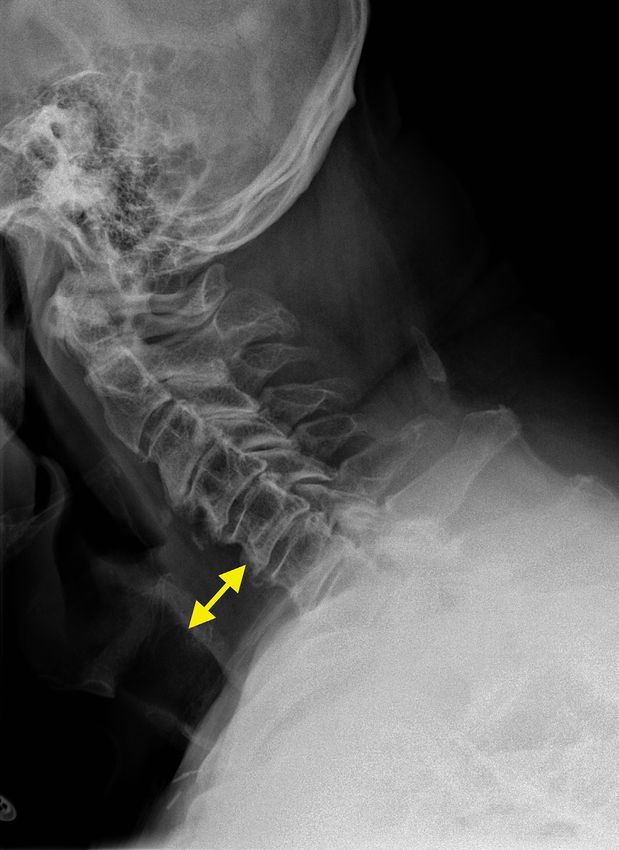
Vierendeels et al. Journal of the Belgian Society of Radiology DOI: 10.5334/jbsr.2263 2
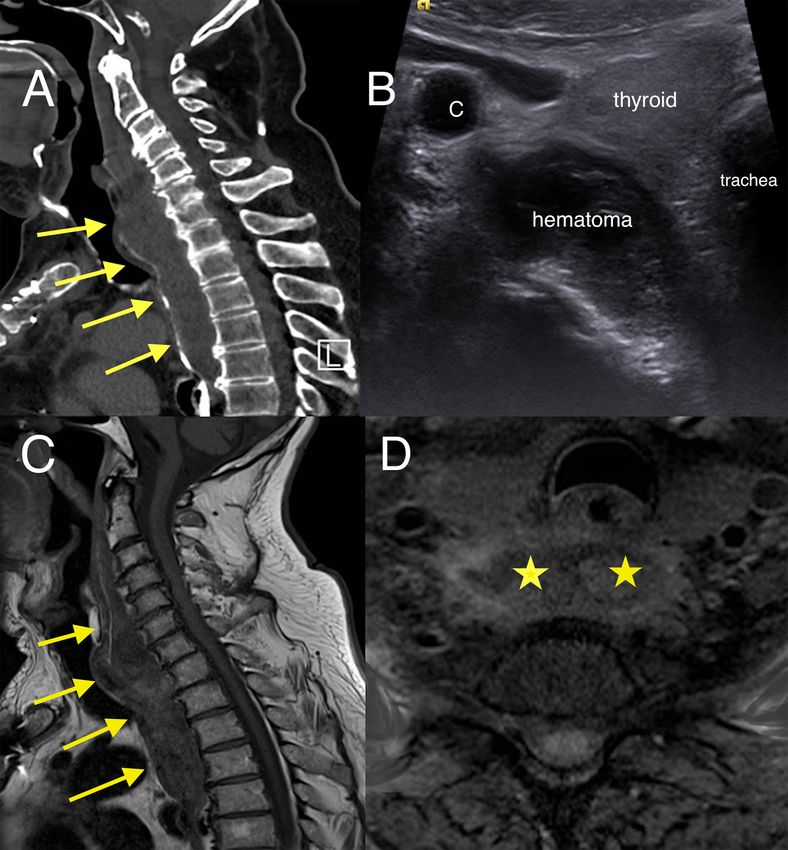
CASE The hematoma was not drained due to its good
tolerance. The patient was monitored closely. On
A 74-year-old man was admitted to the emergency magnetic resonance imaging (MRI) performed seven
department for progressive dysphagia and mild dysphonia days later, T1-weighted sagittal (Figure 2C, arrows) and
in the context of minor bicycle trauma that had occurred axial sequence with fat saturation (Figure 2D, stars)
four days earlier. He had no significant swelling nor any confirmed methemoglobin hyperintensity. A control MRI
cervical spine pain and no limitation of neck movement. carried out three months later showed the disappearance
He had no signs of airways being compromised. of the hematoma.
Previous medical history included anticoagulation
with rivaroxaban for atrial fibrillation and aortic valve
replacement four months before the bicycle trauma. COMMENT
Examination of the oropharynx was unremarkable. He
was afebrile, with normal blood pressure. Retropharyngeal hematoma is a rare entity that can
Lateral cervical X-ray showed increased soft tissue cause airway obstruction rapidly with a potentially fatal
thickness of 30 mm in front of C5 (Figure 1, double arrow). outcome. The diagnosis is often delayed because of the
Contrast-enhanced computed tomography (CECT) with absence of objective signs and diagnostic laboratory
oral gastrografin administration revealed hematoma data.
extending from the retropharyngeal to the superior Many circumstances can lead to its development,
mediastinal space without active bleeding or vertebral such as a trauma, intra-thyroid bleeding, hemophilia, and
fracture (Figure 2A, arrows). Axial US image showed more rarely in the context of rivoxaraban medication [1].
a slightly echoic and heterogeneous liquid collection The onset is usually acute but can also be insidious,
pushing forward trachea and thyroid (Figure 2B). and patients may present symptoms only several days
Figure 1 Lateral cervical X-ray showing soft tissue thickness of 30 mm in front of C5 (double arrow).Vierendeels et al. Journal of the Belgian Society of Radiology DOI: 10.5334/jbsr.2263 3
Figure 2 (A) Sagittal CT image with oral gastrografin administration showing a pre-vertebral hematoma displacing the pharynx
(arrows). (B) Axial US image showing the slightly echoic collection pushing forward trachea and thyroid. (C) Sagittal T1-weighted
image and (D) Axial T1-weighted image with fat saturation showing the retropharyngeal hematoma on day 7 with methemoglobin
hyperintensity (arrows and asterisks).
after hematoma has developed. Patients with symptoms COMPETING INTERESTS
such as dysphagia, dysphonia, or dyspnea should receive
immediate attention. The authors have no competing interests to declare.
The diagnosis is suggested by thickening of the pre-
vertebral space on lateral cervical X-ray. CECT is the
examination of first choice in order to confirm and AUTHOR AFFILIATIONS
measure the retropharyngeal hematoma. Patients may Charlotte Vierendeels
also show active bleeding. Any associated vertebral Clinique Saint-Pierre Ottignies, BE
fracture may also be ruled out. Ultrasound (US) can be Xavier Peeters
done at the patient’s bedside. MRI offers the advantage of Clinique Saint-Pierre Ottignies, BE
anatomical precision and is recommended for follow-up. Pierre Bosschaert
Non-compressive hematoma can be treated Clinique Saint-Pierre Ottignies, BE
conservatively by cervical spine immobilization and close
clinical monitoring. For a retropharyngeal hematoma
producing significant airway obstruction, a tracheostomy REFERENCE
may be necessary. Surgical evacuation is generally
reserved for hematomas that develop quickly, obstruct 1. McCarter JA, Bell PR, McCadden L, et al. Rivaroxaban and
mechanical ventilation, or do not resolve themselves retropharyngeal haemorrhage. BMJ Case Rep. 2016. DOI:
traditionally. https://doi.org/10.1136/bcr-2015-212446Vierendeels et al. Journal of the Belgian Society of Radiology DOI: 10.5334/jbsr.2263 4 TO CITE THIS ARTICLE: Vierendeels C, Peeters X, Bosschaert P. Retropharyngeal Hematoma under Rivaroxaban: A Rare Entity to Know for Its Risk of Airway Obstruction. Journal of the Belgian Society of Radiology. 2021; 105(1): 15, 1–4. DOI: https://doi.org/10.5334/jbsr.2263 Submitted: 29 July 2020 Accepted: 04 November 2020 Published: 16 March 2021 COPYRIGHT: © 2021 The Author(s). This is an open-access article distributed under the terms of the Creative Commons Attribution 4.0 International License (CC-BY 4.0), which permits unrestricted use, distribution, and reproduction in any medium, provided the original author and source are credited. See http://creativecommons.org/licenses/by/4.0/. Journal of the Belgian Society of Radiology is a peer-reviewed open access journal published by Ubiquity Press.
You can also read