SURVEY AND SUMMARY Current and emerging roles of Cockayne syndrome group B (CSB) protein - Oxford Academic Journals
←
→
Page content transcription
If your browser does not render page correctly, please read the page content below
2418–2434 Nucleic Acids Research, 2021, Vol. 49, No. 5 Published online 15 February 2021
doi: 10.1093/nar/gkab085
SURVEY AND SUMMARY
Current and emerging roles of Cockayne syndrome
group B (CSB) protein
*
Vinod Tiwari , Beverly A. Baptiste, Mustafa N. Okur and Vilhelm A. Bohr
Laboratory of Molecular Gerontology, National Institute on Aging, National Institutes of Health, Baltimore, MD 21224,
Downloaded from https://academic.oup.com/nar/article/49/5/2418/6137297 by guest on 11 December 2021
USA
Received November 04, 2020; Revised January 26, 2021; Editorial Decision January 28, 2021; Accepted February 01, 2021
ABSTRACT CS patients are genetically heterogeneous, carrying muta-
tions in genes encoding Cockayne syndrome B (CSB), exci-
Cockayne syndrome (CS) is a segmental premature sion repair cross-complementing protein group 6 (ERCC6)
aging syndrome caused primarily by defects in the on chromosome 10q11, and Cockayne syndrome A (CSA),
CSA or CSB genes. In addition to premature ag- ERCC8 gene on chromosome 5q11. Mutations in the
ing, CS patients typically exhibit microcephaly, pro- ERCC3 gene on chromosome 2q21 [xeroderma pigmento-
gressive mental and sensorial retardation and cuta- sum complementation group B (XPB)], ERCC4 gene on
neous photosensitivity. Defects in the CSB gene were chromosome 16p13 (XPF), and ERCC5 on chromosome
initially thought to primarily impair transcription- 13q33 (XPG) show some overlap with features of CS (1–5).
coupled nucleotide excision repair (TC-NER), pre- Approximately 70% of CS-affected individuals have a mu-
dicting a relatively consistent phenotype among CS tation in CSB, and the majority of the remaining cases har-
patients. In contrast, the phenotypes of CS patients bor CSA mutations, with relatively few from the other listed
genes. A total of 108 distinct mutations are documented in
are pleiotropic and variable. The latter is consistent
CS patients. In general, CS group A patients, carrying mu-
with recent work that implicates CSB in multiple cel- tations in CSA, present with less severe phenotypes than
lular systems and pathways, including DNA base ex- CS group B patients (6,7). CS has a prevalence of 2–3 per
cision repair, interstrand cross-link repair, transcrip- million globally, including the US, although the incidence
tion, chromatin remodeling, RNAPII processing, nu- is greater in some European countries (7–9). Based on the
cleolin regulation, rDNA transcription, redox home- severity and the age of onset, CS is classified into 3 types:
ostasis, and mitochondrial function. The discovery type I includes early-onset CS, where patients typically show
of additional functions for CSB could potentially ex- clinical signs in the first year after birth; type II CS includes
plain the many clinical phenotypes of CSB patients. early-onset cases with more severe symptoms; and type III
This review focuses on the diverse roles played by CS includes patients with a late-onset, mild clinical presen-
CSB in cellular pathways that enhance genome sta- tation (8,10,11).
bility, providing insight into the molecular features of
this complex premature aging disease. PROTEIN BIOCHEMISTRY
CSA is a 44-kDa protein, consisting of 396 amino acids.
INTRODUCTION
CSA lacks any detectable enzymatic function, belonging to
Normal aging is believed to be a manifestation of molecular the “Trp-Asp (WD) 40 repeat” family of structural and reg-
damage that gradually accumulates in cells and organisms ulatory proteins (12). The WD40 domain of CSA consists
over time, leading to increasing dysfunction and eventually of a helix-loop-helix motif and seven well ordered WD40
death. A complex interplay of genetic, epigenetic and en- -propeller structures (13). CSA is part of a multi-subunit
vironmental factors determine the course of aging for any E3 ubiquitin ligase complex (CRL4), comprised of Cullin
individual/organism. Cockayne syndrome (CS) is a rare au- 4A (CUL4A), a regulator of cullins-1 (Rbx1/ROC1) and
tosomal recessive genetic disorder characterized by prema- DNA damage binding protein 1 (DDB1) (14,15). Stalled
ture aging, and one of the most studied segmental progeroid RNA polymerases activate CSA in a CSB-dependent man-
syndromes. ner, which in turn targets several proteins for ubiquitination,
* To whom correspondence should be addressed. Tel: +1 410 558 8223; Email: vbohr@nih.gov
Published by Oxford University Press on behalf of Nucleic Acids Research 2021.
This work is written by (a) US Government employee(s) and is in the public domain in the US.
Nucleic Acids Research, 2021, Vol. 49, No. 5 2419

including CSB and RNA polymerase II (RNAPII) (14,16–           CSA. The cellular phenotype associated with CSA W361C
18). Mutations of CSA predominantly cause type I CS (7).       involves sensitivity to UV but not to oxidative damage (37).
   CSB is a 168-kDa protein with 1493 amino acids. The N-      In this case, the retention of effective repair of oxidative
terminal domain is followed by an acidic stretch, a glycine-   damage could possibly explain a mild CS phenotype.
rich region, a central helicase domain, two putative nu-          CSA is recruited to the RNAPII-CSB complex by the
clear localization signal sequences, and a number of ser-      CIM in CSB (27). Once recruited, CSA promotes associ-
ine phosphorylation sites (Figure 1). CSB belongs to the       ation of UVSSA and stalled RNAPII through ubiquitina-
SWI2/SNF2-family of DNA-dependent ATPases and con-             tion and helps recruit the transcription initiation factor IIH
tains the highly-conserved canonical seven ATPase motifs       (TFIIH) complex (17,27). A number of proteins, including
in the helicase region, characteristic of DNA and RNA          XAB2, nucleolin and UVSSA, are shared interacting part-
helicases (19). The ATPase activity, but not the acidic re-    ners of CSA and CSB and these interactions are thought to
gion, of CSB is important for its role in DNA repair (20).     help maintain cellular homeostasis (17,27,36,38,39). Given
CSB does not appear to possess helicase activity, but does     the emerging diverse roles of CSA and CSB, the neu-

                                                                                                                                Downloaded from https://academic.oup.com/nar/article/49/5/2418/6137297 by guest on 11 December 2021
possess ATP-dependent chromatin remodeling activity (21–       ropathological features of CS may be more tightly linked
23). CSB has ssDNA strand annealing and exchange activ-        to defects in these roles than to their roles in TC-NER.
ities (24). A ubiquitin binding domain (UBD) is part of the
larger winged-helix domain (WHD) located in C-terminus
                                                               Transcription coupled nucleotide excision repair (TC-NER)
and is important for recruitment of CSB to double strand
breaks (DSBs) (25) and TC-NER (26). CSB interacts with         CS has traditionally been associated with and linked to
CSA through its CSA-interaction motif (CIM) upstream of        defects in TC-NER (Figure 2) (40,41). Helix-distorting le-
the UBD, to recruit CSA to DNA damage-stalled RNAPII           sions in active genes can physically block or stall RNAPII
(27) (Figure 1). CSB functions as a homodimer (28), where      progression, which initiates TC-NER to rescue stalled
each CSB subunit wraps around the DNA helix, altering          RNAPII. The region within the DNA footprint of stalled
DNA conformation, which in turn alters protein-DNA in-         RNAPII, ∼35 nucleotides, can no longer be accessed by
teractions in the affected DNA region (29).                    DNA repair enzymes (42). Stalled RNAPII recruits CSB
                                                               to the lesion site, leading to CSB-dependent deployment
                                                               of the CSA/E3 ubiquitin ligase complex (CRL4), and re-
CSB FUNCTIONS
                                                               cruitment of other NER factors and TC-NER specific pro-
A broad range of DNA lesions are caused by endoge-             teins such as UVSSA, ubiquitin-specific processing pro-
nous cellular molecules, such as metabolism-driven reac-       tease 7 (USP7), and XAB2 (36,43). TFIIH is involved in
tive oxygen species (ROS) as well as exogenous agents or       DNA opening and lesion verification (44–46). XPA coordi-
compounds, including ionizing radiation (IR) and ultravi-      nates with TFIIH and the pre-incision complex allowing the
olet (UV) light. UV induces the formation of cyclobutane       XPF-ERCC1 complex to incise 5 of the lesion site to initi-
pyrimidine dimers (CPDs) and 6-pyrimidine-4-pyrimidone         ate DNA repair synthesis. Subsequently, XPG generates a
products (6,4-PP) in DNA. Sensitivity to UV light is a char-   5 phosphate group 3 to the incision, which is required for
acteristic feature of CS. Many DNA lesions in the template     ligation (44,46,47). Following the removal of the damaged
strand of actively transcribed regions of the genome are       oligonucleotides, DNA polymerases ␦ and ε, with the acces-
preferentially repaired by TC-NER, a subpathway of NER         sory proteins proliferating cell nuclear antigen (PCNA) and
(30,31). CS cells are deficient in TC-NER and demonstrate      replication factor C (RFC), fill the gap and DNA LIG1 or
increased sensitivity towards UV irradiation (32). However,    LIG3␣ ligate the nick (48–50).
many clinical phenotypes of CS cannot be explained solely         Several studies have reported CSB and RNAPII interac-
based on the TC-NER defect. Evidence demonstrating that        tion in various ways. In some observations, this interaction
CSA and CSB play roles in multiple cellular pathways, as       was via a complex of proteins, whereas others showed direct
opposed to a singular role in TC-NER, is presented below.      interaction of CSB with RNAPII. The interaction between
   Although this review is primarily focused on CSB, a brief   CSB and RNAPII is independent of DNA, and forms un-
discussion of current understanding of the biological roles    der multiple conditions (51–53). A recent study of the Sac-
of CSA is needed. For example, a better understanding of       charomyces cerevisiae Pol II-Rad26 (the yeast homologue of
CSA-CSB interaction would provide insight into the func-       human CSB) complex solved by cryo-electron microscopy
tion of this complex and the underlying reason for the sim-    demonstrated that Rad26 alters the RNAPII path by bind-
ilarity between CSA and CSB human phenotypes. At the           ing DNA upstream of it. The core ATPase domain of Rad26
molecular level, CSA is a cofactor of an E3 ubiquitin lig-     promotes the forward translocation of RNAPII, which sug-
ase complex, which plays an important role in the ubiqui-      gests a role of CSB (Rad26) in TC-NER and transcription
tination (direct and indirect) and degradation of proteins     elongation (54). The other domains of CSB also play impor-
involved in TC-NER. Further, CSA directly promotes UV-         tant roles in regulating its functions. Under normal growth
dependent proteasome-mediated degradation of CSB (16).         conditions, the N-terminal region prevents stable CSB in-
Besides TC-NER, CSA is involved in transcription and ri-       teraction with chromatin whereas the C-terminal region sta-
bosomal biogenesis (33–35). CSA forms a complex with           bilizes CSB chromatin interaction in the presence of lesion-
XPA-binding protein 2 (XAB2) (36) and promotes interac-        stalled transcription. The central ATPase domain interacts
tion between CSB and stalled RNAPII (17,27). A patient         with the N- and C-terminal regions of CSB (55,56). The N-
with mild UV-sensitive syndrome (UVSS) was diagnosed           terminal region of CSB negatively regulates chromatin as-
with a novel amino acid substitution mutation (W361C) in       sociation of CSB by hiding a DNA-binding region within
2420 Nucleic Acids Research, 2021, Vol. 49, No. 5

                                                                                                                                                          Downloaded from https://academic.oup.com/nar/article/49/5/2418/6137297 by guest on 11 December 2021
Figure 1. Schematic representation of the CSB protein. Some of the important mutations of the CSB are depicted here. AD represents acidic domain, blue
color is glycine stretch, NLS is nuclear localization signal, NBF is nucleotide binding fold, CIM is CSA-interaction motif and UBD is ubiquitin binding
domain.

                                                                              the C-terminal region of CSB. Thus, ATP hydrolysis is dis-
                                                                              pensable for chromatin binding under normal growth con-
                                                                              ditions (56). However, following UV-induced DNA dam-
                                                                              age, the C-terminal region of CSB disengages from the AT-
                                                                              Pase domain of CSB. The C-terminal of CSB contains the
                                                                              WHD and UBD. Both are involved in interaction with the
                                                                              CSB ATPase domain (55). Possibly, ATP hydrolysis drives a
                                                                              conformational change in CSB thereby disrupting engage-
                                                                              ment of the WHD and UBD domains with the ATPase
                                                                              domain, which exposes a DNA-binding region within the
                                                                              C-terminal region of CSB for stable chromatin association
                                                                              (55,56). Thus, it helps overcoming autorepression of CSB
                                                                              association to chromatin through its N-terminal region in
                                                                              an ATP-dependent process (56). This CSB-RNAPII inter-
                                                                              action initiates TC-NER. CSA gets recruited to CSB and to
                                                                              the stalled RNAPII complex by direct interaction with CSB
                                                                              through the CIM region of CSB (27). CSB dynamically as-
                                                                              sociates with RNAPII under normal conditions and this as-
                                                                              sociation is stabilized upon UV irradiation (56,57). Hence,
                                                                              CSA association to the CSB-RNAPII complex becomes
                                                                              part of a CRL4CSA complex (14,58). UVSSA recruitment to
                                                                              the lesion stalled RNAPII is dependent on both CS proteins
                                                                              (27). CSA helps recruiting UVSSA to the stalled RNAPII
                                                                              complex by interacting with a N-terminal VHS domain of
                                                                              UVSSA and this interaction is then stabilized by CSB (27).
                                                                              Some studies suggest that recruitment of UVSSA to DNA
                                                                              damage-stalled RNAPII is independent of the CS proteins
                                                                              (43,59). However, UVSSA was found to be involved in stabi-
                                                                              lization of CSB in TC-NER and facilitates ubiquitination of
                                                                              stalled RNAPII at DNA damage sites (60,61). Damage in-
                                                                              duced ubiquitination of RNAPII occurs before recruitment
                                                                              of UVSSA and TFIIH but UVSSA-K414 ubiquitination is
                                                                              required for the efficient transfer of TFIIH from UVSSA
                                                                              to the stalled RNAPII (17,27). CSB enhances UV induced
                                                                              RNAPII ubiquitination but delays its turnover (62). A sin-
                                                                              gle ubiquitylation site in RPB1 (K1268) (a larger subunit
                                                                              of RNAPII) was found to regulate DNA damage-induced
                                                                              degradation of RNAPII in human cells. This ubiquitylation
                                                                              affects TC-NER, the global transcription recovery to UV
Figure 2. The role of CSB in TC-NER. In TC-NER, blocking lesions (red
star) are identified by stalled RNA polymerase. Persistent stalled RNA
                                                                              irradiation and the RNAPII pool in cells (17,63). RPB1-
polymerase leads to recruitment of the TC-repair factors, CSB and CSA.        K1268 ubiquitination does not affect the association of CS
Following lesion recognition, it is verified by TFIIH and excision is per-    protein with stalled RNAPII, but rather CS protein facil-
formed by XPG and XPF-ERCC1. Nick is sealed by ligase after gap-filling.      itates RPB1-K1268 ubiquitination (17). These findings ex-
Figures were created using artwork of Servier Medical Art and Chemdraw.       plain the role of CSB in recognition of stalled RNAPII and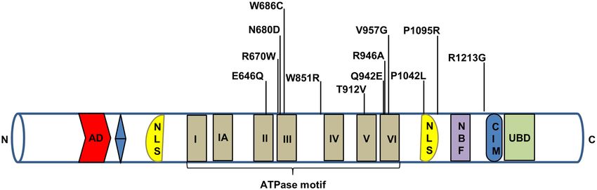
Nucleic Acids Research, 2021, Vol. 49, No. 5 2421

modulating its fate (processing) and downstream TC-NER          (XRCC1) (84). CSB deficiency leads to a three-fold hyper-
process. This area of CSB function has been widely covered      sensitivity to methyl methane sulfonate (MMS), which pro-
in recent reviews (64–68) and here we will focus on emerging    duces primarily N7-methylguanine and N3-methyadenine
areas of roles for CSB in other repair pathways.                DNA adducts repaired by BER. Because CSB stimulates
                                                                APE1 activity in vitro, defects in CSB may decrease BER
                                                                efficiency, and increase sensitivity to alkylating agents such
Base excision repair (BER)
                                                                as MMS (85).
DNA base excision repair (BER) acts selectively on small,          Poly (ADP-ribose) polymerase-1 (PARP1) is a molecu-
non-helix distorting lesions caused primarily by oxidative      lar sensor of DNA damage that binds single-strand DNA
DNA damage. The biochemistry of BER is well studied and         breaks (SSB) with high affinity. Upon binding to SSBs,
has been extensively reviewed (69–71). Briefly, in the first    PARP1 covalently modifies its target proteins, adding linear
steps of BER, a DNA glycosylase scans the genome and            or branched chains of ADP-ribose (PARylation) using the
searches for damaged and inappropriate bases (Figure 3).        cofactor nicotinamide adenine dinucleotide (NAD+ ). In ad-

                                                                                                                                  Downloaded from https://academic.oup.com/nar/article/49/5/2418/6137297 by guest on 11 December 2021
The DNA glycosylase removes the damaged base forming            dition to auto-PARylation, PARP1 substrates include core
an abasic (AP) site, while leaving the DNA backbone in-         histones and CSB (86,87). In general, PARylation alters the
tact. Mammalian cells express 11 different damage-specific      binding and function of the modified protein (86).
DNA glycosylases, seven of which are also expressed in             The N-terminal domain of CSB interacts with unmodi-
the mitochondria. After a DNA glycosylase removes the           fied and PARylated PARP-1 in the presence and absence of
damaged base, bifunctional glycosylases cleave the sugar-       oxidative stress. PARylation of CSB by PARP1 inhibits its
phosphate backbone. When a DNA lesion is incised by a           ATPase activity, making cells sensitive to oxidative stress.
monofunctional glycosylase, the next step is AP endonucle-      In addition, formation of CSB foci is inhibited by PARP in-
ase APE1-mediated cleavage of the DNA backbone, to gen-         hibition, suggesting that PARP-1 and/or PARylation pro-
erate a gapped intermediate. After AP site cleavage, repair     motes formation of CSB foci (88). Under conditions of
proceeds via either a ‘short-patch’ (single-nucleotide) BER     oxidative stress, the CSB/PARP-1 complex recognizes and
or ‘long-patch’ (multi-nucleotide) BER. Gap-filling is per-     binds to DNA lesions and promotes repair (89). When CSB-
formed by a DNA polymerase and the nick is sealed by a          proficient cells are treated with PARP inhibitors, the re-
DNA ligase (72–74).                                             pair of oxidative lesions is decreased, suggesting that PARP
   Several lines of evidence support a role for CSB in the      stimulates BER in a CSB-dependent manner (90). CSB
repair of oxidative DNA damage. First, CSB-deficient cell       plays an important role in displacing activated PARP1 from
lines and patients incise 8-oxoguanine (8-oxoG) less effi-      DNA damage. Therefore, CSB-deficient cells have higher
ciently and express lower levels of OGG1, the glycosylase       levels of PAR and more PARP-1 foci (91). These observa-
that removes this lesion from DNA, than corresponding           tions indicate that CSB plays a critical role in regulating
normal control cells (75,76). Next, primary fibroblasts from    PARP1-dependent DNA repair.
11 different CS patients accumulate significant levels of 8-       The 70-kDa XRCC1 protein acts as a non-enzymatic
oxoG and 8-oxoadenine (8-oxoA) after exposure to IR,            scaffold that recruits other proteins to SSBs generated as
while six normal control fibroblast cultures do not accumu-     BER-intermediates, and promotes processing of the nicked
late detectable levels of these DNA lesions (75–77), which      DNA (84). Binding of XRCC1 to SSBs is stimulated by
are induced by oxidative stress and repaired by BER. Con-       CSB and transcription and tightly linked to BER, although
sistent with this, the putative helicase activity of CSB, es-   XRCC1 binds SSBs generated by pathways other than BER
pecially functions encoded by helicase motifs V and VI,         is independent of CSB and transcription (92). Taken to-
has been shown to play an important role in repair of 8-        gether, these findings establish a role for CSB in facilitating
oxoG and 8-oxoA (77–80). OGG1 is present and functional         BER at several types of DNA lesions and promoting their
in both the nucleus and mitochondria. Although mito-            efficient repair.
chondrial BER is independent from nuclear BER, the pro-
teins responsible for mtBER are encoded by nuclear genes.
                                                                DNA Double-strand break repair
The mitochondrial extracts of CSB-deficient cells and CSB-
knockout mouse liver cells exhibited reduced 8-oxoG inci-       Double-strand DNA breaks (DSBs) are generated directly
sion activity, consistent with CSB’s role in transcription of   by IR or through processing events (referred to as two-
OGG1 (81).                                                      ended DSBs) or as intermediates when the replication fork
   In addition to OGG1, CSB functionally interacts with         encounters a persistent SSB or other bulky lesion (referred
several other BER proteins (38), one of which is NEIL1, a       to as one-ended DSBs). Based on the phase of the cell cycle
glycosylase that excises formamidopyrimidine DNA lesions        and on the cell type, DSBs are resolved by either nonho-
including Fapy-G, Fapy-A, and 5-hydroxycytosine. Consis-        mologous end joining (NHEJ) or homologous recombina-
tent with this, CSB-deficient mouse cells showed elevated       tion (HR). In S and G2 phases, HR is favored over NHEJ
levels of Fapy-G and Fapy-A in DNA (82). CSB stimulates         (30). NHEJ repairs broken ends in the absence of sequence
NEIL1 incision activity and AP lyase activity in a dose-        homology and is often error prone. HR repairs DSBs us-
dependent manner through its N-terminal domain (82).            ing sister chromatid homology in S/G2 phases and is con-
APE1 plays an important role in processing repair inter-        sidered to be almost error free. Ataxia-telangiectasia mu-
mediates produced by OGG1 and other glycosylases. (83).         tated (ATM) is an important signaling molecule in dou-
The N-terminal domain of APE1 interacts with CSB, which         ble strand break repair. It recognizes DSBs and phospho-
also interacts with X-ray cross-complementing protein 1         rylates hundreds of DNA damage response proteins includ-
2422 Nucleic Acids Research, 2021, Vol. 49, No. 5
Downloaded from https://academic.oup.com/nar/article/49/5/2418/6137297 by guest on 11 December 2021
Figure 3. CSB roles in BER, transcription and chromatin remodeling. The classical BER (nuclear) is initiated by damaged base removal (blue star) which
creates an AP site (blue circle). AP site is processed by AP lyase and induce incision at AP site. Following incision, the incision is processed by either short
or long patch repair depending upon nature of the substrate and cellular environment and subsequently nick is sealed by ligase. CSB interacts with some
of the integral player of BER like OGG1, NEIL1, APE1 and PARP1. Similar kinds of BER events also take place in response to damage in mitochondria
(mitochondrial BER). In response to UV induced DNA damage, firstly RNAPII is ubiquitinated and degraded which leads to transcription repression
then ATF3 recruites to inactive promoters. ATF3 is ubiquitinated in presence of CSB and leads to its proteasomal degradation. This allows recruitment
of RNAPII and transcription reactivation. The transcription factor like c-Jun targets CSB to TREs in normal condition. This allows CSB to regulate
transcription by ATP dependent chromatin remodeling which results in recruitment of other transcription regulators (see text for detail).Nucleic Acids Research, 2021, Vol. 49, No. 5 2423
ing breast cancer 1 (BRCA1) (93). During HR repair, resec- The ATPase domain also regulates UV-induced apoptosis
tion of DNA occurs at DSBs to produce 3 single-stranded in CSB-deficient cells. Cells harboring the E646Q ATPase
DNA (ssDNA). Initially, this ssDNA is protected by repli- mutation or a null allele of CSB die by apoptosis at simi-
cation protein A (RPA), and later replaced by RAD51. lar rates (112). Both CSB-deficient and the Q942E mutant
This filamentous RAD51-ssDNA is important for homol- CSB cells express low levels of OGG1 protein and corre-
ogy search and strand invasion (94). The MRN complex spondingly low OGG1 incision activity (113).
(MRE11, DNA repair protein RAD50 and Nibrin/NBS1), In contrast, the phenotype of cells with deletions in the
along with its cofactor CtIP, is involved in DNA resection. acidic domain of CSB was relatively mild (112), with nearly
BRCA1 binds phosphorylated CtIP and forms a complex normal levels of gene-specific repair of CPDs and PCNA
with MRN-CtIP. This BRCA1- MRN-CtIP complex per- relocation after UV irradiation (114). Although CSB is
forms DNA end resection through the endonuclease and unlikely to directly recruit PCNA as it interacts directly
exonucleolytic activities of MRN (95–98). UV exposure with upstream NER proteins (CSA, UVSSA and TFIIH),
stimulates interaction between BRCA1 and CSB. BRCA1 PCNA complex formation was reduced in CSB deficient
Downloaded from https://academic.oup.com/nar/article/49/5/2418/6137297 by guest on 11 December 2021
polyubiquitinates CSB, leading to CSB degradation (99). cells in response to oxidative- and UV-induced DNA dam-
ATM phosphorylates 53BP1, promoting RIF1 recruitment age (115,116). In general, the acidic domain of CSB is
to DSBs, preventing DNA end-resection, which in turn fa- dispensable, with the exception of its role in facilitating
vors the error-prone NHEJ pathway. CSB is recruited to protein–protein interactions (114). An interesting hypoth-
FokI nuclease-induced DSBs through its interaction with esis is that the protein-protein interacting acidic region may
RIF1, an effector of 53BP1, via its WHD during S phase. regulate proteins that control chromatin structure, poten-
Phosphorylation on S10 and S158 amino acids of CSB tially by modulating the rate of transcription and/or acti-
serve as molecular signals and governs chromatin remodel- vation of a subset of target genes. More evidence is needed
ing activity of CSB at DSBs. CSB can remove histones from to validate this hypothesis.
damaged chromatin, promote efficient HR-dependent DSB CSB ATPase and its chromatin remodeling activity
repair, and can restrict RIF1-mediated NHEJ (100,101). (23,117), are required for CSB’s contribution to transcrip-
Thus, CSB regulates DSB repair pathway choice and check tion and DNA repair. The CSB nucleosome remodeling ac-
point activation (102). tivity is 10-fold less active than the well characterized hu-
man ATP-dependent chromatin assembly factor (ACF) re-
modeling complex. However, CSBs interaction with nucle-
Interstrand crosslink (ICL) repair
osome assembly protein 1(NAP1)-like histone chaperones
Interstrand crosslinks (ICLs) are DNA lesions that cova- (NAP1L1 or NAP1L4) enhances its nucleosome remodel-
lently link opposite strands of dsDNA. Many chemothera- ing activity to a level comparable to ACF, potentially by
peutic agents including cisplatin and mitomycin C, as well weakening the interaction between CSB and undamaged
as natural processes like lipid peroxidation cause ICLs. DNA. Under conditions of cellular stress, activation and
ICLs are repaired by multiple repair pathways including translocation of CSB increase, which in turn stimulates TC-
NER and the Fanconi anemia (FA) pathway. The FA path- NER (117,118). Recent studies show that CSB-deficient
way involves at least 14 gene products (103). CSA- or CSB- cells downregulate H3K9me3-specific methyltransferases
deficient cells have increased sensitivity towards cisplatin SUV39H1 and SETDB1. SETDB1 expression is thought to
and mitomycin C (104–106). CSB-deficient cells repair ICLs be regulated by CSB through activating transcription fac-
less efficiently than control cells during G1 (107). CSB is tor 3 (ATF3). The downregulation of methyltransferases in
rapidly and robustly recruited to ICLs (108). CSB inter- CSB-deficient cells results in loss of heterochromatin and in-
acts with 5 to 3 exonuclease DNA crosslink repair 1A creased PARylation in highly-transcribed regions. Increased
(DCLRE1A) (also known as sensitive to nitrogen mustard PARylation by PARP-1 depletes cellular NAD+ , leading to
1A (SNM1A)). CSB stimulates the SNM1A exonuclease ac- mitochondrial dysfunction (see below) (119).
tivity, recruits it to ICLs and facilitates ICL repair. Addi- Hypoxia-inducible factor-1 (HIF-1) induces CSB to com-
tional studies will be needed, before we understand CSB’s pete with p53 for histone acetyltransferase (HAT) p300 in
role in ICL repair at the molecular level (109). response to hypoxia (120). Highly condensed and packed
(‘closed’) chromatin sterically hinders access to and repair
of genomic DNA. In contrast, CSB-mediated chromatin
Chromatin structure/remodeling
remodeling promotes an ‘open’ chromatin conformation,
CSB is a SWI/SNF – protein with DNA-dependent ATPase which modulates association of HAT p300 and HMGN1
activity. The central ATPase domain of CSB mediates CSB to enhance unwind/relax chromatin structure and facilitate
homodimerization, which is essential for its chromatin re- DNA repair (120–123).
modeling activity (Figure 3) (23,28). Mutations in the CSB
ATPase domain range from the E646Q mutation in motif II,
Transcription regulation
which shows no ATPase activity at all to the T912/913V and
Q942E mutations in motifs V and VI, respectively, which It has been known for >20 years that CSB-deficient cells
have low but measurable ATPase activity. CSB ATPase ac- have a nearly 50% lower rate of transcription than some
tivity is also regulated by post-translational modification control cells (124,125). Similarly, transcriptome analysis of
(110). Phosphorylation by casein kinase or Abelson murine CSB-deficient human fibroblasts showed dysregulation of
leukemia viral oncogene homolog 1 (ABL1) and PARyla- thousands of genes including neuronal genes (126). In one
tion by PARP1 negatively regulate CSB ATPase (88,111). study of CSB-deficient cells under conditions of oxidative2424 Nucleic Acids Research, 2021, Vol. 49, No. 5
stress, global transcription appears to be impaired. For ex- CSB interacts with RNAPII resulting in sequential re-
ample, 122 genes (∼1.8% of all genes analyzed in this study) cruitment of CSA, UVSSA and transcription factor TFIIH
were differentially-expressed in CS cells (127). Interestingly, to promote transcription recovery after UV (17,27). The
CSB ATPase domain was found to play an important role first evidence suggesting a role for CSB in transcription
in regulating transcription (127). These results suggest that was the inability of UV-irradiated CS cells to restore global
CSB may regulate transcription in cells with or without RNA synthesis (139). This led to two hypotheses: that CS
DNA damage. played a general role in transcription, or alternatively, that
The tumor suppressor p53 is a master regulator of the CSB played a role in transcription restart after UV expo-
transcriptional response to genotoxic stress, which regu- sure. The fact that CSA and CSB could be depleted in cells
lates cell cycle progression and the initiation of apoptosis. without transcriptional failure and that antibodies against
MDM2, an E3 ubiquitin ligase, promotes polyubiquitina- CSA or CSB did not upset basal transcription favor the
tion and degradation of p53 in unstressed cells, while p53 is latter hypothesis (53,140). However, later studies demon-
phosphorylated, stabilized and more abundant under con- strated that CSB-deficient cells were unable to restore RNA
Downloaded from https://academic.oup.com/nar/article/49/5/2418/6137297 by guest on 11 December 2021
ditions of cellular stress. Several post-translational modifi- synthesis after UV in both damaged and undamaged re-
cations regulate p53, modulating its binding to the promot- gions of the genome, suggesting a more global role for CSB
ers of a variety of genes including genes involved in cell cycle in transcription in addition to TC-NER (141,142). Indeed,
and apoptosis (128). Early studies suggest a direct physical several proposed models linking CSB and CSA to general
interaction between p53 and CSB’s C-terminal region (129– dysregulation of transcription in cells exposed to UV have
131). In CS cells under conditions of stress, the duration of emerged (17,63).
the p53-induced transcriptional response and the frequency Upon UV irradiation, many genes are repressed. The
of apoptosis increase (112,132,133). CSB-deficient primary promoters of UV-repressed genes bind and are repressed
fibroblasts cells express a higher level of p53 than control by ATF3 in response to UV stress (Figure 3) (143,144).
cells, potentially due to decreased expression of MDM2, In normal cells exposed to UV, ATF3 reaches its max-
which in turn would limit the rate of p53 degradation (134). imum expression ∼8 h after UV irradiation, and then
Both CSA and CSB proteins play an important role in gene expression is maximally repressed. By 12–24 h, ATF3
polyubiquitination-mediated p53 degradation (134). Thus, is degraded, RNAPII is recruited and transcripts begins
CSB indirectly regulates the abundance of p53 in the cell. to resume. In contrast, in CSB-deficient cells, ATF3 re-
Following in vitro reconstitution with purified proteins, mains bound to promoters for extended periods of time,
CSB interacts with RNAPII and the transcription elon- leading to persistent transcriptional arrest and downreg-
gation complex, resulting in a 3-fold stimulation of tran- ulation of ∼85% of all genes (143,144). CSB and CSA
scription elongation (51,53,135). Furthermore, chromatin promote ubiquitin-mediated proteasomal degradation of
immunoprecipitated sequencing (ChIP-seq) using anti-CSB ATF3, restoring transcription after UV-induced damage
antibody revealed higher occupancy of CSB at promoter (143). In fact, this effect is so pronounced in CS cells that
and enhancer sites which suggests that CSB plays a di- it has been suggested that ATF3-responsive genes could be
rect role in transcription initiation (136). This study showed used as markers for the diagnosis of CS (145). However,
that CSB was enriched at 12-O-tetradecanoylphorbol-13- ATF3 is activated in response to other genotoxins such as
acetate (TPA) response elements (TREs) that contain bind- IR, but CS cells are not as sensitive to these genotoxins
ing motifs for the activator protein 1 (AP-1), which belongs as to UV irradiation (146,147). This suggests that CSB-
to a family of bZIP transcription factors, including c-Jun dependent ubiquitin-mediated proteasomal degradation of
(Figure 3). c-Jun plays an important role in cell cycle pro- ATF3 may not be the principal mechanism for transcrip-
gression and has an anti-apoptotic activity. The transcrip- tion recovery from exposure to UV. Therefore, other roles
tion factor (c-Jun) can recruit CSB to TRE-containing sites for CSB in UV-exposed cells should be explored in future
to regulate gene expression at transcription initiation stage studies. Indeed, an alternative mechanism involving the in-
(136). fluence of CSB on RNAPII stability was recently proposed
Similarly, in response to oxidative stress, CSB regu- (17,63).
lates genes involved in stress response, cell cycle, tran-
scription, and translation (127). Another study of cells
RNAPII processing
with menadione-induced oxidative DNA damage showed
that CSB occupancy at target promoters was 6-fold (11%) Some DNA lesions stall RNAPII, which initiates DNA re-
higher than in unexposed cells (2%). For example, the occu- pair to remove the damage and also triggers global tran-
pancy of transcriptional regulator CCCTC-binding factor scriptional shutdown to avoid an aberrant expression of
(CTCF) went from 1% in normal cells to 11% in menadione- genes. However, the mechanism of transcriptional arrest is
treated cells. CTCF is involved in long-range chromatin not clear. A newly identified ubiquitination site (K1268) on
interactions (137,138). In vitro protein interaction studies RBP1 may provide insight into this question, as it plays a
showed that CSB and CTCF interact with each other and key role in both DNA damage and the transcriptional re-
that this interaction is stimulated by oxidative stress (137). sponse to UV irradiation (17,63). Mechanistically, K1268
Hence, these studies suggest that CSB may associate with ubiquitination induces the binding of the TFIIH core com-
CTCF and that it regulates transcription in response to ox- plex with the stalled RNAPII in a process also involving
idative stress. Nevertheless, the effect of oxidative stress on UVSSA mono-ubiquitination. When K1268 ubiquitination
TREs is not dependent on CSB, indicating a distinct regu- is impaired, two main consequences have been noted: (i)
latory pathway for basal TRE transcription. stalled RNAPII is not effectively processed/degraded, pre-Nucleic Acids Research, 2021, Vol. 49, No. 5 2425
venting global transcription shutdown and leading to dys- the direct interaction between CSB and G9a was established
regulation in transcription, (ii) TC-NER is impaired as using coimmunoprecipitation and a role for this interaction
UVSSA and TFIIH fail to interact with RNAPII. The role in regulating RNAPI transcription is suggested (153,154).
of CSA or CSB in K1268 ubiquitination, on the other hand, H3K9 modification promotes binding of the heterochro-
is less well understood. After the insult, CSA and CSB matin protein 1 gamma (HP1␥ ), which regulates rRNA syn-
are both recruited to the stalled RNAPII and this process thesis initiation and elongation. Furthermore, CSB ATPase
does not require RNAPII K1268 ubiquitination. However, activity is required for formation of H3K9me by G9a (153).
the subsequent recruitment of UVSSA and TFIIH to the Recent studies demonstrate that CSA and CSB deficiency
stalled RNAPII in the process of TC-NER depends on cause defects in ribosomal DNA transcription (155). CSB’s
CSA and CSB. K1268 ubiquitination on RBP1 is medi- UBD domain interacts with the most abundant nucleo-
ated by cullin-ring type E3 ligases (CRLs) as suppression lar protein, nucleolin, which plays a key role in rDNA
of CRLs completely abolishes UV-induced RBP1 ubiqui- transcription and pre-rRNA synthesis. Indeed, mutations
tination (17). Despite CSA being a part of the CRL4 E3 in the CSB UBD results in inhibition of the synthesis of
Downloaded from https://academic.oup.com/nar/article/49/5/2418/6137297 by guest on 11 December 2021
ubiquitin ligase complex, it is not clear whether CSA is pre-rRNA. More detailed mechanistic studies showed that
directly involved in the K1268 ubiquitination process as CSA, as a part of the E3 ubiquitin ligase complex, promotes
there are many types of CRLs. However, CSA and CSB the ubiquitination of nucleolin and enhances the interac-
contribute to the K1268 ubiquitination of RNAPII, po- tion of CSB with nucleolin (39). Nucleolin is present at the
tentially enhancing processing and degradation of stalled dense fibrillar and the granular center compartment of the
RNAPII (17,61). Indeed, mice expressing ubiquitination- nucleolus. rDNA transcription, pre-rRNA processing, and
resistant RBP1 K1268R in a NER-compromised back- ribosome subunit assembly occur predominantly in these
ground displayed CS-like phenotypes such as growth retar- nucleolar compartments. CSB and CSA increase RNAPI
dation and neurodegeneration (17). These results suggest a loading to the coding region of rDNA, which is dependent
model that CSB promotes degradation of stalled RNAPII, on nucleolin. Thus, CSB and its interaction with nucleolin
which is also supported by other reports (61,148). However, regulate rRNA transcription and ribosome biogenesis (39).
in contrast, there are other findings showing lower levels G4 structures occur at a high frequency in rDNA genes and
of RNAPII in UV-treated CSB- deficient cells, resulting in play a role in regulation of both replication and transcrip-
sustained depletion of RNAPII and defective post-UV re- tion. CSB promotes resolution of G4 structures via its heli-
covery of global transcription (63). Indeed, the introduc- case activity and also reduces transcriptional pausing at G4
tion of non-degradable RBP1 partially restores transcrip- structures (155).
tion restart in CSB-deficient cells, suggesting that the lack
of transcription recovery after UV irradiation is due to ex-
Mitochondria
cessive degradation and depletion of RNAPII (63). Further
studies will be needed to elucidate the mechanism by which There is significant interest and a body of recent evidence
CSA or CSB regulates K1268 ubiquitination and RNAPII supporting a role for mitochondrial dysfunction in many
stability. diseases as well as normal human aging. CS patients ex-
perience mitochondrial disease and neurological dysfunc-
tion (156–158). A recent cross-species transcriptomic anal-
rDNA transcription
ysis suggested that mitochondrial dysfunction is a common
In general, RNA translation depends upon rRNA and ribo- feature found in CS postmortem brain tissue, CS mice and
some biosynthesis. rDNA genes encode rRNA, which are CS nematodes (159). The mitochondrial theory of aging is
present in the nucleolus in multiple copies and are tran- based on that ROS, predominantly produced in mitochon-
scribed by RNAPI and RNAPIII. A subset of these genes dria, damage various macromolecules including mitochon-
are transcriptionally active at any given time to support cell drial DNA (mtDNA), leading to mitochondrial dysfunc-
growth (149). CSB is localized in the nucleolus and is part of tion (160). Lack of abundant protective histones and fewer
a complex that includes RNAPI, TFIIH, XPG and TIF-1B DNA repair pathways have been documented in mitochon-
(150,151). Both CSB and TFIIH are required for RNAPI dria than in the nucleus (161–163); however, mitochondrial
transcription (151). Chromatin remodeling and epigenetic transcription factor TFAM binds strongly to DNA and may
changes regulate rRNA transcription (152). CSB regulates substitute functionally for nuclear histones.
both these activities. Transcription termination factor 1 Mitochondria are a major source of endogenous ROS
(TTF-1) binds to the promoter-proximal terminator site and CSB deficiency might lead to accumulation of muta-
and helps recruit chromatin remodeling factors that facili- tions in mtDNA. This idea is supported by the observa-
tate RNAPI transcription. Co-immunoprecipitation exper- tion that CSB interacts with the mitochondrial transcrip-
iments revealed that TTF-1, CSB and RNAPI were part of tion protein TFAM in vivo (164). Mitodb.com is a bioin-
the same complex. A direct physical interaction between formatics tool that collects and analyzes clinical and ge-
CSB and TTF-1 has been observed, but direct interac- netic information, using clustering algorithms, to determine
tion between CSB and RNAPI has not been demonstrated. whether a disease clusters with known mitochondrial dis-
The CSB-TTF-1 complex competes with other factors of eases, and the probability that it is linked to mitochondrial
the chromatin remodeling complex to regulate rDNA tran- dysfunction. The Mitodb algorithms show that CS clusters
scription. Furthermore, G9a, a methyl transferase, epige- tightly with known mitochondrial diseases (165). As de-
netically regulates RNAPI transcription by mono- and di- scribed below, CSB-associated mitochondrial dysfunction
methylating histone 3 lysine 9 (H3K9). In the same study, might reflect a direct role for CSB in mitochondria or might2426 Nucleic Acids Research, 2021, Vol. 49, No. 5
reflect an indirect role, where CSBs nuclear role in DNA cluding glycolysis, the electron transport chain (ETC), the
repair propagates through nuclear to mitochondrial signal- tricarboxylic acid (TCA) cycle, and DNA repair pathways.
ing. CSB does not have a classic mitochondrial localization NAD+ abundance in various cellular compartments plays
signal, but it may utilize TOM20 or similar transport sys- an important role regulating several metabolic pathways
tem. Mitochondrial dysfunction in CSB cells could poten- (Figure 4). For example, the NAD+ –SIRT1–PGC1␣ axis
tially explain many poorly understood clinical features of regulates nucleus to mitochondria signaling. PARP1 utilizes
CSB, including hearing loss, severe neurological deficien- NAD+ as its substrate to PARylate itself and other target
cies, dysfunction in skeletal muscle and heart, as well as pre- repair proteins in response to DNA damage. There are re-
mature aging (81,166–168). ports that PARP1 localizes to mitochondria, though there
BER plays a prominent role in protecting the nervous is debate over the functional significance of this observation
system against oxidative DNA damage. Increased mtDNA (181–183). The sirtuin family of proteins (SIRT1–7) also
damage and defective nuclear or mitochondrial BER cor- uses NAD+ as a substrate, primarily for deacetylase activi-
relate with neurodegeneration and aging (169,170). BER ties. Sirtuins localize to the mitochondrial or nuclear com-
Downloaded from https://academic.oup.com/nar/article/49/5/2418/6137297 by guest on 11 December 2021
in mitochondria is associated with the inner mitochondrial partments and serve important roles in DNA damage re-
membrane (Figure 3) (171,172). Menadione-induced oxida- sponse and genomic stability (184–188). Of the seven SIRT
tive stress induces CSB mitochondrial localization. In the proteins, SIRT1 is the most studied and has a well vali-
absence of CSB, BER activity at the mitochondrial mem- dated role in mitochondrial biogenesis through deacetyla-
brane is decreased, resulting in an increased mutation rate tion of the transcription factor, PGC-1␣ (189,190). In in-
(164). CSB-deficient cells and mice have increased mito- stances of limiting NAD+ , the activity of all seven sirtuins
chondrial content and higher overall ROS, perhaps due is inhibited. As discussed earlier, CSB interacts with PARP1
to dysregulation of mitochondrial BER. This could cause and both are present in mitochondria. PARP1 PARylates its
damaged mitochondria to accumulate, which then must be substrates in response to DNA damage, whereas CSB me-
removed via mitophagy (105,132,173,174). CSB-deficient chanically displaces PARP1 to limit its activity (91). How-
cells have increased oxygen consumption rates and FCCP- ever, in the absence of CSB, this regulation of PARP1 is
uncoupled respiration, probably due to increased ATP de- lost, leading to persistent PARP1 activity and subsequent
mand. FCCP is a protonophore which mimics physiolog- NAD+ deficiency. It was observed that NAD+ supplemen-
ical energy demand to investigate the role of mitochon- tation activates SIRT1 and rescues CS-associated pheno-
dria in cellular function and CSB-deficient cells showed types in mice and cells (91,191). AMP-activated protein
increased mitochondrial respiration to meet energy de- kinase (AMPK), which acts as an energy sensor of cells,
mand. The NIX/PINK1-Parkin-mediated mitophagy path- upon activation can phosphorylate a number of proteins
way recycles dysfunctional mitochondria. Damaged, ubiq- including SIRT1, PGC-1␣, and HIF-1␣ (192,193). Inter-
uitinated mitochondria recruit the scaffolding protein, P62, estingly, lower AMPK activity was observed in CS patient-
which facilitates mitophagy by fusion with an autophago- derived brain samples and restored with NAD+ augmen-
some coated by the linkage protein LC-3B isoform II. In tation in CSB deficient cells. AMPK plays an important
response to stress, CSB-deficient cells demonstrated de- role in mitochondrial biogenesis and maintains mitochon-
creased colocalization of LC3, P62 and ubiquitin to the mi- drial homeostasis through NAD+ (194). This influences
tochondria, resulting in decreased mitophagy (173,175). NAD+ consumption and the interaction between these pro-
CSB’s role in relation to nuclear transcription is described teins helps to regulate nuclear to mitochondrial crosstalk
above. CSB also plays a role in mitochondrial transcription. (Figure 4). Based on our studies in other premature ag-
Mitochondrial RNA polymerase (mtRNAP) is a single sub- ing and DNA repair-deficient syndromes including XPA,
unit enzyme similar to the bacteriophage T7 RNA poly- ATM, and Werner syndrome, we speculate that NAD+ de-
merase (176). In vitro experiments showed that CSB stim- pletion due to dysregulated PARylation may be the most
ulates mtRNAP elongation similarly to RNAPII. Further- significant cause of mitochondrial dysfunction in CS cells
more, transcription from the mitochondrial heavy strand (185,195,196). Partial mitochondrial dysfunction in CS cells
promoter was lower in CSB-deficient cells than in control could result from defective CSB in the mitochondria. How-
cells (177). Although the exact mechanism of this regulation ever, mitochondrial dysfunction also occurs in CSA- and
by CSB is still unknown, the DNA annealing and transloca- XPA-deficient cells (185,197), which are not reported in
tion activities of CSB were required for this regulation (178). the mitochondria. Thus, it is likely that the major cause
The majority of mitochondrial proteins are encoded in the of mitochondrial dysfunction in CS is defective nuclear-
nucleus and translocated to mitochondria in response to mitochondrial crosstalk in response to DNA damage.
environmental stimuli. Some studies demonstrate nuclear- Neurodegeneration is a major clinical feature of CS, but
mitochondrial crosstalk through various proteins in the mi- its etiology and mechanism are not fully understood. How-
tochondria (179). This crosstalk, from the mitochondria to ever, it is clear that defects in TC-NER can not fully ac-
the nucleus or nucleus to mitochondria, is essential for mi- count for neurodegenerative features in CS, because pa-
tochondrial and cellular health (180). Thus, changes in the tients with UV-sensitive syndrome and defective TC-NER
cellular environment, abundance of NAD+ , and response to manifest mild UV-sensitivity and lack the neuropatholog-
stress play roles in this type of crosstalk, ultimately regulat- ical features of CS (198). Recent studies show that mito-
ing mitochondrial homeostasis. chondrial dysfunction contributes significantly to neurode-
Nicotinamide adenine dinucleotide (NAD) and its ox- generation. In a Caenorhabditis elegans model of CS, CSB
idized (NAD+ ) and reduced (NADH) forms are major mutant worms showed mitochondrial defects and compro-
metabolites and coenzymes involved in critical pathways in- mised respiratory activity and neurodegeneration (199). In-Nucleic Acids Research, 2021, Vol. 49, No. 5 2427
Downloaded from https://academic.oup.com/nar/article/49/5/2418/6137297 by guest on 11 December 2021
Figure 4. NAD+ supplementation as a therapeutic approach to CS. CSB play important role in regulation of repair and transcription under stressed
and normal conditions through chromatin remodeling and maintains normal cellular homeostasis. In CSB-deficient cells, in response to endogenous
and exogenous DNA damage, cells are unable to process DNA damage-stalled RNAPII which lead to prolonged transcription arrest and dysregulation of
number of proteins. PARP1 gets activated and persist at damage sites and PARylates number of proteins utilizing NAD+ . Eventually this leads to decreased
level of NAD+ which affects number of NAD+ dependent proteins like sirtuins. Inactivation of sirtuins causes increase in mitochondrial reactive oxygen
species in response to decreased downstream activation of mitochondrial factors like AMPK, HIF1␣ and PGC1␣ resulting in mitochondrial dysfunction.
Decreased repair, stalled transcription and mitochondrial dysfunction all together contribute to neurodegeneration in CSB deficient patient. Thus, impaired
cellular metabolite concentration can be replenished by NAD+ precursors such as nicotinamide riboside (NR) to overcome some of premature aging
phenotypes (see text).
triguingly, as mentioned above, features of mitochondrial hearing loss in CS mice. However, it is not yet know whether
diseases and CS strongly overlap. Indeed, mitochondrial NAD+ supplementation alleviates other CS neurological
dysfunction due to persistent PARylation leading to NAD+ features, such as progressive retinal degeneration or neu-
depletion is observed in CS cells. Notably, NAD+ augmen- ropathy (200). Nevertheless, these findings suggest CSB’s
tation corrected many aspects of mitochondrial abnormal- role in mitochondrial function as a new emerging mecha-
ities in CS and prevented the progression of sensorineural nism linking CSB deficiency to neurodegeneration.2428 Nucleic Acids Research, 2021, Vol. 49, No. 5
Table 1. Comparison of clinical features in CSA and CSB contains a C-terminal UBD and CIM. CSB binds to CSA
Clinical features through CIM in response to UV induced DNA damage due
to a conformational change triggered by association of CSB
Similar prevalence (in CSA & Variable prevalence (in CSA & with lesion stalled RNAPII (27). Protein-protein interac-
CSB mutations) CSB mutations)
tion of CSB with other transcription factors or repair pro-
Growth failure Low birth weight teins might also be involved in the activation and regula-
Cachexia Cataract tion of CSB. A study on yeast Rad26 using cryo-electron
Mental retardation Microphthalmia
Microcephaly Hearing loss microscopy shed some light on its role in TC-NER and
Retinal degeneration Dental anomalies transcription elongation (54). There is a need for further
Photosensitivity structure-function studies to understand CSB regulation.
Mitochondrial dysfunction is another area where re-
search on CSB’s role is emerging and advancing. A study
showed the presence of CSA inside mitochondria and its in-
Downloaded from https://academic.oup.com/nar/article/49/5/2418/6137297 by guest on 11 December 2021
Closing remarks
volvement with mtBER upon oxidative stress (203). It is in-
CS is a multisystem progeroid disorder, exhibiting some triguing that generally the NER proteins are not present in
features of normal human aging. This review provides a the mitochondria, yet CS patients demonstrate mitochon-
broad picture of the current understanding of CS focusing drial disease phenotypes. Clearly, in the case of CSB, we
on emerging roles for CSB. This knowledge has expanded can speculate that the loss of mtBER activity and result-
our understanding and role of CSA in TC-NER. One third ing increase in mtDNA mutations contributes to this mi-
of CS cases are associated with CSA mutations and two tochondrial phenotype. However, how this relates directly
thirds of these CSA patients are classified as type I CS (7). to CS cases caused by CSA mutations is a subject of active
No obvious genotype-phenotype correlation can be drawn research.
between CSA and CSB mutations and clinical phenotypes Finally, therapeutic intervention is another goal of our re-
due to overlapping symptoms. Even though there are no search. Previously, the use of antioxidants (204) and phar-
specific clinical symptoms that differentiate CSA from CSB, macological chaperones (205) have been suggested. There
there are subtle differences in these phenotypes, as shown in are also some drugs currently under clinical trials (Pro-
Table 1 (201). Since both patient types have similar clinical darsan and Sirolimus). Our research indicates that NAD+
features, it is likely that CSA and CSB regulate some of the supplementation demonstrates promise for restoring mito-
same process(es) in the cell. Research demonstrating differ- chondrial homeostasis, promoting DNA repair and energy
ences between the other phenotype of XP patients deficient balance, and reversing CS-associated hearing loss (Figure
in TC-NER and CS patients makes clear that other DNA 4) (157,167,175,185). There is tremendous scientific inter-
repair pathways are important for CS deficiencies. More est in the potential of NAD+ supplementation as a thera-
research is needed to understand the relationship between peutic approach for CS, and for understanding how NAD+
CSB and especially CSA in those pathways. abundance regulates multiple cellular pathways, including
Initially, CSB was only considered as a TC-NER protein, DNA repair, transcription, chromatin structure and mito-
but the CS- deficient TC-NER defect is not sufficient to ex- chondrial bioenergetics.
plain many of the clinical features of CS. The developmental
defect of CS can be explained by the transcriptional role of ACKNOWLEDGEMENTS
CSB whereas neurodegeneration is more likely attributed to
defective oxidative DNA damage processing. CSA and CSB We thank Drs Deborah Croteau, Burcin Duan Şahbaz and
mouse models are considered mild CS models as they only Seoyun Choi (NIA) for their helpful comments on the arti-
have few features of the patients. The severe CS phenotype cle.
is precipitated only when these genes are inactivated in XPC
or XPA –null background (202). Therefore, the role of CS FUNDING
proteins must be more than DNA damage repair. Thus, it is
Intramural Research Program of the NIH; National Insti-
important to understand the principle molecular functions
tute on Aging. Funding for open access charge: NIH.
of CS proteins and their contribution and correlation to dif-
Conflict of interest statement. None declared.
ferent clinical pathologies. CSB participates in several crit-
ical aspects of DNA repair, RNAPII processing, transcrip-
tion, signaling, chromatin structure and cellular bioener- REFERENCES
getics (e.g. mitochondrial health). Thus, patient-to-patient 1. Cleaver,J.E., Thompson,L.H., Richardson,A.S. and States,J.C.
variation in CS severity may reflect CSBs diverse functions (1999) A summary of mutations in the UV-sensitive disorders:
manifesting at different stages of development. xeroderma pigmentosum, Cockayne syndrome, and
CSB is involved in both RNAPI and RNAPII transcrip- trichothiodystrophy. Hum. Mutat., 14, 9–22.
2. Mallery,D.L., Tanganelli,B., Colella,S., Steingrimsdottir,H., van
tion. CS cells demonstrate BER deficiency (166), which may Gool,A.J., Troelstra,C., Stefanini,M. and Lehmann,A.R. (1998)
reflect reduced transcription of BER proteins or perhaps in- Molecular analysis of mutations in the CSB (ERCC6) gene in
dicate that CSB directly contributes to BER. The potential patients with Cockayne syndrome. Am. J. Hum. Genet., 62, 77–85.
role of CSB in regulating mitochondrial transcription is in- 3. Weeda,G., van Ham,R.C., Vermeulen,W., Bootsma,D., van der
Eb,A.J. and Hoeijmakers,J.H. (1990) A presumed DNA helicase
triguing and in need of further research. encoded by ERCC-3 is involved in the human repair disorders
More details about the regulation of CSB itself are also xeroderma pigmentosum and Cockayne’s syndrome. Cell, 62,
needed. CSB is phosphorylated in response to UV and also 777–791.You can also read