Tethered TGF-β1 in a Hyaluronic Acid-Based Bioink for Bioprinting Cartilaginous Tissues - MDPI
←
→
Page content transcription
If your browser does not render page correctly, please read the page content below

International Journal of
               Molecular Sciences

Article
Tethered TGF-β1 in a Hyaluronic Acid-Based Bioink for
Bioprinting Cartilaginous Tissues
Julia Hauptstein 1 , Leonard Forster 2 , Ali Nadernezhad 2 , Jürgen Groll 2 , Jörg Teßmar 2                              and Torsten Blunk 1, *

                                            1   Department of Trauma, Hand, Plastic and Reconstructive Surgery, University of Würzburg,
                                                97080 Würzburg, Germany; hauptstein_j@ukw.de
                                            2   Department for Functional Materials in Medicine and Dentistry, Bavarian Polymer Institute (BPI),
                                                University of Würzburg, 97070 Würzburg, Germany; leonard.forster@fmz.uni-wuerzburg.de (L.F.);
                                                ali.nadernezhad@fmz.uni-wuerzburg.de (A.N.); juergen.groll@fmz.uni-wuerzburg.de (J.G.);
                                                joerg.tessmar@fmz.uni-wuerzburg.de (J.T.)
                                            *   Correspondence: blunk_t@ukw.de; Tel.: +49-931-201-37115

                                            Abstract: In 3D bioprinting for cartilage regeneration, bioinks that support chondrogenic develop-
                                            ment are of key importance. Growth factors covalently bound in non-printable hydrogels have been
                                            shown to effectively promote chondrogenesis. However, studies that investigate the functionality
                                            of tethered growth factors within 3D printable bioinks are still lacking. Therefore, in this study, we
                                            established a dual-stage crosslinked hyaluronic acid-based bioink that enabled covalent tethering
                                            of transforming growth factor-beta 1 (TGF-β1). Bone marrow-derived mesenchymal stromal cells
                                            (MSCs) were cultured over three weeks in vitro, and chondrogenic differentiation of MSCs within
                                            bioink constructs with tethered TGF-β1 was markedly enhanced, as compared to constructs with
                                            non-covalently incorporated TGF-β1. This was substantiated with regard to early TGF-β1 signaling,
                                            chondrogenic gene expression, qualitative and quantitative ECM deposition and distribution, and

                                     resulting construct stiffness. Furthermore, it was successfully demonstrated, in a comparative anal-
Citation: Hauptstein, J.; Forster, L.;      ysis of cast and printed bioinks, that covalently tethered TGF-β1 maintained its functionality after
Nadernezhad, A.; Groll, J.; Teßmar, J.;     3D printing. Taken together, the presented ink composition enabled the generation of high-quality
Blunk, T. Tethered TGF-β1 in a              cartilaginous tissues without the need for continuous exogenous growth factor supply and, thus,
Hyaluronic Acid-Based Bioink for            bears great potential for future investigation towards cartilage regeneration. Furthermore, growth
Bioprinting Cartilaginous Tissues.          factor tethering within bioinks, potentially leading to superior tissue development, may also be
Int. J. Mol. Sci. 2022, 23, 924. https://   explored for other biofabrication applications.
doi.org/10.3390/ijms23020924

Academic Editors: Magali                    Keywords: biofabrication; bioink; chondrogenic differentiation; dual-stage crosslinking; hyaluronic
Cucchiarini and Henning Madry               acid; tethering; transforming growth factor-beta 1

Received: 23 December 2021
Accepted: 12 January 2022
Published: 15 January 2022
                                            1. Introduction
Publisher’s Note: MDPI stays neutral
                                                 In recent years, 3D biofabrication including bioprinting has evolved as a fast-growing
with regard to jurisdictional claims in
                                            research field in regenerative medicine and for the development of disease models [1–3].
published maps and institutional affil-
                                            3D bioprinting enables precise patterning of cells and hydrogel materials, i.e., bioinks, and
iations.
                                            is investigated as a promising alternative approach for treatment of cartilage defects in
                                            trauma and degenerative diseases. To obtain functional cartilage transplants, bioinks that
                                            support chondrogenic development are of key importance [4–7].
Copyright: © 2022 by the authors.
                                                 Hyaluronic acid represents a promising and attractive material for cartilage regen-
Licensee MDPI, Basel, Switzerland.          eration, as it is a main component of the natural cartilage extracellular matrix (ECM),
This article is an open access article      and it enables diverse chemical modifications for crosslinking reactions with other bioink
distributed under the terms and             components [4,8–10]. Frequently applied functionalizations are, for example, thiol [11],
conditions of the Creative Commons          methacrylate [12], glycidyl methacrylate [13], tyramine [14], or norbornene [15] among sev-
Attribution (CC BY) license (https://       eral others. Nevertheless, studies that show flexible hyaluronic acid inks with a high initial
creativecommons.org/licenses/by/            shape stability after printing as well as convincing long-term development of cartilaginous
4.0/).                                      constructs are still rare [8,16].

Int. J. Mol. Sci. 2022, 23, 924. https://doi.org/10.3390/ijms23020924                                              https://www.mdpi.com/journal/ijms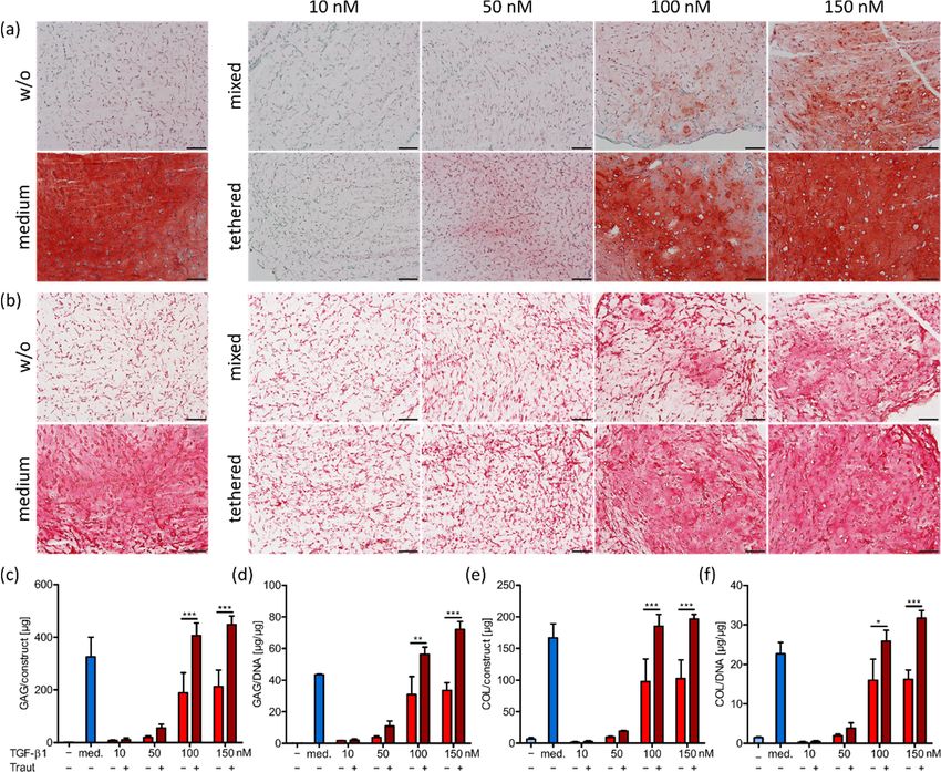
Int. J. Mol. Sci. 2022, 23, 924                                                                                             2 of 20

                                        Apart from a suitable ink material, the growth factor TGF-β is crucial for chondro-
                                  genic differentiation of mesenchymal stromal cells (MSCs) and cartilage homeostasis. It
                                  stimulates ECM production and can enhance cartilage repair, while its absence results in
                                  osteoarthritis (OA)-like changes [17,18]. However, application of TGF-β to the synovial
                                  tissue of the joint, for example via injections, results in a short half-life [19] and can induce
                                  fibrosis and osteophyte formation in adjacent tissues [20,21]. Therefore, a precise local
                                  supplementation and dose control appears desirable. TGF-β immobilization within hy-
                                  drogel scaffolds could be achieved, e.g., by direct loading, encapsulation into particulate
                                  carriers which are incorporated into the scaffold, reverse binding, for example via electro-
                                  static interactions, or covalent tethering [22,23]. Especially the covalent approach holds
                                  great promise for functional cartilage transplants with respect to prolonged availability for
                                  embedded MSCs and low release rates into the surrounding tissue [24–26]. With regard
                                  to 3D bioprinting, covalent incorporation into the used bioink may avert the necessity for
                                  repeated injections in vivo, likely enhancing the clinical potential of the inks for cartilage
                                  regeneration. Over the last couple of years, the impact of shear stress during 3D printing
                                  on cell survival and differentiation has been extensively studied [27–30]. Furthermore, it
                                  is known that shear stress can also strongly impact protein structure and function [31,32].
                                  However, surprisingly, studies that investigate whether the 3D printing process may have
                                  an impact on the functionality of covalently tethered growth factors are still lacking. For
                                  this reason, this study focused on the protein function of soluble and covalently tethered
                                  TGF-β1 in a comparative analysis of cast and printed bioinks.
                                        In a recent study, we established a novel hyaluronic acid-based bioink platform for
                                  cartilage 3D biofabrication [16]. Within this platform, we identified bioinks which enabled
                                  high shape fidelity during and after printing, but still had a very low polymer content of
                                  2% and a highly porous network. This facilitated effective chondrogenic differentiation
                                  of MSCs, a homogeneous distribution of newly produced extracellular matrix (ECM) and
                                  thereby a superior quality of resulting cartilaginous constructs. However, these novel
                                  bioinks were not suitable for growth factor binding and TGF-β1 had to be supplemented
                                  with each medium change.
                                        Therefore, in this study, we investigated a new hyaluronic acid-based bioink which
                                  maintained the beneficial properties of the established bioink platform and further enabled
                                  covalent TGF-β1 tethering. Specifically, TGF-β1 was thiol-modified with Traut’s reagent
                                  and covalently tethered to a newly integrated crosslinker, polyethylene glycol-octaacrylate
                                  (8-arm PEG-acryl), via Michael addition. In the first step of hydrogel formation, the re-
                                  maining acryl groups of 8-arm PEG-acryl reacted via Michael addition with thiol-modified
                                  hyaluronic acid (HA-SH), resulting in a highly viscous and 3D printable ink. After fabrica-
                                  tion, a UV-induced thiol-ene click reaction between polyethylene glycol-diallyl carbamate
                                  (2-arm PEG-allyl) and the residual thiol groups of HA-SH ensured final crosslinking. We
                                  characterized the inks with regard to rheological properties, swelling behavior, and TGF-β1
                                  release. MSC chondrogenesis was assessed in cast constructs with varying amounts of
                                  covalently or non-covalently incorporated TGF-β1, with distinct advantages achieved by
                                  TGF-β1 tethering. Subsequently, constructs with 150 nM TGF-β1, either tethered or non-
                                  covalently incorporated, were cast or 3D bioprinted. Chondrogenic differentiation of MSCs
                                  was extensively evaluated regarding early TGF-β1 signaling, gene expression, qualitative
                                  and quantitative ECM deposition and distribution, and resulting construct stiffness. The
                                  presented HA-based bioink with tethered TGF-β1 yielded superior chondrogenic differen-
                                  tiation, as compared to constructs with non-covalently incorporated TGF-β1. Furthermore,
                                  it was successfully demonstrated in the comparative analysis of cast and printed bioinks
                                  that covalently tethered TGF-β1 maintained its functionality after 3D printing.

                                  2. Results and Discussion
                                  2.1. Bioink Composition and Dual-Stage Crosslinking
                                       This study presents a novel 3D printable hyaluronic acid-based bioink that allows
                                  for biofunctionalization with tethered TGF-β1 to generate advanced cartilaginous 3D con-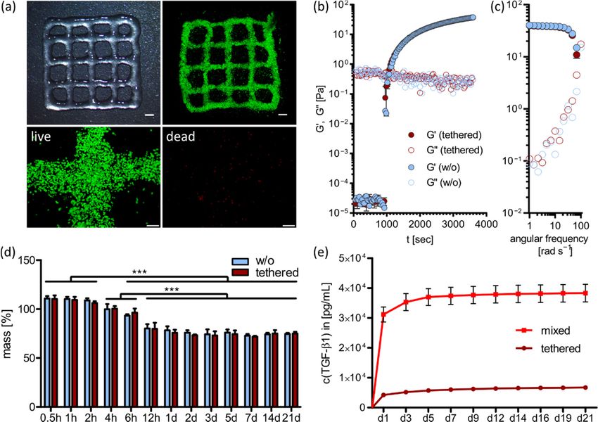
Int. J. Mol. Sci. 2022, 23, 924                                                                                                   3 of 20

                                   structs without the need for an exogenous supply of the differentiation factor during long-
                                   term culture. Previously, TGF-β1 has been bound to hydrogels via Traut’s reagent and other
                                   covalent approaches to induce differentiation and ECM production of embedded MSCs or
                                   chondrocytes, however, only in non-printable hydrogels [24–26,33–35]. The HA-based ink
                                   presented here utilized a recently established dual-stage crosslinking mechanism [16], with
                                   adapted composition in order to facilitate TGF-β1 tethering. Ink components encompassed
                                   thiol-modified hyaluronic acid (HA-SH, 465 kDa), acryl-modified polyethylene glycol
                                   (8-arm PEG-acryl, 10 kDa), and allyl-modified PEG (2-arm PEG-allyl, 6 kDa). Successful
                                   synthesis of all ink components was verified via GPC and 1 H-NMR (Figure S1, Supplemen-
                                   tary Materials). For the newly established 8-arm PEG-acrylate it could be demonstrated
                                   that the acrylation was successful and quantitative using the enzymatic catalysis and no
                                   oligomers were obtained during modification (Figure S1b, Supplementary Materials).
                                        Figure 1 shows the detailed functionalization and crosslinking mechanism of the used
                                   ink composition. At first, free lysine residues of TGF-β1 were thiol-modified using Traut’s
                                   reagent in a 4:1 molar excess of Traut to TGF-β1, as described previously [24–26]. Subse-
                                   quently, 8-arm PEG-acryl was added to react with thiol-modified TGF-β1 in a spontaneous
                                   Michael addition at pH 7.4 (Figure 1a). In the next step of bioink formulation, all other
                                   ink components (HA-SH, 2-arm PEG-allyl, I2959) as well as human MSCs (20 × 106 mL−1 )
                                   were mixed with the TGF-β1-functionalized PEG-acryl solution. Within 30 min of incu-
                                   bation at 37 ◦ C, free acryl groups of PEG-acryl reacted in a Michael addition at pH 7.4
                                   with the main fraction of HA-SH thiol groups to generate a highly viscous and printable
                                   bioink (Figure 1b). After 3D printing, constructs were UV-irradiated at 365 nm to finally
                                   crosslink the residual HA-SH thiol groups with allyl groups of PEG-allyl via thiol-ene
                                   click chemistry in the presence of the photoinitiator Irgacure I2959 (Figure 1c). Figure 1d
                                   presents a schematic graphical overview to visualize the ink composition with its two
                                   different crosslinking processes. The used ink had an overall polymer content of 1.4% (0.5%
                                   HA-SH, 0.5% 8-arm PEG-acryl, 0.4% 2-arm PEG-allyl and 0.05% I2959; all w/v), and still
                                   enabled stand-alone printability. Such low polymer content has previously been shown to
 Int. J. Mol. Sci. 2022, 23, 924                                                                                          4 of 21
                                   be favorable with regard to diffusion properties and penetrability of cellularly produced
                                   ECM in non-printed [36,37] and printed constructs [16,38].

                                    Figure1.1.Ink
                                   Figure      Inkcrosslinking
                                                    crosslinking mechanism
                                                               mechanism     and
                                                                           and   functionalization
                                                                               functionalization    with
                                                                                                 with    TGF-β1.
                                                                                                      TGF-β1.     (a) Thiol-functionaliza-
                                                                                                              (a) Thiol-functionalization
                                    tion of TGF-β1   with  Traut’s reagent and Michael addition  with 8-arm PEG-acryl.
                                   of TGF-β1 with Traut’s reagent and Michael addition with 8-arm PEG-acryl. (b) Michael(b) Michael  addi-
                                                                                                                              addition of
                                    tion of TGF-β1-modified PEG-acryl with HA-SH (pre-crosslinking), resulting in a 3D printable ink.
                                    (c) UV-induced final crosslinking of residual HA-SH thiol groups with 2-arm PEG-allyl in the pres-
                                    ence of Irgacure I2959 at 365 nm. (d) Graphical overview of the dual-stage crosslinking mechanism
                                    and workflow of TGF-β1 tethered construct generation for chondrogenic tissue culture.

                                    2.2. 3D Printing and Ink Characterization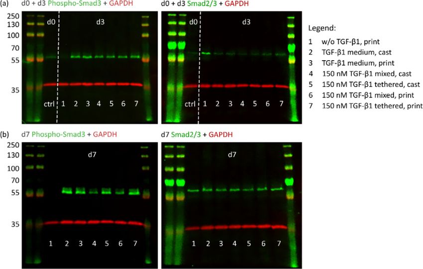
Int. J. Mol. Sci. 2022, 23, 924                                                                                             4 of 20

                                  TGF-β1-modified PEG-acryl with HA-SH (pre-crosslinking), resulting in a 3D printable ink. (c) UV-
                                  induced final crosslinking of residual HA-SH thiol groups with 2-arm PEG-allyl in the presence of
                                  Irgacure I2959 at 365 nm. (d) Graphical overview of the dual-stage crosslinking mechanism and
                                  workflow of TGF-β1 tethered construct generation for chondrogenic tissue culture.

                                  2.2. 3D Printing and Ink Characterization
                                        We have previously characterized printability of dual-stage crosslinked platform
                                  bioinks in detail [16]. In order to prove printability of the adapted ink with the new
                                  component 8-arm PEG-acrylate presented in this study, filament fusion tests and strand
                                  thickness evaluations were performed accordingly (Figure S2a,b, Supplementary Materials).
                                  Filaments fused at the smallest distance of 0.5 mm and partially at 0.75 mm, strand thickness
                                  measurements yielded the average strand thickness of 650 µm and a strand intersection
                                  diagonal of around 1.1 mm, which was well in accordance with the previously reported
                                  results of the printable platform inks [16]. Additionally, as a proof-of-principle, a large
                                  construct with 21 layers was 3D printed, resembling human femoral condyles (Figure S2c,
                                  Supplementary Materials).
                                        Furthermore, we analyzed biocompatibility of the bioink and possible physical impacts
                                  of TGF-β1 tethering on bioink and construct properties. Therefore, the bioink was prepared
                                  as described above and printed in 2-layered constructs to enable imaging with a low
                                  background signal and quantification of single cells. Figure 2a shows an overview of the
                                  printed grid structures and survival of MSCs directly after printing. To visualize living and
                                  dead cells separately, the split channels of a representative image with higher magnification
                                  is shown in the second row. Quantification of overall cell survival yielded around 98%
                                  living cells and only 2% dead cells after printing. This proved well-adapted printing
                                  conditions and confirmed the bioink as a suitable cell carrier.
                                        To assess the potential influence of covalent TGF-β1 tethering to 8-arm PEG-acryl on
                                  the gelation and, eventually, viscoelastic properties of the ink, we evaluated the kinetics
                                  of the first stage of crosslinking (i.e., the Michael addition). Rheological characterization
                                  of time and frequency sweeps were performed on samples without the protein or with
                                  150 nM tethered TGF-β1 (Figure 2b,c). Time sweep measurements revealed a drastic in-
                                  crease in ink viscosity after 15 min and almost complete Michael addition after 30 min.
                                  This corresponded to empirically defined pre-crosslinking times of this ink in 3D printing
                                  experiments for cell culture. Furthermore, covalent incorporation of TGF-β1 did not affect
                                  the mobility and reactivity of 8-arm PEG-acryl, and the kinetics of progression of Michael
                                  addition was almost identical in both formulations (i.e., without or with tethered TGF-β1).
                                  Moreover, the results of frequency sweep experiments showed that after the same incuba-
                                  tion time at 37 ◦ C, the formed covalent bonds during Michael addition yielded a similar
                                  state of long-range interactions between polymer chains. These results demonstrated that
                                  the presence of covalently bound TGF-β1 did not compromise gelation during the first
                                  stage of crosslinking.
                                        For further construct characterization, we examined gel stability after final crosslinking
                                  for constructs without or with 150 nM tethered TGF-β1 (Figure 2d). Therefore, ink was
                                  prepared without cells, cast in a defined cylindrical geometry (5 mm diameter, 40 µL
                                  volume), and incubated in PBS over three weeks. No significant differences between the
                                  constructs with or without tethered TGF-β1 could be detected at all time points (0.5, 1, 2,
                                  4, 6, 12 h, 1, 2, 3, 5, 7, 14, 21 d). Both gel systems initially swelled slightly to 110% within
                                  the first two hours and then started to shrink to around 80%, reaching their equilibrium
                                  state at 12 h after preparation. The swelling process was due to initial water inflow into
                                  the hydrogel system and shrinkage may be explained by formation of disulfide bonds
                                  between unreacted HA-SH thiol groups. Statistically, all thiol groups should be saturated
                                  by PEG-acryl or PEG-allyl crosslinking, but it could not be excluded that some functional
                                  groups were not in close proximity to react with each other and accordingly thiols remained
                                  available for disulfide formation.
Int. J. Mol. Sci. 2022, 23, 924                                                                                                                      5 of 20
  Int. J. Mol. Sci. 2022, 23, 924                                                                                                                   6 of 21

                                     Figure 2. 3D printing and ink characterization. (a) Overview of 3D printed grids and survival of
                                    Figure 2. 3D printing and ink characterization. (a) Overview of 3D printed grids and survival of
                                     MSCs after printing. Living cells are labeled with calcein-AM (green) and dead cells with EthD-III
                                    MSCs after printing. Living cells are labeled with calcein-AM (green) and dead cells with EthD-III
                                     (red). Scale Bars of overview images represent 2 mm, scale bars of split channel images at higher
                                    (red).  Scale Bars
                                     magnification       of overview
                                                      represent        images
                                                                 200 µm.        represent
                                                                          (b) Time  sweep2andmm,(c)scale  bars of
                                                                                                    frequency       split measurements
                                                                                                                  sweep   channel images  ofat
                                                                                                                                             thehigher
                                                                                                                                                  pre-
                                    magnification     represent   200 µm.   (b) Time  sweep   and   (c) frequency     sweep   measurements
                                     crosslinking reaction (Michael addition) showing the progression and gelation state of inks with            of150
                                                                                                                                                    the
                                    pre-crosslinking
                                     nM tethered TGF-β1 reaction  (Michael
                                                              or without theaddition)
                                                                             protein. showing   the progression
                                                                                      (d) Swelling                   and gelation
                                                                                                     analysis of constructs        state or
                                                                                                                               without   of with
                                                                                                                                            inks with
                                                                                                                                                   150
                                     nMntethered
                                    150             TGF-β1
                                           M tethered   TGF-β1over
                                                                 orthree weeks.
                                                                    without       Data are(d)
                                                                              the protein.  represented    as the percentage
                                                                                               Swelling analysis                deviation
                                                                                                                      of constructs without from   the
                                                                                                                                               or with
                                     original
                                    150        wet weight
                                        nM tethered    TGF-β1(=100%)
                                                                 overas mean
                                                                      three    ± standard
                                                                            weeks.         deviation
                                                                                     Data are         (n = 3).asSignificant
                                                                                              represented                   differences
                                                                                                                 the percentage          are marked
                                                                                                                                  deviation   from the
                                     with ***wet
                                    original   (p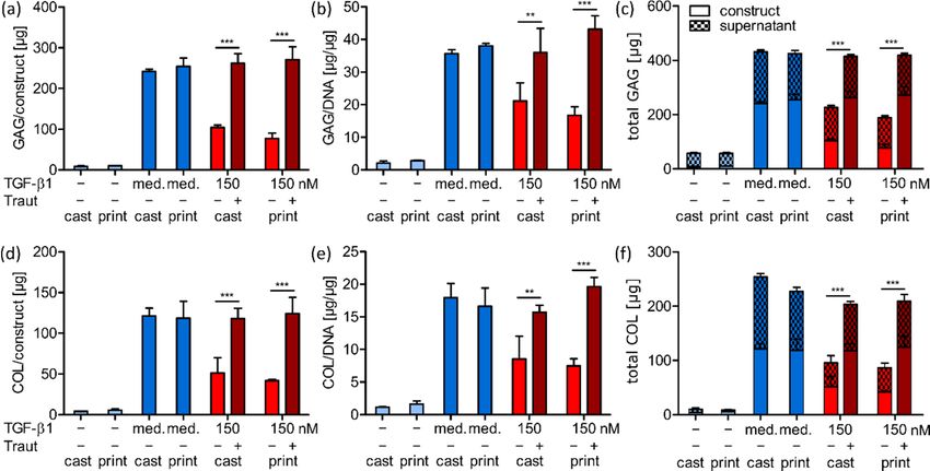
Int. J. Mol. Sci. 2022, 23, 924                                                                                          6 of 20

                                  might be the reason for the relatively low protein release of 25.6% in the mixed group.
                                  However, significantly lower TGF-β1 release of 4.5% was observed in the tethered
                                  group confirming effective growth factor thiol-functionalization with Traut’s reagent
                                  and PEG-acryl tethering via Michael addition.

                                  2.3. Impact of TGF-β1 Concentration and Administration on MSC Differentiation
                                       After successful material characterization, we analyzed the chondrogenic potential
                                  of the biofunctionalized ink. To examine the optimal growth factor concentration for
                                  chondrogenic differentiation of human MSCs, constructs were prepared and cast as
                                  described above with either covalently tethered or only mixed TGF-β1, i.e., with or with-
                                  out Traut’s reagent, in different concentrations (10 n M , 50 n M , 100 n M and 150 n M ) and
                                  cultured in a TGF-β1-free differentiation medium for 21 days. As a control, constructs
                                  without incorporated TGF-β1 were cultured in differentiation medium supplemented
                                  with 10 ng mL−1 TGF-β1 at each medium change, or without the supplementation
                                  as a negative control. Cell survival was very good under all conditions after con-
                                  struct preparation on day 1 as well as after 21 days in the respective culture conditions
                                  (Supplementary Materials, Figure S3). Glycosaminoglycans (GAG) and collagens, as
                                  the main components of natural cartilage ECM, were analyzed in the MSC-laden con-
                                  structs after 21 days (Figure 3). Constructs without TGF-β1 supplementation showed
                                  no GAG (Figure 3a) and collagen (Figure 3b) production, while control constructs in
                                  medium supplemented with TGF-β1 showed strong differentiation. In the constructs
                                  with incorporated TGF-β1, the production of both GAG and collagen increased with
                                  increasing growth factor concentrations (10–150 n M ), and importantly, tethered TGF-
                                  β1 induced markedly higher ECM production compared to the corresponding mixed
                                  conditions. These results were observed in histological stainings (Figure 3a,b), and
                                  further confirmed by quantification of total GAG and collagen amounts in the constructs
                                  (Figure 3c,e) as well as amounts normalized to DNA content (Figure 3d,f). The concen-
                                  trations of 10 n M and 50 n M TGF-β1 (tethered (+Traut) and mixed ( − Traut)) were not
                                  sufficient to induce adequate chondrogenic differentiation of MSCs. The concentrations
                                  of 100 n M and 150 n M tethered TGF-β1 allowed for high amounts of newly produced
                                  GAG and collagen, while non-tethered (mixed) TGF-β1 at 100 n M and 150 n M resulted
                                  in significantly reduced differentiation capacity.
                                       In general, TGF-β1 concentration of 150 n M exhibited the best results regarding
                                  ECM production and distribution. Therefore, this concentration was used in the follow-
                                  ing printing studies. Constructs with 150 n M tethered TGF-β1 and control constructs in
                                  medium supplemented with TGF-β1 demonstrated that a homogeneous ECM distri-
                                  bution was achievable. This was likely due to the low overall polymer content of the
                                  used hydrogel composition (1.4%) and validated this ink as a beneficial cell carrier for
                                  cartilage biofabrication [36,37,43]. Concentration dependence of MSC or chondrocyte
                                  differentiation induced by TGF-β1 tethered via Traut’s reagent has been previously
                                  analyzed in not-printable hydrogels [24–26]. Only one study compared tethered with
                                  mixed TGF-β1 incorporation and detected very similar differences as those found in
                                  this study [24]. All presented results clearly indicated the superior effect of covalently
                                  tethered TGF-β1 into the ink compared to non-covalently incorporated growth factor
                                  (mixed). This can be partially explained by the differences in TGF-β1 release profiles
                                  between the conditions (Figure 2e). A further explanation might be that free TGF-β1
                                  can undergo endocytosis and degradation when bound to the cell surface receptor on
                                  MSCs, thus reducing the available protein concentration [44,45]. In contrast, covalently
                                  tethered protein might trigger prolonged signaling without potential internalization.
(Figure 3d,f). The concentrations of 10 nM and 50 nM TGF-β1 (tethered (+Traut) and mixed
(−Traut)) were not sufficient to induce adequate chondrogenic differentiation of MSCs.
Int. J. Mol. Sci. 2022, 23, 924 The concentrations of 100 nM and 150 nM tethered TGF-β1 allowed for high amounts 7 ofof
20
newly produced GAG and collagen, while non-tethered (mixed) TGF-β1 at 100 nM and
150 nM resulted in significantly reduced differentiation capacity.
Figure 3. Histological staining and quantification of ECM components in cast constructs after 21
Figure 3. Histological staining and quantification of ECM components in cast constructs after
days. (a) Staining for GAG (safranin O) and (b) collagen (picrosirius red) in constructs with 10, 50,
21 days. (a) Staining for GAG (safranin O) and (b) collagen (picrosirius red) in constructs with 10,
100 or 150 nM mixed (−Traut) or tethered (+Traut) TGF-β1, as well as control groups cultured with-
50, M mixed (−Traut) or tethered (+Traut) TGF-β1, as well as control groups cultured
out100 or 150
(w/o, −) ornwith TGF-β1 (med.) as medium supplement. Scale bars represent 100 µm. (c) Quan-
without (w/o, − ) or with TGF-β1
tification of glycosaminoglycan (med.)
(GAG) as medium
content supplement.
in the constructs and Scale bars represent
(d) normalized 100 µm.
to DNA. (e)
(c) Quantification
Quantification ofofcollagen
glycosaminoglycan (GAG)
(COL) content in thecontent in theand
constructs constructs and (d) normalized
(f) normalized to DNA. Datato DNA.
are
represented
(e) as mean
Quantification ± standard
of collagen deviation
(COL) content(n =in3).
the Significant
constructsdifferences are markedtowith
and (f) normalized DNA.* (pData
< 0.05),
are
** (p < 0.01) as
represented and *** (p
mean ±Int. J. Mol. Sci. 2022, 23, 924 8 of 20
2.4.1. TGF-β1 Signaling and Chondrogenic Gene Expression
TGF-β1 induces the canonical Smad-dependent signaling cascade through binding
to cell surface receptors of MSCs. Upon receptor binding, Smad2/3 is phosphorylated
and translocates to the nucleus where it is involved in chondrogenic target gene expres-
Int. J. Mol. Sci. 2022, 23, 924 9 of 21
sion [50–52]. To detect the possible impact of the printing process on TGF-β1 functionality,
Smad phosphorylation was analyzed via Western blotting (Figure 4).
Figure4.4.Western
Figure Westernblotblotanalysis
analysisofofTGF-β1/Smad
TGF-β1/Smadsignaling
signalingin incast
castand
andprinted
printedconstructs.
constructs.Cast
Castand
and
printed constructs with 150 nM mixed (−Traut) or tethered (+Traut) TGF-β1 were analyzed for their
printed constructs with 150 nM mixed (−Traut) or tethered (+Traut) TGF-β1 were analyzed for their
protein expression. Constructs cultured without or with TGF-β1 as medium supplement served as
protein expression. Constructs cultured without or with TGF-β1 as medium supplement served
controls. Samples were analyzed directly after fabrication (d0, ctrl) and all test conditions after (a) 3
as
andcontrols. Samples
(b) 7 days. were analyzedactive
TGF-β1-induced directly after
Smad fabrication
signaling (d0, ctrl)
is shown byand all test
staining ofconditions after
phosphorylated
(a) 3 and(P-Smad3)
Smad3 (b) 7 days.and
TGF-β1-induced
can be compared active Smad
with signaling is shown levels
the unphosphorylated by staining
of the of phosphorylated
transcription factor
Smad3 (P-Smad3)
(Smad2/3) and M
(expected canW be
52 compared
kDa). GAPDH with the unphosphorylated
served levels
as loading control of the transcription
(expected factor
MW 38 kDa). The
protein ladder
(Smad2/3) (expected MW on
is labeled 52 the
kDa).leftGAPDH
(protein served
MW 35–250 kDa). control (expected MW 38 kDa). The
as loading
protein ladder is labeled on the left (protein MW 35–250 kDa).
As a further step in the signaling cascade, expression of the chondrogenic genes ag-
As aand
grecan firstcollagen
response to TGF-β1
type II, mainbinding,
componentsSmad2/3 phosphorylation
of natural was expected
cartilage ECM, early
was evaluated.
during culture. Furthermore, the initial burst release of unbound TGF-β1
Cast and printed constructs with 150 nM tethered or mixed TGF-β1 were analyzed after 3, was determined
during the21first
7, 14 and daysthree
anddays (Figure
compared to 2e). Forconstructs
control these reasons, we analyzed
cultured without orcast and
with theprinted
protein
constructs with 150 n M covalently
as medium supplement (Figure 5). tethered or mixed TGF-β1 for activated phospho-Smad3
(P-Smad3) proteins
In general, all after 3 and 7with
constructs daysMSCs
of culture. As atocontrol,
subjected TGF-β1 constructs without TGF-β1
showed strongly elevated
were harvested
expression directly
levels after preparation,
compared i.e., beforecultured
to MSCs in constructs incubation in culture
without medium
TGF-β1. (d0).
Aggrecan
After 3 days, activated P-Smad3 was found in all constructs which
(ACAN) and collagen type II (COL2A1) expression during the first week of culture re-had contact to TGF-
β1, independent
vealed of application
no significant differences (lanes 2–7). the
between Only the constructs
TGF-β1 without
conditions, TGF-β1
however, after(lane 1)
14 and
lacked P-Smad3. Basal levels of unphosphorylated Smad2/3 were detected
21 days, constructs with tethered TGF-β1 exhibited increased ACAN and COL2A1 expres- in all constructs,
although to a distinctly
sion, as compared to thelesser extent with
constructs in themixed
conditions
TGF-β1with TGF-β1
(Figure (Figure
5a,b). 4a). After
Furthermore, no
7impact
days, medium controls (lanes 2 and 3), as well as the constructs
of the printing process was observed for both genes in all conditions. with 150 n M covalently
tethered TGF-β1 (lanes 5 and 7) contained high levels of P-Smad3, while constructs with
150 nM mixed TGF-β1 (lanes 4 and 6) showed distinctly lower concentrations (Figure 4b).
Constructs without TGF-β1 (lane 1) completely lacked the activated P-Smad3 protein.
Unphosphorylated Smad2/3 protein was detected evenly in all conditions. Importantly,
no obvious differences in Smad phosphorylation could be determined between cast and
printed constructs (Figure 4).Int. J. Mol. Sci. 2022, 23, 924 9 of 20
As a further step in the signaling cascade, expression of the chondrogenic genes
aggrecan and collagen type II, main components of natural cartilage ECM, was evaluated.
Cast and printed constructs with 150 nM tethered or mixed TGF-β1 were analyzed after 3,
Int. J. Mol. Sci. 2022, 23, 924 10 of 2
7, 14 and 21 days and compared to control constructs cultured without or with the protein
as medium supplement (Figure 5).
Figure5. 5.
Figure Gene
Gene expression
expression analysis
analysis of and
of cast castprinted
and printed constructs.
constructs. qRT-PCR qRT-PCR was performed
was performed on gene on gen
expression
expression of of MSCs
MSCs in cast
in cast and and printed
printed constructs
constructs with 150with
nM150
mixednM(− mixed
Traut)(−Traut) or (+Traut)
or tethered tethered (+Trau
TGF-β1.Printed
TGF-β1. Printedconstructs
constructs cultured
cultured without
without or with
or with TGF-β1
TGF-β1 as medium
as medium supplement
supplement served
served as as con
trols. Expression
controls. Expressionofof(a) (a)aggrecan (ACAN)and
aggrecan (ACAN) and(b)(b)collagen
collagen type
type II (COL2A1)
II (COL2A1) waswas determined afte
determined
3, 7,3,14
after 7, and
14 and2121days
daysandandnormalized
normalized to toEEF1A1
EEF1A1 and
and MSCs
MSCs in 2Din on
2Ddayon 0.day
Data0. are
Data are presented a
presented
mean ± standard deviation (n = 3). Significant differences between
as mean ± standard deviation (n = 3). Significant differences between groups are marked with groups are marked with ** (p
0.01) and *** (p < 0.001). Legend: a) Significantly different to corresponding value
** (p < 0.01) and *** (p < 0.001). Legend: (a) Significantly different to corresponding value of the same of the same grou
at day
group at 7day
(at7least p < 0.05),
(at least b) significantly
p < 0.05), (b) significantlydifferent
differenttotocorresponding
corresponding value valueofofthethe same
same group at da
group
at14 (at14least
day p < 0.05),
(at least c) significantly
p < 0.05), different
(c) significantly differenttotocorresponding
corresponding valuevalueofofthethe same
same group
group at day 21 (a
at day
least p < 0.05).
21 (at least p < 0.05).
In ECM
2.4.2. general, all constructs
Production andwith MSCs subjected to TGF-β1 showed strongly elevated
Distribution
expression levels compared to MSCs in constructs cultured without TGF-β1. Aggrecan
An important feature of bioinks for cartilage engineering is the support of MSC di
(ACAN) and collagen type II (COL2A1) expression during the first week of culture revealed
ferentiation
no and the necessary
significant differences between porosity
the TGF-β1forconditions,
ECM distribution. Figure
however, after 6 shows
14 and histologica
21 days,
and immunohistochemical stainings of cartilage-specific ECM components
constructs with tethered TGF-β1 exhibited increased ACAN and COL2A1 expression, as produced b
embedded
compared MSCs
to the duringwith
constructs 21 days.
mixedControl images5a,b).
TGF-β1 (Figure of day 1 are presented
Furthermore, in the
no impact of Supple
mentary
the printingMaterials
process was(Figure S5). for both genes in all conditions.
observed
MSCs cultured in constructs without TGF-β1 were not able to differentiate and pro
2.4.2. ECM Production and Distribution
duced hardly any ECM components, while constructs in medium supplemented wit
An important feature of bioinks for cartilage engineering is the support of MSC differ-
TGF-β1 showed marked differentiation (Figure 6, left). A concentration of150 nM tethere
entiation and the necessary porosity for ECM distribution. Figure 6 shows histological and
TGF-β1 yielded strong ECM production and a homogeneous distribution comparable t
immunohistochemical stainings of cartilage-specific ECM components produced by em-
the medium
bedded control,
MSCs during while
21 days. the same
Control imagesprotein
of day 1 concentration
are presented in without covalent bindin
the Supplementary
(mixed) was distinctly
Materials (Figure S5). less effective (Figure 6, right). This observation was valid for a
stained ECM components, i.e., total GAG and collagen stained by safranin O an
picrosirius red (Figure 6a,c), and specific IHC staining of aggrecan and collagen type
(Figure 6b,d). The fibrocartilage marker collagen type I was produced to a low extent i
all groups receiving TGF-β1 (Supplementary Materials, Figure S6). Importantly, no differ
ence between cast and printed constructs were determined in any condition.Int. J. Mol. Sci. 2022, 23, 924 10 of 20
Int. J. Mol. Sci. 2022, 23, 924 11 of 21
Figure6.6.Histological
Figure Histologicaland
andimmunohistochemical
immunohistochemical(IHC)(IHC)staining
stainingofofMSC-derived
MSC-derivedECM ECMcomponents
components
in cast and printed constructs. Cast and printed constructs with 150 nM mixed (−Traut) or tethered
in cast and printed constructs. Cast and printed constructs with 150 nM mixed (−Traut) or tethered
(+Traut) TGF-β1 were analyzed after 21 days. Constructs cultured without (w/o) or with TGF-β1 as
(+Traut) TGF-β1 were analyzed after 21 days. Constructs cultured without (w/o) or with TGF-β1
medium supplement (medium) served as controls. GAG production and distribution is depicted by
as medium supplement (medium) served as controls. GAG production and distribution is depicted
(a) safranin O staining and (b) IHC staining of aggrecan. Collagen production and distribution is
by (a) safranin
visualized by O
(c)staining andred
picrosirius (b)staining
IHC staining ofIHC
and (d) aggrecan.
stainingCollagen production
of collagen and distribution
type II. Nuclei were coun-
isterstained
visualizedwith
by (c) picrosirius red staining and
DAPI. Scale bars represent 100 µm.(d) IHC staining of collagen type II. Nuclei were
counterstained with DAPI. Scale bars represent 100 µm.
To further validate the visual observations, we quantified GAG and collagen contents
MSCs
of all cast cultured in constructs
and printed constructswithout
after 21TGF-β1 werebiochemical
days with not able to differentiate
assays (Figureand7).pro-
The
duced hardly any ECM components, while constructs in medium supplemented
analysis revealed very low GAG and collagen deposition in constructs cultured without with
TGF-β1
TGF-β1, showed
while marked differentiation
constructs (Figure
with continuous 6, left).medium
TGF-β1 A concentration of150 nM produced
supplementation tethered
TGF-β1 yielded strong ECM production and a homogeneous distribution comparable to the
high ECM amounts (Figure 7a,d, blue bars). GAG and collagen contents in constructs withInt. J. Mol. Sci. 2022, 23, 924 11 of 20
medium control, while the same protein concentration without covalent binding (mixed)
was distinctly less effective (Figure 6, right). This observation was valid for all stained
ECM components, i.e., total GAG and collagen stained by safranin O and picrosirius red
(Figure 6a,c), and specific IHC staining of aggrecan and collagen type II (Figure 6b,d). The
fibrocartilage marker collagen type I was produced to a low extent in all groups receiving
TGF-β1 (Supplementary Materials, Figure S6). Importantly, no difference between cast and
printed constructs were determined in any condition.
To further validate the visual observations, we quantified GAG and collagen contents
of all cast and printed constructs after 21 days with biochemical assays (Figure 7). The
Int. J. Mol. Sci. 2022, 23, 924 12 of 21
analysis revealed very low GAG and collagen deposition in constructs cultured without
TGF-β1, while constructs with continuous TGF-β1 medium supplementation produced
high ECM amounts (Figure 7a,d, blue bars). GAG and collagen contents in constructs
150 n
with M tethered
150 TGF-β1
nM tethered werewere
TGF-β1 comparable
comparable to theto positive control
the positive (medium),
control although
(medium), althoughno
additional growth factor was added during the three weeks of cell
no additional growth factor was added during the three weeks of cell culture. MSCs culture. MSCs in con-
structs
in with 150
constructs with mixed
nM150 TGF-β1TGF-β1
nM mixed produced significantly
produced lower amounts
significantly of ECM compo-
lower amounts of ECM
components compared to constructs in which the same protein concentrationwas
nents compared to constructs in which the same protein concentration wascovalently
covalently
bound(Figure
bound (Figure7a,d,
7a,d,red
red bars).
bars). The same significant differences
differences werewere detected
detectedwhen
whenGAG GAG
andcollagen
and collagencontents
contents were
were normalized
normalized to the DNA content content (Figure
(Figure 7b,e)
7b,e)as
aswell
wellasaswhen
when
totalproduction
total productionwaswascompared,
compared, i.e.,
i.e., the combined ECM contentscontents in in the
theconstruct
constructand andthethe
supernatantcollected
supernatant collectedduring
duringculture
culturetime time(Figure
(Figure7c,f).
7c,f).All
Allinitial
initialvalues
valuesatatday
day1 1asas well
well as
as the
the analyzed
analyzed DNA DNA contents
contents cancan be found
be found in the
in the Supplementary
Supplementary Materials
Materials (Figure
(Figure S7).
S7). In
In general,
general, the quantitative
the quantitative results
results wellwell confirmed
confirmed the histological
the histological findings
findings and again,
and again, there
there was no difference detected between cast and
was no difference detected between cast and printed approaches.printed approaches.
Figure7.7.Quantification
Figure Quantification of of MSC-derived
MSC-derived ECM ECM components
components in in cast
cast and
and printed
printedconstructs.
constructs.Cast
Castand
and
printed constructs with 150 nM mixed (−Traut) or tethered (+Traut) TGF-β1 were analyzed after 21
printed constructs with 150 nM mixed (−Traut) or tethered (+Traut) TGF-β1 were analyzed after
days. Constructs cultured without (−) or with TGF-β1 as medium supplement (med.) served as con-
21 days. Constructs cultured without (−) or with TGF-β1 as medium supplement (med.) served
trols. GAG content is shown for (a) the constructs and (b) normalized to DNA. (c) Total GAG pro-
as controls.
duction wasGAG content
quantified by is shown
adding thefor (a) the
content of constructs andand
the constructs (b) the
normalized
collectiveto DNA.superna-
culture (c) Total
GAG
tant. production was quantified
Collagen content is shown for by(d)
adding the content
the constructs andof(e)
the constructstoand
normalized the (f)
DNA. collective culture
Total collagen
supernatant.
production wasCollagen content
quantified is shown
by adding thefor (d) theofconstructs
content and and
the constructs (e) normalized to culture
the collective DNA. (f) Total
super-
natant. Data
collagen are represented
production as mean
was quantified by±adding
standardthedeviation
content of(n the
= 3).constructs
Significantand
differences are marked
the collective culture
with ** (p < 0.01)
supernatant. Dataand
are *** (p < 0.001).as mean ± standard deviation (n = 3). Significant differences are
represented
marked with ** (p < 0.01) and *** (p < 0.001).
2.4.3. Correlation of ECM Production and Construct Stiffness
2.4.3. Correlation of ECM Production and Construct Stiffness
As an important mechanical feature of engineered cartilage, we analyzed the Young’s
As anof
modulus important mechanical
cast and printed featurewith
constructs of engineered cartilage,
150 nM tethered we analyzed
or mixed TGF-β1the Young’s
after con-
modulus of cast and printed constructs with 150 n M tethered or mixed TGF-β1
struct preparation at day 1 as well as after 21 days of chondrogenic differentiation. Con- after
construct preparation
structs cultured at TGF-β1
without day 1 asorwell
withasthe
after 21 days
soluble of chondrogenic
protein as continuousdifferentiation.
medium sup-
Constructs
plementationcultured
servedwithout TGF-β1
as controls or with
(Figure the soluble
8). Initially, protein asappeared
all constructs continuous medium
very weak
and without significant differences between the conditions (all around 2.5–5.0 kPa at day
1). Constructs cultured without TGF-β1 maintained their weak appearance or even de-
creased in stiffness within 21 days to around 1.6 kPa. MSCs in constructs with TGF-β1
addition in the culture medium differentiated well and produced high amounts of ECM.Int. J. Mol. Sci. 2022, 23, 924 12 of 20
supplementation served as controls (Figure 8). Initially, all constructs appeared very weak
and without significant differences between the conditions (all around 2.5–5.0 kPa at day 1).
Constructs cultured without TGF-β1 maintained their weak appearance or even decreased
in stiffness within 21 days to around 1.6 kPa. MSCs in constructs with TGF-β1 addition in
the culture medium differentiated well and produced high amounts of ECM. This correlated
with a significant increase in Young’s modulus to 73.2 kPa for cast and 76.0 kPa for printed
constructs (Figure 8, blue bars). Covalently tethered TGF-β1 also induced a significant
increase in stiffness to 105.5 kPa for cast and 112.1 kPa for printed constructs after 21 days,
Int. J. Mol. Sci. 2022, 23, 924 while the mixed condition only resulted in 35.6 kPa and 38.2 kPa, respectively (Figure 13 of8,
21
red bars).
Figure8.8.Mechanical
Figure Mechanicalcharacterization
characterizationofofcast
castand
andprinted
printedconstructs.
constructs.Young’s
Young’smodulus
modulusof ofcast
castand
and
printed constructs with 150 nM mixed (−Traut) or tethered (+Traut) TGF-β1 after 1 and 21 days.
printed constructs with 150 nM mixed (−Traut) or tethered (+Traut) TGF-β1 after 1 and 21 days.
Constructs cultured without (−) or with TGF-β1 as medium supplement (med.) served as controls.
Constructs cultured without (−) or with TGF-β1 as medium supplement (med.) served as controls.
Data are presented as mean ± standard deviation (n = 6). Significant differences between groups are
Data are presented as mean ± standard deviation (n = 6). Significant differences between groups
marked with * (p < 0.05), ** (p < 0.01) and *** (p < 0.001). Stars above bars on day 21 indicate significant
are marked with
differences to the* corresponding
(p < 0.05), ** (pvalue
< 0.01) andsame
of the *** (pgroup
< 0.001). Stars
on day 1. above bars on day 21 indicate
significant differences to the corresponding value of the same group on day 1.
In general, bioinks with high polymer contents result in initially stiffer hydrogel net-
In general, bioinks with high polymer contents result in initially stiffer hydrogel
works with good 3D printability, but are often associated with limited cell growth, differ-
networks with good 3D printability, but are often associated with limited cell growth, dif-
entiation and ECM distribution [43]. In a previous study of our group, we demonstrated
ferentiation and ECM distribution [43]. In a previous study of our group, we demonstrated
that the distribution of newly produced ECM molecules was significantly improved in
that the distribution of newly produced ECM molecules was significantly improved in
bioinks with a low polymer content which was associated with an increased construct
bioinks with a low polymer content which was associated with an increased construct
stiffness after chondrogenic differentiation [38]. This was shown for bioinks with 3% pol-
stiffness after chondrogenic differentiation [38]. This was shown for bioinks with 3% poly-
ymer content which needed PCL-support structures for 3D printability. The dual-stage
mer content which needed PCL-support structures for 3D printability. The dual-stage
crosslinkedbioink
crosslinked bioinkpresented
presentedhere hereisisstand-alone
stand-alone3D 3Dprintable
printableandandstill
stillallows
allowsfor forananeven
even
lower polymer content of 1.4%. Although it exhibited a comparably
lower polymer content of 1.4%. Although it exhibited a comparably weak network in the weak network in the
beginning, this bioink enabled homogenous ECM distribution
beginning, this bioink enabled homogenous ECM distribution and thereby significantly and thereby significantly
increasedconstruct
increased constructstiffness
stiffness after
after three
three weeks
weeks in in constructs
constructs with
with strong
strong MSC MSC differentia-
differentiation
tion (tethered TGF-β1 and medium control). Compared to constructs
(tethered TGF-β1 and medium control). Compared to constructs with non-covalently with non-covalently
incorporatedTGF-β1,
incorporated TGF-β1, MSCs
MSCs in constructs
in constructs withwith tethered
tethered TGF-β1 TGF-β1 expressed
expressed distinctly distinctly
higher
higher amounts of cartilaginous ECM with a more homogeneous
amounts of cartilaginous ECM with a more homogeneous distribution throughout the con- distribution throughout
the constructs
structs (Figures (Figures
6 and 7),6which
and 7), which correlated
correlated well withwell with a markedly
a markedly higher construct
higher construct stiffness
stiffness
(Figure 8).(Figure
This was 8). again
This was again independent
independent of bioink processing,
of bioink processing, i.e., no differences
i.e., no differences between
between
cast cast and
and printed printed constructs
constructs were observed.were observed.
In conclusion, we established
In conclusion, we established a dual-stagea dual-stagecrosslinked
crosslinkedhyaluronic
hyaluronicacid-based
acid-basedbioinkbioink
which is 3D printable and enables covalent tethering of TGF-β1. In previous
which is 3D printable and enables covalent tethering of TGF-β1. In previous reports, other reports, other
printable materials were also described for growth factor administration
printable materials were also described for growth factor administration in chondrogenic or in chondrogenic
or osteogenic
osteogenic regeneration
regeneration approaches,
approaches, however,
however, proteins
proteins werewere only mixed
only mixed within within the bi-
the bioinks
oinks
or or administered
administered via nanospheres
via nanospheres but notbut not covalently
covalently tetheredtethered
[53–57].[53–57]. In our itstudy,
In our study, was
it was that
shown shown boththat both processes,
processes, tetheringtethering and 3D did
and 3D printing, printing, did protein
not affect not affect protein func-
functionality or
tionality
bioink or bioinkCovalent
properties. properties. Covalent
TGF-β1 TGF-β1
tethering tethering
enabled enabled
prolonged prolonged
availability for availability
embedded
for embedded
MSCs and markedlyMSCsenhanced
and markedly enhanceddifferentiation,
chondrogenic chondrogenic differentiation,
as compared toas thecompared
growth
to thenon-covalently
factor growth factor non-covalently
incorporated inincorporated in the ink.the
the ink. Furthermore, Furthermore,
low polymer thecontent
low polymer
of the
presented
content ofink thecomposition
presented ink facilitated a penetrable
composition network
facilitated with chondro-supportive
a penetrable network with chondro- prop-
supportive properties and allowed for homogenous ECM distribution and a distinct in-
crease of construct stiffness during chondrogenic differentiation of embedded MSCs.
Taken together, this ink composition enabled the generation of high-quality cartilaginous
tissues without the need for continuous exogenous growth factor supply and, thus, bears
great potential for future investigation towards cartilage regeneration. Moreover, the ob-Int. J. Mol. Sci. 2022, 23, 924 13 of 20
erties and allowed for homogenous ECM distribution and a distinct increase of construct
stiffness during chondrogenic differentiation of embedded MSCs. Taken together, this ink
composition enabled the generation of high-quality cartilaginous tissues without the need
for continuous exogenous growth factor supply and, thus, bears great potential for future
investigation towards cartilage regeneration. Moreover, the observation that a tethered
growth factor within a printed bioink can lead to superior tissue development may also be
explored in other applications in biofabrication.
3. Materials and Methods
3.1. Materials
All chemicals were purchased from Sigma Aldrich (St. Louis, MO, USA) if not stated
differently. Acryloyl chloride stabilized with phenothiazine (abcr GmbH, Karlsruhe, Ger-
many), 1-(3-dimethylaminopropyl)-3-ethylcarbodiimide HCl (EDC, Biosynth CarboSynth,
Compton, UK), 1,4-dithiothreitol (DTT, Biosynth CarboSynth, Compton, UK), 2-hydroxy-1-
[4-(hydroxyethoxy)-phenyl]-2-methyl-1-propanone (I2959; BASF, Ludwigshafen, Germany),
4-(dimethylamino)benzaldehyde (DAB; Carl Roth, Karlsruhe, Germany), basic fibroblast
growth factor (bFGF; BioLegend, London, UK), chloroform-d1 (Eurisotope, St-Aubin Cedex,
France), DAPI mounting medium ImmunoSelect® (Dako, Hamburg, Germany), deuterium
oxide (Deutero GmbH, Kastellaun, Germany), regenerated cellulose dialysis tubes MWCO
3500 Da (Carl Roth, Karlsruhe, Germany), diethylether (Chemobar University of Würzburg,
Würzburg, Germany), dimethyl-3,30 -dithiodipropionate (TCI Chemical Industry Co. Ltd.,
Tokyo, Japan), di-potassium hydrogen phosphate (Merck KGaA, Darmstadt, Germany),
di-sodium hydrogen phosphate (Merck KGaA, Darmstadt, Germany), Dulbecco’s Mod-
ified Eagle’s Medium high glucose 4.5 g L−1 (DMEM; Thermo Scientific, Waltham, MA,
USA), Dulbecco’s Modified Eagle’s Medium/Ham’s F-12 (DMEM/F12; Thermo Scientific,
Waltham, MA, USA), ethanol (99%, TH Geyer, Renningen, Germany), fetal calf serum
(FCS; Thermo Scientific, Waltham, MA, USA), formaldehyde (37%, Carl Roth, Karlsruhe,
Germany), hyaluronic acid sodium salt (MW 1–2 MDa; Biosynth CarboSynth, Compton,
UK), hydrochloric acid (HCl; 32%, 37%, Merck KGaA, Darmstadt, Germany), isopropanol
(VWR, Radnor, PA, USA), ITS+ premix (Corning, New York, NY, USA), L-hydroxyprolin
(Merck KGaA, Darmstadt, Germany), live/dead cell staining kit (PromoKine, Heidel-
berg, Germany), methanol (Fisher Scientific, Schwerte, Germany), N-hydroxysuccinimide
(NHS, Biosynth CarboSynth, Compton, UK), papain (Worthington, Lakewood, CA, USA),
penicillin-streptomycin (PS; 100 U mL−1 penicillin, 0.1 mg mL−1 streptomycin; Thermo
Scientific, Waltham, MA, USA), perchloric acid (60%, Merck KGaA, Darmstadt, Germany),
phosphate-buffered saline (PBS; Life Technologies, Carlsbad, CA, USA), polyethylene gly-
col (2-arm PEG, 6 kDa: Sigma Aldrich, St. Louis, MO, USA; 8-arm PEG, 10 kDa: JenKem®
Technologies USA, Plano, TX, USA), potassium dihydrogen phosphate (Merck KGaA,
Darmstadt, Germany), Proteinase K (Digest-All 4, Life Technologies, Carlsbad, CA, USA),
sodium hydrogen carbonate (Merck KGaA, Darmstadt, Germany), sodium hydroxide
(Merck KGaA, Darmstadt, Germany), Tissue Tek® O.C.T. (Sakura Finetek, Tokyo, Japan),
toluene (Fisher Scientific, Schwerte, Germany), transforming growth factor β1 (TGF-β1;
Novoprotein Inc., CA59, Summit, NJ, USA, purchased from PELOBIOTECH GmbH, Mu-
nich, Germany), tris(carboxyethyl)phosphine HCl (TCEP, Biosynth CarboSynth, Compton,
UK), trypsin-EDTA (0.25%, Life Technologies, Carlsbad, CA, USA).
3.2. Synthesis of the Different Bioink Components
The synthesis of the different components of the previously established hyaluronic
acid-based bioink (3,30 -dithiobis(propanoic dihydrazide) (DTPH), thiolated hyaluronic acid
(HA-SH), polyethylene glycol-diamine, and polyethylene glycol-diallyl carbamate (2-arm
PEG-allyl)) was performed as previously published [16]. Synthetical details are given in
the Supplementary Materials.Int. J. Mol. Sci. 2022, 23, 924 14 of 20
3.3. Synthesis of Polyethylene Glycol Octaacrylate (8-Arm PEG-Acryl)
The synthesis of the branched PEG-acrylate necessary for growth factor immobilization
was performed using enzyme catalyzed transesterification [58,59]. In brief, 8-arm PEG
(5.0 g, 1.0 eq.) was dried in vacuo at 100 ◦ C for 1 h, cooled to 60 ◦ C under argon atmosphere
and dissolved in dry toluene (50 mL). Vinyl acrylate (3.0 eq.) and Novozyme 435 (50.0 mg)
were added, the suspension was incubated for 3 days at 50 ◦ C and subsequently filtered.
After precipitation and washing with diethyl ether, the product was dried in vacuo to form
a bulky white solid.
3.4. NMR Analysis
1 H-NMR measurements were performed at a 300 MHz Bruker Biospin spectrometer
(Bruker, Billerica, MA) using CDCl3 (PEG), d6 -DMSO (DTP) and D2 O (HA-SH) as solvents.
The solvent peak was set to δ = 7.26 ppm for CDCl3 , 2.50 ppm for DMSO and 4.79 ppm for
D2 O to which all chemical shifts refer. The degree of substitution of HA-SH was determined
by the ratio of the integrals of the thiol carrying substituent and the acetyl amide signal
in combination with the anomeric protons of the saccharide backbone. The quantitative
PEG-modification was verified by the absence of alterations in the spectra after the addition
of trifluoroacetic anhydride to the NMR sample solution.
3.5. GPC Analysis
A GPC system from Malvern (Herrenberg, Germany) with a triple detection containing
a refractive index detector (VE 3580), a viscometer (270 dual detector) and a multi angle light
scattering detector (SEC-MALS 20) was used for GPC analysis. Depending on the molecular
weight, different column sets (Malvern, Herrenberg, Germany) were used. For HA-SH
samples, two A6000M mixed-bed columns, and for PEG samples, a set of A2000/A3000
columns were chosen. The eluent was prepared using deionized water containing 8.5 g L−1
NaNO3 and 0.2 g L−1 NaN3 and the columns were calibrated with PEG standards (Malvern,
Herrenberg, Germany). HA-SH samples were dissolved in deionized water with 0.5 g L−1
TCEP over 6 h at rt and PEG samples were dissolved in deionized water over 6 h at rt. The
obtained data was processed with OmniSEC 5.12 (Malvern, Herrenberg, Germany).
3.6. Ink Preparation, TGF-β1 Tethering and 3D Printing
Final composition of the used ink is: 0.5% HA-SH (465 kDa), 0.5% 8-arm PEG-acryl
(10 kDa), 0.4% 2-arm PEG-allyl (6 kDa) and 0.05% Irgacure (I2959); all concentrations are
indicated as (w/v). For ink preparation, HA-SH was dissolved in HEPES buffer (pH 7.6,
154 mM), all other components were dissolved in PBS. Three different experimental condi-
tions were prepared: constructs without TGF-β1, constructs with non-covalently bound
TGF-β1 (10 nM, 50 nM, 100 nM and 150 nM) and constructs with covalently tethered TGF-β1
(10 nM, 50 nM, 100 nM and 150 nM). For TGF-β1 tethered hydrogels, Traut’s reagent was
used for thiol-modification of the protein (4:1 molar excess of Traut’s reagent, 1 h incuba-
tion at rt). Subsequently, different amounts of thiolated TGF-β1 were tethered to 8-arm
PEG-acryl via Michael addition (1 h at 37 ◦ C). For control constructs with mixed TGF-β1
(no tethering) the same procedure was performed without the addition of Traut’s reagent.
Afterwards, all ink components were mixed with or without MSCs (20 × 106 cells mL−1 ,
passage 4), transferred to a 3 cc printing cartridge (Nordson EFD, Westlake, OH, USA) and
incubated for 30 min at 37 ◦ C to achieve the necessary viscosity for 3D printing via Michael
addition. 3D bioprinting as well as printing simulation was performed with an Inkredible+
3D bioprinter (Cellink, Boston, MA, USA) at 50 kPa through a 330 µm steel nozzle (Nordson
EFD, Westlake, OH, USA). G-codes for 3D printed grids and filament fusion tests were
generated with the HeartWare software (Cellink, Boston, MA, USA). The NIH ImageJ Fiji
software (version 1.52a) was utilized for strand thickness measurements. The CAD file
source of the human femoral condyles was MakerBot Thingiverse (object 5820, created by
BME_sundevil). The CAD file was sliced with the Heartware Software and printed with a
BioX 3D bioprinter (Cellink, Boston, MA, USA). For the printing simulation, bioink wasYou can also read