Skeleton-secreted PDGF-BB mediates arterial stiffening - The ...
←
→
Page content transcription
If your browser does not render page correctly, please read the page content below
The Journal of Clinical Investigation                                                                                                                          RESEARCH ARTICLE

Skeleton-secreted PDGF-BB mediates arterial stiffening
Lakshmi Santhanam,1,2,3 Guanqiao Liu,4,5 Sandeep Jandu,1 Weiping Su,4,6 Bulouere P. Wodu,7 William Savage,3 Alan Poe,2
Xiaonan Liu,4,5 Lacy M. Alexander,8 Xu Cao,4 and Mei Wan4
Department of Anesthesiology and Critical Care Medicine and 2Department of Biomedical Engineering, The Johns Hopkins University School of Medicine, Baltimore, Maryland, USA. 3Department of Chemical
1

and Biomolecular Engineering, Whiting School of Engineering, The Johns Hopkins University, Baltimore, Maryland, USA. 4Department of Orthopaedic Surgery, The Johns Hopkins University School of
Medicine, Baltimore, Maryland, USA. 5Department of Orthopaedics and Traumatology, Nanfang Hospital, Southern Medical University, Guangzhou, Guangdong, China. 6Department of Orthopaedic Surgery,
The Xiangya Hospital of Central South University, Changsha, Hunan, China. 7Department of Biotechnology, The Johns Hopkins University, Baltimore, Maryland, USA. 8Department of Kinesiology, Penn State
University, University Park, Pennsylvania, USA.

             Evidence links osteoporosis and cardiovascular disease but the cellular and molecular mechanisms are unclear. Here we
             identify skeleton-secreted platelet-derived growth factor–BB (PDGF-BB) as a key mediator of arterial stiffening in response
             to aging and metabolic stress. Aged mice and those fed high-fat diet (HFD), relative to young mice and those fed normal
             chow food diet, respectively, had higher serum PDGF-BB and developed bone loss and arterial stiffening. Bone/bone marrow
             preosteoclasts in aged mice and HFD mice secrete an excessive amount of PDGF-BB, contributing to the elevated PDGF-BB in
             blood circulation. Conditioned medium prepared from preosteoclasts stimulated proliferation and migration of the vascular
             smooth muscle cells. Conditional transgenic mice, in which PDGF-BB is overexpressed in preosteoclasts, had 3-fold higher
             serum PDGF-BB concentration and developed simultaneous bone loss and arterial stiffening spontaneously at a young age.
             Conversely, in conditional knockout mice, in which PDGF-BB is deleted selectively in preosteoclasts, HFD did not affect serum
             PDGF-BB concentration; as a result, HFD-induced bone loss and arterial stiffening were attenuated. These studies confirm
             that preosteoclasts are a main source of excessive PDGF-BB in blood circulation during aging and metabolic stress and
             establish the role of skeleton-derived PDGF-BB as an important mediator of vascular stiffening.

Introduction                                                                                           Bisphosphonate therapy for osteoporosis decreases the risk of
Accumulating evidence supports a link between bone metabo-                                             aortic valve and thoracic aorta calcification (14). These findings
lism and the vascular system. Cross-sectional and longitudinal                                         strongly suggest that bone-derived cues may directly affect the
studies have shown a direct association between osteoporosis                                           vascular system. Furthermore, accumulating clinical studies have
and cardiovascular disease (CVD) (1–5), 2 primary conditions                                           demonstrated an association between low bone mass and vascu-
that cause substantial morbidity and death in older people. In                                         lar calcification (15–17), a well-defined independent risk factor
fact, the correlation between these 2 disorders is independent of                                      for CVD and mortality. Vascular calcification and bone mineral-
age. Particularly, bone mineral density (BMD) is inversely and                                         ization are both actively regulated processes that may share com-
independently correlated with atherosclerosis and its established                                      mon pathogenetic mechanisms. Multiple factors including modi-
marker, aortic calcification (6–10). Low BMD has been associ-                                          fied low-density lipoprotein (LDL), inflammatory cytokines, Wnt
ated with cardiovascular morbidity and mortality. Although the                                         signaling, bone morphogenetic proteins, matrix proteins (such as
concept of a bone/vascular axis has long been proposed (3, 4, 11),                                     thrombospondin, tenascin, osteopontin, osteocalcin, osteoprote-
the exact cellular and molecular basis for the interplay between                                       gerin, matrix Gla protein, cathepsins, and DMP-1), parathyroid
the skeletal and vascular systems is poorly understood. Several                                        hormone, phosphate, and vitamins D and K are implicated in
hypotheses have been proposed to explain the link between oste-                                        both bone and vascular metabolism, suggesting the interaction of
oporosis and CVD, including shared risk factors, common patho-                                         these 2 pathological conditions.
logical mechanisms and genetic factors, and a causal association                                           The skeleton is not only a recipient for hormonal input but
(12). However, high bone turnover is associated with cardiovascu-                                      also an endocrine organ that regulates the homeostasis of periph-
lar death in the elderly, independent of sex and overall health (13).                                  eral organs (18, 19). For example, osteoclasts regulate the activity
                                                                                                       of other cells by secreting “clastokines” or releasing factors from
                                                                                                       bone matrix via bone resorption (20–23). Osteoclasts are multi-
      Related Commentary: https://doi.org/10.1172/JCI153644                                            nucleated cells that have the ability to degrade mineralized matri-
                                                                                                       ces, such as bone and calcified cartilage. Osteoclasts in adults
Authorship note: LS, GL, and SJ contributed equally to this work.                                      are derived mainly from bone marrow monocytes/macrophages
Conflict of interest: The authors have declared that no conflict of interest exists.
                                                                                                       (Mo/Macs). During osteoclastogenesis, Mo/Macs sequentially
Copyright: © 2021, American Society for Clinical Investigation.
                                                                                                       express colony-stimulating factor-1 receptor followed by receptor
Submitted: December 23, 2020; Accepted: August 24, 2021; Published: October 15, 2021.
Reference information: J Clin Invest. 2021;131(20):e147116.                                            activator of nuclear factor κB (RANK) and tartrate-resistant acid
https://doi.org/10.1172/JCI147116.                                                                     phosphatase (TRAP) in response to stimulation with macrophage

                                                                                                                                                                                                  1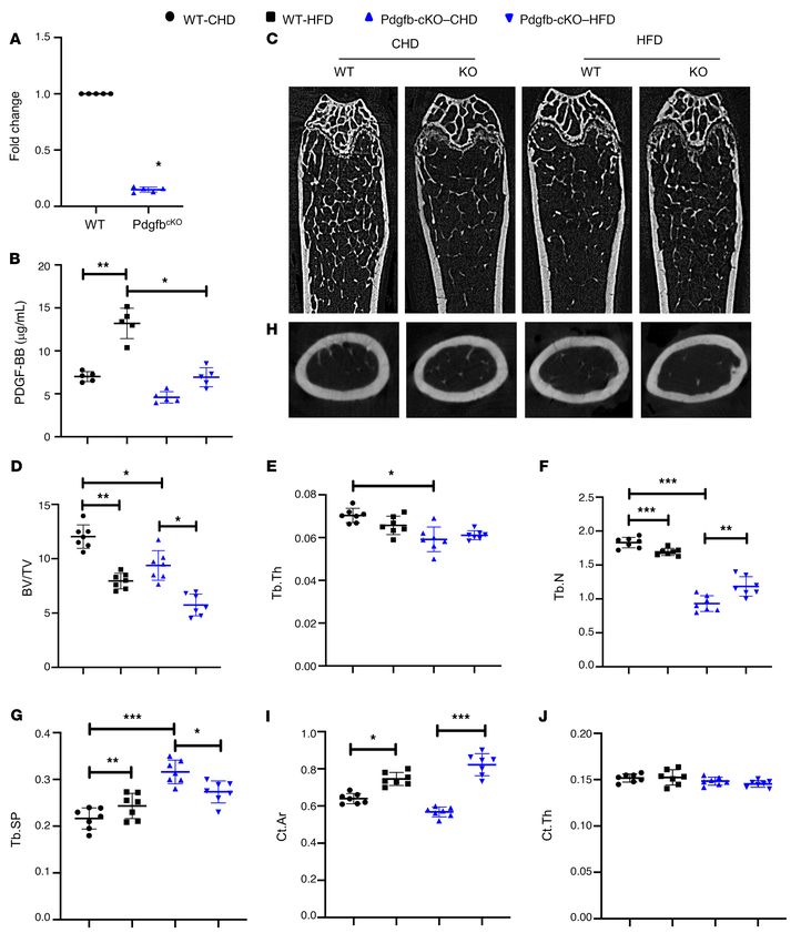
RESEARCH ARTICLE                                                                                          The Journal of Clinical Investigation

Figure 1. Aged mice and HFD-challenged mice develop low bone mass and an arterial stiffening phenotype. (A–E) Representative μCT images (A) and
quantitative analysis (B–E) of the trabecular bone area of the distal femur from 4- and 20-month-old male C57BL/6 mice. Bone volume per tissue volume
(BV/TV) (B), trabecular bone thickness (Tb.Th) (C), trabecular bone number (Tb.N) (D), and trabecular bone separation (Tb.Sp) (E). (F and G) Pulse-wave
velocity (PWV) and systolic, diastolic, and mean blood pressure (BP) measurements of 4- and 20-month-old male mice. (H–L) Representative μCT images
(H) and quantitative analysis (I–L) of the trabecular bone area of the distal femur from 3-month-old male C57BL/6 mice fed a Western HFD or normal CHD
for 5 months. BV/TV (I), Tb.Th (J), Tb.N (K), and Tb.Sp (L). (M and N) PWV and BP measurements of the mice fed HFD or CHD. n = 5 to 9. Data are mean ±
SD, *P < 0.05, *P < 0.01, ***P < 0.005, as determined by Student’s t tests.

   2                                     J Clin Invest. 2021;131(20):e147116 https://doi.org/10.1172/JCI147116
The Journal of Clinical Investigation                                                                             RESEARCH ARTICLE

colony-stimulating factor (M-CSF) and RANK ligand. Eventual-               Results
ly, mononuclear preosteoclasts fuse to form multinuclear osteo-            Animals develop low bone mass and an arterial stiffening phenotype in
clasts (20, 24, 25). Osteoclast lineage cells normally have a much         response to aging and HFD challenge. We first assessed the chang-
shorter life span (2 weeks) relative to osteoblasts (3 months) and         es in bone mass and arterial stiffness in mice with advancing age.
other bone cells (26). After osteoclasts have eroded bone to a par-        Twenty-month-old C57B/L6 mice had a low bone mass pheno-
ticular depth from the surface, they die quickly. During estrogen          type relative to young mice (4 months of age), as detected by μCT
deficiency or aging, the life span of this lineage of cells is pro-        analysis (Figure 1A). Although the difference in trabecular number
longed through an antiapoptosis mechanism (26, 27), resulting in           (Tb.N) in aged versus young mice was not significant (Figure 1D),
increased bone resorption. It has been demonstrated that bone/             the differences in the remaining 3 parameters were significant.
bone marrow mononuclear preosteoclasts secrete platelet-de-                Bone volume per tissue volume (BV/TV; Figure 1B) and trabecu-
rived growth factor–BB (PDGF-BB) to maintain normal bone                   lar thickness (Tb.Th; Figure 1C) were less, and trabecular space
homeostasis in healthy, young mice (28), whereas abnormally                (Tb.Sp; Figure 1E) was greater in 20-month-old mice compared
high production of PDGF-BB from preosteoclasts leads to skeletal           with 4-month-old mice. We also measured blood pressure (BP)
disorders, such as osteoarthritis (29).                                    and pulse-wave velocity (PWV), an index of in vivo vascular stiff-
      With advancing age, complex structural and functional chang-         ness. PWV was significantly higher in 20-month-old mice than
es occur in the arterial system. The large compliance vessels,             in 4-month-old mice (Figure 1F). Consistently, systolic, diastolic,
including the aorta and its major branches, stiffen with age, and          and mean BPs of the old mice were all higher than those of the
this stiffening can be accelerated by comorbidities, including obe-        young mice (Figure 1G).
sity and atherosclerosis (30–35). Increased aortic stiffness increas-           Next, to observe accelerated deterioration of the bone and vas-
es central arterial pressure and pulse pressure and is an indepen-         culature, we used a HFD challenge, because a HFD induces bone
dent risk factor for cardiovascular morbidity and death (36–41).           loss and increases aortic stiffness and endothelial dysfunction in
Moreover, arterial aging is characterized by accelerated develop-          mice (10, 60–62). Baseline PWV was measured, after which we
ment of atherosclerotic lesions and neointima formation during             placed mice on a HFD. We then examined the phenotypic changes
atherosclerosis (42). Hallmarks of the stiff vessel include intimal        of bone and vasculature in HFD-challenged and control mice. μCT
and medial thickening and an increased collagen/elastin ratio in           analysis showed less bone mass (Figure 1H), lower BV/TV (Figure
the arterial wall, as well as elastin fracture (43–46). Traditional-       1I), lower Tb.N (Figure 1K), and greater Tb.Sp (Figure 1L) in HFD
ly, it was suggested that the remodeling and accumulation of the           mice compared with CHD mice. The difference in Tb.Th between
vascular matrix is the main element of vascular stiffening. How-           groups was not significant (Figure 1J). Greater arterial stiffness
ever, recent studies have recognized that vascular smooth muscle           was also observed in HFD mice relative to CHD mice, as indicated
cell (VSMC) dysfunction and stiffening are major contributors to           by higher PWV (Figure 1M) and BPs (Figure 1N). Therefore, simul-
vascular stiffening (47–49). Thus, augmented VSMC motility, pro-           taneous bone loss and arterial stiffening occur with advancing age
liferation, and dedifferentiation are critical to vascular stiffening.     and under HFD challenge.
      PDGFs are important serum factors that stimulate smooth                   Aging mice, rats, and humans and mice with HFD challenge have
muscle cell migration and proliferation (50, 51). The PDGF family          elevated serum PDGF-BB concentration. Because PDGF-BB has been
consists of 5 members: PDGF-AA, PDGF-BB, PDGF-CC, PDGF-                    implicated in the fibrosis of organs (50, 63–65) and the modulation
DD, and PDGF-AB. Genetic manipulations combined with var-                  of extracellular matrix of the arteries (66–68), we tested the possi-
ious inhibitory strategies have provided strong evidence for the           ble involvement of PDGF-BB in regulating arterial stiffness in our
prominent role of PDGF-BB in the development of neointimal                 model. We measured the change in serum PDGF-BB concentra-
hyperplasia after injury and in atherosclerosis (52–56). Although          tion in aged animals and human subjects. A markedly higher lev-
PDGF and its receptors are detected in many cultured vascular              el of serum PDGF-BB was detected in 20-month-old mice versus
cells and in arteries after injury, PDGF-BB is expressed at very low       3-month-old mice (Figure 2A) and in 25-month-old rats versus
or undetectable levels in normal vessels (52). Increased expres-           4-month-old rats (Figure 2B). To determine the potential transla-
sion of PDGF receptors was detected in VSMCs of aged arteries              tional relevance, we measured serum PDGF-BB levels in young and
(57). Serum PDGF levels increase in hypertension (58) and hyper-           aged human subjects and found higher levels of serum PDGF-BB in
cholesterolemia (59). Thus, PDGF-BB likely serves as a vascular            aged subjects compared with young subjects (Figure 2C). In addi-
aging–inducing factor.                                                     tion, serum PDGF-BB concentration was elevated in HFD-fed mice
      In the current study, we aimed to determine whether circulat-        (Figure 2D). These results suggest that PDGF-BB may be associated
ing PDGF-BB is elevated in arterial stiffening associated with aging       with age- and diet-induced arterial stiffness.
and high-fat diet (HFD) and whether or how bone/bone marrow                     Bone/bone marrow preosteoclasts are a main source of elevated
preosteoclasts are involved in this process. In aged mice and mice         PDGF-BB in response to aging and HFD challenge. We previously
fed a Western HFD, mononuclear preosteoclasts in bone/bone                 showed that mononuclear TRAP+ preosteoclasts are a prima-
marrow produced markedly more PDGF-BB relative to young mice               ry cell type in bone/bone marrow–secreting PDGF-BB (25). To
and mice fed a chow-food diet (CHD), respectively. We generated            determine whether PDGF-BB production in bone/bone marrow
conditional knockout and transgenic mice, in which PDGF-BB was             changes with age, we detected PDGF-BB protein expression by
deleted and overexpressed, respectively, in TRAP+ preosteoclasts,          immunofluorescence staining of frozen femoral bone tissue sec-
and found that preosteoclast-derived PDGF-BB was both sufficient           tions. Consistent with our previous study (28), PDGF-BB+ cells
and required for HFD-induced augmented arterial stiffness.                 were detected in bone/bone marrow of young (3-month-old) mice

                                       J Clin Invest. 2021;131(20):e147116 https://doi.org/10.1172/JCI147116                               3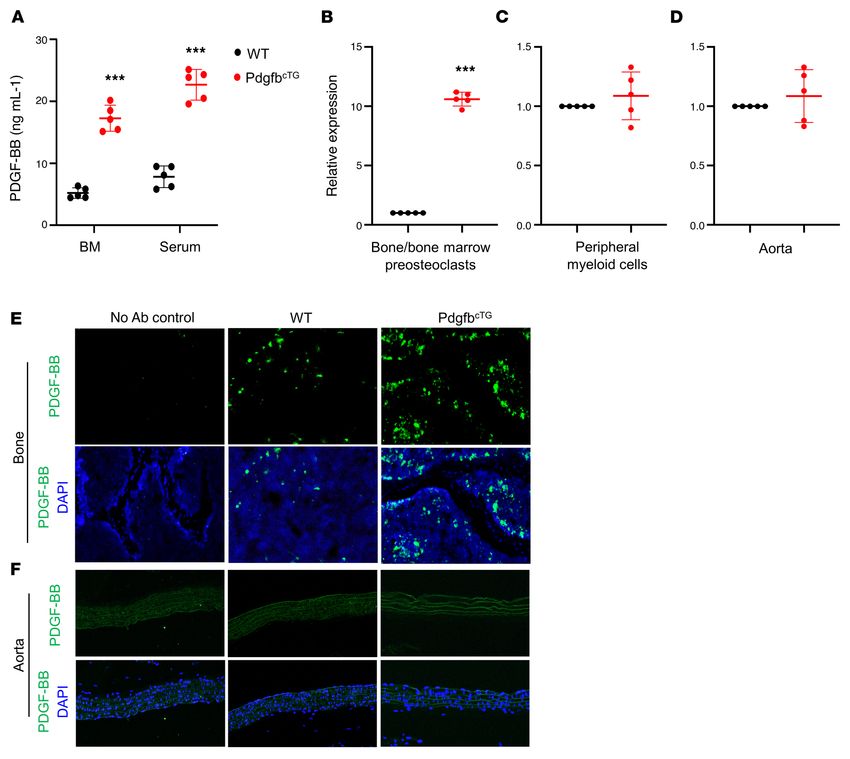
RESEARCH ARTICLE The Journal of Clinical Investigation
Figure 2. Aged mice, rats, and humans and HFD-challenged mice have elevated serum PDGF-BB concen-
tration. (A) ELISA measurement of serum PDGF-BB concentrations in 3- and 20-month-old mice. (B) ELISA
measurement of serum PDGF-BB concentrations in 4- and 25-month-old rats. (C) ELISA measurement of
serum PDGF-BB concentrations in young and aged humans. Old, ages 58 to 71 years; young, ages 21 to 26
years. (D) ELISA measurement of serum PDGF-BB concentrations in HFD mice and CHD mice. n = 5 to 10.
Data are mean ± SD, *P < 0.05, as determined by Student’s t tests.
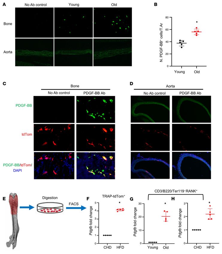
(Figure 3A). The number of PDGF-BB–expressing cells markedly clast precursors from 20-month-old mice versus 6-month-old mice
increased in the bone/bone marrow of 20-month-old mice rela- (Figure 3G) and HFD mice versus CHD mice (Figure 3H). Together,
tive to 3-month-old mice (Figure 3, A and B). PDGF-BB+ cells were the results suggest that bone/bone marrow preosteoclasts secrete
not detected in aorta tissue in either 3- or 20-month-old mice. excessive PDGF-BB in response to aging or HFD challenge.
To determine whether the increased PDGF-BB is produced by Preosteoclast-derived PDGF-BB stimulates VSMC proliferation
preosteoclasts in bone/bone marrow, we generated TRAP/tdTom and migration. One of the important functions of PDGF-BB is to
mice, in which tdTomato is expressed under TRAP-cre. Therefore, stimulate proliferation and migration of VSMCs, favoring patholog-
TRAP+ preosteoclasts and their descendants are labeled by tdTom ical vascular remodeling and arterial stiffening (66, 67, 69, 70). We
fluorescence in the mice. tdTom+ cells were abundant in bone tis- investigated whether preosteoclast-secreted PDGF-BB is sufficient
sue, and the majority of PDGF-BB–expressing cells are tdTom+ to induce phenotypic change of VSMCs using conditioned media
cells (Figure 3C). We did detect a few tdTom+ cells in the aorta tis- (CM) of preosteoclast cultures. Bone marrow Mo/Macs isolated
sue. However, none of the cells in the aorta expressed PDGF-BB from mice differentiate into TRAP+ mononuclear preosteoclasts 3
(Figure 3D). These results suggest that although there is a nonspe- days after treatment with M-CSF and RANK ligand, and most cells
cific Cre expression, cells in aortic tissue do not produce PDGF- differentiate into TRAP+ multinuclear mature osteoclasts 7 days
BB. Therefore, the effect of local aorta tissue–produced PDGF-BB after treatment (Figure 4A). We collected CM from cells at 0, 3,
can be excluded by using this TRAP-Cre line. and 8 days of M-CSF and RANK ligand treatment, which represent
Moreover, we performed FACS sorting to isolate the TRAP/ Mo/Mac CM, preosteoclast CM, and osteoclast CM, respectively.
tdTom+ cells from femoral bone/bone marrow cells (Figure 3E) Dramatically elevated PDGF-BB concentration was detected in
and conducted real-time qPCR analysis. TRAP/tdTom+ cells iso- preosteoclast CM relative to Mo/Mac CM, whereas PDGF-BB con-
lated from HFD-challenged mice had much higher Pdgfb expres- centration in osteoclast CM was lower compared with preosteo-
sion compared with those from CHD mice (Figure 3F). To further clast CM (Figure 4B). Importantly, rat VSMCs showed increased
validate the abnormally high expression of Pdgfb in the osteoclast proliferation (Figure 4C) and migration (Figure 4D) when the cells
precursors, we detected Pdgfb expression in bone/bone marrow were incubated with preosteoclast CM relative to the cells with Mo/
RANK+ cells with exclusion of the CD3/B220/Ter119+ cells (the Mac CM. These effects of preosteoclast CM were antagonized by
sum of T cells, B cells, and erythrocytes). We detected markedly PDGF-BB–neutralizing antibody. Therefore, PDGF-BB secreted by
greater expression of Pdgfb in CD3/B220/Ter119– RANK+ osteo- preosteoclasts can stimulate VSMC proliferation and migration.
4 J Clin Invest. 2021;131(20):e147116 https://doi.org/10.1172/JCI147116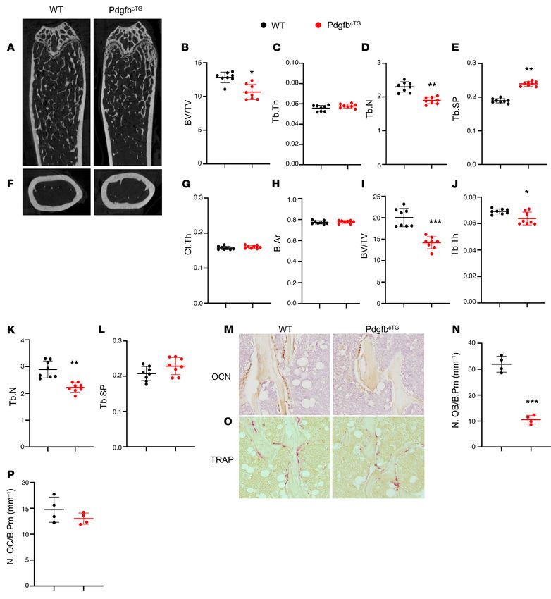
The Journal of Clinical Investigation                                                                                  RESEARCH ARTICLE

Figure 3. Bone/bone marrow preosteoclasts in aged mice and HFD mice are a main source of elevated circulating PDGF-BB. (A and B) Immunofluo-
rescence staining of femoral bone tissue from 3- and 20-month-old mice. Representative PDGF-BB staining image (A) and quantitative analysis of the
number of PDGF-BB+ cells per tissue area (B). (C and D) Frozen femoral bone (C) and aorta tissue sections (D) from TRAP/tdTom mice were subjected to
immunofluorescence staining using specific PDGF-BB antibody. Fluorescence imaging of tdTom+ cells (red), PDGF-BB+ cells (green), and double positive
cells (yellow) are shown. (E–H) Measurement of Pdgfb mRNA in bone/bone marrow preosteoclasts. Diagram showing the procedure for the isolation of
bone/bone marrow cells from femoral bone using our previously described approach (E) (also see description in Methods). Cell suspension collected from
TRAP/tdTom mice with CHD and HFD was subject to FACS to isolate tdTom+ cells. mRNA expression levels of Pdgfb were measured by qRT-PCR (F). Cell
suspension collected from C57B/L6 mice was subject to FACS to isolate CD3/B220/T119–RANK+ cells. The mRNA levels of Pdgfb in aged mice versus young
mice (G) and HFD mice versus CHD mice (H) were measured by qRT-PCR. n = 5. Data are mean ± SD. *P < 0.001, as determined by Student’s t tests.

                                         J Clin Invest. 2021;131(20):e147116 https://doi.org/10.1172/JCI147116                                   5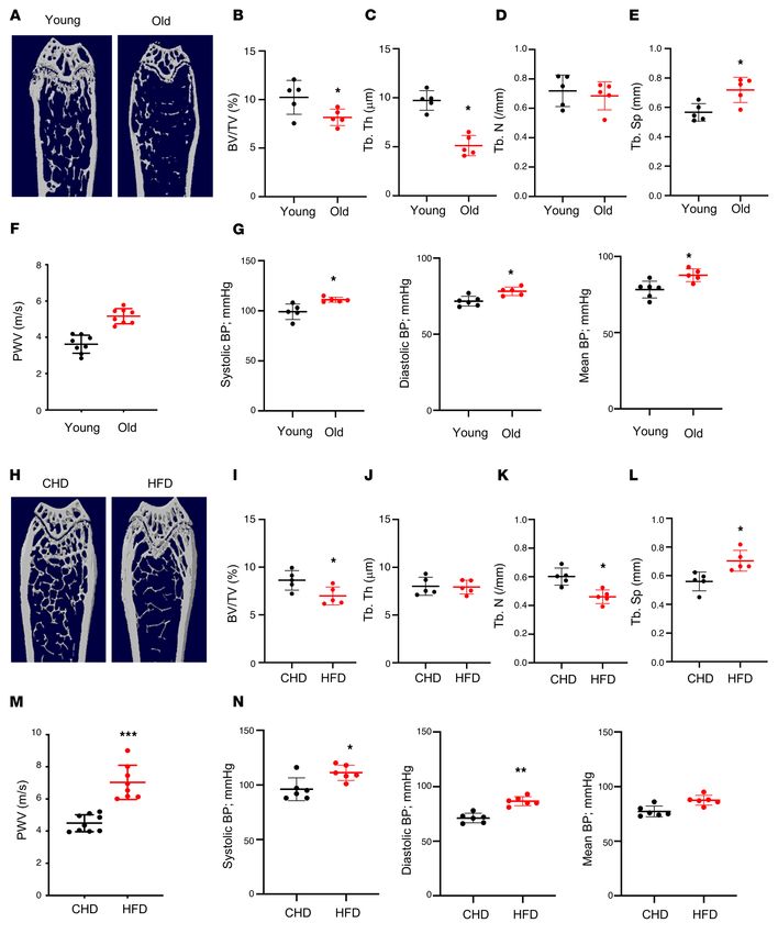
RESEARCH ARTICLE                                                                                          The Journal of Clinical Investigation

Figure 4. Preosteoclast-derived PDGF-BB stimulates VSMC proliferation and migration. (A) Schematic diagram showing the in vitro isolation of bone
marrow Mo/Macs and the induction of osteoclast differentiation. (B) Conditioned medium (CM) was collected from Mo/Mac, preosteoclast, and osteoclast
cultures, as described in Methods. PDGF-BB protein concentration in different CMs was measured using ELISA. (C) Rat VSMCs were incubated with CM
from Mo/Mac, preosteoclasts, and osteoclasts for 48 hours. Cell proliferation was assessed using the MTT method. (D) Transwell assays for preosteoclast
CM–induced migration of VSMCs. n = 5. Data are mean ± SD. *P < 0.05, 1-way ANOVA with Bonferroni post hoc test.

    Conditional Pdgfb transgenic mice recapitulate low bone mass              harvested from PdgfbcTG mice and WT littermates. Although the
and an arterial stiffening phenotype. To determine whether                    expression of Pdgfb was also detected, the expression levels were
increased production of PDGF-BB from preosteoclasts is suffi-                 not significantly elevated in both periphery blood myeloid cells
cient to induce vascular stiffening, we generated conditional Pdgfb           (Figure 5C) and aorta tissue (Figure 5D) from PdgfbcTG mice rel-
transgenic mice (PdgfbcTG), in which PDGF-BB is overexpressed in              ative to WT mice. Consistently, the PDGF-BB protein expression
TRAP+ cells by ligation of a 2.8-kb full-length human Pdgfb gene              was dramatically increased in bone/bone marrow cells as detect-
with a TRAP+ cell-specific promoter, TRACP5 (29). No abnormal                 ed by immunofluorescence staining of femoral bone tissue sec-
appearance or behavior was found in the PdgfbcTG mice relative                tions (Figure 5E). Increased PDGF-BB expression was not found in
to their WT littermates. Intriguingly, bone marrow and serum                  aortic walls from PdgfbcTG mice relative to WT mice (Figure 5F). Of
PDGF-BB levels were more than 3-fold higher in the PdgfbcTG mice              note, PDGF-BB+ cells were not detected in any of the aortas where
compared with the age-matched WT mice (Figure 5A). We then                    calcification was found in the PdgfbcTG mice. Therefore, the elevat-
assessed whether Pdgfb is specifically overexpressed in bone/                 ed circulating PDGF-BB in transgenic mice is primarily produced
bone marrow preosteoclasts in the transgenic mice by measuring                by bone/bone marrow preosteoclasts rather than a local effect
the mRNA expression in isolated bone/bone marrow CD3/B220/                    derived from blood vessels and blood myeloid cells.
Ter119 –RANK+ cells, which are primarily precursors of osteoclast                  We conducted a systemic bone phenotypic analyses of the
lineage (23, 71, 72). As we expected, quantitative RT-PCR analysis            transgenic mice at 6 months of age. MicroCT analyses of the dis-
shows that Pdgfb expression was markedly higher in preosteoclasts             tal femur in 6-month-old male PdgfbcTG mice revealed a low bone
from PdgfbcTG mice relative to WT mice (Figure 5B). To assess                 mass phenotype (Figure 6A) with reduced BV/TV (Figure 6B)
whether circulating myeloid cells and vascular resident cells may             and Tb.N (Figure 6D) and increased Tb.Sp (Figure 6E) relative to
also be the sources of elevated circulating PDGF-BB in the trans-             their WT littermates. Tb.Th was not changed in the PdgfbcTG mice
genic mice, periphery blood myeloid cells and aorta tissue were               compared with the WT mice (Figure 6C). Therefore, young Pdg-

   6                                     J Clin Invest. 2021;131(20):e147116 https://doi.org/10.1172/JCI147116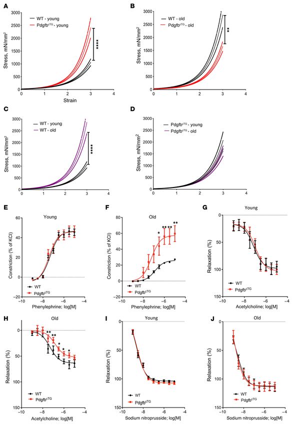
The Journal of Clinical Investigation                                                                               RESEARCH ARTICLE

                                                                                           Figure 5. Conditional Pdgfb transgenic mice have
                                                                                           increased PDGF-BB expression in bone/bone marrow
                                                                                           and elevated serum PDGF-BB concentration. (A) ELISA
                                                                                           measurements of bone marrow (BM) and serum PDGF-
                                                                                           BB concentrations in PdgfbcTG and WT littermates.
                                                                                           (B–D) Bone/bone marrow CD3/B220/T119–RANK+ cells
                                                                                           (B), peripheral blood myeloid cells (C), and aorta tissue
                                                                                           (D) were collected from 6-month-old PdgfbcTG mice
                                                                                           and WT littermates as described in Methods. mRNA
                                                                                           expression of Pdgfb was measured by qRT-PCR. (E
                                                                                           and F) Representative PDGF-BB immunofluorescence
                                                                                           staining of the femoral bone (E) and aorta (F) tissue
                                                                                           sections from 6-month-old PdgfbcTG mice and WT
                                                                                           littermates. n = 5. Data are mean ± SD. ***P < 0.001, as
                                                                                           determined by Student’s t tests.

fbcTG mice mirrored aging-associated trabecular bone changes.            er in old PdgfbcTG relative to young PdgfbcTG mice, indicating an
Cortical thickness (Ct.Th) and bone area (B. Ar) were not differ-        age-dependent progression of arterial stiffening in the conditional
ent in the PdgfbcTG mice compared with the WT mice (Figure 6,            transgenic mice. As expected, PWV increased significantly with
F–H). We also evaluated 9-month-old female mice and found that           age in WT control mice. In the aged mice, a significant sex effect
female PdgfbcTG mice had a similar low bone mass phenotype in            was noted as old female mice had significantly higher PWV than
trabecular compartment relative to their age- and sex-matched            corresponding age-matched males (Figure 7E). Systolic, diastolic,
WT littermates (Figure 6, I–L). Therefore, overexpression of Pdgfb       and mean BPs were all significantly higher in young PdgfbcTG mice
in the preosteoclasts results in decreased bone mass in trabecular       than those in age-matched WT mice (Figure 7, B–D). However, dif-
but not in cortical bone compartments. Histomorphometry anal-            ferences in BP were not noted in the old PdgfbcTG mice versus age-
ysis shows that the number of bone surface osteocalcin (OCN)+            matched WT mice. Aged female WT mice had significantly lower
osteoblasts were significantly reduced (Figure 6, M and N) but the       systolic, diastolic, and mean pressures than did aged male WTs.
number of bone surface TRAP+ osteoclasts remained unchanged              However, no sex differences were noted in the BP of PdgfbcTG old
(Figure 6, O and P) in the PdgfbcTG mice relative to WT mice, sug-       mice (Figure 7, F–H).
gesting that overexpression of Pdgfb in the preosteoclasts primari-          It has been reported that there is an age-dependent increase
ly impaired osteoblast bone formation.                                   in the lumen diameter and wall thickness of the aorta (73, 74).
     We then measured PWV, the gold-standard index for aortic            Consistent with these previous reports, the thoracic aortic lumen
stiffness. Both young (3–4 months old) and old (>18 months old)          diameter was greater in both aged (versus young) WT mice and
PdgfbcTG mice had significantly higher PWV compared with their           aged (versus young) PdgfbcTG mice (Figure 8, A and B). Importantly,
age-matched WT mice (Figure 7A). Moreover, PWV is also high-             aged PdgfbcTG mice, relative to their age-matched WT littermates,

                                     J Clin Invest. 2021;131(20):e147116 https://doi.org/10.1172/JCI147116                                     7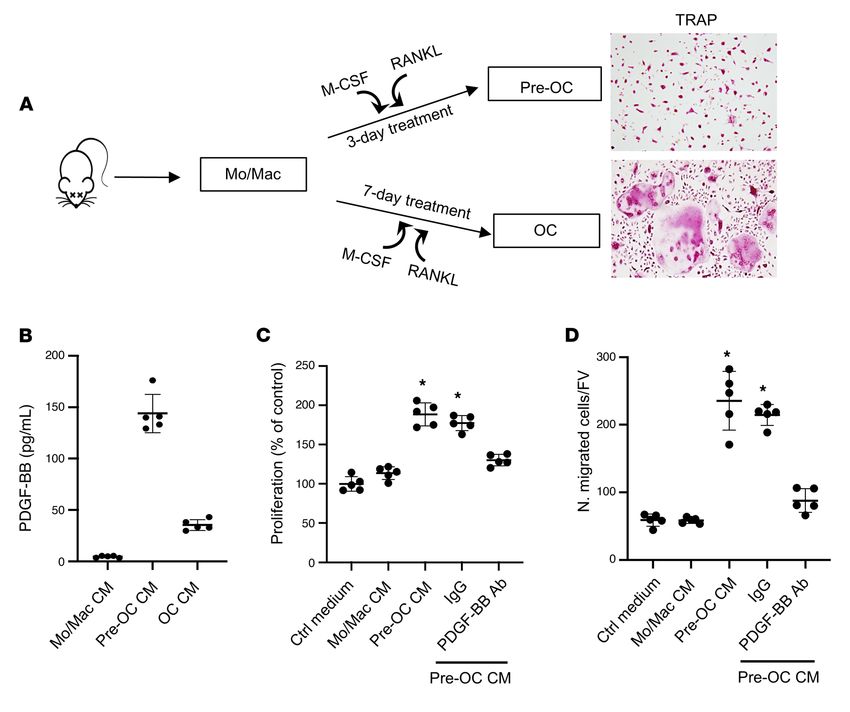
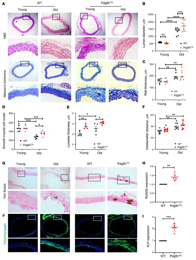
RESEARCH ARTICLE                                                                                          The Journal of Clinical Investigation

Figure 6. Conditional Pdgfb transgenic mice recapitulate an aging-associated bone phenotype. (A–E) Representative μCT images (A) and quantita-
tive analyses (B–E) of the trabecular bone area of the distal femur from male 6-month-old PdgfbcTG mice and WT littermates. BV/TV (B), Tb.Th (C),
Tb.N (D), and Tb.Sp (E). Representative μCT images (F) and quantitative analysis (G and H) of the cross-sections of femoral mid-diaphysis of mice.
Ct.Th, cortical bone thickness; B. Ar, bone area. (I–L) Quantitative μCT analyses of the trabecular bone area of the distal femur from female 9-month-
old PdgfbcTG mice and WT littermates. BV/TV (I), Tb.Th (J), Tb.N (K), and Tb.Sp (L). (M and N) Representative immunohistochemical staining (M) and
quantitative analysis of osteocalcin (OCN) (N) in femur sections. (O and P) Representative TRAP staining (O) and quantitative analysis of TRAP+ cells in
femur sections (P). N.OB/B.Pm, number of osteocalcin+ osteoblasts per bone perimeter; N.OC/B.Pm, number of TRAP+ osteoclasts per bone perimeter.
*P < 0.05, **P < 0.01, ***P < 0.001 as determined by Student’s t tests.

   8                                     J Clin Invest. 2021;131(20):e147116 https://doi.org/10.1172/JCI147116
The Journal of Clinical Investigation                                                                                RESEARCH ARTICLE

                                                                                 Figure 7. Conditional transgenic mice expressing PDGF-BB in
                                                                                 preosteoclasts recapitulate aging-associated artery phenotype.
                                                                                 PWV (A), systolic BP (B), diastolic BP (C), and mean BP (D) were
                                                                                 measured in young (3- to 4-month old) and old (>18 months old)
                                                                                 PdgfbcTG and WT littermates. PWV (E), systolic BP (F), diastolic BP
                                                                                 (G), and mean BP (H) were measured in old (>18 months old) male
                                                                                 and female PdgfbcTG and WT littermates. n = 5 to 14. Data are mean
                                                                                 ± SEM, *P < 0.05, **P < 0.01, ***P < 0.001, ****P < 0.0001 as
                                                                                 determined by 1-way ANOVA with Bonferroni post hoc test.

had increased aortic lumen diameter, indicating an exacerbat-             In addition, we examined the activation of an osteogenic trans-dif-
ed age-related morphological change of the aorta when Pdgfb is            ferentiation program in the aortas of the PdgfbcTG mice. Both osteo-
overexpressed. Aortic wall was significantly thicker in aged WT           blast differentiation markers RUNX2 (Figure 8H) and ALP (Figure
and PdgfbcTG mice than in young WT and PdgfbcTG mice, respec-             8I) were upregulated at mRNA level in the aortas of PdgfbcTG mice
tively (Figure 8, A and C). The aortas from aged WT and PdgfbcTG          compared with WT mice, indicating that vascular calcification may
mice, relative to young mice, showed smooth muscle cell nuclei            contribute to arterial stiffening induced by preosteoclast-secreted
loss (Figure 8D), a characteristic of vascular aging. Moreover, the       PDGF-BB. We also examined whether the expression of PDGFRβ,
VSMC nuclei loss is more in the WT mice than in the PdgfbcTG mice,        the receptor of PDGF-BB, is changed in aorta tissues of old (versus
indicating that the old PdgfbcTG mice may have increased PDGFB/           young) and transgenic mice (versus WT mice). Markedly increased
PDGFRβ signaling in the arterial tissue. Lamellar thickness and           expression of PDGFRβ in smooth muscle cells of the aorta wall
intralamellar distance both increased significantly with age in the       was found in both aged mice and PdgfbcTG mice, as compared with
WT and PdgfbcTG mice, indicating a significant accumulation of            young mice and WT mice, respectively (Figure 8J).
matrix in the vascular wall (Figure 8, E and F). Vascular calcifica-           We then measured the passive stiffness of the vessels. Tensile
tion is a key link between osteoporosis and CVD. We then assessed         testing showed greater stiffness of the descending aorta in both
whether the transgenic mice have vascular calcification by per-           young and aged PdgfbcTG mice compared with their age-matched
forming von Kossa staining of aorta tissue sections. Positive signal      WT littermates, with the difference in young mice being of high-
was found in 1 of 4 old mice (24 months old) but in 0 of 6 young          er magnitude (Figure 9, A and B). While there was a greater ves-
mice (4 months old). Importantly, positive signal was found in 2 of       sel stiffness in the aged WT mice relative to young WT mice, this
8 PdgfbcTG mice at 6 months of age, whereas none of the 7 aortas          age-dependent difference was not significant in the PdgfbcTG mice
from the littermates (WT) showed positive signaling (Figure 8G).          (Figure 9, C and D). The significantly higher passive stiffness of the

                                      J Clin Invest. 2021;131(20):e147116 https://doi.org/10.1172/JCI147116                                    9
RESEARCH ARTICLE The Journal of Clinical Investigation 10 J Clin Invest. 2021;131(20):e147116 https://doi.org/10.1172/JCI147116
The Journal of Clinical Investigation                                                                                  RESEARCH ARTICLE

Figure 8. Conditional Pdgfb transgenic mice develop pathological              fed a HFD compared with WT mice fed a CHD, but this elevation
aortic morphology and vascular calcification. (A) Representative his-         was not detected in Pdgfb-cKO mice after HFD challenge (Figure
tological staining analysis (10×; inset 40×) showing H&E and Masson’s         10B). Of note, serum PDGF-BB concentration in HFD-challenged
trichrome staining of aorta from 4- and 18-month-old PdgfbcTG and
WT littermates. (B) Lumen diameter, (C) vessel wall thickness, and (D)
                                                                              Pdgfb-cKO mice was reduced to a similar level as in the WT mice
smooth muscle cell nuclei, Lamellar thickness (E), and intralamellar          without HFD challenge. The results further validated that bone/
distance (F) in aortas were calculated. n = 5 to 11. *P < 0.05, **P < 0.01,   bone marrow preosteoclasts are a main source of elevated PDGF-
***P < 0.001, and ****P < 0.0001 as determined by 1-way ANOVA with            BB in blood circulation in mice during aging or under HFD.
Bonferroni post hoc test. (G) Representative micrographs of von Kossa              We conducted an analysis of bone phenotype in Pdgfb-cKO
stained sections of the thoracic aorta from young (4-month-old) and old
(20-month-old) mice (left panels) and from 6-month-old PdgfbcTG and
                                                                              mice (Figure 10C). MicroCT analyses shows that trabecular BV/
WT littermates (right panels). (H and I) Aorta tissues were harvested         TV, Tb.N, and Tb.Th were all lower, and Tb.Sp was bigger in the
from 6-month-old PdgfbcTG and WT littermates. mRNA expressions of             distal femur of Pdgfb-cKO mice relative to their Pdgfbfl/fl littermates
RUNX2 (H) and alkaline phosphatase (ALP) (I) were measured by qRT-            (WT; Figure 10, D–G). The results are consistent with our previous
PCR. n = 6. Data are mean ± SD, **P < 0.01, ***P < 0.001 as determined        work showing that healthy, unchallenged Pdgfb-cKO mice exhib-
by Student’s t tests. (J) Immunofluorescence staining of aortic tissue
sections with antibody against PDGFRβ from young (4-month-old) and
                                                                              ited a low-bone-mass phenotype (28). We then evaluated whether
old (20-month-old) mice (left panels) and from 6-month-old PdgfbcTG           deletion of Pdgfb from preosteoclasts affects bone phenotype in
and WT littermates (right panels).                                            HFD-challenged mice, in which PDGF-BB concentrations in both
                                                                              bone marrow and serum were aberrantly elevated compared with
                                                                              mice fed normal CHD. Whereas HFD induced reduction in BV/
PdgfbcTG mouse aorta determined by tensile testing suggests that the          TV and Tb.N and increase in Tb.Sp in WT mice, the changes of
increase in PWV noted in these mice is not solely due to the effect of        these parameters induced by HFD were, at least partially, rectified
higher BP, and there is substantial arterial stiffening as well.              by Pdgfb deletion (Figure 10, D–G). HFD induced an increase in
     We also tested the contraction and relaxation responses of the           cortical bone area (Ct. Ar) without changing cortical bone thick-
aorta. We did not detect differences in the contraction response to           ness (Ct.Th) in both WT and Pdgfb-cKO mice (Figure 10, H–J).
phenylephrine (Figure 9E) and endothelial relaxation response to              Therefore, preosteoclast-derived PDGF-BB plays a paradoxical
acetylcholine (Figure 9G) of the vessels from the young PdgfbcTG              role specifically in trabecular bone regulation. While PDGF-BB is
mice compared with those from the young WT mice. However,                     required for the maintenance of bone homeostasis under normal
phenylephrine-induced contraction was higher in the vessels from              physiological conditions, excessive production of PDGF-BB from
the aged PdgfbcTG mice relative to the age-matched WT mice (Fig-              preosteoclasts leads to trabecular bone loss in pathological condi-
ure 9, F and H), suggesting that Pdgfb overexpression sensitizes              tions (e.g., aging and metabolic dysregulation).
the vessels to agonist-induced contraction in aging, as has been                   We next determined whether targeting PDGF-BB secretion
previously shown (75). This suggests the possibility of augmented             by preosteoclasts can prevent deterioration of vascular mechan-
tone of the vascular smooth muscle cells with increased circulat-             ics and function. To this end, we used a HFD challenge to accel-
ing PDGF-BB. Moreover, acetylcholine-mediated relaxation of                   erate vascular stiffening and deterioration of bone as a rapid
preconstricted vessels was notably lower in the aorta from aged               alternative to natural aging, which takes at least 18 months in the
PdgfbcTG mice when compared with age-matched WT controls                      mouse model. The HFD challenge is shown to cause an increase
when Cox pathways were inhibited using indomethacin. No dif-                  in PWV prior to the onset of systolic hypertension, as is the case
ferences were noted in the endothelial-independent sodium nitro-              in aging (35). Here, we measured PWV in Pdgfb-cKO mice fed a
prusside–induced (SNP-induced) relaxation in both young and                   HFD for different time periods. Pdgfbfl/fl (WT) mice had a slight
aged PdgfbcTG mice relative to age-matched WT mice (Figure 9, I               increase in PWV after 8 weeks and a significant increase in PWV
and J). This suggests that Pdgfb either induces a larger deficit in           after 12 to 14 weeks of HFD feeding. Importantly, Pdgfb-cKO
age-associated endothelial dysfunction, or that the larger precon-            mice were partially protected from the diet-induced elevation
striction caused by phenylephrine is not fully countered by the               in PWV noted in the WT mice (Figure 11A). The difference in
endothelial-mediated relaxation response. Together, these find-               PWV levels at baseline between Pdgfb-cKO mice and WT mice
ings suggest that Pdgfb overexpression in preosteoclasts causes               was not significant. HFD-induced elevation of systolic, diastol-
endothelial dysfunction, VSMC dysregulation, and vascular stiff-              ic, and mean BPs in the WT mice was also not detected in the
ening during aging.                                                           Pdgfb-cKO mice (Figure 11, B–D). Increased stiffness of both the
     Conditional Pdgfb knockout mice are protected from HFD-induced           matrix and VSMC dysfunction are known to occur in response
bone loss and arterial stiffening. We tested whether increased circu-         to HFD, contributing to vascular stiffening in vivo. Therefore,
lating PDGF-BB is required for HFD-induced bone loss and arteri-              we next examined the mechanical and functional properties of
al stiffening by generating conditional Pdgfb knockout mice (Pdg-             the aorta at the end of 14 weeks of HFD. Tensile testing of the
fb-cKO), in which Pdgfb is deleted selectively in the TRAP+ cells by          descending aorta showed a significantly more compliant vessel
crossing Pdgfbfl/fl mice with Trap-Cre mice (29). We detected a dra-          in HFD Pdgfb-cKO mice compared with HFD WT mice (Figure
matically lower mRNA level of Pdgfb in CD3/B220/Ter119–RANK+                  11E). Vascular contractility studies showed an exaggerated con-
preosteoclasts isolated from the bone/bone marrow of Pdgfb-cKO                tractility response to increasing concentrations of phenylephrine
mice compared with Pdgfbfl/fl littermates (WT; Figure 10A), validat-          in WT mice compared with Pdgfb-cKO mice (Figure 11F). The
ing the efficiency of Pdgfb deletion in preosteoclasts in the knockout        endothelial-dependent relaxation to acetylcholine after precon-
mice. Importantly, the serum PDGF-BB level was higher in WT mice              striction with phenylephrine was higher in Pdgfb-cKO mice than

                                          J Clin Invest. 2021;131(20):e147116 https://doi.org/10.1172/JCI147116                                11
RESEARCH ARTICLE The Journal of Clinical Investigation 12 J Clin Invest. 2021;131(20):e147116 https://doi.org/10.1172/JCI147116
The Journal of Clinical Investigation                                                                                RESEARCH ARTICLE

Figure 9. Aortic plasticity and vasoreactivity are impaired in conditional    aging or under metabolic stress remain unclear. Preosteoclasts
Pdgfb transgenic mice. (A–D) Tensile testing of aortic rings was measured     may develop a unique secretory phenotype during aging and are
in 4- and 18-month-old PdgfbcTG and WT littermates. n = 10 rings. Data        likely a primary cell type producing clastokines to regulate other
are mean ± SEM, ****P < 0.0001 versus WT mice, as determined by 1-way
ANOVA with Bonferroni post hoc analysis. Aortic constriction in response
                                                                              cell types or tissues. Further analysis of the other factors released
to increasing doses of phenylephrine was measured in the young (E) and        from preosteoclasts in addition to PDGF-BB will be important to
aged (F) PdgfbcTG mice vs. WT littermates. n = 8 aortic rings. Data are       define the secretory function of this cell type within the bone mar-
shown as mean ± SEM, *P < 0.05, **P < 0.01, as determined by one-way          row microenvironment under disease conditions.
ANOVA with Bonferroni post hoc test. Endothelium-dependent aortic                  Our data suggest that PDGF-BB exerts paradoxical bone
relaxation in response to increasing doses of acetylcholine was measured
in young (G) and aged (H) PdgfbcTG mice versus WT littermates. n = 8 aortic
                                                                              effects depending on the concentration of PDGF-BB in the
rings. Data are mean ± SEM, *P < 0.05, **P < 0.01, as determined by 1-way     bone microenvironment. We previously showed that a normal
ANOVA with Bonferroni post hoc test. Endothelium-independent vasore-          range of PDGF-BB is essential for the maintenance of bone
laxation in response to sodium nitroprusside was measured in young (I)        homeostasis in young, healthy mice because deletion of Pdg-
and aged (J) PdgfbcTG mice versus WT littermates. n = 8 aortic rings. Data    fb from TRAP+ preosteoclasts led to reduced trabecular and
are mean ± SEM.
                                                                              cortical bone mass (28). Consistently, here we also found a
                                                                              low-bone-mass phenotype in the young, healthy Pdgfb-cKO
                                                                              mice relative to their WT littermates. However, during aging
in WT mice (Figure 11G). The endothelial-independent relax-                   or under metabolic stress conditions such as HFD challenge,
ation of the vessels to SNP was similar between the 2 groups                  aberrantly elevated PDGF-BB in bone marrow microenviron-
(Figure 11H), suggesting that the blunted acetylcholine response              ment exerts an adverse bone effect. Importantly, deletion of
of WT mice is caused by a greater decline in the endothelial func-            Pdgfb in preosteoclasts normalized the PDGF-BB concentration
tion of WT mice in response to a HFD than in the Pdgfb-cKO                    and largely rectified the pathological bone phenotype induced
mice. Together, these findings suggest that PDGF-BB promotes                  by aging and HFD. Furthermore, the young PdgfbcTG mice,
diet-induced vascular stiffening by mediating changes in cellular             resemblant of the old mice, have a much-elevated PDGF-BB
function, including augmented vascular contractility in conjunc-              concentration in bone marrow. As a result, the mice develop an
tion with endothelial dysfunction in the HFD WT mice.                         aging-associated trabecular bone loss phenotype. Together, our
                                                                              finding imply that PDGF-BB is required for the maintenance of
Discussion                                                                    bone homeostasis under normal physiological conditions, and
The regulatory mechanisms of the vascular system through                      aberrantly elevated PDGF-BB in bone marrow microenviron-
bone-derived cues during aging are poorly understood. Here we                 ment leads to bone loss in pathological conditions (e.g., aging
showed that with advancing age or under metabolic stress, mono-               and metabolic dysregulation). At the cellular level, we found
nuclear preosteoclasts in bone/bone marrow, as a main source                  a substantially reduced osteoblast number but an unchanged
of excessive circulating PDGF-BB, contribute to arterial stiffen-             osteoclast number in the trabecular compartment of long bone
ing (Figure 12). Our study provides new insight into the cellular             in the PdgfbcTG mice relative to WT mice. The data suggest that
and molecular mechanisms underlying the bone/vascular axis.                   excessive PDGF-BB produced by preosteoclasts negatively reg-
Despite the well-recognized role of PDGF-BB in aging-associated               ulates osteoblast differentiation activity in a paracrine manner,
arterial stiffness and atherosclerosis development, we are aware              leading to impaired bone formation.
of no studies of the role of PDGF-BB as a systemic proaging fac-                   Our results further reveal that skeletal preosteoclasts are a
tor. We detected elevated bone marrow and serum PDGF-BB con-                  main cell type contributing to the elevation of PDGF-BB in blood
centration in aged mice, rats, and human subjects relative to their           circulation during aging and under metabolic stress. While aged
young counterparts. More importantly, the data from our condi-                mice and those fed HFD had higher serum PDGF-BB relative to
tional transgenic and knockout mouse models suggest that aber-                young mice and CHD mice respectively, mice with Pdgfb dele-
rantly elevated PDGF-BB secreted by preosteoclasts is a driving               tion from preosteoclasts had a normalized serum PDGF-BB con-
force for the pathological changes of both the skeletal and vascular          centration. Moreover, young conditional Pdgfb transgenic mice,
systems (i.e., bone loss and arterial stiffening) in response to aging        resemblant of the aged mice, had a much higher serum PDGF-
and HFD challenge.                                                            BB level relative to their WT littermates. Therefore, skeletal
     Multinuclear osteoclasts have been considered an orches-                 preosteoclast-derived PDGF-BB is both sufficient and required
trator, with more functions beyond bone resorption. Osteoclasts               to cause circulating PDGF-BB elevation. We are aware that the
secrete clastokines that regulate the activity of neighboring cells           increased PDGF-BB may also be produced from tissues other
within the bone/bone marrow microenvironment (20). It was                     than bone. However, we detected increased expression of Pdg-
reported that PDGF-BB in bone/bone marrow microenvironment                    fb mRNA and PDGF-BB protein only in preosteoclasts but not in
is primarily produced by osteoclast precursors but not by uncom-              the periphery blood myeloid cells and aorta tissue from PdgfbcTG
mitted Mo/Macs and multinuclear osteoclasts in healthy, young                 mice relative to WT mice. The results suggest that the elevated
mice (28). The present study agrees with this finding and further             circulating PDGF-BB in response to aging or metabolic stress is
demonstrates that mononuclear TRAP+ preosteoclasts secrete                    primarily produced by bone/bone marrow preosteoclasts rath-
much more PDGF-BB in aged mice and HFD-challenged mice rel-                   er than a local effect derived from arterial vessel wall or blood
ative to young mice and CHD mice, respectively. The mechanisms                myeloid cells. We are aware that the Trap-Cre line causes dele-
by which preosteoclasts secrete a high amount of PDGF-BB during               tion of PDGF-BB in all Trap+ osteoclast lineage cells, including

                                          J Clin Invest. 2021;131(20):e147116 https://doi.org/10.1172/JCI147116                              13
RESEARCH ARTICLE The Journal of Clinical Investigation Figure 10. Deletion of Pdgfb in preosteoclasts attenuates HFD-induced bone loss. (A) Measurement of PDGF-BB mRNA in isolated CD3/B220/T119– RANK+ cells from bone/bone marrow of Trap-Cre Pdgfbf/f mice (Pdgfb-cKO) and Pdgfbf/f littermates (WT). (B) ELISA measurements of serum PDGF-BB concentrations in Pdgfb-cKO and WT littermates fed a HFD or CHD. n = 5. Data are mean ± SD. *P < 0.05, **P < 0.01, as determined by Student’s t tests and 1-way ANOVA with Bonferroni post hoc test. (C–G) Representative μCT images (C) and quantitative analysis of the trabecular bone area of the distal femur from 3-month-old Pdgfb-cKO mice and WT littermates after 14 weeks of a Western HFD or chow diet (CHD). BV/TV (D), Tb.Th (E), Tb.N (F), and Tb.Sp (G). Representative μCT images (H) and quantitative analysis (I and J) of the cross-sections of femoral mid-diaphysis of mice. n = 7. Data are mean ± SD. *P < 0.05, **P < 0.01, ***P < 0.001, as determined by 1-way ANOVA with Bonferroni post hoc analysis. 14 J Clin Invest. 2021;131(20):e147116 https://doi.org/10.1172/JCI147116
The Journal of Clinical Investigation                                                                             RESEARCH ARTICLE

TRAP+ mononuclear preosteoclasts and TRAP+ multinuclear                     culating PDGF-BB attenuated phenylephrine contractility and
mature osteoclasts. However, PDGF-BB is secreted specifical-                augmented acetylcholine-induced vasorelaxation in the PdgfbcTG
ly by Trap+ mononucleate preosteoclasts, and a very low level               mice. These findings are also consistent with global knockout
of PDGF-BB is secreted by mature osteoclasts (ref. 25 and Fig-              of the Pdgfb gene, which showed a loss of functional contractil-
ure 4). Moreover, our data from the TRAP/tdTom mice clearly                 ity of VSMCs, causing a remarkable dilation of the aorta rather
demonstrate that although there is a nonspecific Cre expression,            than producing structural deficits in the large compliance ves-
cells in aortic tissue do not produce PDGF-BB. Therefore, the               sels (82). In young PdgfbcTG mice, despite the vascular stiffen-
effect of local aorta tissue–produced PDGF-BB can be excluded               ing noted, there is no compromised contraction or endothelial
by using the TRAP-Cre line, and the reduced PDGF-BB concen-                 relaxation response. Prior studies have shown that PDGF-BB
tration in the Trap-Cre–driven knockout mice is caused mainly               caused severe and chronic vasoconstriction (83). Interestingly,
by the deletion of PDGF-BB from preosteoclasts.                             however, we did not note a change in contraction responses in
     PDGF-BB overexpression accelerated aging-associated vascu-             the young mice. One reason for the unchanged contraction or
lar stiffening. A well-known VSMC mitogen, PDGF-BB can elicit               endothelial relaxation response in young transgenic mice may
VSMC migration and proliferation in the aging aorta, leading to             be that PDGF receptor is not expressed highly in the young ves-
structural/compositional changes that are characteristic of the             sels. However, in the old PdgfbcTG mice, a significantly higher
aged, stiff vessel. For example, aging VSMCs express higher levels          contraction response was noted. This is likely due to increased
of PDGF-BB receptor (57) and exhibit augmented proliferation in             expression of PDGF-R in the aged vessels (Figure 8J) in good
response to PDGF-BB (76, 77). Thus, PDGF-BB can cause diffuse               agreement with the literature (84). We also found more VSMC
intimal changes in the aging vessel, which is a hallmark of aging           nuclei loss in the aged WT mice than in the aged PdgfbcTG mice.
(78). In this study, we show that PDGF-BB secreted by preosteo-             This can also be attributed to the upregulated PDGF-R expres-
clasts contributes to vascular aging. In vitro, conditioned media           sion and Pdgfb signaling that may lead to VSMC proliferation in
from preosteoclasts overexpressing Pdgfb promoted VSMC prolifer-            the old conditional transgenic mice. Our studies further show
ation. In vivo, young PdgfbcTG mice have stiffer vessels as reflected       that the lumen diameter of the aged PdgfbcTG mice is higher
by the augmented PWV, and higher systolic and diastolic BPs. The            than the WT littermates. Furthermore, while wall thickness was
passive stiffness of the vessels measured by tensile testing shows          not significantly higher in the aged PdgfbcTG mice, the number
a strikingly higher stiffness of the young PdgfbcTG mice compared           of cells in the vessel wall was higher. Collectively, our findings
with their littermates. The augmented PWV is likely to be a com-            from young and old PdgfbcTG mice imply that conditional Pdgfb
bination of increased BP and passive stiffening in the conditional          transgenic mice not only have an accelerated stiffening response
transgenic mice. Notably, while the passive stiffness of WT mice            to age, but also exaggerated endothelial and vascular dysfunc-
increased remarkably with age, this did not occur in the PdgfbcTG           tion. More importantly, arterial stiffening induced by a HFD
mice, in which the young vessels were markedly stiffer to begin             was significantly alleviated in the PDGF-BB knockout mice,
with. Moreover, with age, there was a marked increase in PWV, sys-          confirming the critical role of preosteoclast-derived PDGF-BB
tolic BP, and diastolic BP in the WT mice, but not in the PdgfbcTG          in the increase in vascular stiffness. Thus, our study supports a
mice. At the histology level, PdgfbcTG mice exhibited increased col-        growing body of evidence showing that in addition to the extra-
lagen fibers in the extracellular matrix and calcification of the aorta,    cellular matrix stiffness and remodeling, aortic tone and VSMC
suggesting that vascular fibrosis and calcification are major contrib-      stiffness are critical determinants of in vivo vascular stiffness.
utors to PDGFB/PDGFRβ-associated arterial stiffening. Interest-                  The observation that young conditional Pdgfb transgen-
ingly, aging resulted in a higher PWV in female PdgfbcTG mice when          ic mice fed a CHD develop both low bone mass and an arte-
compared with age-matched males, while BP was not notably dif-              rial stiffening phenotype spontaneously is intriguing. This
ferent between the sexes. This is an intriguing finding, particularly       phenomenon implies that preosteoclast-derived increases in
in the context of higher incidence of osteoporosis in elderly females.      PDGF-BB, which are sufficient to cause bone loss and arterial
We previously noted that in rodents, the stiffening trajectory is to        stiffening, function as a molecular link for the bone/vascular
have a steep increase in stiffness between 3 to 12 months, and then         axis. Of great relevance for the clinic, our finding reveals that
a plateau of values (79). Considering these prior reports, we postu-        serum PDGF-BB can be used as a biomarker to determine who
late that PdgfbcTG mice have an accelerated vascular stiffening and         are at the greatest risk for age-associated vascular diseases. Our
reach a plateau much earlier in their lifespan than do the WT mice.         observations also provide the basis for future investigations to
Therefore, when we compare the aged (>18 months old) WT and                 determine whether targeting preosteoclasts or PDGF-BB sig-
PdgfbcTG mice, the differences are no longer as remarkable because          naling is an efficient strategy to prevent or treat cardiovascular
both cohorts have reached the maximal plateau.                              disease in the elderly population, especially those who develop
     Accumulating evidence points to VSMC stiffening and tone               cardiovascular disease together with osteoporosis. The use of
as key mediators of overall vascular stiffness. This is particu-            intervention to inhibit or neutralize PDGF-BB in the aging and
larly interesting in this study, as PDGF-BB also acts as a vaso-            under HFD conditions is a goal of future studies.
constrictor (75, 80, 81). Our study further shows that PDGF-BB
contributes to the functional contractility of VSMCs because                Methods
aging resulted in a marked sensitization of the agonist-induced             Animals and treatment. Male C57BL/6J mice were purchased from The
vasoconstriction response and an attenuated vasorelaxation                  Jackson Laboratory. Male Sprague-Dawley rats were purchased from
response in the PdgfbcTG mice. Conversely, reduced levels of cir-           Charles River Laboratories. All animals were bred and housed in the

                                        J Clin Invest. 2021;131(20):e147116 https://doi.org/10.1172/JCI147116                            15
RESEARCH ARTICLE The Journal of Clinical Investigation Figure 11. Deletion of Pdgfb in preosteoclasts attenuates HFD-induced vascular stiffening. PWV (A), systolic BP (B), diastolic BP (C), and mean BP (D) were measured in Pdgfb-cKO and WT littermates after 8 and 14 weeks of a Western HFD. n = 6 to 11. Data are mean ± SEM. *P < 0.05, **P < 0.01 versus WT mice, as determined by 1-way ANOVA with Bonferroni post hoc test. (E) Tensile testing of aortic rings was measured after 14 weeks of a Western HFD diet. n = 10 rings. Data are mean ± SEM. ***P < 0.001 versus WT mice, as determined by 1-way ANOVA with Bonferroni post hoc analysis. (F) Phenyleph- rine-induced contractility was measured after 14 weeks of a HFD in Pdgfb-cKO and WT littermates. n = 8 to 10 rings. Data are mean ± SEM. **P < 0.01, as determined by 1-way ANOVA with Bonferroni post hoc analysis. (G) Acetylcholine-induced endothelium-dependent relaxation of phenylephrine-pre- constricted aortic rings was measured after 14 weeks of a HFD in Pdgfb-cKO and WT littermates. n = 8 to 10 rings. Data are mean ± SEM. **P < 0.01, as determined by 1-way ANOVA with Bonferroni post hoc analysis. (H) The endothelial-independent sodium nitroprusside–induced relaxation was measured after 14 weeks of a HFD in Pdgfb-cKO and WT littermates. 16 J Clin Invest. 2021;131(20):e147116 https://doi.org/10.1172/JCI147116
The Journal of Clinical Investigation RESEARCH ARTICLE
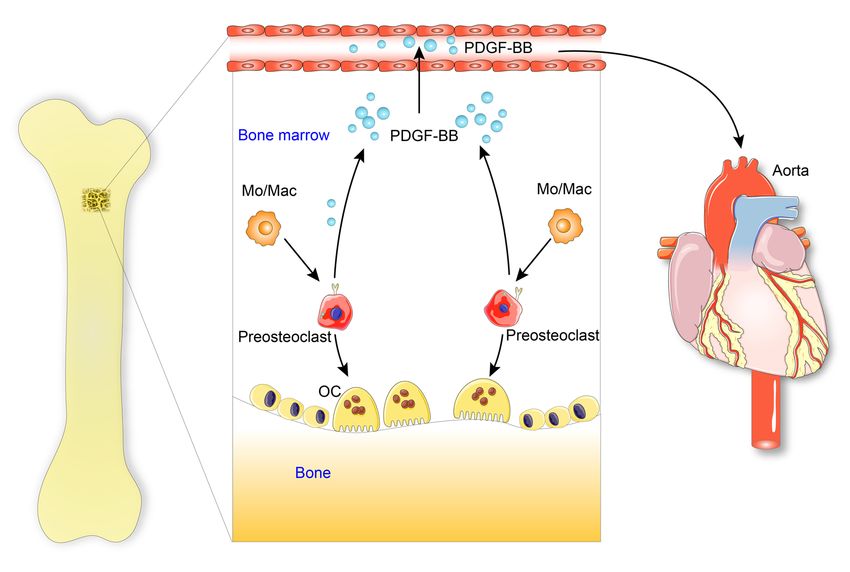
Figure 12. Schematic model illustrating the bone-vascular interplay during aging. With advancing age or under metabolic stress, preosteoclasts in
bone secrete a high amount of PDGF-BB, which infuses into blood circulation. Elevated circulating PDGF-BB, serving as a systemic progeronic factor,
drives arterial stiffening.
animal facility at our institution. At 10 to 12 weeks of age, mice were osteoblasts per bone perimeter (N.OB/B.Pm) and number of osteoclasts
placed on a Western HFD (21.2% fat by weight) (TD 88137, Harlan per bone perimeter (N.OC/B.Pm) in 5 randomly selected visual fields
Laboratories) or normal CHD for 8 to 20 weeks. per specimen, in 4 specimens per mouse in each group were measured.
Micro-CT and histomorphometric analyses of femoral bone. Mice Immunofluorescence staining of bone and aorta tissue sections. Femo-
were anesthetized by inhalation of 2.5% isoflurane (Abbott Labo- ra and aortas were dissected after mouse sacrifice and fixed in PBS (pH
ratories) mixed with O2 (1.5 L/min). For μCT analysis, mice femora 7.4) containing 4% paraformaldehyde for 48 hours. Femora were then
were dissected free of soft tissue, fixed overnight in 10% formalin decalcified in 0.5M EDTA (pH 7.4) with constant shaking for 8 days.
at 4°C, and analyzed by high-resolution μCT (Skyscan 1172, Bruker For dehydration, the decalcified bones and aortas were immersed in
MicroCT). The scanner was set at 65 kV, 153 μA, and a resolution of a solution of 20% sucrose and 2% polyvinylpyrrolidone for 24 hours.
9.0 μm/pixel. We used NRecon image reconstruction software, ver- The tissues were embedded in OCT, and 10 μm–thick longitudinal-
sion 1.6 (Bruker MicroCT), CTAn data analysis software, version 1.9 ly oriented sections of bone were collected for immunofluorescence
(Bruker MicroCT), and CTVol 3D model visualization software, ver- staining as previously described (86). Transverse and longitudinal
sion 2.0 (Bruker MicroCT) to analyze parameters of trabecular bone sections of aorta were also prepared. The tissue sections were incubat-
in the metaphysis. To perform 3D histomorphometric analysis of tra- ed with primary antibody to PDGF-BB (ab178409, Abcam, 1:50, poly-
becular bone, we selected the regions of interest from 1 mm below the clonal) or PDGFRβ (ab32570, Abcam, 1:100, monoclonal) followed by
distal epiphyseal growth plate and extended distally for proximally 2 fluorescence-conjugated secondary antibodies. Nuclei were counter-
mm. Trabecular bone was analyzed to determine trabecular BV/TV, stained with DAPI (Sigma-Aldrich). The sections were mounted with
Tb.Th, Tb.N, and Tb.Sp. Cortical morphometry was analyzed within a the ProLong Antifade Kit (Molecular Probes) and observed under a
600 μm long section at mid-diaphysis of the femur and included mea- Zeiss LSM 780 confocal microscope (Carl Zeiss AG).
surements of average thickness and cross-sectional area. Isolation of bone/bone marrow preosteoclasts and blood myeloid
For histomorphometric analysis, the femora were resected and cells. To isolated bone/bone marrow preosteoclasts, 2 approaches
fixed in 4% paraformaldehyde for 48 hours, decalcified in 0.5M EDTA were used. As the first approach, a mixture of bone and bone mar-
(pH 7.4) at 4°C, and embedded in paraffin. Five μm–thick longitudi- row cell suspensions was prepared from C57B/L6 mice as previously
nally oriented sections of bone were processed for OCN immunohis- described (86), with modifications. Briefly, the epiphysis was removed
tochemical staining (for osteoblast analysis), and TRAP staining (for from the distal femora and proximal tibia, and bone marrow was
osteoclast analysis). All sections were observed using an Olympus BX51 flushed. Moreover, the metaphyseal region of bone tissue was har-
microscope. Quantitative histomorphometry analyses were performed vested, crushed in ice-cold PBS with a mortar and pestle, and digested
as described previously (85) in a blinded fashion using OsteoMeasure with collagenase I (3 mg/mL), dispase I (4 mg/mL), and deoxyribonu-
Software (OsteoMetrics, Inc.). The sample area selected for calculation clease (1 U/mL) in PBS at 37°C for 30 minutes. The resultant single
was a 1 mm2 area within the metaphyseal trabecular bone. Number of bone cell suspensions were combined with the flushed bone marrow
J Clin Invest. 2021;131(20):e147116 https://doi.org/10.1172/JCI147116 17You can also read