The Selenium Yeast vs Selenium Methionine on Cell Viability, Selenoprotein Profile and Redox Status via JNK/ P38 Pathway in Porcine Mammary ...
←
→
Page content transcription
If your browser does not render page correctly, please read the page content below
ORIGINAL RESEARCH
                                                                                                                                                  published: 01 April 2022
                                                                                                                                          doi: 10.3389/fvets.2022.850935

                                             The Selenium Yeast vs Selenium
                                             Methionine on Cell Viability,
                                             Selenoprotein Profile and Redox
                                             Status via JNK/ P38 Pathway in
                                             Porcine Mammary Epithelial Cells
                                             Caichi Wu 1,2,3 , Chang Cui 1 , Xiaoyu Zheng 1 , Jun Wang 1 , Ziwei Ma 1 , Pengwei Zhu 1 ,
                                             Gang Lin 4 , Shihai Zhang 1,2,3 , Wutai Guan 1,2,3* and Fang Chen 1,2,3*
                                             1
                                               Guangdong Provincial Key Laboratory of Animal Nutrition Control, College of Animal Science, South China Agricultural
                                             University, Guangzhou, China, 2 College of Animal Science and National Engineering Research Center for Breeding Swine
                                             Industry, South China Agricultural University, Guangzhou, China, 3 Guangdong Laboratory for Lingnan Modern Agriculture,
                           Edited by:        South China Agricultural University, Guangzhou, China, 4 Key Laboratory of Agrifood Safety and Quality, Institute of Quality
                           Bing Dong,        Standards and Testing Technology for Agricultural Products, Chinese Academy of Agricultural Sciences, Ministry of
   China Agricultural University, China      Agriculture and Rural Affairs, Beijing, China
                       Reviewed by:
                                Shu Li,
                 Northeast Agricultural      Comprehensive studies have been conducted to compare the effect of organic and
                      University, China      inorganic selenium previously, but there is still limited knowledge about the difference
                            Binlin Shi,
            Inner Mongolia Agricultural
                                             between organic selenium (Se) from varied sources despite the widely use of organic Se
                      University, China      in both animal and human being nutrient additives. In the present study, we systemically
                 *Correspondence:            compared the effect of two different types of organic Se including selenium yeast (SeY)
                        Fang Chen
                                             and selenium methionine (Sel-Met) on cell viability, selenoprotein transcriptome, and
          chenfang1111@scau.edu.cn
                        Wutai Guan           antioxidant status in porcine mammary epithelial cells (PMECs) and the results indicated
                wtguan@scau.edu.cn           that appropriate addition of SeY and Sel-Met both significantly promoted cell viability
                                             and up-regulated the mRNA expression of most selenopreoteins including DIOs, GPXs,
                    Specialty section:
         This article was submitted to       and TrxRs family et al. (P < 0.05). Besides, two different sources of Se supplementation
     Animal Nutrition and Metabolism,        both greatly improved redox status with higher levels of T-AOC, SOD, and CAT (P <
                a section of the journal
                                             0.05), while less content of MDA (P < 0.05), and reduced protein expression of cleaved-
        Frontiers in Veterinary Science
                                             caspase-3 (P < 0.05) to mitigate cell apoptosis. Furthermore, the key proteins related
         Received: 08 January 2022
        Accepted: 14 February 2022           to p38/JNK pathway including p38, p-p38, JNK, and p-JNK were apparently reduced
           Published: 01 April 2022          in the groups with both of SeY and Sel-Met (P < 0.05). Interestingly we found that the
                              Citation:      changes induced by SeY supplementation in cell viability, selenoprotein transcriptome,
Wu C, Cui C, Zheng X, Wang J, Ma Z,
  Zhu P, Lin G, Zhang S, Guan W and
                                             antioxidative capacity, and anti-apoptosis were comprehensively greater compared with
Chen F (2022) The Selenium Yeast vs          same levels addition of Sel-Met in PEMCs (P < 0.05). In conclusion, both SeY and Sel-
 Selenium Methionine on Cell Viability,
                                             Met promoted cell viability and attenuated cell apoptosis by regulating the selenoprotein
     Selenoprotein Profile and Redox
      Status via JNK/ P38 Pathway in         expression and antioxidative capacity via p38/JNK signaling pathway in PMEC, but SeY
    Porcine Mammary Epithelial Cells.        has more efficient benefits than that of Sel-Met.
            Front. Vet. Sci. 9:850935.
     doi: 10.3389/fvets.2022.850935          Keywords: antioxidant, cell viability, porcine mammary epithelial cells, organic selenium, selenoprotein

Frontiers in Veterinary Science | www.frontiersin.org                                1                                               April 2022 | Volume 9 | Article 850935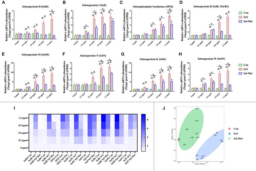
Wu et al.                                                                                                                Comparitive Study of Organic Selenium

INTRODUCTION                                                                          nowadays. This study therefore was conducted to compare the
                                                                                      effects of two different sources (SeY and Sel-Met) of organic
Lactating sows showed higher nutrition and energy demands                             Se using pig mammary gland epithelial cells (PMECs) as an in
due to the extensive metabolism in mammary glands to produce                          vitro model by evaluating cell viability, gene transcriptome of
milk, associated with adaption of a whole animal body system.                         selenoproteins, which are the main forms of Se in mammals,
It has been reported that 632 genes related to amino acids,                           and antioxidant status, with the purpose to provide theoretical
fatty acids, and glucose metabolism were differentially expressed                     foundations for organic Se application in lactating sow diets.
in livers during lactation (1). Increased metabolic burdens in
lactating sows result in great oxygen consumption, oxygen free
radicals, and lipid peroxides, leading to aggravation of oxidative
                                                                                      MATERIALS AND METHODS
stress (2, 3). Comprehensive studies have reported the damage                         Cell Culture
of mammary tissues caused by oxidative stress (4), and acute                          The PMECs used in this study were previously isolated and
redox imbalance induced by failed adaption of mammary glands                          characterized from mammary glands of lactating sows in our lab
during the transition from pregnancy to lactation was a main                          and were used to evaluate the synthesis and/or transport of amino
cause of mastitis accompanying impaired mammary functions                             acids, fatty acids, and lactose of sows in our previous studies (32–
(5). Further studies suggested that the damages of mammary                            36) as the following: the mammary gland tissue was cut into 1-
glands induced by severe oxidative stress negatively impacted                         mm3 fragments with sterile scissors and washed repeatedly with
offspring’s systemic redox balance and health, as well as boosted                     D-Hanks solution to wash away blood cells and other connective
inflammatory response through breast feeding (6). Therefore, it                       tissue. Then the tissue fragments were digested with collagenase
is of importance to relieve oxidative stress in mammary tissue                        II at 37◦ C and 5% CO2 for 1 h. The isolated cells were cultured
during lactation for sow health and production.                                       in DMED/F12 medium supplemented with 10% fetal bovine
    In recent decades, antioxidants either from nature or artificial                  serum, and the medium was changed every 24 h. Additionally,
synthesis have been widely accepted and used to improve animal                        fluorescence-activated cell sorting (FACS) analysis of cytokeratin
production. This beneficial effect is attributed to their capacity of                 expression was used to verify the purity of mammary epithelial
reactive oxygen species (ROS) detoxication to maintain dynamic                        cells at more than 90%. In the present study, isolated cells were
redox balance (7, 8). Se is a well-known component of many                            incubated at 37◦ C, 5% CO2 and cultured in a complete medium
enzymatic proteins participating in chemical reactions related                        according to the formula of Jaeger et al. (37), which consists
to ROS neutralization (9). It is also commonly considered as                          of Dulbecco’s Modified Eagle Medium/Nutrient Mixture F-12
an essential trace element for animal health (10–12) as it is                         (DMEM/F12, GIBCO), 10% fetal bovine serum (FBS, PAA),
involved in multiple biological functions such as reproduction                        1% antibiotic/antimycotic solution (10,000 U/mL penicillin, 10
(13), muscle metabolism (14–18), and immune response (19),                            mg/mL streptomycin sulfate, 25 µg/mL amphotericin B, GIBCO,
as well as antiaging (20). Se deficiency has been shown to                            I-15240), 10 µg/mL insulin (Sigma, I 6634), and 1 µg/mL
cause reduced animal health and performance especially during                         hydrocortisone (Sigma-Aldrich).
lactation in various animal species (21, 22). Instead, appropriate
Se supplementation resulted in increased milk production (15–                         Preparation of Selenium Compounds
17) and milk quality (10, 17, 23, 24).                                                The protease solution was prepared by adding 2 mg protease XIV
    Based on those study results, various forms of Se were                            (Sigma-Aldrich) into 0.5 mL 10 mM Tris-HCl (Sigma-Aldrich)
added in diets with the purpose to improve animal systemic                            buffer. Dissolve 40 mg selenium yeast (SeY, Nicholasville, KY) in
redox status and consequently enhance lactating performance.                          the prepared protease solution and mix well. The samples were
In general, Se was added in animal diets in two different                             ultrasonic 25 s with 80% amplitude on ice and cleaned with ultra-
forms, which are inorganic salts mainly represented as sodium                         pure water. The extraction power was 30 W and the samples were
selenite, and organic selenium mainly represented as selenium                         run for 15 min. The obtained sample was centrifuged at 14,000
methionine (Sel-Met) and selenium yeast (SeY). Comprehensive                          rpm for 3 min, and the supernatant was taken. The precipitation
studies have been conducted to evaluate the relative efficiency                       was cleaned and mixed with ultrapure water until resuspension,
of different types of Se on animal performance and achieved a                         and the supernatant was centrifuged again at 14,000 rpm for
common understanding that organic Se has an overall advantage                         3 min to obtain the supernatant. The concentration of selenium
in absorption rate (25), antioxidant capacity (10, 26–28), low                        in the sample was determined by inductively coupled plasma
toxicity (29), and animal health and performance improvement                          mass spectrometry (ICP-MS) (38).
(30, 31). To date, the main gap in knowledge regarding the                               A total of 12 mg of Sel-Met were dissolved in 10 L DMED/F12
Se nutritional regulation is the comparison of different sources                      to prepare 1.2 ppm stock solution. The original solution was
of organic and underlying mechanisms considering their wide                           added to 5, 3.75, 2.5, 1.25, and 0 mL volumes of DMED/F12 to
application in both human being and animal feeding additives                          prepare the working solution at concentrations of 0, 0.3, 0.6, 0.9,
                                                                                      and 1.2 ppm, respectively.
Abbreviations: Se, Selenium; SeY, Selenium yeast; Sel-Met, Selenium methionine;
PMECs, Porcine mammary epithelial cells; TRX, Thioredoxin reductase;
                                                                                      Cell Viability Assay
GPX, Glutathione peroxidases; TXNRD, Thioredoxin reductase family; DIOs,              Cell viability was tested using the CCK-8 assay (Dojindo, Japan)
Deiodinase family; JNK, Jun-n-kinase; MAPKs, Mitogen-activated protein kinases.       according to the manufacturer’s instruction. Briefly, PMECs

Frontiers in Veterinary Science | www.frontiersin.org                             2                                       April 2022 | Volume 9 | Article 850935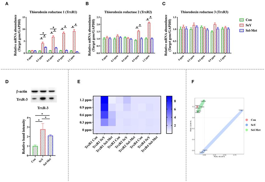
Wu et al.                                                                                                                      Comparitive Study of Organic Selenium

TABLE 1 | Primers used for RT-PCR.                                                           RNA Isolation and Quantitative Real-Time
Gene          Accession number           Primer pairs (5’ to 3’ direction)
                                                                                             PCR
                                                                                             PMECs were seeded into 6-well plates at 2 mL/well with 5×10⁴
DIO1          AY533206                   F: CATGGCCAAGAACCCTCACT                             cells/mL and were cultured in complete medium at 37◦ C,
                                         R: CCAGAAATACTGGGCACTGAAGA                          5% CO2 for 48 h. Then, the cells were treated with various
DIO2          AY533207                   F: CGCTGCATCTGGAAGAGCTT                             levels of SeY and Sel-Met (0, 0.3, 0.6, 0.9, or 1.2 ppm) for
                                         R: TGGAATTGGGTGCATCTTCA                             24 h. After that, total RNA was extracted from PMECs using
DIO3          AY533208                   F: TGAAGTGGAGCTCAACAGTGATG
                                                                                             TRIZOL (Invitrogen catalog no. 15596–026), according to the
                                         R: TGTCGTCAGACACGCAGATAGG
                                                                                             manufacturer’s instructions. The quality and quantity of RNA
                                                                                             were analyzed by Agilent Bioanalyzer 2100 using an RNA 6000
GPX1          AF532927                   F: GATGCCACTGCCCTCATGA
                                                                                             Labchip kit. Potential DNA contamination of the extraction
                                         R: TCGAAGTTCCATGCGATGTC
                                                                                             was eliminated using the DNA-free kit (Ambion, catalog no.
GPX2          DQ898282                   F: AGAATGTGGCCTCGCTCTGA
                                                                                             AM1906), and the RNA quality was verified by both agarose
                                         R: GGCATTGCAGCTCGTTGAG
                                                                                             gel (1%) electrophoresis, and spectrometry (A260/A280). First-
GPX4          NM_214407                  F: TGAGGCAAGACGGAGGTAAACT                           strand cDNA synthesis was performed by using a Prime Script
                                         R: TCCGTAAACCACACTCAGCATATC                         RT reagent kit with gDNA eraser (Takara, Dalian, China).
GPX6          NM_001137607               F: GAGCTGAAGCCTTTTGGTGTAGTT                         The cDNA was synthesized from 1 µg total RNA using Super
                                         R: CTTTGCTGGTTCTTGTTTTCCA                           Script III reverse transcriptase according to the manufacturer’s
SELH          HM018602                   F: TGGTGGAGGAGCTGAAGAAGTAC                          instructions. The mRNA levels of 25 selenoprotein genes were
                                         R: CGTCATAAATGCTCCAACATCAC                          analyzed by qPCR using the SYBR Green PCR Master Mix
SELR          NT_033777.3                F: GAACCACTTTGAGCCAGGTGTCTAC                        according to the manufacturer’s instructions (Cat# RR047A,
                                         R: GCCTTTAGGGATGAACTTCAGGGAAC                       TakaRa). Primers for the 25 selenoprotein genes were referenced
SELW          NM_213977                  F: CACCCCTGTCTCCCTGCAT                              from the study of Zhao et al. (39) (Table 1), and primer for the β-
                                         R: GAGCAGGATCACCCCAAACA
                                                                                             actin gene (Actb) was from our previous study (40). The 2−11Ct
                                                                                             method (41) was used for the quantification with the β-actin gene
SPS2          BM489698.1                 F: CGTTGGGTATCGGAACTGAC
                                                                                             as a reference gene, and the relative abundance was normalized to
                                         R: CGTCCACCAGAGGGTAGAAA
                                                                                             the control.
SELI          EST                        F: GATGGTGTGGATGGAAAGCAA
                                         R: GCCATGGTCAAAGAGTTCTCCTA                          Western Blot Analysis
SELK          DQ372075                   F: CAGGAAACCCCCCTAGAAGAA                            PMECs were seeded into 6-well plates at 2 mL/well with 5×10⁴
                                         R: CTCATCCACCGGCCATTG                               cells/mL and were cultured in complete medium at 37◦ C 5% CO2
SELM          FJ968780                   F: CAGCTGAATCGCCTCAAAGAG                            for 48 h. Then, the cells were treated with 0.6 ppm SeY and Sel-
                                         R: GAGATGTTTCATGACCAGGTTGTG                         Met for 24 h. After that, cells were collected and homogenized
SELN          EF113595                   F: ACCTGGTCCCTGGTGAAAGAG                            in RIPA lysis buffer (Beyotime, Nanjing, China). The Western
                                         R: AGGCCAGCCAGCTTCTTGT                              blot analysis was done according to the procedures described
TXNRD1        AF537300                   F: GATTTAACAAGCGGGTCATGGT                           in our previous study (33). In general, the same number of
                                         R: CAACCTACATTCACACACGTTCCT                         samples were electrophoretic ally separated and transferred to
TXNRD2        GU181287                   F: TCTTGAAAGGCGGAAAAGAGAT
                                                                                             PVDF membrane, which was sealed at room temperature with
                                                                                             5% skim milk for 2 h. The primary antibody of the target protein
                                         R: TCGGTCGCCCTCCAGTAG
                                                                                             was added and incubated at 4◦ C for 12 h, then the secondary
TXNRD3        BX918808                   F: GTGCCCTACGTTTATGCTGTTG
                                                                                             antibody was incubated for 1.5 h, and the chemiluminescence
                                         R: TCCGAGCCACCAGCTTTG
                                                                                             reaction was carried out. The primary antibody was diluted
ACTB, beta actin; DIO, iodothyronine deiodinase; GPX, glutathione peroxidase; SELH, I,       according to the instructions: GPX1 antibody (1:1000, 3206,
K, M, selenoproteins H, I, K, M; SEPHS2 (SPS2), selenophosphate synthetase 2; SELW,          Cell Signaling Technology, United States), TrxR3 antibody
selenoprotein W; TXNRD, thioredoxin reductase.
                                                                                             (1:1000, 19517-1-AP, Proteintech, United States), JNK (1:1000,
                                                                                             66210-1-lg, Proteintech, United States), P-JNK (1:1000, 80024-
                                                                                             1-RR, Proteintech, United States), Bax (1:1000, 50059-2-
                                                                                             lg, Proteintech, United States), Bcl-2 (1:2000, 60178-1-lg,
were seeded into 96-well microplates at 200 µL/well with                                     Proteintech, United States), P-p38 (1:1000, 4511, Cell Signaling
2×10⁴ cells/mL and were cultured in complete medium at                                       Technology, United States), p38 (1:1000, 8690, Cell Signaling
37◦ C 5% CO2 for 48 h. Then, selenium yeast (SeY; Alltech Inc,                               Technology, United States), Caspase 3 (1:1000, 9662, Cell
Nicholasville, KY) and selenium methionine (Sel-Met, sigma,                                  Signaling Technology, United States), and β-actin (1:2000, bs-
3211-76-5, United States) were successively added to the 96-well                             0061R, Bioss, China).
plate at concentration gradients of 0, 0.3, 0.6, 0.9, and 1.2 ppm,
respectively. At 24 h post-treatment 20 µL, CCK-8 was added to                               Antioxidant Enzymes Assay
each well, incubated for 4 h at 37◦ C, and then measured using a                             PMECs were seeded into 6-well plates at 2 mL/well with 5 ×
microplate reader at the wavelength of 450 nm.                                               104 cells/mL and were cultured in complete medium at 37◦ C

Frontiers in Veterinary Science | www.frontiersin.org                                    3                                       April 2022 | Volume 9 | Article 850935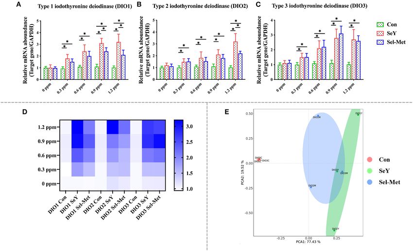
Wu et al. Comparitive Study of Organic Selenium
5% CO2 for 48 h. Then, the cells were treated with 0.6 ppm group were significantly higher than those in the control group
SeY and Sel-Met for 24 h. The collected cells were cleaned with (Figures 3A–D) (P < 0.05). For Sel-Met group, the GPX1 mRNA
PBS for two times and centrifuged to retain cell precipitation. expression level of different supplemental levels was higher than
The cells were suspended with 0.5 mL isotonic PBS buffer, and that of the control group (P < 0.05), but lower than that of the
the broken cells were ground in the grinding machine to obtain SeY group (P < 0.05). The mRNA expression levels of GPX2 and
PMECs cell suspension, which was used to detect the content of GPX4 were similar in the Sel-Met group and the SeY group at
superoxide dismutase (SOD), malondialdehyde (MDA), catalase 0.3 and 0.6 ppm. In particular, 1.2 ppm Sel-Met inhibited GPX4
(CAT), and the total antioxidant capacity (T-AOC) of cells. The expression in PMECs (P < 0.05). At 0.6 ppm, GPX6 mRNA level
antioxidant capacity of cells was determined using the kit of in the Sel-Met group was higher than that in the control group,
Nanjing JianCheng Institute of Biological Engineering, and the except that the mRNA expression levels of other supplemental
detection method was according to the instructions of the kit. levels were the same as that in the control group (P < 0.05).
Combined with heat map (Figure 3F) observation, it can be
Statistical Analysis concluded that the SeY group has a much better promotional
The data of CCK-8, PCR, Western blot, and antioxidant status effect on the mRNA expression level of the GPX family in PMECs
were analyzed by one-way ANOVA, LSD multiple comparison, than the Sel-Met group. By PCA figure can be seen that the SeY
and Pearson correlation analysis using SPSS 22.0 software in the and Sel-Met groups have obvious distinctions (Figure 3G) that
present study. The establishment of PCA and heatmap models both in different addition amounts of GPXs mRNA expression of
was performed on the Tutools platform (https://www.cloudtutu. family has a significant difference. This result was also verified by
com). The P-value < 0.05 was used as the criterion to judge the Western blot assay (Figure 3E) (P < 0.05).
significance of the difference. Observe Figure 4, which depicts mRNA and protein
expression of the thioredoxin reductase family (TrxR). The
RESULTS addition of SeY and Sel-Met at different concentrations had
no effect on TrxR3 mRNA expression (P > 0.05), but TrxR1
Appropriate Se Supplementation Promoted and TrxR2 mRNA expression were different. TrxR1 mRNA
PMEC Cell Viability expression in SeY groups was significantly higher than that in the
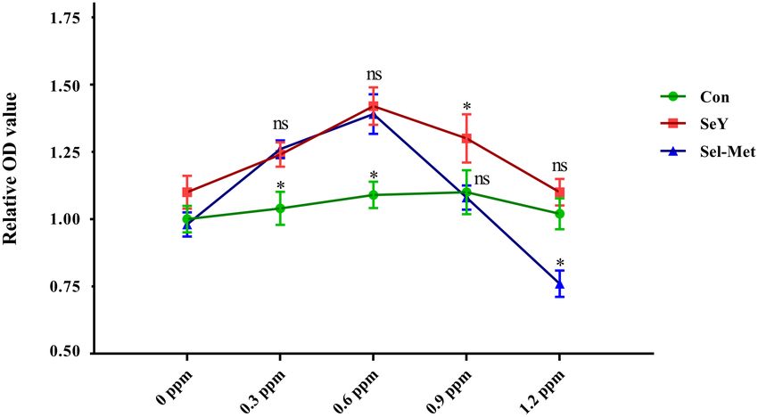
As shown in Figure 1, compared with the control group, both 0.3 control group (P < 0.05). Sel-Met significantly increased TrxR1
and 0.6 ppm SeY and Sel-Met resulted in higher cell viability (P < mRNA expression at 0.3 and 0.6 ppm, while it was similar to
0.05). At the increased concentrations of 0.9 ppm SeY still led to the control group at 0.9 and 1.2 ppm. TrxR2 mRNA expression
greater cell viability, but Sel-Met had no affect (P > 0.05), while was significantly increased in 0.9 and 1.2 ppm SeY groups (P
at 1.2 ppm SeY was not different from the control and Sel-Met < 0.05), while there was no significant change in the Sel-Met
treated cells had lower viability (P < 0.05). group (Figures 4A–C). In the PCA diagram, Sel-Met and SeY
groups did not overlap at all, and the above results indicated
Se Supplementation Altered Selenoprotein that there was a significant difference in addition effect between
Gene Profile of PMECs the two groups (Figure 4F). The heat map clearly shows the
Figure 2 describes the mRNA expression of DIOs family in addition effect of SeY, which is significantly better than Sel-Met
PMECs cells after SeY and Sel-Met treatment. The gene (Figure 4E). Western blot detection showed that TxrR3 protein
expression of DIO1, DIO2, and DIO3 mRNA were significantly expression in both Se supplemental groups was better than that
up-regulated in 0.3 and 0.6 ppm of two different sources of in the control group (Figure 4D), but the effect of SeY addition
organic Se treated cells (Figures 2A–C) (P < 0.05), and there was more significant (P < 0.05).
was no significant difference between SeY and Sel-Met. When As presented in Figure 5, the mRNA expression of SeIH
Se concentration was increased up to 0.9 and 1.2 ppm, the was greater with all SeY concentration levels compared with
supplementation of both forms of Se lead to higher mRNA control. Increasing concentrations of SeY from 0.3 to 1.2 ppm
expression of DIO1, DIO2, and DIO3 (P < 0.05), but SeY groups significantly and consistently elevated the mRNA expression of
showed higher mRNA expression levels of DIO1 and DIO2 SeIR, SeIW, and SPS2 (Figures 5C,D,H) (P < 0.05). However,
compared with those with Sel-Met at the same concentration Sel-Met did not affect the mRNA expression of SeIH (Figure 5A)
(P < 0.05). The PCA (Figure 2E) diagram can represent the (P > 0.05) from 0.3 to 0.9 ppm, and lowered (P < 0.05)
difference between different groups found. The farther the spatial the mRNA expression of SeIH when added at 1.2 ppm. The
distance between different samples is, the greater the data mRNA expression of SelR, SelW, and SPS2 increased significantly
difference between them is. The non-overlapping area between after adding 0.6 and 0.9 ppm Sel-Met (Figures 5C,D,H) (P <
SeY group and Sel-Met group was larger than the overlapping 0.05). At 1.2 ppm, Sel-Met increased the mRNA expression
area, indicating that there was a significant difference in the of SelW and SPS2 (P < 0.05) but had no effect on SelR
addition effect of the two groups on the mRNA expression of (P > 0.05). At the same amount of addition, the expression
DIOs family. Taken together, these data suggest that SeY is more of SelH, SelR, SelW, SPS2 in Sel-Met cells was the same as
effective in increasing DIOs expression than addition of Sel-Met. that of SeY or lower than that of SeY (P < 0.05). With the
Figure 3 illustrates the changes of GPX family gene and increase of SeY and Sel-Met levels from 0.3 to 1.2 ppm, the
protein expression after SeY and Sel-Met treatment. The mRNA mRNA expression levels of Sell, SelK, and SelM were higher
expression levels of GPX1, GPX2, GPX4, and GPX6 in SeY than those in the control group (Figures 5B,E,G). SelN mRNA
Frontiers in Veterinary Science | www.frontiersin.org 4 April 2022 | Volume 9 | Article 850935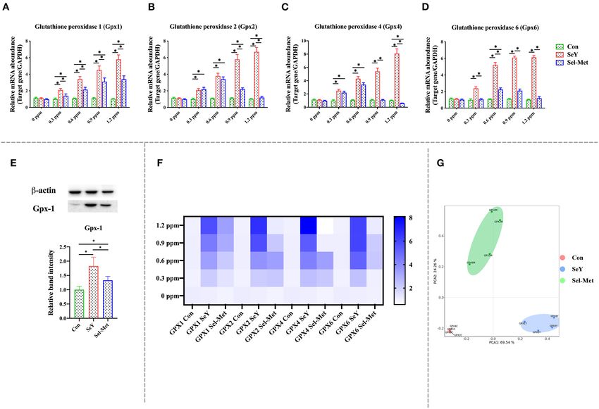
Wu et al.                                                                                                                       Comparitive Study of Organic Selenium

 FIGURE 1 | Effects of SeY and Sel-Met supplementation on cell viability in PMECs. Cells were incubated with different concentrations of SeY and Sel-Met (0, 0.3, 0.6,
 0.9, and 1.2 ppm) for 24 h, respectively. Cell viability was analyzed using the CCK-8 assay. Data are expressed as mean ± SEM (n = 12). Superscript * indicates
 significant (P < 0.05).

 FIGURE 2 | Supplement SeY and Sel-Met to mRNA expression of DIOs family in PMECs. The cells were incubated with SeY and Sel-Met at different concentrations
 (0, 0.3, 0.6, 0.9, and 1.2 ppm) for 24 h, respectively, and then collected to detect mRNA expression. Data were expressed as mean ± SEM (n = 6), and different
 superscripts * indicate statistically significant differences (P < 0.05). (A–C) Are the mRNA expression level of DIOs detected by qPCR. (D) Heat map comparison of
 DIOs mRNA levels. (E) PCA score plot results compared the mRNA expression of the DIOs family among the three groups.

Frontiers in Veterinary Science | www.frontiersin.org                             5                                               April 2022 | Volume 9 | Article 850935
Wu et al.                                                                                                                           Comparitive Study of Organic Selenium

 FIGURE 3 | Effects of SeY and Sel-Met addition on the mRNA expression of glutathione peroxidase family in PMECs. The cells were incubated with different
 concentrations of SeY and Sel-Met (0, 0.3, 0.6, 0.9, and 1.2 ppm) for 24 h, respectively, and then collected for detection. Data were expressed as mean ± SEM (n =
 6), and * indicated statistically significant difference (P < 0.05). (A–D) mRNA expression levels of GPXs family were detected by qPCR. (E) The relative protein levels of
 GPX1 were analyzed by Western blotting. (F) Heat map comparison of mRNA expression levels of GPXs. (G) PCA score plot results compared the mRNA expression
 of GPXs between the three groups.

expression increased with the increase of concentration except                           compared with the control. Furthermore, the MDA levels
0.3 ppm (Figure 5F) (P < 0.05). The expression of SelN at 0.9                            are significantly higher (P < 0.05) with Sel-Met compared
and 1.2 ppm was significantly higher than that in the control                            to SeY.
group. Except for the same expression of Sell at 1.2 ppm, the
effect of the SeY group was better than the Sel-Met group (P
< 0.05). In the PCA diagram (Figure 5J), the Sel-Met group                               JNK/p38 Pathway Was Inhibited by
overlapped with the control group and was clearly distinguished                          Supplementation of Se
from the SeY group. As can be seen, the difference between                               Figure 7 showed that the ratio of p-JNK/JNK and p-p38/p38,
the Sel-Met group and the control group was small, but there                             and the abundance of cleaved-caspase3 were significantly lower
was significant difference between the Sel-Met group and the                             in the SeY group and Sel-Met groups compared to the control,
SeY group.                                                                               with SeY being lower (P < 0.05) than Sel-Met. The abundance
                                                                                         of Bcl-2 was significantly higher (P < 0.05) with both Se sources
Se Supplementation Promoted Cellular                                                     compared to the control, with SeY being higher (P < 0.05) than
Antioxidative Capacity                                                                   Sel-Met.
As can be seen in Figure 6, it is clear that T-AOC, SOD,                                    To further explore the interaction between selenoproteins,
and CAT levels were significantly higher with either Se source                           correlation analysis was conducted. As shown in Figure 8,
(P < 0.05) and compared with Sel-Met, the groups with SeY                                the expressions of various selenoproteins except TrxR3 were
addition had significantly higher (P < 0.05) T-AOC, SOD, and                             obviously positively correlated (Figure 8A). There was a negative
CAT. In contrast, the MDA levels are significantly lower (P                              correlation between apotheosis index and antioxidant index
< 0.05) for both groups supplemented with selenium addition                              (Figure 8B).

Frontiers in Veterinary Science | www.frontiersin.org                                6                                                April 2022 | Volume 9 | Article 850935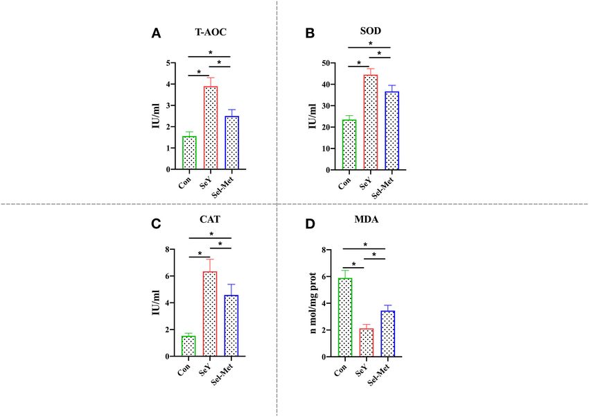
Wu et al.                                                                                                                     Comparitive Study of Organic Selenium

 FIGURE 4 | Effects of SeY and Sel-Met addition on mRNA expression of TrxRs family in pMECs. The cells were incubated with different concentrations of SeY and
 Sel-Met (0, 0.3, 0.6, 0.9, and 1.2 ppm) for 24 h, respectively, and then collected for detection. Data were expressed as mean ± SEM (n = 6), and * indicated
 statistically significant difference (P < 0.05). (A–C) mRNA expression level of TrxRs family was detected by qPCR. (D) The relative protein levels of TrxR3 were
 analyzed by Western blotting. (E) Heat map comparison of TrxRs mRNA expression levels. (F) PCA score plot results compared the mRNA expression of the DIOs
 family among the three groups.

DISCUSSION                                                                           SeY having no effect on cell viability in the 1.2 ppm range, while
                                                                                     Sel-Met began to show inhibition. In previous studies, organic Se
Se has been shown to have beneficial effects in cell proliferation                   has been shown less toxic due to its higher retention in animal
and anti-apoptosis process due to its antioxidative properties in                    tissues and lower concentration levels in plasma compared with
previous studies (42). Zeng reported that Se deficiency resulted                     inorganic Se (47), but there is a lack of data regarding negative
in a decreased number of G1 phase cells that corresponded to                         effect of too much excessive organic Se and comparison of toxic
increased numbers of G2 and sub-G1 phase cells, while 0.25                           effects of different sources of organic Se. The result of the present
µmol/L Sel-Met addition significantly enhanced the expression                        study suggested that excessive Sel-Met addition might be toxic
of cell cycle-related genes and reversed this cell growth trend                      to cells and potentially induce cellular damage. The absence of
(43). In addition, several sources of Se including Na2 SeO3 , Sel-                   impaired cell growth caused by high concentration of SeY in the
Met, and selenite containing Se compounds all showed positive                        current study implied that cells probably have higher tolerance to
effects on cell viability in various cells including human immature                  SeY than Sel-Met at the super nutritional level, which mechanism
dendritic cells, human lens epithelial cells (44), chondrocyte                       should be paid more attention to in the future.
ATDC5 cells (42), fibroblast HT1080 cells, and primary porcine                           Se acts as an antioxidant regulating cellular redox balance
macrophages cells (45, 46). Thus, it is not unexpected that                          mainly in the form of selenoprotein including the glutathione
reasonable levels of both SeY and Sel-Met supplementation                            peroxidase family (GPXs), thioredoxin reductase family (TrxR),
improved the cell viability of PMECs in the present study                            and deiodinase family (DIOs), as well as SelH, SelI, SelM, SelK,
(Figure 1). Interestingly, we found inconsistent trends between                      etc. (48, 49). The GPX family is a group of antioxidant enzymes
the two groups of cells when treated with 0.9 and 1.2 ppm, with                      with Se as the active component to decompose peroxides into

Frontiers in Veterinary Science | www.frontiersin.org                            7                                              April 2022 | Volume 9 | Article 850935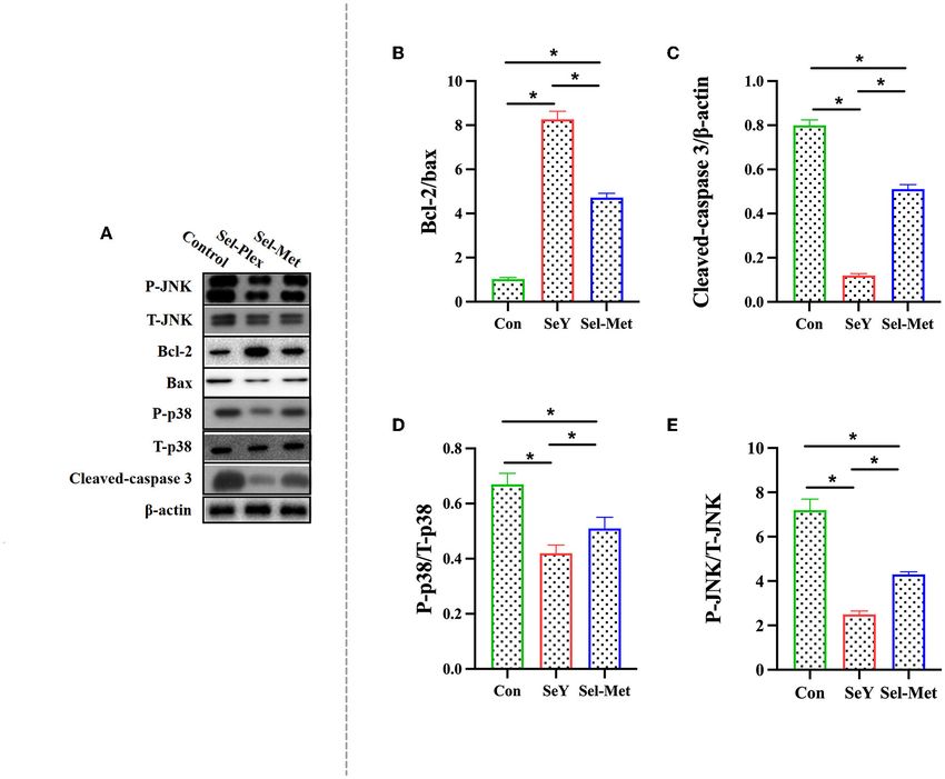
Wu et al.                                                                                                                        Comparitive Study of Organic Selenium

 FIGURE 5 | Effects of SeY and Sel-Met addition on mRNA expression of SelH, Sell, SPS2, SelM, SelN, SelK, SelR, and SelW in PMECs. The cells were incubated
 with different concentrations of SeY and SEL-Met (0, 0.3, 0.6, 0.9, and 1.2 ppm) for 24 h, respectively, and then collected for detection. Data were expressed as mean
 ± SEM (n = 6), and * indicated statistically significant difference (P < 0.05). (A–H) qPCR was used to detect the mRNA expression levels of SelH, Sell, and other
 selenium proteins. (I) Heat map comparison of mRNA expression levels of SelH, Sell, and eight other selenoproteins. (J) PCA score plot results the mRNA expression
 levels of SelH, Sell, SPS2, SelM, SelN, SelK, SelR, and SelW were compared among the three groups.

non-toxic hydroxyl compounds thus preventing peroxide from                             SELH, SELN, SELP, and SELW in ATDC5 cells, and GPX1,
peroxidation of the cell membrane lipids (50–52). In cells, ROS                        SELH, SELN, SELP, SELW, and GPX3 in C28/I2 cells, while
could be neutralized by glutathione under catalyzation of GPX                          decreased the mRNA abundance of SPS2 and SELO in ATDC5
to generate oxidized glutathione (GSSH), and then GR catalyzes                         cells, and SPS2, SELO, TRXR2 in C28/I2 cells (58). Stolwijk
the regeneration of GSH from GSSH, which recycle of GSH                                et al. (59) showed that the activities of GPX1 and GPX4 were
producing were defined as glutathione system playing important                         significantly up-regulated in the exponentially growing cells
roles in cellular redox maintenance. Similar to the glutathione                        cultured in the medium supplemented with 200 nM Seleno-L-
system, thioredoxin (Trx) and TrxR make up the thioredoxin                             methionine. Similarly, Doroshow et al. (60) also found that 30 nM
system and were involved in multiple ROS scavenging processes                          sodium selenite addition to the cell culture medium significantly
through reversible Trx oxidation/reduction reaction catalyzed by                       increased the activity of GPX using NCI/ADR-RES cancer cells
TrxR (53, 54). Current research mainly focuses on TrxR1 and                            as an in vitro model. In addition, dietary Se supplementation also
TrxR2, both of which play a key role in protecting cells from                          has been reported to upregulate selenoprotein transcriptome in
oxidative stress damage, while TrxR3 does not play a significant                       chicken embryonic neurons, liver, and muscle (61). Furthermore,
role in antioxidant effects. In studies on antioxidants, most of the                   Se deficiency disease has been shown to be related to decreased
supplements had an effect on the mRNA expression of TrxR1                              mRNA expression of several selenoprotein genes (GPX1, GPX4,
and TrxR2, but had no significant change in the expression of                          SEPW1, SEPN1, SEPP1, SELO, and SELK) in liver and muscle
TrxR3 (55, 56). It was well-established that Se status within cells                    (62). Dietary Se deficiency also has been reported to significantly
and tissues is a key regulator of selenoproteins expression both                       reduce the mRNA expression levels of DIO, GPX3, and TXNRD2
in vivo and in vitro (57). For instance, Se supplementation in                         in the muscle of broilers (63). In the present study, we compared
cell medium greatly increased the mRNA abundance of GPX1,                              the effects of two different organic Se to gene expression of

Frontiers in Veterinary Science | www.frontiersin.org                              8                                              April 2022 | Volume 9 | Article 850935
Wu et al.                                                                                                                    Comparitive Study of Organic Selenium

 FIGURE 6 | Effects of SeY and Sel-Met supplementation on (A) T-AOC, (B) SOD, (C) CAT, and (D) MAD in PMECs. Cells were incubated for 24 h with 0.6 ppm SeY
 and Sel-Met, and then were collected for T-AOC, SOD, CAT, and MAD activity analysis. Data are expressed as mean ± SEM (n = 6). *Indicates that the difference was
 statistically significant (P < 0.05).

selenoproteins and results showed that both SeY and Sel-Met                            It is well-known that Se can repair damage caused by
addition in cell medium increased gene expression of most                           oxidative stress and promote cellular antioxidant capacity (68).
selenoproteins including GPX1, GPX2, GPX4, GPX6, and TrxR1,                         Liu et al. showed that dietary selenium-enriched yeast apparently
TrxR2, which is consistent with previous findings. In addition,                     promoted piglet antioxidant status and attenuated oxidative
in terms of protein expression, the addition of two selenium                        stress-induced growth retardation (69). An investigation
sources significantly increased the protein expression of GPX1                      conducted on chickens also reported that Se administration
and TrxR3. We found that SeY had a better beneficial effect than                    significantly relieved oxidative stress and testicular toxicity
Sel-Met at the same level of addition, combining gene and protein                   induced by lead exposure, associated with increased activity of
experimental results. To our best knowledge, this result is the first               antioxidant enzymes, and reduced inflammatory response (70).
time to show the different impact of different sources of organic                   To further compare the antioxidant effect of various sources
Se on global selenopreoteins using an in vitro model.                               of Se on PMECs, we tested the contents of SOD, MDA, CAT,
    SeY, not as Sel-Met, is a complex of multiple macronutrients                    and T-AOC, which are key makers (T-AOC, SOD, CAT, and
including Sel-Met as the main component, unknown forms of                           MDA) of antioxidant status in PMCEs cells. It is clear that both
organic Se, and other bioactive elements originally from yeast.                     organic Se have a positive effect on the antioxidant status of
Accumulating evidence showed that selenium could interact                           PMECs with increased T-AOC, SOD, and CAT, while decreased
with kinds of elements such as vitamin C (64), zinc (65),                           MDA, which result is consistent with previous findings that Se
cadmium (66) and others to participate in redox status regulation                   improved levels of antioxidant markers and ameliorated the
and the synergism of multiple components resulted in higher                         damage caused by LPS-induced in bovine mammary epithelial
efficiency than single one element. Salah et al. reported that co-                  cells (71). Similar to the gene expression of selenoproteins, we
treatment of Se and zinc more efficiently attenuated the oxidative                  also observed higher antioxidative capacity of SeY compared
stress induced by NiCl2 in pregnant Wistar rats than Se/Zinc                        with the same supplementation of Sel-Met, which result might
supplementation (67). Chen et al. assessed the effect of vitamin                    be attributed to the synergistic benefit.
E, Se, and co-treatment of vitamin E and Se on sow performance                         Oxidative stress is a result of imbalance between accumulation
and they also overserved synergistic effect of vitamin E and Se                     of ROS and the ability to get rid of it in cells and tissues
(23). We did not find much literature related to Se synergism with                  (72). Severe oxidant stress induced by excessive ROS has been
other substrates using animal models due to the limit in this field,                shown to trigger cell apoptosis associated with damaged DNA,
but we presume that the synergistic effect of multiple components                   oxidation of polyunsaturated fatty acids and amino acids, as
in SeY might make the main contribution to its higher efficiency                    well as mitochondrial dysfunction (73). Previous studies reported
in the present study.                                                               that Se status in cells and tissues plays a key role in the

Frontiers in Veterinary Science | www.frontiersin.org                           9                                             April 2022 | Volume 9 | Article 850935
Wu et al. Comparitive Study of Organic Selenium FIGURE 7 | Effects of SeY and Sel-Met supplementation on (A) is the original band of Western Blot, (B) Bcl-2/Bax, (C) Caspase-3, (D) P-p38/T-p38, and (E) P-JNK/T-JNK protein expression in PMECs. Cells were incubated for 24 h with 0.6 ppm SeY and Sel-Met and then were collected for the determination of protein expression. Data are expressed as mean ± SEM (n = 3). *Indicates that the difference was statistically significant (P < 0.05). regulation of apoptosis process and dietary supplementation of mitogen-activated protein kinases (MAPKs) are involved in Se might be a potential strategy to promote animal health by the mediation of cellular apoptosis induced by oxidative stress reducing apoptosis induced by oxidant stress (74). Wang et al. (77). Yeo et al. found that sodium selenite exerted a profound demonstrated that Se deficiency in cell medium lead to increased preventive effect on cell apoptosis via inhibition of p38 mitogen- intracellular ROS content and activated apoptotic process via activated protein kinase, pSAPK/JNK, and Bax activation in in caspase-3 signal pathway in human uterine smooth muscle vivo and in vitro rat spinal cord injury models (78). A study cells (75). On the other hand, Se supplementation significantly conducted by Wang et al. indicated that Se deficiency induced attenuated the damage and apoptosis caused by bisphenol Se in cell apoptosis by increasing gene expression of p-P38 and p-JNK mice (76). In cells, once apoptosis was initiated, active caspase- in human uterine smooth muscle cells (75). In the present study, 8 directly cleaves pro-caspase-3 and triggers downstream events similarly we also observed the changed expression of key protein of apoptosis signaling. Here we found that both SeY and Sel- in p38/JNK signaling pathway among those cells with different Met supplementation significantly reduced protein expression treatment implying its possible involvement in the modulation of cleaved caspase-3 implying that Se addition protected cells of Se on PMECs apoptotic process. The lower ratio of p-p38/p38 from apoptosis, which results were further confirmed by and p-JNK/JNK in the groups treated with SeY compared with decreased ratio of Bax/Bcl-2, another most used parameter for Sel-Met was consistent with other findings in the current study cellular apoptosis. that SeY has more efficient effect to prevent cells from oxidant A growing body of literature has shown that p38 and stress and apoptotic damage due to syngenetic benefit of multiple c-Jun-N-terminal kinase (JNK), both of which belong to elements included in SeY. Frontiers in Veterinary Science | www.frontiersin.org 10 April 2022 | Volume 9 | Article 850935
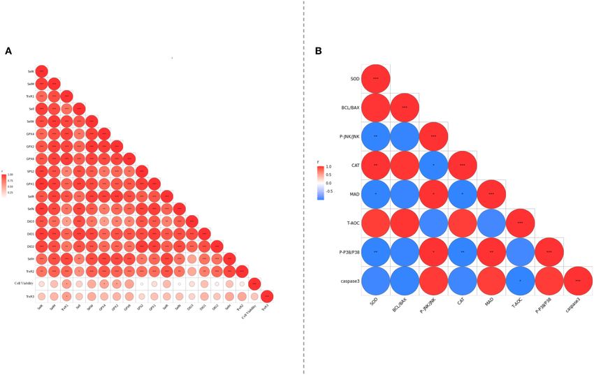
Wu et al.                                                                                                                        Comparitive Study of Organic Selenium

 FIGURE 8 | (A) Analyzed the correlation between selenoprotein mRNA expression levels of PMCES cultured with different additives and different dosage. (B) The
 degree of correlation between PMCEs related indicators of antioxidant and apoptosis was analyzed. The area size and color of the circle in the figure represent the
 correlation, *0.01 < P
Wu et al.                                                                                                                              Comparitive Study of Organic Selenium

 3. Berchieri-Ronchi CB, Kim SW, Zhao Y, Correa CR, Yeum KJ, Ferreira AL.                  21. Nogales F, Ojeda ML, Fenutria M, Murillo ML, Carreras O. Role of selenium
    Oxidative stress status of highly prolific sows during gestation and lactation.            and glutathione peroxidase on development, growth, and oxidative balance in
    Animal. (2011) 5:1774–9. doi: 10.1017/S1751731111000772                                    rat offspring. Reproduction. (2013) 146:659–67. doi: 10.1530/REP-13-0267
 4. Liu K. Changes in oxidative stress status in sows from days                            22. Barnett MP, Bermingham EN, Young W, Bassett SA, Hesketh JE, Maciel-
    100 of gestation to post-partum estrus. Pak Vet J. (2018)                                  Dominguez A, et al. Low folate and selenium in the mouse maternal diet alters
    38:165–8. doi: 10.29261/pakvetj/2018.045                                                   liver gene expression patterns in the offspring after weaning. Nutrients. (2015)
 5. Shahid M, Gao J, Zhou Y, Liu G, Ali T, Deng Y, et al. Prototheca                           7:3370–86. doi: 10.3390/nu7053370
    zopfii isolated from bovine mastitis induced oxidative stress and                      23. Chen J, Han JH, Guan WT, Chen F, Wang CX, Zhang YZ, et al. Selenium
    apoptosis in bovine mammary epithelial cells. Oncotarget. (2017)                           and vitamin E in sow diets: I. Effect on antioxidant status and reproductive
    8:31938–47. doi: 10.18632/oncotarget.16653                                                 performance in multiparous sows. Anim Feed Sci Technol. (2016) 221:111–
 6. Yao L, Chen L, Chen B, Tang Y, Zhao Y, Liu S, et al. Toxic effects of TiO2                 23. doi: 10.1016/j.anifeedsci.2016.08.022
    NPs in the blood-milk barrier of the maternal dams and growth of offspring.            24. Vonnahme KA, Wienhold CM, Borowicz PP, Neville TL, Redmer DA,
    Ecotoxicol Environ Saf. (2021) 208:111762. doi: 10.1016/j.ecoenv.2020.111762               Reynolds LP, et al. Supranutritional selenium increases mammary gland
 7. Turpin CA, Sakyi SA, Owiredu WK, Ephraim RK, Anto EO. Association                          vascularity in postpartum ewe lambs. J Dairy Sci. (2011) 94:2850–
    between adverse pregnancy outcome and imbalance in angiogenic regulators                   8. doi: 10.3168/jds.2010-3832
    and oxidative stress biomarkers in gestational hypertension and preeclampsia.          25. Calamari L, Petrera F, Bertin G. Effects of either sodium selenite or Se yeast (Sc
    BMC Pregn Childb. (2015) 15:189. doi: 10.1186/s12884-015-0624-y                            CNCM I-3060) supplementation on selenium status and milk characteristics
 8. Hong J, Park EA, Kim YJ, Lee HY, Park BH, Ha EH, et al. Association of                     in dairy cows. Livest Sci. (2010) 128:154–65. doi: 10.1016/j.livsci.2009.12.005
    antioxidant vitamins and oxidative stress levels in pregnancy with infant              26. Li JG, Zhou JC, Zhao H, Lei XG, Xia XJ, Gao G, et al. Enhanced
    growth during the first year of life. Public Health Nutr. (2008) 11:998–                   water-holding capacity of meat was associated with increased Sepw1 gene
    1005. doi: 10.1017/S1368980007001322                                                       expression in pigs fed selenium-enriched yeast. Meat Sci. (2011) 87:95–
 9. Kieliszek M, Lipinski B. Pathophysiological significance of protein                        100. doi: 10.1016/j.meatsci.2010.05.019
    hydrophobic interactions: an emerging hypothesis. Med Hypotheses. (2018)               27. Sun P, Wang J, Liu W, Bu DP, Liu SJ, Zhang KZ. Hydroxy-selenomethionine: a
    110:15–22. doi: 10.1016/j.mehy.2017.10.021                                                 novel organic selenium source that improves antioxidant status and selenium
10. Chen J, Zhang F, Guan W, Song H, Tian M, Cheng L, et al. Increasing                        concentrations in milk and plasma of mid-lactation dairy cows. J Dairy Sci.
    selenium supply for heat-stressed or actively cooled sows improves piglet                  (2017) 100:9602–10. doi: 10.3168/jds.2017-12610
    preweaning survival, colostrum and milk composition, as well as maternal               28. Zhan X, Wang M, Zhao R, Li W, Xu Z. Effects of different
    selenium, antioxidant status and immunoglobulin transfer. J Trace Elem Med                 selenium source on selenium distribution, loin quality and
    Biol. (2019) 52:89–99. doi: 10.1016/j.jtemb.2018.11.010                                    antioxidant status in finishing pigs. Anim Feed Sci Technol. (2007)
11. Falk M, Lebed P, Bernhoft A, Framstad T, Kristoffersen AB, Salbu B, et al.                 132:202–11. doi: 10.1016/j.anifeedsci.2006.03.020
    Effects of sodium selenite and L-selenomethionine on feed intake, clinically           29. Juniper DT, Phipps RH, Givens DI, Jones AK, Green C, Bertin G. Tolerance of
    relevant blood parameters and selenium species in plasma, colostrum and                    ruminant animals to high dose in-feed administration of a selenium-enriched
    milk from high-yielding sows. J Trace Elem Med Biol. (2019) 52:176–                        yeast. J Anim Sci. (2008) 86:197–204. doi: 10.2527/jas.2006-773
    85. doi: 10.1016/j.jtemb.2018.12.009                                                   30. Mahan DC. Effect of organic and inorganic selenium sources and levels
12. Lauridsen C, Schonherz AA, Hojsgaard S. Effect of maternal dietary redox                   on sow colostrum and milk selenium content. J Anim Sci. (2000) 78:100–
    levels on antioxidative status and immunity of the suckling off-spring.                    5. doi: 10.2527/2000.781100x
    Antioxidants. (2021) 10:30478. doi: 10.3390/antiox10030478                             31. Zhang S, Wu Z, Heng J, Song H, Tian M, Chen F, et al. Combined
13. Mistry HD, Broughton Pipkin F, Redman CW, Poston L.                                        yeast culture and organic selenium supplementation during late gestation
    Selenium in reproductive health. Am J Obstet Gynecol. (2012)                               and lactation improve preweaning piglet performance by enhancing the
    206:21–30. doi: 10.1016/j.ajog.2011.07.034                                                 antioxidant capacity and milk content in nutrient-restricted sows. Anim Nutr.
14. Meyer AM, Reed JJ, Neville TL, Taylor JB, Hammer CJ, Reynolds LP, et al.                   (2020) 6:160–7. doi: 10.1016/j.aninu.2020.01.004
    Effects of plane of nutrition and selenium supply during gestation on ewe              32. Chen F, Zhang S, Deng Z, Zhou Q, Cheng L, Kim SW, et al.
    and neonatal offspring performance, body composition, and serum selenium.                  Regulation of amino acid transporters in the mammary gland from late
    J Anim Sci. (2010) 88:1786–800. doi: 10.2527/jas.2009-2435                                 pregnancy to peak lactation in the sow. J Anim Sci Biotechnol. (2018)
15. Liu MF, Makarechian M. Optimum test period and associations between                        9:35. doi: 10.1186/s40104-018-0250-4
    standard 140-day test period and shorter test periods for growth rate                  33. Lv Y, Zhang S, Guan W, Chen F, Zhang Y, Chen J, et al. Metabolic
    in station tested beef bulls. J Anim Breed Genet. (1993) 110:312–                          transition of milk triacylglycerol synthesis in response to varying levels
    7. doi: 10.1111/j.1439-0388.1993.tb00743.x                                                 of palmitate in porcine mammary epithelial cells. Genes Nutr. (2018)
16. Zhang L, Liu XR, Liu JZ, An XP, Zhou ZQ, Cao BY, et al. Supplemented                       13:18. doi: 10.1186/s12263-018-0606-6
    organic and inorganic selenium affects milk performance and selenium                   34. Lv Y, Guan W, Qiao H, Wang C, Chen F, Zhang Y, et al. Veterinary medicine
    concentration in milk and tissues in the Guanzhong dairy goat. Biol Trace                  and omics (veterinomics): metabolic transition of milk triacylglycerol
    Elem Res. (2018) 183:254–60. doi: 10.1007/s12011-017-1112-1                                synthesis in sows from late pregnancy to lactation. OMICS. (2015) 19:602–
17. Tufarelli V, Laudadio V. Dietary supplementation with selenium and vitamin                 16. doi: 10.1089/omi.2015.0102
    E improves milk yield, composition and rheological properties of dairy Jonica          35. Shu DP, Chen BL, Hong J, Liu PP, Hou DX, Huang X, et al. Global
    goats. J Dairy Res. (2011) 78:144–8. doi: 10.1017/S0022029910000907                        transcriptional profiling in porcine mammary glands from late pregnancy to
18. Chen J, Tian M, Guan W, Wen T, Yang F, Chen F, et al. Increasing                           peak lactation. OMICS. (2012) 16:123–37. doi: 10.1089/omi.2011.0116
    selenium supplementation to a moderately-reduced energy and protein diet               36. Chen F, Chen B, Guan W, Chen J, Lv Y, Qiao H, et al. Metabolic
    improves antioxidant status and meat quality without affecting growth                      transition of milk lactose synthesis and up-regulation by AKT1 in sows
    performance in finishing pigs. J Trace Elem Med Biol. (2019) 56:38–                        from late pregnancy to lactation. Cell Biochem Biophys. (2017) 75:131–
    45. doi: 10.1016/j.jtemb.2019.07.004                                                       8. doi: 10.1007/s12013-016-0778-x
19. Saad MB, Gertner LR, Bona TD, Santin E. Selenium influence in the                      37. Jaeger A, Bardehle D, Oster M, Gunther J, Murani E, Ponsuksili S, et al.
    poultry immune response–review. Recent Pat Food Nutr Agric. (2009) 1:243–                  Gene expression profiling of porcine mammary epithelial cells after challenge
    7. doi: 10.2174/2212798410901030243                                                        with Escherichia coli and Staphylococcus aureus in vitro. Vet Res. (2015)
20. Cosin-Tomas M, Senserrich J, Arumi-Planas M, Alquezar C, Pallas                            46:50. doi: 10.1186/s13567-015-0178-z
    M, Martin-Requero A, et al. Role of resveratrol and selenium on                        38. Lynch SJ, Horgan KA, White B, Walls D. Selenium source impacts protection
    oxidative stress and expression of antioxidant and anti-aging genes in                     of porcine jejunal epithelial cells from cadmium-induced DNA damage, with
    immortalized lymphocytes from Alzheimer’s disease patients. Nutrients.                     maximum protection exhibited with yeast-derived selenium compounds. Biol
    (2019) 11:81764. doi: 10.3390/nu11081764                                                   Trace Elem Res. (2017) 176:311–20. doi: 10.1007/s12011-016-0828-7

Frontiers in Veterinary Science | www.frontiersin.org                                 12                                                 April 2022 | Volume 9 | Article 850935
Wu et al. Comparitive Study of Organic Selenium
39. Zhao H, Li K, Tang JY, Zhou JC, Wang KN, Xia XJ, et al. Expression of 60. Doroshow JH, Juhasz A. Modulation of selenium-dependent glutathione
selenoprotein genes is affected by obesity of pigs fed a high-fat diet. J Nutr. peroxidase activity enhances doxorubicin-induced apoptosis, tumour cell
(2015) 145:1394–401. doi: 10.3945/jn.115.211318 killing and hydroxyl radical production in human NCI/ADR-RES cancer
40. Zhang Y, Zhang S, Guan W, Chen F, Cheng L, Lv Y, et al. GLUT1 and lactose cells despite high-level P-glycoprotein expression. Free Radic Res. (2019)
synthetase are critical genes for lactose synthesis in lactating sows. Nutr Metab. 53:882–91. doi: 10.1080/10715762.2019.1641602
(2018) 15:40. doi: 10.1186/s12986-018-0276-9 61. Jiang XQ, Cao CY, Li ZY, Li W, Zhang C, Lin J, et al. Delineating
41. Livak KJ, Schmittgen TD. Analysis of relative gene expression data using real- hierarchy of selenotranscriptome expression and their response to selenium
time quantitative PCR and the 2(-Delta Delta C(T)) Method. Methods. (2001) status in chicken central nervous system. J Inorg Biochem. (2017) 169:13–
25:402–8. doi: 10.1006/meth.2001.1262 22. doi: 10.1016/j.jinorgbio.2017.01.002
42. Yan J, Tian J, Zheng Y, Han Y, Lu S. Selenium promotes proliferation 62. Huang JQ, Li DL, Zhao H, Sun LH, Xia XJ, Wang KN, et al. The
of chondrogenic cell ATDC5 by increment of intracellular ATP selenium deficiency disease exudative diathesis in chicks is associated with
content under serum deprivation. Cell Biochem Funct. (2012) downregulation of seven common selenoprotein genes in liver and muscle.
30:657–63. doi: 10.1002/cbf.2845 J Nutr. (2011) 141:1605–10. doi: 10.3945/jn.111.145722
43. Zeng H. Selenite and selenomethionine promote HL-60 cell cycle progression. 63. Yao H, Zhao W, Zhao X, Fan R, Khoso PA, Zhang Z, et al. Selenium
J Nutr. (2002) 132:674–9. doi: 10.1093/jn/132.4.674 deficiency mainly influences the gene expressions of antioxidative
44. Lin CY, Tsai PH, Kandaswami CC, Chang GD, Cheng CH, Huang CJ, et al. selenoproteins in chicken muscles. Biol Trace Elem Res. (2014)
Role of tissue transglutaminase 2 in the acquisition of a mesenchymal- 161:318–27. doi: 10.1007/s12011-014-0125-2
like phenotype in highly invasive A431 tumor cells. Mol Cancer. (2011) 64. Ahmed MA, Hassan KH, Hassanein KM, Waly H. Role of vitamin C
10:87. doi: 10.1186/1476-4598-10-87 and selenium in attenuation of nicotine induced oxidative stress, P53
45. Xia H, Zhang L, Dai J, Liu X, Zhang X, Zeng Z, et al. Effect of selenium and and Bcl2 expression in adult rat spleen. Pathophysiology. (2014) 21:211–
peroxynitrite on immune function of immature dendritic cells in humans. 7. doi: 10.1016/j.pathophys.2014.07.003
Med Sci Monit. (2021) 27:e929004. doi: 10.12659/MSM.929004 65. Ei HH, Zheng T, Farooq MU, Zeng R, Su Y, Zhang Y, et al. Impact of
46. Wu J, Ding J, Shi Y, Fang Y, Li P, Fan F, et al. Inhibition of selenium, zinc and their interaction on key enzymes, grain yield, selenium,
immunotoxicity of Pb(2+)-induced RAW2647 macrophages by zinc concentrations, and seedling vigor of biofortified rice. Environ Sci Pollut
selenium species in selenium-enriched rice. Food Chem Toxicol. (2021) Res Int. (2020) 27:16940–9. doi: 10.1007/s11356-020-08202-8
148:111943. doi: 10.1016/j.fct.2020.111943 66. Al-Saleh I, Al-Rouqi R, Obsum CA, Shinwari N, Mashhour A,
47. Kim YY, Mahan DC. Comparative effects of high dietary levels of organic and Billedo G, et al. Interaction between cadmium (Cd), selenium (Se)
inorganic selenium on selenium toxicity of growing-finishing pigs. J Anim Sci. and oxidative stress biomarkers in healthy mothers and its impact
(2001) 79:942–8. doi: 10.2527/2001.794942x on birth anthropometric measures. Int J Hyg Environ Health. (2015)
48. Zoidis E, Demiris N, Kominakis A, Pappas AC. Meta-analysis of selenium 218:66–90. doi: 10.1016/j.ijheh.2014.08.001
accumulation and expression of antioxidant enzymes in chicken tissues. 67. Salah I, Adjroud O, Elwej A. Protective effects of selenium and zinc
Animal. (2014) 8:542–54. doi: 10.1017/S1751731113002395 against nickel chloride-induced hormonal changes and oxidative damage
49. Pappas AC, Zoidis E, Surai PF, Zervas G. Selenoproteins and maternal in thyroid of pregnant rats. Biol Trace Elem Res. (2021) 2021:1–12.
nutrition. Comp Biochem Physiol B Biochem Mol Biol. (2008) 151:361– doi: 10.1007/s12011-021-02815-x
72. doi: 10.1016/j.cbpb.2008.08.009 68. Aboul-Soud MA, Al-Othman AM, El-Desoky GE, Al-Othman ZA, Yusuf
50. Halliwell B, Gutteridge J. Free Radicals in Biology and Medicine. Amsterdam: K, Ahmad J, et al. Hepatoprotective effects of vitamin E/selenium against
Elsevier (1985). doi: 10.1016/0748-5514(85)90140-0 malathion-induced injuries on the antioxidant status and apoptosis-related
51. Ellwanger JH, Franke SI, Bordin DL, Pra D, Henriques JA. Biological functions gene expression in rats. J Toxicol Sci. (2011) 36:285–96. doi: 10.2131/jts.36.285
of selenium and its potential influence on Parkinson’s disease. Acad Bras 69. Liu L, Chen D, Yu B, Luo Y, Huang Z, Zheng P, et al. Influences of selenium-
Cienc. (2016) 88(3Suppl.):1655–74. doi: 10.1590/0001-3765201620150595 enriched yeast on growth performance, immune function, and antioxidant
52. Labunskyy VM, Hatfield DL, Gladyshev VN. Selenoproteins: capacity in weaned pigs exposure to oxidative stress. Biomed Res Int. (2021)
molecular pathways and physiological roles. Physiol Rev. (2014) 2021:5533210. doi: 10.1155/2021/5533210
94:739–77. doi: 10.1152/physrev.00039.2013 70. Wang S, Hou L, Wang M, Feng R, Lin X, Pan S, et al. Selenium-alleviated
53. Buey RM, Galindo-Trigo S, Lopez-Maury L, Velazquez-Campoy A, Revuelta testicular toxicity by modulating inflammation, heat shock response, and
JL, Florencio FJ, et al. A new member of the thioredoxin reductase family autophagy under oxidative stress in lead-treated chickens. Biol Trace Elem Res.
from early oxygenic photosynthetic organisms. Mol Plant. (2017) 10:212– (2021) 199:4700–12. doi: 10.1007/s12011-021-02588-3
5. doi: 10.1016/j.molp.2016.06.019 71. Zhang B, Guo Y, Yan S, Guo X, Zhao Y, Shi B. The protective effect of selenium
54. Xue J, Jiang W, Chen Y, Gong F, Wang M, Zeng P, et al. Thioredoxin reductase on the lipopolysaccharide-induced oxidative stress and depressed gene
from Toxoplasma gondii: an essential virulence effector with antioxidant expression related to milk protein synthesis in bovine mammary epithelial
function. FASEB J. (2017) 31:4447–57. doi: 10.1096/fj.201700008R cells. Biol Trace Elem Res. (2020) 197:141–8. doi: 10.1007/s12011-019-01961-7
55. Li J, Cheng P, Li S, Zhao P, Han B, Ren X, et al. Selenium 72. Tobola-Wrobel K, Pietryga M, Dydowicz P, Napierala M, Brazert J, Florek E.
status in diet affects acetaminophen-induced hepatotoxicity via Association of oxidative stress on pregnancy. Oxid Med Cell Longev. (2020)
interruption of redox environment. Antioxid Redox Signal. (2021) 2020:6398520. doi: 10.1155/2020/6398520
34:1355–67. doi: 10.1089/ars.2019.7909 73. Sun XD, Tang Y, Jiang CH, Luo SB, Jia HD, Xu QS, et al. Oxidative stress, NF-
56. Rong Y, Gao J, Kuang T, Chen J, Li JA, Huang Y, et al. DIAPH3 kappa B signaling, NLRP3 inflammasome, and caspase apoptotic pathways
promotes pancreatic cancer progression by activating selenoprotein are activated in mammary gland of ketotic Holstein cows. J Dairy Sci. (2021)
TrxR1-mediated antioxidant effects. J Cell Mol Med. (2021) 104:849–61. doi: 10.3168/jds.2020-18788
25:2163–75. doi: 10.1111/jcmm.16196 74. Fan RF, Liu JX, Yan YX, Wang L, Wang ZY. Selenium relieves oxidative stress,
57. Chen XD, Zhao ZP, Zhou JC, Lei XG. Evolution, regulation, and inflammation, and apoptosis within spleen of chicken exposed to mercuric
function of porcine selenogenome. Free Radic Biol Med. (2018) 127:116– chloride. Poult Sci. (2020) 99:5430–9. doi: 10.1016/j.psj.2020.08.031
23. doi: 10.1016/j.freeradbiomed.2018.04.560 75. Wang Y, Li X, Yao Y, Zhao X, Shi X, Cai Y. Selenium deficiency
58. Yan J, Zheng Y, Min Z, Ning Q, Lu S. Selenium effect on induces apoptosis and necroptosis through ROS/MAPK signal
selenoprotein transcriptome in chondrocytes. Biometals. (2013) in human uterine smooth muscle cells. Biol Trace Elem Res.
26:285–96. doi: 10.1007/s10534-013-9610-x (2021). doi: 10.1007/s12011-021-02910-z. [Epub ahead of print].
59. Stolwijk JM, Falls-Hubert KC, Searby CC, Wagner BA, Buettner GR. 76. Kaur S, Saluja M, Aniqa A, Sadwal S. Selenium attenuates bisphenol
Simultaneous detection of the enzyme activities of GPx1 and GPx4 guide A incurred damage and apoptosis in mice testes by regulating
optimization of selenium in cell biological experiments. Redox Biol. (2020) mitogen-activated protein kinase signalling. Andrologia. (2021)
32:101518. doi: 10.1016/j.redox.2020.101518 53:e13975. doi: 10.1111/and.13975
Frontiers in Veterinary Science | www.frontiersin.org 13 April 2022 | Volume 9 | Article 850935You can also read


