AC-DC electropenetrography unmasks fine temporal details of feeding behaviors for two tick species on unsedated hosts - Nature
←
→
Page content transcription
If your browser does not render page correctly, please read the page content below
www.nature.com/scientificreports
OPEN AC–DC electropenetrography
unmasks fine temporal details
of feeding behaviors for two tick
species on unsedated hosts
Kathryn E. Reif1* & Elaine A. Backus2
Ticks are significant nuisance pests and vectors of pathogens for humans, companion animals,
and livestock. Limited information on tick feeding behaviors hampers development and rigorous
evaluation of tick and tick-borne pathogen control measures. To address this obstacle, the present
study examined the utility of AC–DC electropenetrography (EPG) to monitor feeding behaviors of
adult Dermacentor variabilis and Amblyomma americanum in real-time. EPG recording was performed
during early stages of slow-phase tick feeding using an awake calf host. Both tick species exhibited
discernable and stereotypical waveforms of low-, medium-, and high-frequencies. Similar waveform
families and types were observed for both tick species; however, species-specific waveform structural
differences were also observed. Tick waveforms were hierarchically categorized into three families
containing seven types. Some waveform types were conserved by both species (e.g., Types 1b, 1c, 2b,
2c) while others were variably performed among species and individually recorded ticks (e.g., Types
1a, 2a, 2d). This study provides a proof-of-principle demonstration of the feasibility for using EPG
to monitor, evaluate, and compare tick feeding behaviors, providing a foundation for future studies
aimed at correlating specific feeding behaviors with waveforms, and ultimately the influence of
control measures and pathogens on tick feeding behaviors.
Ticks are globally and economically significant pests of humans, companion animals, and livestock. Billions of
dollars are spent annually trying to control tick populations and manage tick-borne diseases (TBDs) of medical
and veterinary concern. In the United States (U.S.), TBDs are the most common vector-borne diseases of people,
with 59,349 TBD cases reported to the Centers for Disease Control and Prevention in 2017, an approximate 200%
increase in the last two d ecades1–4. The annual healthcare cost of Lyme disease alone is estimated at $712 million
illion1,5. Because no TBD vaccines are licensed for humans in the U.S., control measures to prevent TBDs
to $1.3 b
center around repelling or killing ticks. For U.S. companion animals, tick-borne pathogen (TBP) seroprevalence
rates are also high with 5.64%, 3.23%, and 2.94% of dogs seropositive for Lyme disease, anaplasmosis, and ehr-
lichiosis agents, respectively, in 2 0186. In the same year, revenue from the systemically-acting isoxazoline class
of ectoparasiticides represented the major share of the $8.65 billion global animal parasiticide market7. Finally,
ticks are globally the most significant vectors of disease for livestock, with approximately 80% of the world’s cattle
populations infected or at risk for TBPs, resulting in an annual economic loss of $19 b illion8,9. Despite the medi-
cal and economic importance of ticks, effective control measures are limited, and concern over the longevity of
those currently available highlights the imperative need for development of new mitigation strategies.
A significant knowledge gap exists regarding the details of tick feeding behavior and associated host interac-
tions at and within the feeding lesion because activities such as mouthpart movements, tissue damage, and sali-
vation occur within opaque host tissue, masked from ready observation or investigation. This gap in knowledge
presents a significant obstacle to the development and rigorous evaluation of tick and TBP control products.
Further complicating the development and evaluation of tick and TBP control strategies is the long duration of
time over which ticks feed upon their host, taxing the ability to maintain continuous observation10. To facilitate
attachment to the host and feeding, as well as to avoid recognition by the host’s immune system, ticks salivate
a highly coordinated and wide array of pharmacological compounds to achieve their objective—a successful
bloodmeal10. Although coordinated salivation processes are generally understood at a coarse time scale, from
1
Department of Diagnostic Medicine/Pathobiology, College of Veterinary Medicine, Kansas State University,
Manhattan, KS 66506‑5802, USA. 2USDA Agricultural Research Service, San Joaquin Valley Agricultural Sciences
Center, 9611 South Riverbend Ave., Parlier, CA 93648, USA. *email: kreif@vet.k‑state.edu
Scientific Reports | (2021) 11:2040 | https://doi.org/10.1038/s41598-020-80257-6 1
Vol.:(0123456789)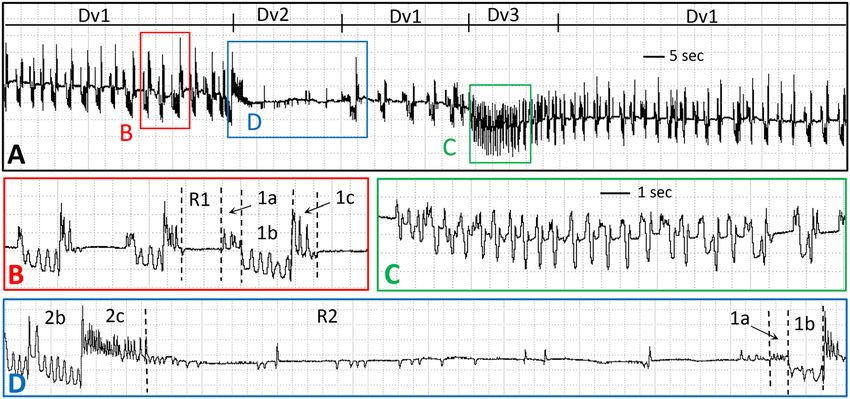
www.nature.com/scientificreports/
numerous molecular and biochemical s tudies11–16, the time-scale temporal dynamics of specific tick salivation
and ingestion events are largely unknown. Until ‘normal’ tick feeding behaviors and tick-host interactions are
delineated, including the temporal sequence of these events, it is nearly impossible to attain a clear understanding
of how tick and TBP intervention methods alter tick feeding behavior and tick-host interactions17.
Electropenetrography (EPG; not to be confused with ‘electrophysiology’ also sometimes abbreviated EPG) is
an electronic technology that allows researchers to observe, record, and quantify feeding behaviors of arthropods
whose mouthparts penetrate into opaque host tissues, and therefore cannot be directly visualized in real-time.
This revolutionary technology was originally i nvented18 to study the feeding behaviors of very small plant sap-
feeding insects on host plants. The technology has subsequently been improved several t imes19–21, but the basic
principle remains the same. An AC (alternating current) or DC (direct current) signal is conveyed to the plant
via a referent electrode in the s oil22. The arthropod (tethered with a recording electrode of thin, solid-gold wire
glued to its dorsum) is placed on the plant. When it inserts its mouthparts, current is conveyed to the instru-
ment for signal processing and then to a computer. Changes in output voltage over time create electrical patterns
(waveforms) displayed on the computer. Waveforms can be correlated with highly specific behaviors, such as
mouthpart movements, direction of fluid flow, salivation, ingestion, puncturing of specific cells, and pathogen
acquisition/inoculation22. Three generations of major EPG technology designs have occurred, in parallel with
advancements in electronics17,22. The first-generation (AC) monitors18,20 (no longer manufactured); used high
AC applied signal and fixed-low amplifier sensitivity (input resistor or Ri; 1 06 Ω). The second-generation (DC)
monitor21 (still marketed); uses low-to-high DC applied signal and fixed, high Ri ( 109 Ω). More recently, a third-
generation (AC–DC) monitor was introduced19 as a culmination of design and signal analysis comparisons
among the previous designs. The AC–DC monitor has selectable AC or DC applied signal and selectable Ri ( 106
to 1010 Ω plus 1013 Ω).
EPG has been widely used in studies of feeding behavior/physiology of plant-feeding, piercing-sucking
hemipteroid insects17,22. EPG has made possible virtually all present knowledge of the role of feeding in vector-
mediated transmission of plant pathogens22. In addition, EPG has been instrumental in gaining insights into
and improvements in pest m anagement17, such as: (i) targeting insecticide modes of a ction23,24; (ii) improving
25,26
host plant r esistance ; and, (iii) targets for R NAi27.
Application of EPG to the study of tick feeding behavior and tick-host interactions could be a profoundly
enabling solution to investigate, in unprecedented detail and real-time resolution, the temporal intricacies of tick
feeding behaviors and tick-host interactions. We hypothesize that EPG can be tailored to investigate on-host tick
feeding and that ticks will exhibit a complex assembly of waveforms representative of specific behaviors while
attached to the host. Therefore, the objectives of this study were to: (i) demonstrate that AC–DC EPG can suc-
cessfully be used to monitor on-host tick feeding behaviors; (ii) determine whether ticks produce distinguishable
EPG feeding waveforms; and, (iii) qualitatively and quantitatively describe/compare tick EPG feeding waveforms
performed by two tick species during the early stages of slow-phase tick f eeding28.
Methods and materials
Ticks and tick maintenance. Pathogen-free, adult female Dermacentor variabilis (American dog tick) and
Amblyomma americanum (Lone star tick) (Ecto Services, Inc., Henderson, NC), ca. 12–14 weeks post-molt, were
used in this study. Ticks were maintained in humidified chambers within environmental incubators at 98% RH,
26 °C, and with a 12:12 L:D cycle until use.
Calf maintenance and tick infestation. All study activities involving animals were reviewed by the
Kansas State University Institutional Animal Care and Use Committee and performed in accordance with the
approved study-specific protocol and ARRIVE guidelines. Four Holstein or Holstein-Jersey steers between
4–8 months of age were used as tick-feeding hosts. Feeding patches for ticks were applied to calves by shaving
an area on their back behind the shoulder and attaching a stockinet using a veterinary-approved adhesive. Ticks
(~ 5 total per patch, 2–3 D. variabilis and 2–3 A. americanum) were placed together in the stockinet and allowed
to attach to the calf. During tick infestation, calves were housed indoors and restrained in stanchions to prevent
them from disturbing the tick-feeding patch. Calves were fed a complete grain diet at 2% bodyweight per day,
hay, and water ad libitum.
Stanchioned calves had free vertical movement and received no pharmacological drugs to avoid reducing
other natural movements; by comparison, electrophysiology studies normally require use of fully sedated animals,
constraining recording durations. During the recording process, these awake calves regularly would stand/lay,
perform normal movements, and other bodily functions within the confines of the stanchion. One head stage
amplifier was mounted on the back of each calf near the wired tick; thus, the amplifier moved with the calf. Inves-
tigators observed real-time data capture of all waveforms. When a calf laid down, such large, jolting movement
occasionally caused a voltage peak in the recordings (which was inserted as an observation in real-time on the
recording), but the other normal calf behavior movements did not affect the recordings.
Electropenetrography. Individual ticks were wired after attachment to the calf host. Ticks were tethered
with a 38.1 µm thin, gold wire (sold as 0.0015 in., Sigmund Cohn Co., Mt. Vernon, NY) following the methods
previously described29, using silver glue whose recipe is described therein. In brief, gold wire was glued to a cop-
per wire soldered to a brass nail, then was bent into a small loop at its other end; the loop was then glued to the
dorsum of the feeding tick. While multiple recordings were made, not all were considered high enough quality
for measurement and publication. Recordings, of different time durations, were made early during slow-phase
tick feeding, between 20 and 48 h post-infestation from a total of four ticks (two D. variabilis and two A. america-
num). Three recordings were analyzed of D. variabilis ticks (tick 1 for 70.2 and 14.0 min, and tick 2 for 24.3 min);
Scientific Reports | (2021) 11:2040 | https://doi.org/10.1038/s41598-020-80257-6 2
Vol:.(1234567890)www.nature.com/scientificreports/
one recording was analyzed for each A. americanum ticks (tick 3 for 276.5 min and tick 4 for 172 min). We
recorded one tick at a time per host (no simultaneous recordings of multiple ticks on the same host at the same
time). The two D. variabilis were recorded on the same calf at different, non-overlapping times on the same day.
The two A. americanum ticks were recorded on two separate calves for overlapping time periods over 2 days. All
recordings made in this study occurred before any tick began to develop any noticeable abdominal expansion.
Recordings were made using a four-channel AC–DC electropenetrograph manufactured by EPG Technolo-
gies, Inc. (Gainesville, FL, andygator3@gmail.com). The instrument is similar to that introduced i n19, described
further in22, with block diagram in30. Head stage amplifiers and applied signal electrodes were secured on the
calf near the tick attachment location using a combination of veterinary-approved adhesive and adhesive tape.
Applied voltage was 350 mV AC and amplifier sensitivity (input resistor or Ri level) was set to 1 08 Ohms for most
recordings. Analog signals were acquired and digitized via a DI-710 board and Windaq Lite software (both from
DATAQ Instruments, Akron, OH) using sample rate of 100 Hz. Waveforms were re-played for measurement
using Windaq Waveform Browser (DATAQ). Pre- and post-rectification signals were simultaneously recorded
and checked to ensure that they were identical; if not, the offset function was used to remove rectifier fold-over
of the output signal and retain native polarity (positive- or negative-going property) of the waveform31. Instru-
ment gain for all the recordings was 2000X. As needed, a piece of a grounded thermal mylar blanket was draped
over the assembly of head-stage amplifier and feeding tick to reduce ambient electrical noise during recordings.
EPG waveform terminology used was similar to the system previously published29. Briefly, a ‘waveform’ is
a series of output voltage changes over time that, together, form a pattern discernible to the human eye. When
describing further structural composition of waveforms, a hierarchical system (from largest to smallest) of
waveform ‘family’ and waveform ‘type’ is used. A waveform family is a recognizable waveform appearance vis-
ible to the human eye at a coarse level, or compression (on the X-axis) level of 1–2 min per vertical division in
the screen view. A family can be a single, undividable pattern, or have obvious components that can be further
subdivided as waveform ‘types’ when compression is spread out to seconds per division. Thus, in the present
study, some waveforms were divided only to the family level, while others were further divided to the type level.
Within a family, types can be single, that is, never repeated in a measured event (see below for definition of
‘event’), or types can be highly repetitive, with several types grouped together to form ‘episodes’ that are consist-
ently repeated within an event of the family.
A waveform ‘event’ is not part of the above hierarchical naming convention, but an independent identifica-
tion that is important for quantitative measurement of waveform durations, counts, and frequencies. An event
consists of a single, uninterrupted occurrence of a waveform family or type, whose duration is measured for
quantification. No matter what aspect of a waveform in an event is measured (duration, frequency), each indi-
vidual measurement ultimately becomes a single observation in a dataset for statistical analysis. Each event has
a sequence-specific location in the recording; thus, each event is temporally unique. A researcher can choose to
name an event at the family level [e.g., all of Dv1 until interrupted by Dv2 (see below)] or at the type level [e.g.,
Dv1a separate from Dv1b, and so on (see below)]. Certain families (e.g., Dv3, see below) are considered events
at the family level, with no further division into types. Others can be considered events at either the family level
(e.g., Dv1 for durations herein) or the type level (e.g., Dv1a for frequencies herein).
Durations and counts of events at the waveform family level were summarized and descriptive statistics were
calculated using the Ebert v. 1.0 and Backus v. 2.0 SAS (v. 9.4) analysis programs (SAS, Cary, North Carolina,
U.S.A.) (downloadable from https://crec.ifas.ufl.edu/extension/epg/epg_workshop.shtml), generating means
and standard errors for the following, standardized variables for each species: (1) Waveform Duration per Event
per Insect (WDEI), (2) Number of Waveform Events per Insect (NWEI), and (3) Waveform Duration per Insect
(WDI)31,32. (The above, standardized variable names were used to be consistent with other EPG studies, even
though ticks are not insects.) In addition, number of episodes (see definition below) of Family 1 waveforms were
manually counted within selected, naturally terminated events, i.e., Family 1 events that both preceded and fol-
lowed recorded Family 2 events (see below). All such naturally-terminated events were counted for D. variabilis,
while 20 randomly-selected Family 1 events per tick were counted for the longer A. americanum recordings that
contained many Family 1 events. Manual episode counts were summarized for each species for the following
variables: (1) mean (naturally terminated) event duration, (2) mean number of episodes per event, (3) mean
number of episodes per sec. While sample sizes of recordings per species were considered sufficient for char-
acterization and descriptive statistics, they were not sufficient for statistical testing of the above duration-based
variables or between species.
In contrast, in-depth measurements of waveform frequency (i.e., number of peaks per sec, or Hz) were statisti-
cally comparable among waveform types in Families 1 and 2, for individual ticks within species. This was done
because frequencies (or frequency-amplitude combinations) have been shown in other EPG studies to be highly
characteristic of different waveform families and types. For Family 1 types, frequencies were measured for 10
representative episodes per tick. For Family 2 types, frequencies were measured for all events in each recording
(See “Results”). The same SAS programs cited above were again used to generate means and standard errors, but
also to perform mixed model Analysis of Variance to statistically compare among untransformed means by and
among ticks and w aveforms31,32. Means were considered significantly different at α = 0.05.
Results
Overview. To demonstrate the application of EPG to study tick feeding behavior, we characterized and com-
pared the feeding of two tick species (D. variabilis and A. americanum) during the early stages of slow-phase
tick feeding (~ 20–48 h post-infestation). Baseline waveform levels for an unattached tick standing on calf were
determined at the end of recording by forcing the tick to detach from the calf. Both on-calf and off-calf base-
line voltage levels were far below the voltage level for all tick feeding waveforms observed. Therefore, all wave-
Scientific Reports | (2021) 11:2040 | https://doi.org/10.1038/s41598-020-80257-6 3
Vol.:(0123456789)www.nature.com/scientificreports/
Figure 1. EPG waveforms for D. variabilis feeding approx. 36 h after attachment to calf. (A) Compressed view
of three Dv1 events and intervening Dv2 and Dv3 events. Labelled boxes contain waveform segments that are
expanded in parts with the same letter label. Windaq gain 64×. (B) Two and a half episodes of Dv1 showing all
four types, Dv1a, Dv1b, Dv1c and DvR1, but with Dv removed from each label to save space. (C) One event
of Dv3. (D) One event of Dv2, plus the start of the next Dv1 event. See narrative for further descriptions of
waveform names. Scale bar in part C and Windaq gain of 128 × same for parts B – D.
Number of episodes counted in selected Family 1 events (all naturally
terminated)
Recording duration Mean event duration Mean no. Frequency (mean
Tick no. Recording no. (min) N (s) episodes per event episodes per s)
D. variabilis
1 1&2 84.2 9 314.2 ± 55.7 125.6 ± 34.7 0.40
2 3 24.3 1 233.4 24.0 0.10
A. americanum
3 4 276.5 20 185.9 ± 20.2 11.3 ± 1.7 0.06
4 5 172.0 20 370.6 ± 66.9 17.3 ± 2.6 0.05
Table 1. Descriptive statistics for early, slow-phase feeding episodes of adult, female D. variabilis and A.
americanum Family 1 waveforms measured within naturally terminated events. Mean ± standard errors.
forms were monophasic positive (i.e., positive-going) in polarity30. Three families of waveforms, based strictly
on appearance, were characterized.
Waveforms were hierarchically categorized into three families and seven types within families, named consist-
ently for both species. Type categorization was supported by the statistical frequency analysis, whose results are
described within each section below. Waveforms were considered very high-frequency (very fast) at 10.0–25 Hz.,
high-frequency (fast) at 6.0–9.9 Hz., medium-frequency at 4.0–5.9 Hz. (medium), low-frequency (slow) at
2.0–3.9 Hz., very low-frequency (very slow) at 0.1–1.9 Hz., or flat at 0.0 Hz. Frequencies for each waveform type
within each tick were highly consistent. Frequencies were significantly different among different waveform types
(P < 0.0001 within each tick species) but, unless otherwise stated below, were not significantly different between
ticks within a species.
Family 1. The most common waveform family, Family 1, occurred in the background throughout all record-
ings with virtually no cessation except interruption by the other two families. Family 1 waveforms were similar
between ticks within each species but were interestingly different between species. Therefore, we chose specific
Family names for each species: Dv1 (for Dermacentor variabilis Family 1) and Aa1 (for Amblyomma americanum
Family 1).
Family 1 was composed of highly stereotypical and repeating episodes, rapidly recurring about every 2.5 s
for dozens of episodes per event of Dv1 for D. variabilis tick 1 (Fig. 1A), but more slowly (every 10 s) for tick 2
(Table 1). Dv1 events lasted 3–5 min each (Table 2). In contrast, both A. americanum ticks (3 and 4) performed
Aa1 similarly. Each Aa1 episode was much longer and less frequent (about once every 20 s) than for Dv1, with
fewer than 20 episodes in each 3–6 min event for many measured events of Aa1 (Fig. 2A, Table 1).
Family 1 episodes were divided into four subtypes, termed 1a, 1b, 1c and R1 (Fig. 1B), although Type 1a (thus,
Dv1a) was only performed by tick 1. It is possible that Dv1a was not seen in the tick 2 recording because that
Scientific Reports | (2021) 11:2040 | https://doi.org/10.1038/s41598-020-80257-6 4
Vol:.(1234567890)www.nature.com/scientificreports/
N WDEI (s) N WDEI (s)
D. variabilis A. americanum
Waveform Dv1 17 309.0 ± 94.5 Waveform Aa1 74 849.8 ± 627.8
Waveform Dv2 15 24.3 ± 7.8 Waveform Aa2 73 126.8 ± 1.4
Waveform Dv3 1 19.9
Table 2. Waveform duration per event per insect (WDEI) means ± standard errors (in sec) during early, slow-
phase feeding of adult, female D. variabilis and A. americanum. N number of events.
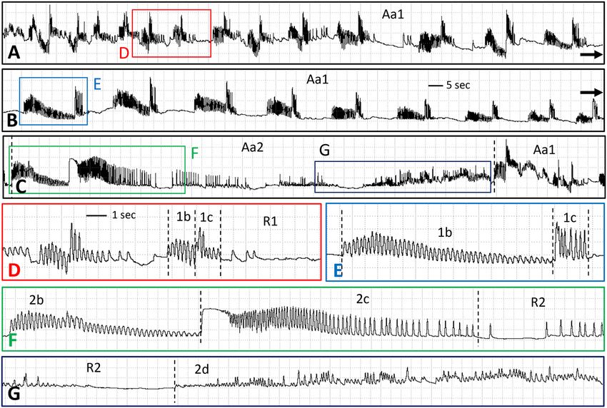
Figure 2. EPG waveforms for A. americanum feeding approx. 36 h after attachment to calf. (A–C) Compressed
view of one event of Aa1, showing appearances of successive episodes during the progression of the event
through three rows of waveforms. Labelled boxes contain waveform segments that are expanded in parts with
the same letter label. Scale bar in part (B) same for parts (A,C). Windaq gain 64×. (D) Two episodes of Aa1
showing four out of four types, Aa1b, Aa1c and AaR1, but with Aa removed from each label to save space. (E)
A later episode of Aa1 showing lengthened Aa1b. (F) First third of one event of Aa2, showing three out of five
types, Aa2b, Aa2c, and the first part of AaR2. (G) Last third of one event of Aa2, showing last part of AaR2 and
all of Aa2d. See narrative for further descriptions of waveform names. Scale bar in part D and Windaq gain of
128 × same for parts (D–G).
recording was slightly noisier than the recording of tick 1; alternatively, the behavior represented by Type 1a may
not be conserved, as described in “Discussion”. Type Dv1a comprised a high-frequency series of peaks (Table 3)
all of the same short amplitude (Fig. 1B). Type 1a was not performed by A. americanum in our recordings.
Type 1b immediately followed Dv1a or was the first component of a Family 1 episode in A. americanum
(Aa1b). Type 1b consisted of medium-frequency, short-to-medium-amplitude peaklets that occurred on a differ-
ent voltage level, either lower (Dv1b; Fig. 1B) or slightly higher (Aa1b; Fig. 2D) than the voltage level preceding
it. Type 1b durations were short in D. variabilis as well as in early episodes of A. americanum (Table 2). However,
Aa1b sections in each episode gradually lengthened in duration over time, also becoming shorter in amplitude
and eventually tapering off (compare Fig. 2D,E). Mean frequencies were significantly different between ticks
within species. Dv1b of ticks 1 and 2 were numerically and significantly different (2.83 ± 0.36 for tick 1; 5.78 ± 0.10
for tick 2) (df = 1, 18, F = 63.18; P < 0.0001). Aa1b for ticks 3 and 4 appeared less numerically different, yet also
significantly different between ticks (4.79 ± 0.10 for tick 3; 4.10 ± 0.11 for tick 4) (df = 1, 18, F = 21.01P = 0.0002).
There was little variation in frequency of Aa1b among episodes within each tick.
For both species, Type 1c was a high-frequency (Table 3) series of peaks, short in duration (Figs. 1B, 2E),
but higher in amplitude than the preceding Type 1b. Frequencies of 1c were not significantly different between
ticks within species.
The fourth component of a Family 1 episode consisted of short or variable durations of flat or near-flat
waveform. Because this waveform was located at a voltage level above baseline, this likely represents a period
of no or reduced behavioral activity while the tick was still attached to the calf. Thus, the waveform was termed
Scientific Reports | (2021) 11:2040 | https://doi.org/10.1038/s41598-020-80257-6 5
Vol.:(0123456789)www.nature.com/scientificreports/
Type 1a Type 1b Type 1c Type R1
N Hz N Hz N Hz N Hz
D. variabilis 10 8.45 ± 0.53 a 20 4.30 ± 0.35 b 20 8.07 ± 0.23 a 20 0.00 ± 0.00
A. americanum 0 20 4.44 ± 0.11 ab 20 6.02 ± 0.32 a 20 0.49 ± 0.17 c
Type 2a Type 2b Type 2c Type R2
N Hz N Hz N Hz N Hz
D. variabilis 5 8.41 ± 0.76 a 8 4.19 ± 0.35 b 8 9.26 ± 0.54 a 8 1.6 ± 0.19 c
A. americanum 6 4.83 ± 0.58 ab 23 3.74 ± 0.07 b 23 5.47 ± 0.11 ab 23 0.38 ± 0.08 d
Type 2d
N Hz
D. variabilis 0
A. americanum 18 1.33 ± 0.35 c
Table 3. Results of frequency analysis of tick waveform types. Mean ± standard errors in Hz. Same lower case
letter indicates values were not significantly different.
Resting (R), Type 1 (R1), for Family 1 (Figs. 1B, 2D). In addition, frequencies for A. americanum ticks 3 and 4,
while very low, were nonetheless significantly different (0.91 ± 0.28 for tick 3; 0.08 ± 0.03 for tick 4) (df = 1, 18,
F = 8.9; P = 0.008). Thus, while no peaks occurred during DvR1, a few stray peaks occurred occasionally during
AaR1 (compare Figs. 1B and 2D).
Family 1 episodes for D. variabilis were uniformly short in duration and rapidly repeated (Fig. 1) before
abruptly transitioning into Family 2. In contrast, Family 1 episodes for A. americanum gradually lengthened
in duration as the episodes were repeated over time between Family 2 events (Fig. 2). This lengthening of Aa1
episodes over time occurred primarily because of the lengthening of Aa1b, described above. Also, R1 resting
intervals gradually lengthened between Aa1 episodes, until they reached maximum duration shortly before the
recording abruptly transitioned to Family 2. Constant performance of Family 1 episodes was the background
tick feeding behavior in our recordings, on which were superimposed two more waveform families.
Family 2. On a predictable and regular cycle for each recorded tick, a distinctive waveform change occurred,
comprising Family 2. This change occurred about every 5 min for both D. variabilis ticks, but every 3 or 6 min for
A. americanum ticks 3 and 4, respectively (Table 2). Despite their regularity, due to varying recording durations
for each tick, varying numbers of Family 2 events were measured, i.e., 6 events (tick 1), 2 (tick 2), 10 (tick 3), and
13 (tick 4). Family 2 was composed of five types.
Type 2a, like Type 1a, was not recorded for every episode of Family 2, i.e., 62.5% of episodes (both ticks 1
and 2) had 2a for D. variabilis, but only 26% of episodes (only tick 4) for A. americanum (Table 3). Because of
its relative rarity in A. americanum recordings, Type 2a is not shown in Fig. 2. However, its appearance was
very similar to Type 1a (for D. variabilis, Fig. 1B), with statistically similar high frequency and short duration.
Type 2b was nearly identical in appearance to Type 1b for both tick species (compare Figs. 1B,D, 2D–F), except
that Aa2b always looked like the longest-duration version of Aa1b. Like 1b, both species performed medium-
frequency peaks. Mean frequencies of Dv2b were significantly different between ticks 1 and 2 (3.72 ± 0.24 for
tick 1; 5.58 ± 0.04 for tick 2) (df = 1, 6, F = 17.83; P < 0.0001) but not significantly different for Aa2b between ticks
3 and 4 (3.66 ± 0.13 for tick 3; 3.79 ± 0.09 for tick 4).
Type 2c was always performed in both tick species, but its appearance varied by species. In D. variabilis
recordings, Dv2c was similar to Dv1c; a very high-frequency peak burst on a higher voltage level, distinctly sepa-
rate from Type 2b and relatively short in duration (Fig. 1D). In A. americanum, however, Aa2c was different in
appearance from Aa1c. Each Aa2c started with an abrupt jump in voltage level from the preceding, low-amplitude
end of Aa2b. This short, flat, then usually declining-voltage-level section of Aa2c abruptly transitioned into a
much longer-duration section distinctly resembling a comb with ever-widening tine-like peaks (Fig. 2F). The
comb started at a very high frequency of 20–25 Hz, then gradually slowed through medium to low frequency
of 3–5 Hz, eventually tapering off to flat or near-flat (R2, below). When calculated over a full duration of Aa1c,
mean frequency was medium (Table 3).
Following Type 2c, another resting section (near-flat R2) occurred. Type R2 was different in appearance for D.
variabilis versus A. americanum. DvR2 was quite long in duration and included frequent (at the beginning), short
dips downward and occasional short, upward peaks (Fig. 1D). After a few sec, the downward dips ended, and the
short peaks sometimes became more frequent (Fig. 1D); however, usually there were no peaks at the end of DvR2.
DvR2 ended abruptly with the start of new Dv1 episodes, but with longer DvR1 resting periods (Fig. 1A). After
four to six of such special Dv1 episodes, a gradual decline in voltage level sometimes led into Dv3 (see below).
AaR2 was different from DvR2. AaR2 began with a distinct lengthening of the flat line between Aa2c peaks,
followed by a short section almost as flat as R1 but with slight undulations that gradually became spikier until
a series of very short, high-frequency spikes erupted, gradually increasing in amplitude but still very irregular
in frequency (Fig. 2G). After a fairly long duration of flat waveform interspersed with short peak bursts, AaR2
abruptly transitioned to Aa2d (see below).
Scientific Reports | (2021) 11:2040 | https://doi.org/10.1038/s41598-020-80257-6 6
Vol:.(1234567890)www.nature.com/scientificreports/
Unsurprisingly given the above differences in R2 waveform appearances, especially varying numbers and
arrangements of peaks, R2 frequencies appeared to be different between ticks in each species. D. variabilis fre-
quencies were very low to low and significantly different between ticks 1 and 2 (1.37 ± 0.15 for tick 1; 2.31 ± 0.17
for tick 2) (df = 1, 6, F = 11.26; P = 0.0153). Similarly, A. americanum frequencies were very low and also different
between ticks 3 and 4 (0.64 ± 0.15 for tick 3; 0.19 ± 0.05 for tick 4) (df = 1, 21, F = 9.86; P = 0.0049).
Uniquely for A. americanum (Table 3), 78% of Aa2 episodes had a variably long duration of irregular-fre-
quency and -amplitude peaks at the end of each event (Fig. 2G) termed Type 2d (thus, Aa2d). Frequencies were
significantly different between the 9 episodes for each tick (2.04 ± 0.61 for tick 3; 0.62 ± 0.12 for tick 4) (df = 1, 16,
F = 5.21; P = 0.0364). Aa2d ended with an abrupt transition to Aa1a, the start of a new cycle of Family 1 episodes.
Family 3. Due to shorter recording durations, only one Dv3 event was found for D. variabilis (Fig. 1D).
Family 3 was strikingly different in appearance from the two previous families, and less uniform in appearance
because its appearance evolved over time; thus, no types were assigned. Dv3 started with a Dv1b-like series of
broad peaks that gradually increased in amplitude, declined in voltage level, and developed into two-peaked,
M-shaped plateau-like structures that were repeated several times (Fig. 1C). These two-horned plateaus grad-
ually evolved into Dv1a and Dv1b, eventually resuming the background behavior of repetitive Dv1. Despite
longer recordings for A. americanum than for D. variabilis, no Family 3 events were observed for A. americanum.
Discussion
A major challenge to the development and rigorous evaluation of host-level tick and TBP intervention strategies is
the lack of transparency of temporal events and behaviors that occur at the tick attachment site—the interface of
tick-host-pathogen interactions. The occurrence of these behaviors within host skin obstructs ready investigation
of ‘normal’ tick-host interactions and impedes evaluation of how chemical control measures or pathogens specifi-
cally alter these interactions to inhibit successful tick feeding or facilitate transmission, respectively. Adapting
AC–DC EPG to study the intricate behaviors of blood-feeding arthropods on-host can potentially transform
blood-feeding arthropod research.
Electronic instruments have previously been used to record the feeding behavior of blood-feeding arthropods
(~ 20 publications), including with argasid t icks33,34. Recent instrument designs use high-sensitivity electromyo-
graphy (EMG) amplifiers, like those used by electrophysiologists. EPG and EMG are easily confused, but there
are distinct differences between them, namely, the placement of the referent electrode and amplifier design. The
electrical circuit for EPG includes both the arthropod and its host. The recording electrode is attached to the
arthropod while the referent electrode is attached to the host. Waveforms are derived from electrical currents
carried by ionized fluids in the mouthpart canals and foregut of the arthropod. The EPG monitor detects a
mixture of (i) resistance to/conductance of current flow (R component); and, (ii) non-neural biopotentials, i.e.,
streaming potentials generated during fluid flow (emf component). In EMG, the electrical circuit includes only
the arthropod; both the recording and referent electrodes are attached to different parts of the arthropod body.
EMG instruments are designed to record strictly biopotentials, i.e., muscle/action potentials nearest the recording
electrode; in EPG parlance, they record only emf component from neural sources. EMG instruments necessitate
higher Ri levels, extensive band pass filtering, and differential amplification to detect these tiny biopotentials,
compared with EPG instruments. More information on EPG/EMG electronics is h ere17,22,35.
Thus, EMG instruments previously used for tick recordings were designed to detect biopotentials originating
in muscles35 controlling ingestion35, not salivation. Far more information is available if recording amplifiers are
also sensitive to electrical resistance/conductivity of fluid (e.g. saliva, blood) flow. These include: (i) electrical
resistance from opening/closing of valves/pumps in the foregut as well as mouthpart movements/depth; and, (ii)
electrical conductivity related to biochemical composition of s aliva20,22. While a few EMG papers have identi-
fied trace salivation and mouthpart movement biopotentials33,34, their electrical resistance information is likely
incomplete. In addition, a major benefit of AC–DC EPG is the far wider range of selectable amplifier sensitivi-
ties, to detect all signal types from resistance-only to biopotential-only and mixtures of b oth19,22. Another major
benefit of AC–DC EPG is that the head stage amplifier is relatively sturdy, and can be attached to a gently-moving,
unsedated host animal (as in our study), allowing for increased recording durations, especially important for
studying ixodid tick feeding behaviors. Previous EMG studies used sedated mice because the amplifier was too
delicate to tolerate host movement. Thus, AC–DC EPG will likely provide a far wider and more operationally
useful view of ixodid tick behaviors and physiologies than EMG.
In our study, we used AC–DC EPG to record and begin to characterize on-host feeding waveforms (behav-
iors) for two ixodid tick species of medical and veterinary significance during periods early in slow-phase tick
feeding (~ 20–48 h post-infestation). We demonstrated that: i) AC–DC EPG can be used to monitor tick feeding
behaviors on an awake animal host; ii) ticks produce a constant series of differentiable and definable waveforms
while feeding on-host; and, (iii) a similar series of recognizable waveforms were produced by two ixodid tick
species at a comparable feeding stage.
Another challenge to studying ixodid tick feeding behaviors is the extreme duration adult ticks feed, normally
6–9 days. This study provides brief ‘snapshots’ of adult tick feeding behaviors within the early stages of slow-phase
feeding; a timeframe during which chemical control measures are expected to work and when pathogen trans-
mission often occurs. During slow-phase feeding, adult ixodid ticks inject a temporally orchestrated cornucopia
of salivary proteins10 to complete attachment, evade host immune responses, prepare the feeding lesion, and
begin feeding/ingesting, all processes masked within host tissue. Previous studies of mechanisms underlying
tick-feeding success have primarily used various combinations of morphology, histology, transcriptome, and
proteome analyses at defined time points post-infestation or attachment. The latter often encompasses a variable
window of time rather than a specific time10,15,28,36–38. Previous studies examining specific tick tissue functions are
Scientific Reports | (2021) 11:2040 | https://doi.org/10.1038/s41598-020-80257-6 7
Vol.:(0123456789)www.nature.com/scientificreports/
rarer, and commonly require: (i) interruption of tick feeding, with or without dissection of the tissue target; (ii)
adaptation of ticks to artificial feeding systems; (iii) use of sedated hosts (argasid tick studies); or, (iv) significant
host discomfort33,34,39–41. Studies on real-time expression of precise on-host ixodid feeding behaviors are lacking,
largely due to absence of tools to investigate them. Addressing this deficit, we demonstrate that AC–DC EPG
can be adapted to investigate on-host ixodid tick feeding behaviors in real-time, following a specific, behavior-
triggered zero time point.
While EPG waveforms were similar enough between D. variabilis and A. americanum to be generally catego-
rized into the same waveform families and types, there were interesting differences between species. Generally,
D. variabilis performed waveforms faster than A. americanum. Episode repetition rate of the constantly-cycling
background waveform, Family 1, was faster (every 2.5 s) for D. variabilis than for A. americanum (every 10 s).
Similarly, although waveform types usually had stereotypical frequencies (peaks per sec), those of D. variabilis
were commonly higher/faster than those of A. americanum.
In addition, certain waveform types were always performed by both species (tentatively termed conserved),
while other types were rarely performed, either by one or both species (tentatively termed variable). For example,
in waveform Family 1, Types 1b, 1c, and R1 were highly conserved, performed dozens to hundreds of times by
all ticks in both species at stereotypical frequencies. In contrast, Type 1a was only performed by D. variabilis tick
1 and no other. It is possible that quality of recordings eliminated 1a in D. variabilis tick 2. However, because
recordings for both the A. americanum ticks (3 and 4) were very high-quality, noise cannot be the sole reason
for the loss of 1a in those ticks.
Similarly, for Family 2 waveforms, Type 2a was recorded in three out of the four ticks, but A. americanum
tick 3 did not perform 2a, and certain events lacked 2a in the other three ticks’ recordings. Interestingly, Type 2d
was exclusively observed in A. americanum. In contrast, Types 2b, 2c, and R2 were performed by all ticks of both
species, in every Family 2 event recorded. Also, like their Family 1 counterparts, the 2b, 2c, and R2 frequencies
were highly stereotypical for both species, again being medium-, high-, and low-frequencies, respectively. Thus,
2b, 2c, and R2 represent highly conserved behaviors; Types 2a and 2d represent variable behaviors.
Due to the repetitive, stereotypical and cycling background behavior of Family 1 waveforms for both tick
species, we hypothesize that these frequent and repetitive waveforms are associated with salivation and specific
mouthpart, pump or valve movements. Consistent salivation at this stage of the tick feeding process is likely
required to prevent rejection from the host immune responses and prepare the feeding lesion10,42–44. We also
hypothesize that different salivary secretions (perhaps different chemistries from different acini cell types) flow
from the mouthparts during conserved 1a, 1b, and 2c. Perhaps the variable waveform 1a represents a less-
commonly secreted type of saliva. During R1 of Family 1, the ticks rest for a few seconds before beginning the
salivation cycle anew.
During a second type of regularly cycling (but less frequent) behaviors, Family 2 waveforms are interspersed
among Family 1 waveforms. It is possible that the Family 2 waveforms represent expulsion of a different salivary
composition because the waveforms resemble Family 1 waveforms, but are longer in duration and more detailed
in fine structure. Alternatively, the regularly cycling Family 2 waveforms may represent brief periods of fluid
uptake and ingestion, which may occur less frequently during the first 48 h of ixodid tick feeding, enabling the
tick to gauge its feeding progress or help replenish depleted resources. Again, the conserved performance of 2b
and 2c suggest that they represent essential behavioral events, while the variable Types 2a and 2d represent less
important or less frequently required behavioral events.
Neural control of tick salivary glands, including: (i) salivary gland innervation patterns; (ii) roles of associ-
ated neurotransmitters; and, (iii) temporal production of transient secretory vesicles, are all areas of i nterest40.
EPG can be used to better understand these mechanisms underpinning salivary gland control, to study their
ultimate functional effect. For comparison, different salivary fractions of hemipteran piercing-sucking insects
can be secreted by each section of the principle salivary glands under voluntary control by the i nsect45–48. Future
work to differentiate conserved versus variable waveforms could help in deciphering the biological meanings of
these tick-feeding waveforms.
Our study is an introductory, proof-of-principle benchmark for future tick EPG research. We demonstrate
the active and intricate behaviors performed by ixodid ticks, even for only a brief period of the extensive tick
feeding process. Additional studies, with longer recordings and larger sample size, will be required to confirm
whether Family 1 and 2 waveforms initially characterized as variable are truly idiosyncratic for the studied tick
life stage and species. Future studies with longer recordings are also needed to fully qualitatively and quantita-
tively characterize the temporal sequence of on-host tick feeding behaviors. Studies deciphering the behavioral
and functional activities associated with individual waveform families and types will be required to interpret
their biological significance.
Despite the medical and economic importance of ticks and other blood-feeding arthropods, effective control
measures are limited. The imperative need for development of new mitigation strategies requires novel means of
closing persistent knowledge gaps about arthropod blood feeding. AC–DC EPG provides a platform to delineate
intricate and temporal feeding behaviors of ticks and other blood-feeding arthropods49. Such fundamental stud-
ies are required to understand how chemical control interventions, pathogens, or host immunity functionally
alter feeding behaviors. Once such benchmark studies are completed, EPG can be used to rigorously evaluate
how management methods, pathogens, etc. alter blood-feeding arthropod feeding behavior(s) using real-time
quantitative measurements. Further, EPG can be used to identify perfect zero time points based on initiation
of a specific behavior rather than using time post-infestation/attachment. For ixodid ticks, successfully captur-
ing a true zero timepoint has been consistently difficult; achieving this task will have profound implications
for interpretation of experimental results. Accordingly, we contend that AC–DC EPG is a uniquely enabling
technology, with transformative potential to widely open previously unrealizable investigative possibilities to
study blood-feeding vector-host–pathogen interactions.
Scientific Reports | (2021) 11:2040 | https://doi.org/10.1038/s41598-020-80257-6 8
Vol:.(1234567890)www.nature.com/scientificreports/
Data availability
All data generated or analyzed during this study are included in this published article.
Received: 16 July 2020; Accepted: 18 December 2020
References
1. Adrion, E. R., Aucott, J., Lemke, K. W. & Weiner, J. P. Health care costs, utilization and patterns of care following lyme disease.
PLoS ONE 10, https://doi.org/10.1371/journal.pone.0116767 (2015).
2. Prevention, C. F. D. C. A. Tickborne Disease Surveillance Data Summary. https://www.cdc.gov/ticks/data-summar y/index.html
(2020).
3. Prevention, C. F. D. C. A. Summary of Notifiable Infectious Diseases (2020).
4. Prevention, C. F. D. C. A. Notifiable Infectious Diseases and Conditions Data Tables (2020).
5. Nelson, C. A. et al. Incidence of clinician-diagnosed lyme disease, United States, 2005–2010. Emerg. Infect. Dis. 21, 1625–1631.
https://doi.org/10.3201/eid2109.150417 (2015).
6. Council, C. A. P. Parasite Prevalence Maps—Dogs—Tickborne disease agents, 2018. https://capcvet.org/maps/#2018/all/lyme-disea
se/dog/united-states/ (2020).
7. Research, Z. M. Global Animal Parasiticides Markets Will Reach USD 12.85 Billion by 2025. https: //www.globen ewswi re.com/news-
release/2019/03/13/1752568/0/en/Global-Animal-Parasiticides-Market-Will-Reach-USD-12-85-Billion-By-2025-Zion-Marke
t-Research.html (2019).
8. Estrada-Peña, A. & Salman, M. Current limitations in the control and spread of ticks that affect livestock: A review. Agriculture
(Switzerland) 3, 221–235. https://doi.org/10.3390/agriculture3020221 (2013).
9. Mack, A. in Forum on Microbial Threats, Board on Global Health, Health and Medicine Division. The National Academies of Sciences,
Engineering, Medicine. (The National Academies Press, 2016).
10. Sonenshine, D. E. & Anderson, J. M. in Biology of Ticks Vol. 1 (eds R. M. Roe & D. E. Sonenshine) Chap. 6, 122–162 (Oxford
University Press, 2013).
11. Karim, S. & Ribeiro, J. M. C. An insight into the sialome of the lone star tick, Amblyomma americanum, with a glimpse on its time
dependent gene expression. PLoS ONE 10, https://doi.org/10.1371/journal.pone.0131292 (2015).
12. Mans, B. J. Quantitative visions of reality at the tick-host interface: biochemistry, genomics, proteomics, and transcriptomics as
measures of complete inventories of the tick sialoverse. Front. Cell. Infect. Microbiol. 10, 574405. https://doi.org/10.3389/fcimb
.2020.574405 (2020).
13. McSwain, J. L., Essenberg, R. C. & Sauer, J. R. Protein changes in the salivary glands of the female lone star tick, Amblyomma
americanum, during feeding. J. Parasitol. 68, 100–106. https://doi.org/10.2307/3281330 (1982).
14. Nuttall, P. A. Wonders of tick saliva. Ticks Tick-borne Dis. 10, 470–481. https://doi.org/10.1016/j.ttbdis.2018.11.005 (2019).
15. Perner, J., Kropáčková, S., Kopáček, P. & Ribeiro, J. M. C. Sialome diversity of ticks revealed by RNAseq of single tick salivary
glands. PLoS Neglect. Trop. Dis. 12, e0006410. https://doi.org/10.1371/journal.pntd.0006410 (2018).
16. Tan, A. W. L., Francischetti, I. M. B., Slovak, M., Kini, R. M. & Ribeiro, J. M. C. Sexual differences in the sialomes of the zebra tick,
Rhipicephalus pulchellus. J. Proteom. 117, 120–144. https://doi.org/10.1016/j.jprot.2014.12.014 (2015).
17. Backus, E. A., Guedes, R. N. C. & Reif, K. E. AC–DC Electropenetrography: Fundamentals, controversies, and perspectives for
pest management. Pest Manag. Sci. in press https://doi.org/10.1002/ps.6087 (2020).
18. McLean, D. L. & Kinsey, M. G. A technique for electronically recording aphid feeding and salivation. Nature 202, 1358–1359. https
://doi.org/10.1038/2021358a0 (1964).
19. Backus, E. A. & Bennett, W. H. The AC–DC correlation monitor: New EPG design with flexible input resistors to detect both R and
emf components for any piercing-sucking hemipteran. J. Insect Physiol. 55, 869–884. https: //doi.org/10.1016/j.jinsph ys.2009.05.007
(2009).
20. Backus, E. A., Devaney, M. J. & Bennett, W. H. in Principles and Applications of Electronic Monitoring and Ohter Techniques in the
Study of Homopteran Feeding Behavior (eds G. P. Walker & E. A. Backus) 102–143 (Entomological Society of America, 2000).
21. Tjallingii, W. F. Electrical recording of aphid penetration. Nod. Entomol. 24, 521–530 (1978).
22. Backus, E. A., Cervantes, F. A., Guedes, R. N. C., Li, A. Y. & Wayadande, A. C. AC–DC electropenetrography for in-depth studies
of feeding and oviposition behaviors. Ann. Entomol. Soc. Am. 112, 236–248. https://doi.org/10.1093/aesa/saz009 (2019).
23. Serikawa, R. H., Backus, E. A. & Rogers, M. E. Effects of soil-applied imidacloprid on Asian citrus psyllid (Hemiptera: Psyllidae)
feeding behavior. J. Econ. Entomol. 105, 1492–1502. https://doi.org/10.1603/ec11211 (2012).
24. Tariq, K. et al. The toxicity of flonicamid to cotton leafhopper, Amrasca biguttula (Ishida), is by disruption of ingestion: An elec-
tropenetrography study. Pest Manag. Sci. 73, 1661–1669. https://doi.org/10.1002/ps.4508 (2017).
25. Mayoral, A. M., Tjallingii, W. F. & Castanera, P. Probing behavior of Diuraphis noxia on five cereal species with different hydroxamic
acid levels. Entomol. Exp. Appl. 78, 341–348 (1996).
26. Yorozuya, H. Analysis of tea plant resistance to tea green leafhopper, Empoasca onukii, by detecting stylet-probing behavior with
DC electropenetrography. Entomol. Exp. Appl. 165, 62–69. https://doi.org/10.1111/eea.12621 (2017).
27. Mutti, N. S. et al. A protein from the salivary glands of the pea aphid, Acyrthosiphon pisum, is essential in feeding on a host plant.
Proc. Natl. Acad. Sci. USA 105, 9965–9969 (2008).
28. Franta, Z. et al. Dynamics of digestive proteolytic system during blood feeding of the hard tick Ixodes ricinus. Parasites Vectors 3,
119. https://doi.org/10.1186/1756-3305-3-119 (2010).
29. Cervantes, F. A. & Backus, E. A. EPG waveform library for Graphocephala atropunctata (Hemiptera: Cicadellidae): Effect of adhe-
sive, input resistor, and voltage levels on waveform appearance and stylet probing behaviors. J. Insect Physiol. 109, 21–40. https://
doi.org/10.1016/j.jinsphys.2018.05.008 (2018).
30. Backus, E. A. & Shih, H. T. Review of the EPG waveforms of sharpshooters and spittlebugs including their biological meanings in
relation to transmission of Xylella fastidiosa. J. Insect Sci. in press https://doi.org/10.1093/jisesa/ieaa055 (2020).
31. Backus, E. A., Cline, A. R., Ellerseick, M. R. & Serrano, M. S. Lygus hesperus (Hemiptera: Miridae) feeding on cotton: New methods
and parameters for analysis of nonsequential electrical penetration graph data. Ann. Entomol. Soc. Am. 100, 296–310. https://doi.
org/10.1603/0013-8746(2007)100[296:LHHMFO]2.0.CO;2 (2007).
32. Ebert, T. A., Backus, E. A., Cid, M. & Fereres, A. A new SAS program for behavioral analysis of electrical penetration graph data.
Comput. Electron. Agric. 116, 80–87. https://doi.org/10.1016/j.compag.2015.06.011 (2015).
33. Costa, G. C. A. et al. Physiological characterization of the hematophagy of Ornithodoros rostratus (Acari: Argasidae) on live hosts.
J. Exp. Biol. 219, 3656–3664. https://doi.org/10.1242/jeb.144246 (2016).
34. Zheng, H. et al. Biological and physiological characterization of in vitro blood feeding in nymph and adult stages of Ornithodoros
turicata (Acari: Argasidae). J. Insect Physiol. 75, 73–79. https://doi.org/10.1016/j.jinsphys.2015.03.005 (2015).
35. Mizrahi, J. Advances in applied electromyography. in InTech (2011).
Scientific Reports | (2021) 11:2040 | https://doi.org/10.1038/s41598-020-80257-6 9
Vol.:(0123456789)www.nature.com/scientificreports/
36. Agyei, A. D. & Runham, N. W. Studies on the morphological changes in the midguts of two ixodid tick species Boophilus microplus
and Rhipicephalus appendiculatus during digestion of the blood meal. Int. J. Parasitol. 25, 55–62. https://doi.org/10.1016/0020-
7519(94)00114-4 (1995).
37. Mudenda, L. et al. Proteomics informed by transcriptomics identifies novel secreted proteins in Dermacentor andersoni saliva. Int.
J. Parasitol. 44, 1029–1037. https://doi.org/10.1016/j.ijpara.2014.07.003 (2014).
38. Narasimhan, S. et al. Host-specific expression of Ixodes scapularis salivary genes. Ticks Tick-borne Dis. 10, 386–397. https://doi.
org/10.1016/j.ttbdis.2018.12.001 (2019).
39. Kim, D., Šimo, L. & Park, Y. Orchestration of salivary secretion mediated by two different dopamine receptors in the blacklegged
tick Ixodes scapularis. J. Exp. Biol. 217, 3656–3663. https://doi.org/10.1242/jeb.109462 (2014).
40. Šimo, L., Zitňan, D. & Park, Y. Neural control of salivary glands in ixodid ticks. J. Insect. Physiol. 58, 459–466. https://doi.
org/10.1016/j.jinsphys.2011.11.006 (2012).
41. Tatchell, R. J., Carnell, R. & Kemp, D. H. Electrical studies on the feeding of the cattle-tick, Boophilus microplus. Z. Parasite. 38,
32–44 (1972).
42. Anderson, J. M. et al. Ticks, Ixodes scapularis, feed repeatedly on white-footed mice despite strong inflammatory response: An
expanding paradigm for understanding tick-host interactions. Front. Immunol. 8, 1784. https: //doi.org/10.3389/fimmu. 2017.01784
(2017).
43. Mulenga, A., Sugimoto, C., Ohashi, K. & Onuma, M. Characterization of an 84 kDa protein inducing an immediate hypersensitivity
reaction in rabbits sensitized to Haemaphysalis longicornis ticks. Biochem. Biophys. Acta. 1501, 219–226. https://doi.org/10.1016/
s0925-4439(00)00026-0 (2000).
44. Trager, W. Acquired immunity to ticks. J. Parasitol. 25, 57–81, https://doi.org/10.2307/3272160 (1939).
45. Ammar, E. D. Ultrastructure of the salivary glands of the planthopper, Peregrinus maidis (ashmead) (Homoptera : Delphacidae).
Int. J. Insect Morphol. Embryol. 15, 417–428. https://doi.org/10.1016/0020-7322(86)90034-6 (1986).
46. Miles, P. Adv. Insect Physiol. 9, 183–255 (1972).
47. Wayadande, A. C., Baker, G. R. & Fletcher, J. Comparative ultrastructure of the salivary glands of two phytopathogen vectors, the
beet leafhopper, Circulifer tenellus (Baker), and the corn leafhopper, Dalbulus maidis Delong and Wolcott (Homoptera: Cicadel-
lidae). Int. J. Insect Morphol. Embryol. 26, 113–120. https://doi.org/10.1016/S0020-7322(97)00009-3 (1997).
48. Miles, P. W. Aphid saliva. Biol. Rev. 74, 41–85. https://doi.org/10.1111/j.1469-185X.1999.tb00181.x (1999).
49. Wayadande, A. C., Backus, E. A., Noden, B. H., Ebert, T. & Hillyer, J. Waveforms from stylet probing of the mosquito Aedes aegypti
(Diptera: Culicidae) measured by AC–DC electropenetrography. J. Med. Entomol. 57, 353–368. https://doi.org/10.1093/jme/tjz18
8 (2020).
Acknowledgements
We acknowledge and sincerely thank Andrew M. Dowell, EPG Technologies, Inc., Gainesville, FL, for his tech-
nical assistance during the research described herein. This study was supported by an award from the USDA
National Institute of Food and Agriculture, Small Business Innovation Research grant program to Dowell [award
no. 2018-33610-28255/project accession no. 1016038]. Salary and travel funds for E. A. Backus were provided
by in-house funds of USDA ARS (appropriation project #2034-22000-010-00D). We also thank two anonymous
reviewers for their helpful suggestions on an earlier draft of this manuscript. Mention of trade names or com-
mercial products in this publication is solely for purpose of providing specific information and does not imply
recommendation or endorsement by the U.S. Department of Agriculture. USDA is an equal opportunity provider
and employer. This article was prepared by a U.S. Department of Agriculture employee as part of his/her official
duties. Copyright protection under U.S. Copyright Law Title 17 U.S.C. § 105 is not available for such works.
Accordingly, there is no copyright to transfer. The fact that the private publication in which the article appears
is itself copyrighted does not affect the material of the U.S. Government, which can be freely reproduced by the
public. Articles and other publications prepared as part of a Federal employee’s official duties are property of
the U.S. Government.
Author contributions
K.E.R. and E.A.B. both conceptualized study design, performed experiments, performed data analysis and inter-
pretation, and co-wrote the manuscript.
Competing interests
E.A.B. co-invented the AC-DC electropenetrograph while an employee of the USDA, which filed a patent granted
as US 8,004,292. As patent owner, the USDA has licensed the patent to Andrew M. Dowell, owner of EPG Tech-
nologies, Inc. By statute, E.A.B is authorized to receive a small portion of royalty payments remitted to the USDA,
if any. Also, with USDA permission and as part of her salaried position, E.A.B. is an advisor to the licensee to
assist in instrument improvements. K.E.R. declares no competing interests.
Additional information
Correspondence and requests for materials should be addressed to K.E.R.
Reprints and permissions information is available at www.nature.com/reprints.
Publisher’s note Springer Nature remains neutral with regard to jurisdictional claims in published maps and
institutional affiliations.
Scientific Reports | (2021) 11:2040 | https://doi.org/10.1038/s41598-020-80257-6 10
Vol:.(1234567890)You can also read