Association of Sonic Hedgehog with the extracellular matrix requires its zinc-coordination center
←
→
Page content transcription
If your browser does not render page correctly, please read the page content below
Jägers and Roelink BMC Molecular and Cell Biology                (2021) 22:22
https://doi.org/10.1186/s12860-021-00359-5
                                                                                                                                       BMC Molecular and
                                                                                                                                       Cell Biology

 RESEARCH ARTICLE                                                                                                                                    Open Access

Association of Sonic Hedgehog with the
extracellular matrix requires its zinc-
coordination center
Carina Jägers and Henk Roelink*

  Abstract
  Background: Sonic Hedgehog (Shh) has a catalytic cleft characteristic for zinc metallopeptidases and has significant
  sequence similarities with some bacterial peptidoglycan metallopeptidases defining a subgroup within the M15A
  family that, besides having the characteristic zinc coordination motif, can bind two calcium ions. Extracellular matrix
  (ECM) components in animals include heparan-sulfate proteoglycans, which are analogs of bacterial peptidoglycan
  and are involved in the extracellular distribution of Shh.
  Results: We found that the zinc-coordination center of Shh is required for its association to the ECM as well as for
  non-cell autonomous signaling. Association with the ECM requires the presence of at least 0.1 μM zinc and is
  prevented by mutations affecting critical conserved catalytical residues. Consistent with the presence of a
  conserved calcium binding domain, we find that extracellular calcium inhibits ECM association of Shh.
  Conclusions: Our results indicate that the putative intrinsic peptidase activity of Shh is required for non-cell
  autonomous signaling, possibly by enzymatically altering ECM characteristics.
  Keywords: Shh signaling, Zinc metallopeptidases, Extracellular matrix, BacHh

Background                                                                              rejected by demonstrating that mutation of a critical
The Hedgehog (Hh) gene was first identified in the                                      residue involved in catalysis (E177) did not impair the
Drosophila melanogaster screen performed by Christiane                                  ability of Shh to activate the Hh response [4], and conse-
Nüsslein-Volhard and Eric Wieshaus in the late 1970s                                    quently the zinc coordination center of Shh is often re-
[1]. Like other segment polarity genes found in this                                    ferred to as its “pseudo active” site [5, 6]. Still, a role for
screen, Hh genes are widely conserved among animals,                                    the zinc coordination center is supported by the finding
and mammals have three Hh paralogs that play roles in                                   that Shh-E177A is unable to mediate non-cell autono-
development [2]. Like all other Hhs, Sonic Hedgehog                                     mous long-range signaling from the notochord to the
(Shh) is synthesized as a pro-protein that undergoes                                    overlying neural plate [7]. Perhaps unsurprisingly, the
autoproteolytic cleavage mediated by the C-terminal part                                zinc-coordination motif is found mutated in some indi-
yielding an N-terminal part (ShhNp) that is the active                                  viduals with the Shh signaling-related birth defect
ligand. Structural analysis of ShhN revealed its similarity                             holoprosencephaly [8, 9], further indicating that the
to zinc-peptidases and Shh coordinates a zinc ion with                                  zinc-coordination center of Shh is important for normal
residues H141, D148, and H183 [3]. The notion that Shh                                  function. This is consistent with structures of Shh com-
signaled through a peptidase activity was quickly                                       plexed with its receptor Patched1 (Ptch1), showing that
                                                                                        the N-terminal 22 residues of Shh that are not part of
* Correspondence: roelink@berkeley.edu                                                  the zinc-coordination motif, mediate binding to Ptch1
University of California, Department of Cell and Molecular Biology, Berkeley,           [10–12] and suffice to regulate Ptch1 activity [13].
CA, USA

                                         © The Author(s). 2021 Open Access This article is licensed under a Creative Commons Attribution 4.0 International License,
                                         which permits use, sharing, adaptation, distribution and reproduction in any medium or format, as long as you give
                                         appropriate credit to the original author(s) and the source, provide a link to the Creative Commons licence, and indicate if
                                         changes were made. The images or other third party material in this article are included in the article's Creative Commons
                                         licence, unless indicated otherwise in a credit line to the material. If material is not included in the article's Creative Commons
                                         licence and your intended use is not permitted by statutory regulation or exceeds the permitted use, you will need to obtain
                                         permission directly from the copyright holder. To view a copy of this licence, visit http://creativecommons.org/licenses/by/4.0/.
                                         The Creative Commons Public Domain Dedication waiver (http://creativecommons.org/publicdomain/zero/1.0/) applies to the
                                         data made available in this article, unless otherwise stated in a credit line to the data.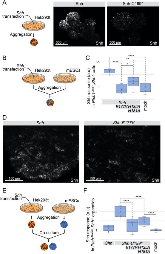
Jägers and Roelink BMC Molecular and Cell Biology   (2021) 22:22                                                   Page 2 of 18

  Some bacterial species have conserved genes coding               fraction (lysate) with a pattern distinct from the lysate
for peptidases that coordinate zinc and calcium                    indicating recovery of extracellular molecules (Fig. 1a).
identically to Shh [14, 15]. These bacterial peptidases            More Shh could be detected in the ECM than in the
(members of the M15A subfamily of zinc D-Ala-D-Ala                 cell-only fraction of Shh-C199* transfected Hek293t cells
carboxypeptidases) cleave murein peptidoglycans,                   (Fig. 1b), showing that entry of ShhN into the ECM is
suggesting that Shh too might cleave a glycan-modified             robust. Here, we are focusing on the association of Shh
protein, possibly a matrix heparan sulfate proteoglycan            with the ECM, and in order to circumvent the complex-
(HSPGs). HSPG are an integral part of the extracellular            ities of Shh maturation and secretion [26], we used Shh-
matrix (ECM) and play in important role in the trans-              C199* (ShhN). This form of Shh is active and secreted
port and presentation of several morphogens, including             independent of Disp1 function [27], and lacks the C-
Hhs [16]. Several HSPGs bind Shh and can both                      terminal sterol modification. We found that ShhN could
negatively and positively affect the Shh response [17–             readily be detected in the ECM extracted from decellu-
20]. Furthermore, mutations in Ext1 and − 2 coding for             larized tissue culture plates (Fig. 1b). Visualizing Shh-
glycosyltransferases that catalyze glycosaminoglycan               C199* by staining of the decellularized plates with the
addition to the core proteins, disrupt Hh signaling in             anti-Shh mAb5E1, “footprints” of Shh producing cells
vertebrates [20, 21] and insects [22].                             were observed (Fig. 1b). Shh-C199* is commonly
  By mutating residues in the zinc-coordination center             thought of as a “soluble” protein and also accumulates in
that are conserved between bacterial Hh-like peptidases            the supernatant of ShhN-producing cells. The non-
and Shh, we provide evidence that this center is required          homogeneous association of Shh with the ECM in
for the association of ShhN to the ECM and for non-cell            apparent “footprints” of the Shh-C199*-expressing cells
autonomous signaling. Release of Shh into the ECM is               indicates that upon secretion Shh enters adjacent ECM
enhanced in the presence of μM amounts of zinc indi-               directly and is not first released into the supernatant,
cating that this ion is an agonist of Shh. The ECM-                which would result in a more homogenous distribution
associated Shh is active in signaling, indicating that the         of the protein across the ECM. Shh-responsive LightII
zinc-coordination center of Shh mediates its release into          cells plated on the decellularized and Shh-conditioned
the ECM to facilitate non-cell autonomous signaling,               ECM showed that it is able to elicit a transcriptional Hh
possibly through an intrinsic metallopeptidase activity of         pathway response similar to that of ShhN-conditioned
Shh.                                                               medium (Fig. 1c). Furthermore, wild-type Shh bound to
                                                                   the ECM also activated the Hh pathway response in
Results                                                            LightII cells (Fig. 1d).
ShhN associates with the extracellular matrix
Due to its very high sequence similarity to bacterial mu-          Mutations in the zinc-coordination motif reduce the
rein peptidases [15], a conceivable function for the Shh           stability of Shh-C199*
zinc-coordination center could be to modify proteogly-             Mutating the residues directly involved in the coordin-
cans, thereby affecting its extracellular matrix (ECM)             ation of zinc (H141, D148, H183) are obvious candidates
association. Cultured cells condition their substrate with         to assess a role for the zinc-coordination center. How-
functional ECM proteins like fibronectin and collagen, at          ever, Shh mutants in the zinc coordination motif could
least some of which is retained by the substrate after re-         barely be detected as the N-terminal processed form
moval of the cells [23]. Shh has a Cardin-Weintraub                (ShhNp) on Western Blots, despite normal detection of
motif that mediates binding to heparan sulfate in the              the Shh pro-protein [28]. We and others [9] initially in-
ECM [24, 25], and we assessed ECM-bound Shh in the                 correctly interpreted this as a failure of auto-processing.
fraction of macromolecules that remain on the tissue               However, testing these mutants in Shh-C199* revealed
culture plate after non-lysing cell removal. Hek293t cells         that even when auto-processing is circumvented, these
were transfected with Shh (mutant) constructs and after            mutants are still detected at lower levels. To assess if
2 days the cells were removed by washing with PBS and              these mutants are unstable, we added protease inhibi-
mild agitation. The cells were then collected and lysed            tors. Addition of the proteasome inhibitor MG132 [29]
with RIPA buffer for protein gel and Western Blot ana-             or inhibitors of endosome acidification (chloroquine or
lysis. The tissue culture dishes were extensively washed           concanamycin A) resulted in ShhN accumulation of
with PBS and remaining material was collected in hot               Shh-C199*/H183A (ShhN/H183A), possibly indicating a
SDS using a scraper for further analysis [23]. We will             misfolded protein-induced degradation of this mutant
refer to this as the ECM fraction. Using gel electrophor-          via the proteasome and to a smaller extend the lysosome
esis followed by SYPRO Ruby protein staining, we de-               (Fig. 1e). The Dynamin inhibitor Dynasore (that inhibits
tected overall fewer proteins in the fraction remaining in         endocytosis) [30] causes strong accumulation of Shh-
the tissue culture dish (ECM) compared to the cell-only            C199*, but not of Shh-C199*/H183A, further indicating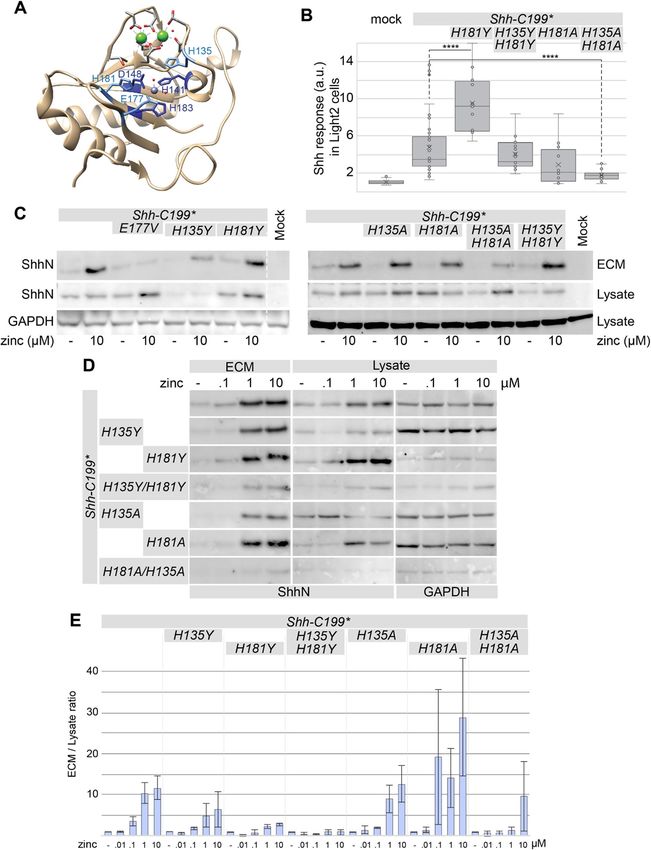
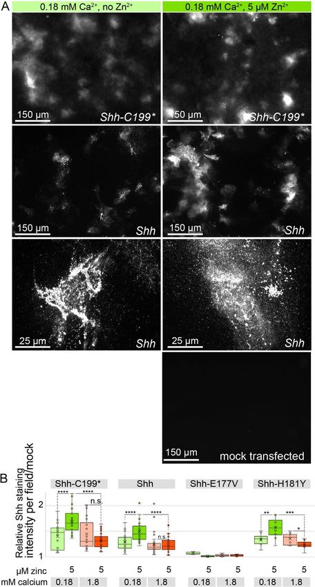
Jägers and Roelink BMC Molecular and Cell Biology   (2021) 22:22   Page 3 of 18

 Fig. 1 (See legend on next page.)

Jägers and Roelink BMC Molecular and Cell Biology        (2021) 22:22                                                                  Page 4 of 18

 (See figure on previous page.)
 Fig. 1 Active ShhN associates with the extracellular matrix. a Lysate and ECM deposited by mock and Shh-C199*-transfected Hek293t cells
 analyzed by SDS-PAGE/SYPRO-Ruby staining and on a Western Blot (H2 α-ShhN). Equal fractions of the total cell lysate and ECM were analyzed.
 ShhN is indicated. b Shh-C199*-transfected Hek293t cells were plated on glass slides and removed after 24 h. The slides were stained with
 mAb5E1, showing the presence of ShhN. Scale bar is 50 μm. c Supernatant and ECM conditioned by Shh-C199*-transfected Hek293t cells. LightII
 cells were either grown on mock or ShhN conditioned ECM. Cells grown on ECM deposited by mock transfected cell were grown in the absence
 or presence of mock or Shh-C199* conditioned supernatant. Box and whisker plots, n ≥ 3. *p < 0.05, ****p < 0.0001. d Hh response in LightII cells
 grown on the decellularized ECM of mock-, Shh-C199*-, or Shh-transfected Hek293t cells. ****p < 0.0001. e Western blot analysis of Hek293t cells
 transfected with the indicated Shh mutants. 100 nM MG-132 (proteasome inhibitor), 100 nM Chloroquine and 100 nM Concanamycin A (inhibitors
 of endosome acidification) were assessed for their ability to affect Shh accumulation. Immune reactive signals are quantified and normalized to
 the untreated and Shh-C199* transfected condition. f Western blot analysis of Hek293t cells transfected with the indicated Shh mutants, and the
 effects of the dynamin inhibitor Dynasore (50 μM) was assessed for its effect on Shh accumulation. Immune reactive signals are quantified and
 normalized to the untreated and Shh-C199* transfected condition. Full-length gels blots are presented in Supplementary Figure 3

that the destabilization of Shh-C199*/H183A occurs be-                     we found the Shh-C199*/E177A and −/E177V mutants
fore it reaches the plasma membrane (Fig. 1f). We found                    to be stable in the lysate (Fig. 2b). Shh-C199*/E177V in
that other zinc coordination mutations as well as several                  decellularized ECM activated the Hh response in LightII
holoprosencephaly-associated point mutations in Shh                        cells to a smaller extend than Shh-C199* (Fig. 2d). This
cause its destabilization, indicating a role for increased                 was likely not caused by lower protein amounts of Shh-
ShhN degradation in this birth defect [28]. In general,                    C199*/E177V in the ECM, as we found that the amount
we will not use these mutants with a reduced half-life.                    of Shh-C199*/E177V in the ECM was similar to that of
                                                                           Shh-C199* under low zinc concentrations but failed to
The zinc-coordination center of Shh is required for                        further accumulate in the ECM under increasing zinc
association with the ECM                                                   concentrations. This demonstrates that ShhE177 is re-
BacHh belongs to a family of metallopetidases that co-                     quired for ECM association (Fig. 2b, c), and importantly
ordinate zinc, and consistent with the absolute conserva-                  that the zinc effects are not primarily mediated by a
tion of the zinc-coordination center, zinc was found in                    Shh-independent zinc sensitive event. These observation
the catalytic cleft of Shh (Fig. 2a, grey sphere). We hy-                  supports the notion that a catalytic activity intrinsic to
pothesized that occupancy of the zinc-coordination cen-                    Shh is required for its association with the ECM.
ter is required for normal Shh function. The Kd for zinc
binding to Shh in the absence of calcium appears to be                     Mutations in additional conserved residues in the zinc-
low [31], but DMEM tissue culture medium has no                            coordination motif of Shh affect association with the ECM
added zinc and is thus expected to have only very small                    A second group of conserved residues in the zinc-
amounts of it. While the amount of protein in the lysate                   coordination motif are two histidine residues with stack-
of Hek293t cells transfected with Shh-C199* remained                       ing sidechains, H135 and H181 (Fig. 3a). These two
relatively unchanged with increasing zinc concentra-                       histidine residues are conserved between Shh and
tions, the amount of ECM-bound Shh-C199* increased                         BacHhs, but either one can be a tyrosine residue in
approximately two and a half times with an EC50 be-                        M15A peptidases, and a tyrosine residue is present in
tween 0.1 and 1 μM zinc (Fig. 2b, c). This indicates that                  the position homologous to H181 in butterfly and moth
there is little effect of zinc on Shh synthesis and intracel-              Hhs (e.g. NCBI PCG69308.1). We mutated either or
lular stability, but that occupancy of the zinc coordin-                   both histidine residues 135/181 into alanine or tyrosine
ation center enhances ECM association. Divalent copper                     residues (Shh-C199*/H135YorA, Shh-C199*/H181YorA)
and magnesium failed to increase the amount of ShhN                        and found that these forms of Shh process normally and
in the ECM (Fig. S2). Calcium, however, did have an ef-                    are stable in the lysate (Fig. 3c). Substituting one or two
fect consistent with its ability to bind to ShhN, and is                   histidines with tyrosines had little effect on the mutant’s
further addressed below.                                                   ability to elicit a Hh response in LightII cells from condi-
  Besides the zinc coordinating residues, the glutamic                     tioned ECM (Fig. 3b). Alanine substitutions reduced the
acid residue at position 177 (E177, mouse numbering) is                    Hh response in the LightII compared to Shh-C199* (Fig.
well-conserved and in close proximity to the zinc-                         3b). Similar to Shh-C199*/E177V, the reduced Hh re-
coordinating residues (Fig. 2a). In Shh-related peptidases                 sponse coincided with lower protein levels in the ECM.
the E177 equivalent is required for catalytic activity as it               Mutants with one or two tyrosine substitutions as well
strips a proton from water yielding a reactive hydroxide.                  as single alanine substitutions could be rescued under
The mutants Shh-C199*/E177A and −/E177V are pre-                           high zinc conditions (1, 10 μM) suggesting that tyrosine
dicted to be able to coordinate zinc but lack the putative                 residues, as they are found in butterfly and moth Hhs,
catalytic activity. Unlike the zinc coordination mutants,                  are largely synonymous mutations. Only Shh-C199*/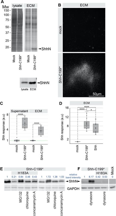
Jägers and Roelink BMC Molecular and Cell Biology        (2021) 22:22                                                                   Page 5 of 18

 Fig. 2 ECM-association of ShhN requires zinc and the catalytic E177V residue. a Diagram of the Shh structure (PDB: 3D1M) with relevant residues
 indicated. b Western blot analysis of the lysate and ECM of Hek293t cells transfected with Shh-C199* and Shh-C199*/E177V and cultured in DMEM
 containing 0.18 mM calcium and the indicated concentrations of zinc. Uncropped blots can be found in the supplement. c Quantification of
 band intensities shown in C as a fraction of ECM over lysate, normalized to the respective mock treatment to better illustrate the effects of zinc
 treatment. n = 2. d Hh response in LightII cells grown on the conditioned and decellularized ECM of Hek293t cells transfected with Shh-C199* or
 Shh-C199*/E177V. n ≥ 3, *p < 0.05, ****p < 0.0001. Full-length blots are presented in Supplementary Figure 4

H135A/H181A poorly associated with the ECM in the                           peptidoglycan peptidases we tested if this bacterial pep-
presence of zinc. We found that all H135 and H181 mu-                       tidase activity could substitute for the putative peptidase
tants have a similar EC50 for zinc (Fig. 3d and quantified                  activity intrinsic to ShhN. The conservation between
in Fig. 3e), consistent with the notion that these residues                 BacHhs and Shh involves the calcium and zinc binding
are not directly involved in zinc coordination. Together                    motifs, but not the N-terminal domain that binds to
with Shh-C199*/E177V, our findings using Shh-C199*/                         Ptch1 and Heparan Sulfate [24], nor the 10 amino acids
H135A/H181A further support the notion that the zinc-                       that follow this domain in ShhN. Therefore, we made a
coordination center of Shh is required for ECM                              construct coding for a chimeric protein consisting of the
association.                                                                N-terminal 65 residues of Shh, the conserved calcium
                                                                            and zinc binding motifs of Bradyrhizobium paxllaeri
The peptidase domain of a BacHh is unable to facilitate                     BacHh (codon optimized for expression in mammalian
ECM association                                                             cells), followed by an HA tag replacing the bacterial stop
The protein sequences of bacterial Hhs (BacHh) are                          codon, followed by the last 10 residues of Shh up to
highly similar Shh and all residues of the zinc coordin-                    G198 (Shh/BacHhHA, Fig. 4a diagram). As a control, we
ation motif are identical. As BacHhs are predicted to be                    positioned an HA tag at the same distance (10 residues)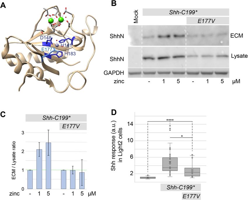
Jägers and Roelink BMC Molecular and Cell Biology   (2021) 22:22   Page 6 of 18

 Fig. 3 (See legend on next page.)

Jägers and Roelink BMC Molecular and Cell Biology        (2021) 22:22                                                                  Page 7 of 18

 (See figure on previous page.)
 Fig. 3 Stacking histidines of the zinc coordination center affect ECM association. a Diagram of the Shh structure (PDB: 3D1M) with relevant
 residues indicated. b Hh response in LightII cells grown on the conditioned and decellularized ECM of Hek293t cells transfected with the
 indicated Shh-C199* constructs. n ≥ 2, *p < 0.05, ****p < 0.0001. c The effect of mutations of the transition state-stabilizing residues H135 and
 H181 to alanine (A) or tyrosine (Y) on the zinc-dependent accumulation in the ECM was analyzed on a Western Blot of the extracted ECM from
 transfected Hek293t cells cultured in 0.18 mM calcium with or without 10 μM zinc. d zinc dose-response analysis of H135 and H181 mutations
 assessed by Western blot of the lysate and ECM of Hek293t cells transfected with the indicated mutants and cultured in 0.18 mM calcium and
 increasing concentrations of zinc (0.1, 1, 10 μM). Uncropped blots can be found in the supplement. e Quantification of band intensities shown in
 D as a fraction of ECM over lysate, normalized to the respective mock treatment. n = 2. Uncropped, full-length blots are presented in
 Supplementary Figure 5

from the C-terminus of Shh-C199* (ShhHA-C199*). We                          residues (Shh-C199*/E90A/E91D/D96A/E127A/D130N/
found that ShhHA-C199* behaved indistinguishable from                       D132L, Shh-C199*-CaFree) and this form of Shh should
Shh-C199* and entered into the ECM and the medium                           be unable to bind calcium. After transfection, more
in a zinc-dependent manner (Fig. 4a). In contrast, al-                      ShhN was detected in lysates of cells cultured in the
though readily detected in the lysate, no Shh/BacHhHA                       presence of higher calcium levels, but that was also ob-
was detected in the ECM or the medium (Fig. 4a). We                         served in the Shh-C199*-CaFree expressing cells, and
detected similar amounts of GAPDH in the medium as                          thus unlikely a direct effect of calcium on Shh (Fig. 5a).
in the cell lysate. GAPDH is commonly used as a loading                     Increased amounts of ShhN in the lysate at higher cal-
control for intracellular proteins but has also been de-                    cium concentrations complicated the interpretation of
scribed as a soluble form found in the extracellular com-                   the effects of calcium on ShhN accumulation in the
partment [32]. Cell lysis in the tissue culture as an                       ECM. Sill, while ShhN accumulation in the ECM varied
explanation for GAPDH in the medium seems unlikely,                         with calcium concentrations, that of the Shh-C199*-
as no Shh/BacHhHA was detectable in the soluble                             CaFree mutant remained at the same level, indicating that
fraction. As ShhN can be internalized by several Shh-                       this mutant is insensitive to extracellular calcium as
binding proteins, [33–35], we assessed whether the                          measured by ECM association (Fig. 5a).
chimeric proteins accumulates on the outside of cells                         One possible mechanism of calcium regulating the
with detergent-free, live staining with an α-HA antibody                    transition from a cell- to an ECM-bound state would be
prior to fixation of transfected receptor-less (Ptch1Lac-                   by affecting zinc coordination, thereby changing its Kd
Z/LacZ
       ;Ptch2−/−;Boc−/−;Cdo−/−;Gas1−/−) fibroblasts. We                     for zinc. We therefore tested if the EC50 of zinc is differ-
found no difference in staining between ShhHA-C199*                         ent under high (1.8 mM, the concentration in regular
and Shh/BacHhHA, indicating that Shh/BacHhHA, simi-                         DMEM) and low (0.18 mM, the lowest concentration
larly to ShhHA-C199*, is being trafficked to the plasma                     the cultured cells appeared normal) calcium. Under low
membrane (Fig. 4b). It is not, however, being released                      calcium conditions, the addition of 5 μM zinc to the
from the cell, indicating that the bacterial zinc-                          medium resulted in increased accumulation of ShhN in
coordination domain is not sufficient for entry into the                    the ECM both of Shh-C199*-CaFree and Shh-C199* (Fig.
ECM. Bradyrhizobium paxllaeri BacHh presumably                              5a). This indicates that Shh-C199*-CaFree is still active,
lacks the specificity for an ECM binding partner that is                    and supports the notion that calcium binding is not re-
recognized by Shh. These results suggest that the ob-                       quired for Shh distribution. E127 is located at the inter-
served presence of Shh and ShhN in the decellularized                       face between the calcium and zinc-binding centers of
tissue culture plate is due to a precise property of the                    Shh, and we tested if restauration of this residue in Shh-
Shh rather than simple cell lysis or ShhN-containing cell                   C199*-CaFree affects ECM localization but found little or
debris.                                                                     no difference (Fig. 5a). To better quantify the effect of
                                                                            calcium and zinc on Shh-C199* and Shh-C199*-CaFree in
Shh-C199* mutants unable to bind calcium remain                             their ability to accumulate in the ECM we used an indir-
sensitive to zinc                                                           ect ELISA protocol directly on the decellularized ECM.
The overall structure of ShhN and the BacHhs indicate                       Hek293t cells were cultured and transfected in 96 well
that they consist of a regulatory calcium-binding and a                     plates and washed off with PBS to allow for subsequent
catalytic zinc coordinating center [14], making up most                     detection of ShhN with HRP-linked antibodies. Under
of ShhN outside the extreme N-terminal Ptch1-binding                        low calcium conditions we found that the response to
domain. With the exception of BacHhs, bacterial M15A                        increasing zinc concentrations was similar between Shh-
metallopeptidases lack the Hh/BacHh-type calcium co-                        C199* and Shh-C199*-CaFree (Fig. 5b). For both, the
ordination center, and this part is thus unlikely to be                     EC50 for zinc appeared to be around 0.1 μM. Instantiated
required for catalytic function per se. We made a Shh-                      in Fig. 5a and quantified over multiple experiments in
C199* mutant that lacked all calcium-coordinating                           Fig. 5b, it appears that Shh-C199*-CaFree is less efficient
Jägers and Roelink BMC Molecular and Cell Biology (2021) 22:22 Page 8 of 18 Fig. 4 The zinc-coordination domain of BacHh is not sufficient for association with the ECM. a Western Blot analysis of the lysate, ECM, and supernatant of ShhN-HA or Shh-BacHh-HA (diagrams) transfected Hek293t cultured in the indicated zinc concentrations. Uncropped blots can be found in the supplement. b Detergent-free live staining with an α-HA antibody (3F10) of transfected Ptch1LacZ/LacZ;Ptch2−/−;Boc−/−;Cdo−/−;Gas1−/− cells. Nuclei were stained with DAPI. Uncropped, full-length blots are presented in Supplementary Figure 6
Jägers and Roelink BMC Molecular and Cell Biology        (2021) 22:22                                                                   Page 9 of 18

 Fig. 5 Calcium alters the sensitivity of Shh to zinc. a Western blot analysis of lysates and ECM of Hek293t cells transfected with Shh-C199* and
 Shh-C199*/E90A/E91D/D96AD130N/D132L, or Shh-C199* and Shh-C199*/E90A/E91D/D96A/E127A/D130N/D132L (CaFree), cultured in the presence of
 0.18 or 1.8 mM calcium, and in the absence or presence of 5 μM added zinc. b ECM-associated Shh-C199*N or Shh-C199*/CaFree deposited by
 transfected Hek293t cells was assessed by ELISA in the presence of .18 mM calcium (left panel) or 1.8 mM calcium (right panel) and increasing
 zinc concentrations as indicated. Shown are means and standard errors, n = 6. Uncropped, full-length blots are presented in
 Supplementary Figure 7

in entering the ECM than Shh-C199*. This effect was                           The effects of zinc on Shh distribution in the presence
more profound in the presence of 1.8 mM calcium, and                        of 1.8 mM calcium was much less pronounced, further
much more Shh-C199* was detected in the ECM than                            supporting the finding that high calcium negatively af-
Shh-C199*-CaFree in the absence of added zinc. The                          fects the zinc-dependent activity of Shh.
addition of zinc had a bigger effect on Shh-C199*-CaFree                      To assess if the observed effects require the zinc-
than on Shh-C199*. These results indicate that Shh-                         coordination center of Shh we tested Shh-E177V, and
C199*-CaFree behaves similarly in high and low calcium                      -H181Y for their ability to associate with the ECM.
and resembles Shh-C199* under low calcium. Thus,                            Consistent with the biochemical observations using Shh-
whereas the behavior of Shh-C199* changes as a func-                        C199* (Figs. 1, 2, 3), we could barely visualize ShhE177V
tion of calcium, that of Shh-C199*-CaFree does not,                         in the ECM (Fig. 6b). In contrast, ShhH181Y distribution
indicating that binding of calcium to Shh alters its in-                    into the ECM was indistinguishable from Shh, further
trinsic properties as measured by its ECM association.                      indicating that this “butterfly version” of Shh is func-
The calcium concentration in the endoplasmic reticulum                      tional (Fig. 6b).
and the Golgi apparatus is variable, with values ranging
from .2-1 mM [36]. These concentrations that Shh en-                        Mutations in the putative peptidase domain reduce non-
counters in these organelles are within the range in                        cell autonomous signaling
which we observe changes in zinc sensitivity of ShhN.                       To test if the increased accumulation of Shh in the ECM
This is consistent with the notion that the activity of                     is correlated with the non-cell autonomous signaling ef-
ShhN could be regulated by calcium.                                         ficacy, we assessed the signaling activity of ShhN
                                                                            expressing cells embedded in three-dimensional cell ag-
Distribution of cholesterol-modified ShhNp in the ECM                       gregates of responding cells (Fig. 7a). Live staining with
differs from cholesterol-unmodified ShhN but remains                        5E1 prior to fixation and cell permeabilization showed
zinc sensitive                                                              that ShhNp and ShhC199* were detected around a large
While ShhN could readily be detected in the ECM of                          proportion of cells in transfected Hek293t aggregates. In
Hek293t cells, ShhNp was poorly detectable on Western                       mixed aggregates consisting of 80% Hh-responsive
Blots of the ECM fraction of Hek293t cells. We therefore                    Ptch1LacZ/+;Shh−/− mouse embryonic stem cell (mESC)
turned to staining of decellularized ECM of our line of                     reporter cells and 20% transfected Hek293t cells (Fig.
fibroblasts lacking Shh binding partners (Ptch1LacZ/LacZ;                   7b), we observed that both Hek293t-derived ShhNp and
Ptch2−/−;Boc−/−;Cdo−/−;Gas1−/−) that were found to accu-                    Shh-C199* induced the Shh response (Fig. 7c). Shh pep-
mulate ShhNp in the ECM to comparable levels as                             tidase mutants (Shh-E177V and Shh-H135A/H181A)
ShhN, presumably due to a failure to re-internalize                         failed to elicit a Hh-response, further supporting the no-
ShhN(p) (Fig. 6a and b). Staining for Shh on decellular-                    tion that the peptidase activity is required for non-cell
ized plates showed that ShhN was present in small                           autonomous signaling (Fig. 7c). The pattern of Shh
puncta that gave a cloudy appearance at lower magnifi-                      extracellular distribution was similar for Shh and Shh-
cations, whereas cholesterol-modified ShhNp was de-                         E177V as seen in detergent-free live staining with 5E1,
tected in larger puncta in more restricted areas (Fig. 6a).                 although slightly weaker staining for Shh-E177V was ob-
5 μM zinc increased ShhN and ShhNp association with                         served (Fig. 7d). In a more stringent non-cell autono-
the ECM as measured by fluorescence intensity across                        mous assay, aggregated of transfected Hek293t cells
the entire image area (Fig. 6b).                                            were co-cultured with 3D spinal cord organoids (SCOs)
Jägers and Roelink BMC Molecular and Cell Biology (2021) 22:22 Page 10 of 18 Fig. 6 (See legend on next page.)
Jägers and Roelink BMC Molecular and Cell Biology       (2021) 22:22                                                                Page 11 of 18

 (See figure on previous page.)
 Fig. 6 Cholesterol-modified ShhNp associates with the ECM in a zinc and peptidase-dependent manner. a In situ staining with anti ShhN
 mAb5E1 of ECM deposited by Ptch1LacZ/LacZ;Ptch2−/−;Boc−/−;Cdo−/−;Gas1−/− cells that were transfected with Shh or Shh-C199* in the presence or
 absence of 5 μM zinc and presence of 0.18 mM calcium. b Quantification of ECM-bound Shh shown in A. Box and whisker plots of mean
 fluorescence intensity per image of 10 microscope fields per experiment was measured in ImageJ and normalized to the ECM of mock
 transfected cells. n = 3, ****p < 0.0001)

derived from the mESC Ptch1LacZ/+;Shh−/− reporter cells                    strongly suggest they have similar functions. It is striking
(Fig. 7e). Whereas the Hh response to ShhNp was below                      that the conservation between BacHhs and animal Hhs
detection level in this assay, Shh-C199* induced the Shh                   is much greater than between Hedglings and Hhs (Fig.
response in reporter cells (Fig. 7f), likely via a soluble                 S1 A,B,D), with mollusk, echinoderm and cephalochord-
form of ShhN. Putative peptidase mutants failed to show                    ate Hedglets even less conserved (Fig. S1 A). The con-
any activity in this assay, further supporting the idea that               servation between BacHhs and animal Hhs, but not
an intact zinc-coordination center of Shh is necessary to                  Hedglings and Hedglets, includes all residues critical for
mediate the release from Shh-producing cells and conse-                    catalysis and calcium binding further supporting the no-
quently, the facilitation of long-range signaling.                         tion that both are calcium regulated peptidases. The
                                                                           organization of bacterial genomes into operons helps in
Discussion                                                                 the assignment of possible functions of unknown
Here, we provide evidence for a function of the zinc-                      proteins. The putative role of BacHh (as a M15A
coordination center of Shh for the association of Shh to                   peptidase [37]) in the modification of the bacterial
the ECM and non-cell autonomous signaling. The zinc                        peptidoglycans is further supported by the observation
coordination domain of Shh appears to be involved as                       that in Mesorhizobium and Bradyrhizobium the BacHh
μM amounts of zinc are required for ECM association,                       gene is surrounded by genes (likely constituting an
while mutants in the zinc coordination domain are in-                      operon) that code for proteases, including lysozyme, N-
sensitive to zinc in the medium and have reduced activ-                    acetylmuramoyl-L-alanine amidase, a peptidoglycan
ity in non-cell autonomous signaling. The observation                      endopeptidase (peptidase M23A), several Trypsin homo-
that Shh-E177A is unable to mediate signaling from the                     logs (peptidase S1), a zinc Matrix Metalloprotease
notochord to the overlying neural tube (a textbook ex-                     (MMP) homolog (peptidase M54), an endonuclease,
ample of non-cell autonomous Shh signaling), but is                        peptidase S53, and possibly a Phytase (DUF3616). This
fully capable to induce the Hh response when expressed                     complex of enzymes might be involved in bacterial feed-
in the developing neural tube (likely cell-autonomously)                   ing or scavenging. BacHhs in Rhizobiacea are not part of
[7], provides in vivo evidence that the zinc-coordination                  the core genome [38], as the majority of these bacteria
domain of Shh is required for non-cell autonomous sig-                     do not carry BacHh, a further indication that BacHhs
naling, although not for the productive binding of Shh                     provide a niche-specific specialized function. It remains
to its cognate receptors. The initial experiments that                     plausible that this niche function in bacteria was useful
demonstrated that E177 is dispensable for the activation                   in animal development after a gene transfer event into a
of the Hh response is easily explained as this mutant lig-                 cnidarian ancestor. Hedgling distribution in porifera and
and was added to the responding cells as a purified and                    choanoflagellates [39] indicates that they are phylogenet-
soluble fraction [4], thus bypassing the requirement for                   ically older, while the curious distribution of Hedglets in
the function of the zinc-coordination domain. This is                      mollusks and basal deuterostomes remains enigmatic.
further supported by structural analysis of the Ptch1/Shh                  Still the loss of catalytically important residues in Hedg-
complex demonstrating that the extreme N-terminus of                       lings and Hedglets further emphasize that Hhs share a
Shh interacts with Ptch1 [10–12] and suffices to alter                     catalytic function with (the likely ancestral) BacHhs.
Ptch1 activity [13]. These observations further demon-                        Possible mechanisms of catalysis of zinc peptidases
strate the dispensability of the zinc-coordination domain                  have been elucidated with the help of structural models
to activate the Shh response in the responding cell.                       of enzyme-inhibitor complexes. Thermolysin is a well-
                                                                           studied zinc metallopeptidase structurally related to Shh
Do bacterial Hhs and Shh share a peptidase activity?                       [3, 14]. Shh and Thermolysin coordinate zinc via two
Our observations indicate that Shh distribution away                       histidine and an aspartic acid residue. A catalytic glu-
from the sites of synthesis and non-cell autonomous Shh                    tamic acid residue initiates catalysis (E177 in mouse
signaling can be enhanced under low-calcium and high                       Shh) by accepting a proton from water to form the nu-
zinc conditions. The surprising sequence similarity be-                    cleophilic hydroxide that attacks the carbonyl carbon,
tween bacterial and mammalian Hedgehog proteins                            further stabilized by the coordinated zinc. With the two
Jägers and Roelink BMC Molecular and Cell Biology (2021) 22:22 Page 12 of 18 Fig. 7 (See legend on next page.)
Jägers and Roelink BMC Molecular and Cell Biology         (2021) 22:22                                                                  Page 13 of 18

 (See figure on previous page.)
 Fig. 7 The zinc coordination center of Shh is required for long-range signaling. a Experimental setup and immunofluorescent staining of Shh-
 expressing Hek293t aggregates. Hek293t cells were transfected with Shh or Shh-C199*, washed off the culture dish with PBS and placed on a
 rotating platform in DMEM without serum for 2 days, followed by live staining with 5E1. Shades of brown represent differences in transfection
 efficiency. b Experimental setup: Hek293t cells were transfected with Shh-constructs, washed off the culture dish with PBS, and mixed with mESC
 Ptch1LacZ/+;Shh−/− reporter cells in a 1:4 ratio. Chimeric aggregates were then formed on a rotating platform in the absence of serum. The
 Ptch1:LacZ expression was measured after 3 days and normalized to total protein content. c Ptch1:LacZ expression in chimeric aggregates in
 response to the indicated versions of Shh expressed by Hek293t cells. Box-and-Whisker plot, n = 2. *p < 0.05, **p < 0.01, ****p < 0.0001. d 5E1 live
 staining of Shh in the chimeric aggregates described in B and C. e Experimental setup: Hek293t cells were transfected with Shh-constructs,
 washed off the culture dish with PBS, and incubated on a rotating platform. Separate mESC Ptch1LacZ/+;Shh−/− reporter aggregates were added to
 the dish in a 1:4 ratio and Ptch1:LacZ expression in mESCs was measured after 3 days. f Ptch1:LacZ expression normalized to total protein in
 mESC aggregates co-cultured with Shh-expressing Hek293t aggregates. Box-and-Whisker plot, n = 4, ****p < 0.0001

stacking histidine (or occasionally tyrosine) residues a                     the Ur-eukaryote, but more likely are products of more
pentacoordinate transition state is formed and resolved                      recent gene transfers from bacteria. The distribution
into the hydrolyzation products [40, 41].                                    among eukaryotes of Glypicans and Hs is overlapping,
   M15 peptidases cleave peptidoglycans, the major com-                      and both are first observed in Cnidarians and present in
ponent of the bacterial periplasmic space, and a major                       all bilaterians. A more recent evolutionary relationship
component of detritus. Peptidoglycans are analogs of                         between BacHh and Hhs is further supported by the ob-
proteoglycans that are common in extracellular matrix                        servation that the C-terminal residue of many BacHhs
(ECM) of animals. Therefore, it is possible that func-                       perfectly aligns with the exon 2 splice donor site in Hh
tional conservation between BacHhs and Shh is reflected                      genes, thereby providing a parsimonious explanation
in the ability of Shh to cleave or modify proteoglycans,                     how a BacHh gene was incorporated in a eukaryotic
thus affecting the Shh response and/or distribution, in-                     genome giving rise to Hh. This is in contrast to the
dependent of its binding to the canonical Hh receptors.                      much less conserved Hh domain in Hedgling that is
Although any Shh antagonist could be a possible tar-                         encoded within a single large exon. Given the central
get for the putative Shh peptidase activity, the Hh-                         role of Gpcs in the distribution of and response to Hhs
interacting protein (Hhip) is an unlikely substrate                          (including Shh), Glypicans and Hhs might have co-
candidate as it binds to Shh via the zinc ion, thereby                       evolved possibly as a peptidase/substrate combination,
replacing the catalytic water. This mode of binding is                       co-opting the peptidoglycan activity of BacHhs to cleave
akin to that of a metalloprotease/inhibitor interaction                      the proteoglycan Glypican. Shh binds heparan sulfate
[5], and thus likely to inhibit the putative catalytic                       (HS), the O-linked glycosaminoglycan sidechain of Gpcs
function of Shh instead of being a substrate. Still, it                      and other proteoglycans [44], via the N-terminal Cardin-
leaves open the possibility that the main mechanism                          Weintraub motif, which plays a crucial role in the re-
by which Hhip inhibits Hh signaling is not only via                          lease of Shh from the producing cell [24]. Although HS
ligand sequestration [42, 43], but actually by repres-                       is thought to merely aid in scaffolding of a release com-
sing the peptidase activity of Shh.                                          plex [45, 46], our results could hint to an additional role
                                                                             for HS in guiding Shh to its peptidase target. We cannot
Are Hhs proteoglycan peptidases?                                             exclude that at least part of the signal detected in the
Hedglings and Hedglets are related to Hhs, and the con-                      Sypro-ruby staining of the ECM extract of ShhN-trans-
served domains possibly homologous. All animals that                         fected cells (Fig. 1a) is in fact cleaved substrate.
have Hedglet also have a Hh gene and it is thus plausible                       Hh-like bacterial peptidases (M15A) are predicted
that Hedglets are derived from Hh. Hhs are not found in                      to be carboxy (trans) peptidases, cleaving adjacent to
any eukaryote except cnidarians and bilaterians. The                         the D-ala that is linked to the murein glycans [47,
distribution of Hedgling and Hhs only overlaps in cni-                       48]. By analogy, Shh might cleave an unusually modi-
darians, but Hedgling can also be found in sponges and                       fied C-terminal residue. It is intriguing that the C-
choanoflagellates (Fig. S1). This suggests two evolution-                    termini of Glypicans are linked to the GPI anchor
ary events giving rise to these proteins; one occurring in                   that restricts them to the cell surface [49].
a Choanoflagellate ancestor that originated the gene                         Solubilization of Shh-sequestering glypicans by GPI
coding for Hedgling, and an independent event in a Cni-                      removal would elegantly reconcile the observed
darian ancestor that gave rise to modern Hh. The ab-                         peptidase-dependent entry of Shh into the ECM with
sence of both Hedgling and Hh from algae, plants, fungi,                     the important effects of glypicans on Shh signaling
in addition to almost all unicellular eukaryotes makes it                    and distribution. If Shh remains attached to its poten-
unlikely that both Hh and Hedgling linearly evolved                          tial substrate after cleavage and enters the ECM in a
from a BacHh protein that could have been present in                         complex or alone remains unresolved.
Jägers and Roelink BMC Molecular and Cell Biology (2021) 22:22 Page 14 of 18
Drosophila is the exception peptidase activity that can modify ECM components. In
It is perhaps unfortunate that Hh was discovered in addition, Shh is subject to further N-terminal processing,
Drosophila, as of all animals sequenced, only Hh in Dro- or “shedding”, prior or concomitant to its release from
sophilids is divergent for two of the three residues that the cell [53], but this event can be mediated by a family
coordinate zinc and has a valine residue at the equiva- of zinc-metalloproteases called a disintegrin and metal-
lent position of the critical E177. The predicted lack of loprotease (ADAM), in concert with the scaffolding pro-
peptidase activity in Drosophilid Hhs is remarkable and tein Scube2 [54]. These metalloproteases share
further supports the observation that the putative pep- overlapping functions and are therefore sufficient, but
tidase activity is not required for the Hh/receptor inter- not essential, for Shh shedding [55], hinting at the var-
action. Perhaps stricter reliance on cytonemes in iety of paths Shh can follow to enter the extracellular
Drosophila that detect Hh at its source [50] renders the space.
ancestral peptidase activity obsolete. Nevertheless, this Although mutations of the central zinc coordinating
loss of the putative peptidase activity is unique to Droso- triad are unstable and thus cannot be easily assessed for
philids, as all other (sequenced) animals retain the typ- loss of peptidase function, mutations of several other
ical zinc coordination motif and the associated E177 catalytically important residues (E177, H135 and H181)
equivalent that are required for catalytic activity. Based are not destabilized and show a loss in the ability of Shh
on the loss-of-function of several mutants, this intrinsic to enter the ECM. Together with the observation by
property is likely a zinc metallopeptidase activity, just Himmelstein and colleagues [7] that ShhE177A cannot
like the bacterial counterparts of Shh. Still, the observa- signal from the notochord to the overlying neural plate
tion that substitution of the Shh calcium/zinc sequences strongly supports the idea that a Shh-associated peptid-
with those of BacHh results in a protein that does not ase activity is required for non-cell autonomous signal-
enter the ECM, indicates that their substrates are not ing by promoting its distribution away from the source
interchangeable. cells. The implications of a peptidase function that is in-
trinsic to Shh for normal, non-cell autonomous signaling
Is Shh oligomerization preventing ECM association? are significant. The Zn-coordination domain is found
The zinc coordination center of Shh and in particular mutated in some individuals with the Shh signaling-
E177 are disadvantageous for Shh multimerization [7]. related birth defect holoprosencephaly [8, 9], further
Zinc prevents oligomerization and we find that zinc is a indicating that the intrinsic peptidase activity of Shh is
potent agonist of ECM association and putative peptid- important for normal function. Furthermore, a catalytic
ase activity. Furthermore, while the E177A mutant pre- interaction between Shh and HSPGs that affects non-cell
vents ECM association it enhances oligomerization [7]. autonomous signaling would provide significant new in-
These observations are consistent with the idea that the sights in developmental and tumor-inducing mecha-
putative peptidase activity of Shh prevents or reverts nisms, including the roles of Ext1 and Ext2 as tumor
multimerization. This notion is further supported by the suppressors [56], roles of Gpcs as tumor suppressors
structural observation that the cholesterol-modified C- [57, 58] a role for extracellular calcium as an inhibitor of
terminus is in close proximity to the zinc coordination Shh that contributes to Shh-mediated morphogenesis, or
center, raising the hypothesis that Shh could have that affects tumor formation or progression, and a re-
intermolecular autoproteolytic activity that prevents quirement for μM amounts of zinc for Shh activity in
oligomerization [3]. Indeed, multimer size has been re- general.
ported to be negatively correlated with long-range sig- Our findings challenge the dogma of the so called
naling activity of Shh [51]. This observation could “pseudo catalytic domain” of Shh, and we provide a
explain why cholesterol-unmodified Shh-C199*, which function for the apparent peptidase activity intrinsic to
would be expected to form multimers less efficiently due ShhN. None of our observations are in conflict with pre-
to the absence of cholesterol, is more readily detectable vious reports, as we confine the activity of the Shh-
in the ECM of transfected Hek293t cells and is an effi- associated peptidase upstream of the interaction between
cient non-cell autonomous ligand. Although an inter- or Shh and its receptors.
intramolecular autoproteolytic activity has not been
demonstrated for ShhNp, many peptidases are produced Conclusions
as pro-proteins that undergo either an intra- or intermo- Our observations support the notion that the remarkable
lecular activation event. The intramolecular activation sequence similarity between the BacHhs and Shh reflects
events are often auto-catalyzed by the intrinsic peptidase to a conserved function as a glycopeptidase. All ShhN
domain [52]. If Shh has retained these bacterial charac- mutants that are predicted abolish its intrinsic peptidase
teristics it is possible that the autocatalytic de- activity fail to bind to the ECM and have an impaired
oligomerization event results in a form of ShhN with a ability to signal non-cell autonomously. However, BacHhJägers and Roelink BMC Molecular and Cell Biology (2021) 22:22 Page 15 of 18
cannot mediate the activities of ShhN, indicating that from IDT DNA, and cloned into pcDNA3.1(+). Both the
the substrates for BacHh are not conserved in Shh-C199* vector backbone including the Shh N- and C-
metazoans. terminus as well as the calcium and zinc coordination
motifs of Bradyrhizobium paxllaeri BacHh were PCR
Methods amplified, separated on a 1% agarose gel, and extracted
Sequence analysis with MinElute columns (QIAGEN). The fragments were
Bacterial Hedgehogs, Hedglings and Hedglets were iden- cut with BsaI and ligated with T4 DNA ligase according
tified via protein-protein BLAST (NCBI) and HMMER to the Golden Gate cloning protocol (New England
(ensemble) searches [59] using the peptide sequence of Biolabs).
the Shh N-terminal domain as the initial query se-
quence. Conserved sequences were manually curated to Immunostaining
contain only the calcium and zinc coordination motifs Ptch1−/−;Ptch2−/− fibroblasts were plated on 12 mm glass
(around 105 residues). Sequences (supplementary file) cover slips and transfected with Shh-C199* the following
were aligned in Clustal Omega (EMBL-EBI). An average day and subsequently allowed to recover for 24 h. The
distance tree and a PCA plot were generated in Jalview transfected cells were then incubated for 24 h in serum-
[60], using the BLOSUM62 algorithm. Visualizations of free DMEM containing varying concentrations of CaCl2
the ShhN structure were generated in UCSF Chimera or ZnCl2, the cells were detached from the cover slip
using Protein Database (PDB) ID 3D1M [61]. with PBS. The cover slips were washed with PBS at least
5 times and blocked with 10% heat-inactivated goat
Materials serum in PBS with 0.1% TritonX (PBS-T). Mouse α-Shh
MG-132 and Concanamycin A were from Calbiochem, (5E1, Developmental Studies Hybridoma Bank) was used
Chloroquine and ZnCl2 from Sigma, CaCl2 from Fisher at 1:30 in blocking solution and goat α-mouse Alexa568
Scientific, and Dynasore from Abcam. secondary antibody (Invitrogen) at 1:1000 in blocking
solution. Shh distribution was visualized with a Zeiss
Cell culture Observer at 10x and 63x magnification.
Ptch1−/−;Ptch2−/− fibroblasts were derived from mouse For live staining, transfected cells were incubated in
embryonic stem cells received from Allen Bradley (AB1) serum-free medium for 20 h. An α-HA antibody 3F10
and are described elsewhere [28]. Ptch1LacZ/LacZ;Ptch2−/−; (Sigma) was added for another 4 h (1:1000) before cells
Boc−/−;Cdo−/−;Gas1−/−;Shh−/− were derived from were fixed with 4% PFA in PBS and blocked in 10%
Ptch1LacZ/LacZ;Ptch2−/−;Shh−/− cells and are described heat-inactivated goat serum in PBS. A goat α-rat
elsewhere [28]. No mice were directly involved in this Alexa488 secondary antibody (Invitrogen) was used at 1:
study. Hek293t cells were purchased from ATCC. All 1000 in blocking solution and nuclei stained with DAPI.
cells were cultured in DMEM (Invitrogen) supplemented
with 10% FBS (Atlas Biologicals). Mouse embryonic stem Western blot/SYPRO ruby staining
cells were cultured in DMEM (Invitrogen) supplemented Hek293t cells were plated in 12 well plates and trans-
with 20% FBS (Atlas Biologicals), 2 mM L-Glutamine fected with Shh mutants as indicated the next day.
(Gibco), 1X MEM non-essential amino acids (Gibco), Twenty-four hours after transfection, the medium was
0.001% 2-Mercaptoethanol (Gibco), and Leukemia In- switched to serum free DMEM with the indicated cal-
hibitory Factor (LIF) titrated to optimal growth support cium and zinc concentrations overnight. Cells were then
(0.001%). Cells were transfected using Lipofecta- detached from the plate with PBS and lysed in a micro-
mine2000 reagent (Invitrogen) according to the manu- centrifuge tube with RIPA buffer (150 mM NaCl, 50 mM
facturer’s protocol. Tris-HCl, 1% Igepal, 0.5% Sodium Deoxycholate, and
protease inhibitors). The lysate was incubated for 30 min
DNA constructs on ice and cleared by centrifugation. For isolation of
The following mutations were created via site-directed ECM-bound Shh, the decellularized tissue culture dish
mutagenesis: Shh-C199*/E177V, Shh-C199*/H135A, Shh- was washed with PBS and deionized water at least 5
C199*/H135Y, Shh-C199*/H181A, Shh-C199*/H181Y, times and scraped with a cell scraper and 5X SDS sam-
Shh-C199*/H135A/H181A, Shh-C199*/H135Y/H181Y, ple buffer heated to 95 °C, as described [23]. A fifth of
Shh-C199*/E90A/E91D/D96A/D130N/D132L, Shh-C199*/ the sample was run on a 12% SDS-PAGE gel and trans-
E90A/E91D/D96A/E127A/D130N/D132L. Bradyrhizo- ferred to a 0.45 μ nitrocellulose membrane. Membranes
bium paxllaeri BacHh (EnsemblBacteria: LMTR21_38280, were blocked in 5% milk in Tris-buffered saline with
NCBI: WP_065756078.1) was codon optimized for 0.1% Tween-20 (TBS-T) and incubated with a polyclonal
eukaryotic expression using the IDT DNA Codon rabbit α-Shh antibody (H2, 1:10,000) [62] in blocking so-
Optimization Tool, ordered as a gBlocks gene fragment lution, followed by incubation with a goat α-rabbitJägers and Roelink BMC Molecular and Cell Biology (2021) 22:22 Page 16 of 18
Alexa647 secondary antibody (Invitrogen, 1:10,000) in once in PBS, and lysed in 100 mM potassium phosphate,
blocking solution. GAPDH was used as a loading control pH 7.8, 0.2% Triton X-100. The lysates were cleared by
(Rabbit α-GAPDH, 14C10, Cell Signaling Technologies). centrifugation and 20 μl analyzed in triplicates for Ptch1:
Western Blots were visualized with a ChemiDoc LacZ expression using the Galacto-Light chemilumines-
visualization system (Bio-Rad). cent kit (Applied Biosciences). Lysates were normalized
Alternatively, the SDS-PAGE gel was stained with for total protein using the Bradford reagent (BioRad).
SYPRO-Ruby gel stain (Thermo-Fisher) according to the For the long-range signaling assay, Hek293t cells were
manufacturer’s instructions and visualized with a Che- plated in 12 well plates and transfected with the indi-
miDoc visualization system (BioRad). cated Shh-C199* constructs the next day. Twenty-four
hours after transfection, cells were washed off with PBS
Elisa and collected in a conical tube. Aggregates were allowed
Hek293t cells were plated in 96 well plates and trans- to form for 48 h in DFNB medium in non-treated tissue
fected with Shh-C199* and Shh-C199*/E90A/E91D/ culture plates rotated at 1 Hz. Similarly, four times as
D96A/E127A/D130N/D132L in triplicates the next day. many mESC Ptch1LacZ/+;Shh−/− reporter cells as Hek293t
Twenty-four hours after transfection, the medium was cells were aggregated in DFNB medium for 48 h rotated
replaced with DMEM containing 0.18 mM or 1.8 mM at 1 Hz. Hek293t aggregates and 2 μM Retinoic Acid
Calcium and Zinc concentrations ranging from 0.001 to (RA) were added to the mESC organoids for another 48
1 μM for 48 h. The cells were removed from the plate h. Ptch1:LacZ expression was measured as described
with PBS and deionized water. The plates were blocked above.
with PBS + 5% heat-inactivated goat serum, incubated
with mAB5E1, followed by an HRP conjugated α-mouse Genome editing
secondary antibody (Invitrogen). Western-Lightning Ptch1LacZ/LacZ;Ptch2−/−;Boc−/−;Cdo−/−;Gas1−/−;Shh−/− were
Plus-ECL (Perkin Elmer) was added to the wells and lu- derived from Ptch1LacZ/LacZ;Ptch2−/−−;Shh−/− cells [64].
minescence was measured in a Wallac Victor3 plate TALEN constructs targeting the first exon of mouse
reader (Perkin Elmer). Cdo and Gas1 were designed and cloned into the
pCTIG expression vectors containing IRES puromycin
ECM signaling assay and IRES hygromycin selectable markers [65]. The
Hek293t cells were plated in 24 well plates and trans- following repeat variable domain sequences were gen-
fected with the indicated Shh-C199* variants the next erated: Cdo, 5′ TALEN: NN HD NI NG HD HD NI
day. Twenty-four hours after transfection, cells were NN NI HD HD NG HD NN NN; 3′ TALEN: HD NI
washed off with PBS and each well washed with PBS ex- HD NI NI NN NI NI HD NI NG NI HD NI NN;
tensively to remove residual cells. LightII reporter cells Gas1, 5′ TALEN: NN NI NN NN NI HD NN HD
[63] were added and the medium was switched to HD HD NI NG NN HD HD; 3′ TALEN: NN NN NI
DMEM as soon as the cells were adherent. Forty-eight NI NI NI NN NG NG NG NN NG HD HD NN NI.
hours later, the cells were lysed and luciferase activity Two CRISPR constructs targeting a double strand
was measured using the Dual Luciferase Reporter Assay break flanking the first exon of mouse Boc were
System (Promega). Firefly luciferase measurements were cloned into pSpCas9 vector with an IRES puromycin
normalized against Renilla luciferase measurements for selectable marker [66]. The Boc CRISPRs targeted the
each technical replicate to control for differences in cell following forward genomic sequences (PAM se-
growth. Firefly/Renilla luciferase values were then nor- quences underlined): Upstream of first exon 5′
malized to the mock control average for each CCTGTCCTCGCTGTTGGTCCCTA 3′; Downstream
experiment. of first exon 5′ CCCACAGACTCGCTGAAGAGCTC
3′. Ptch1LacZ/LacZ;Ptch2−/−;Shh−/− mouse embryonic
Non-cell autonomous signaling assays stem cells [64] were transfected with 6 genome edit-
For the non-cell autonomous signaling assay in chimeric ing plasmid. One day after transfection, ES medium
three-dimensional aggregates, Hek293t cells were plated with 100 μg/mL hygromycin and 0.5 μg/mL puromycin
in 12 well plates and transfected with Shh-C199* con- was added for 4 days. Surviving mESC colonies were
structs the next day. Twenty-four hours after transfec- isolated, expanded and genotyped by sequence PCR
tion, cells were washed off the culture dish with PBS and products spanning TALEN and CRISPR-binding sites.
mixed with four times as many mESC Ptch1LacZ/+;Shh−/− PCR screening was performed on cell lysates using
reporter cells in non-treated tissue culture plates. Cells primers flanking the TALEN or CRISPR binding sites
were placed in DFNB medium on a rotating platform at for the Boc, Cdo, and Gas1 loci. Boc, (5′) CATCTA
1 Hz for 48 h and 2 μM Retinoic Acid (RA) was added ACAGCGTTGTCCAACAATG and (3′) CAAGGTGG
for another 48 h. The cells were then collected, washed TATTGTCCGGATC; Cdo, (5′) CACTTCAGTGYou can also read