The Role of MicroRNAs in Therapeutic Resistance of Malignant Primary Brain Tumors - Frontiers
←
→
Page content transcription
If your browser does not render page correctly, please read the page content below
REVIEW
                                                                                                                                                published: 07 October 2021
                                                                                                                                            doi: 10.3389/fcell.2021.740303

                                            The Role of MicroRNAs in
                                            Therapeutic Resistance of Malignant
                                            Primary Brain Tumors
                                            Ilgiz Gareev 1† , Ozal Beylerli 1† , Yanchao Liang 2,3† , Huang Xiang 2,3 , Chunyang Liu 2,3 ,
                                            Xun Xu 2,3 , Chao Yuan 2,3 , Aamir Ahmad 4* and Guang Yang 2,3*
                                            1
                                             Central Research Laboratory, Bashkir State Medical University, Ufa, Russia, 2 Department of Neurosurgery, The First
                                            Affiliated Hospital of Harbin Medical University, Harbin, China, 3 Institute of Brain Science, Harbin Medical University, Harbin,
                                            China, 4 Interim Translational Research Institute, Academic Health System, Hamad Medical Corporation, Doha, Qatar

                        Edited by:
                        Ata Abbas,          Brain tumors in children and adults are challenging tumors to treat. Malignant primary
    Case Western Reserve University,
                     United States
                                            brain tumors (MPBTs) such as glioblastoma have very poor outcomes, emphasizing the
                       Reviewed by:
                                            need to better understand their pathogenesis. Developing novel strategies to slow down
                 Mohd Wasim Nasser,         or even stop the growth of brain tumors remains one of the major clinical challenges.
       University of Nebraska Medical
                                            Modern treatment strategies for MPBTs are based on open surgery, chemotherapy,
                 Center, United States
                      Maria Braoudaki,      and radiation therapy. However, none of these treatments, alone or in combination,
           University of Hertfordshire,     are considered effective in controlling tumor progression. MicroRNAs (miRNAs) are
                      United Kingdom
                                            18–22 nucleotide long endogenous non-coding RNAs that regulate gene expression
                 *Correspondence:
                       Guang Yang
                                            at the post-transcriptional level by interacting with 30 -untranslated regions (30 -UTR)
           yangguang1227@163.com            of mRNA-targets. It has been proven that miRNAs play a significant role in various
                      Aamir Ahmad
                                            biological processes, including the cell cycle, apoptosis, proliferation, differentiation,
                aahmad9@hamad.qa
     † These                                etc. Over the last decade, there has been an emergence of a large number of
               authors have contributed
                    equally to this work    studies devoted to the role of miRNAs in the oncogenesis of brain tumors and the
                                            development of resistance to radio- and chemotherapy. Wherein, among the variety of
                    Specialty section:
         This article was submitted to
                                            molecules secreted by tumor cells into the external environment, extracellular vesicles
       Epigenomics and Epigenetics,         (EVs) (exosomes and microvesicles) play a special role. Various elements were found
                a section of the journal    in the EVs, including miRNAs, which can be transported as part of these EVs both
  Frontiers in Cell and Developmental
                                Biology     between neighboring cells and between remotely located cells of different tissues using
            Received: 12 July 2021          biological fluids. Some of these miRNAs in EVs can contribute to the development
      Accepted: 17 September 2021           of resistance to radio- and chemotherapy in MPBTs, including multidrug resistance
        Published: 07 October 2021
                                            (MDR). This comprehensive review examines the role of miRNAs in the resistance of
                               Citation:
          Gareev I, Beylerli O, Liang Y,
                                            MPBTs (e.g., high-grade meningiomas, medulloblastoma (MB), pituitary adenomas (PAs)
         Xiang H, Liu C, Xu X, Yuan C,      with aggressive behavior, and glioblastoma) to chemoradiotherapy and pharmacological
Ahmad A and Yang G (2021) The Role          treatment. It is believed that miRNAs are future therapeutic targets in MPBTs and such
         of MicroRNAs in Therapeutic
Resistance of Malignant Primary Brain       the role of miRNAs needs to be critically evaluated to focus on solving the problems of
                                Tumors.     resistance to therapy this kind of human tumors.
       Front. Cell Dev. Biol. 9:740303.
      doi: 10.3389/fcell.2021.740303        Keywords: malignant primary brain tumors, miRNAs, resistance, therapy, oncogenesis, exosomes

Frontiers in Cell and Developmental Biology | www.frontiersin.org                    1                                            October 2021 | Volume 9 | Article 740303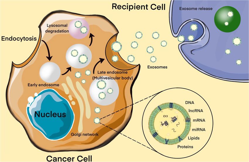
Gareev et al.                                                                                                        miRNAs and Tumor Resistance

INTRODUCTION                                                             Moreover, the activation or suppression of specific miRNA
                                                                         families is the mechanism by which oncogenes such as epidermal
Malignant primary brain tumors (MPBTs) are one of the                    growth factor receptor (EGFR) and MET or tumor suppressor
most difficult to treat types of tumors, resulting in significant        genes such as phosphatase and tensin homolog deleted on
morbidity and mortality in both children and adults. The most            chromosome 10 (PTEN), adenomatous polyposis coli (APC), and
common MPBTs are glioblastomas, high-grade meningiomas,                  breast cancer type 1/2 (BRCA1/2) induce or inhibit oncogenesis
medulloblastoma (MB), and pituitary adenomas (PAs) with                  (Zhang et al., 2007; Lee and Muller, 2010).
aggressive behavior, as aggressive prolactin PAs (Schiff and                 To date, a lot of evidence has been collected about the
Alyahya, 2020; Thakkar et al., 2021). For example, for patients          aberrant expression of miRNAs in various tumors, particular,
with glioblastoma or MB, the overall survival remains is poor,           malignant (Van Roosbroeck and Calin, 2017). It was shown that
with conventional therapies such as radio- and chemotherapy              miRNAs control the expression of genes of regulatory pathways
only providing marginal benefits to patient survival. Therefore,         that play a key role in tumor development, control apoptosis
new strategies are needed to overcome the barriers to successful         and proliferation of tumor cells, tumor growth in response to
treatment (Thakkar et al., 2021).                                        DNA damage and repair, angiogenesis and response to hypoxia,
   Over the past decades, significant progress has been achieved         the interaction of tumor cells with the microenvironment
in the study of tumor biology, the study of the mechanisms of            (Rupaimoole and Slack, 2017; Van Roosbroeck and Calin, 2017).
control of tumor metastasis, apoptosis, invasion, angiogenesis,          A number of researchers isolate miRNAs directly related to
and proliferation of tumor cells. These data were obtained by            individual processes in a tumor, or stages of development, of a
studying the cellular composition and microenvironment of                disease (Vishnoi and Rani, 2017). Many miRNAs are involved
tumors, various intracellular signaling pathways, and molecular          in key signaling pathways associated with the regulation of the
processes of oncogenesis (Richardson et al., 2020). MicroRNAs            cell cycle and apoptosis. In one of the latest studies, Allahverdi
(miRNAs) are 18–22 nucleotide endogenous non-coding RNAs                 et al. (2020) found that adipose-derived mesenchymal stem cells
that regulate gene expression at the post-transcriptional level          (AD-MSCs) delivering miR-4731 induces apoptosis and cell cycle
by interacting with 30 -untranslated regions (30 -UTR) of mRNA-          arrest in the glioblastoma cell line. Another study provided
targets (Lu and Rothenberg, 2018; Figure 1). It is estimated             evidence that miR-221-3p reduces MB cell proliferation by
that more than 60% of all human protein-coding genes are                 inducing apoptosis and G0/G1 arrest by suppressing eukaryotic
directly regulated by miRNAs (Ha and Kim, 2014; Dexheimer and            translation initiation factor 5A-2 (EIF5A2) (Yang et al., 2019).
Cochella, 2020). It has been proven that miRNAs are involved in              There is also a separate group of miRNAs associated with the
various biological processes, including the cell cycle, apoptosis,       metastatic activity of tumors – metastamiRs. Moreover, among
cell proliferation, and differentiation (Rupaimoole and Slack,           such miRNAs, some promote (miR-9, miR-210, miR-21, miR-
2017; Saliminejad et al., 2019). In addition, miRNAs play a role         218, etc.) tumor metastasis, while others (miR-145, miR-7, miR-
in the oncogenesis of various human tumors, including brain              146-a, etc.), on the contrary, suppress it (Alsidawi et al., 2014; Lu
tumors (Bertoli et al., 2015; Qadir and Faheem, 2017; Ali Syeda          et al., 2015; Lima et al., 2017; Ji et al., 2018; Maryam et al., 2021).
et al., 2020; Balachandran et al., 2020). Recently, most research            In recent years, thanks to advances in molecular oncology,
has focused on the role of miRNAs in resistance to malignant             it has been possible to decipher some of the mechanisms of
human tumors therapy. In MPBTs, the role of miRNAs in radio-             oncogenesis and to determine the signs of a malignant phenotype,
and chemotherapy resistance is an attractive area of research            one of which is angiogenesis. Malignant tumors requires more
and is expected to lead to the development of novel treatment            oxygen and nutrients to grow (Viallard and Larrivée, 2017).
strategies. This review will focus on differential expression of         The solution to this issue is to trigger the mechanism of
miRNAs in MPBTs (e.g., high-grade meningiomas, MB, PAs with              angiogenesis in the tumor. Vascular endothelial growth factor
aggressive behavior, and glioblastoma) with their gene-targets           (VEGF) is extremely important for the formation of an adequate
and their potential role in resistance to radio- and chemotherapy,       functioning vascular system during embryogenesis and in the
and to pharmacological treatment.                                        early postnatal period, but it also plays an important role in
                                                                         pathological angiogenesis. In many types of tumors, increased
                                                                         VEGF expression correlates with poor prognosis, including
MICRORNAs DYSREGULATION IN                                               aggressive tumor growth, recurrence, metastasis, and decreased
MALIGNANT PRIMARY BRAIN TUMORS                                           survival (Melincovici et al., 2018). In addition, VEGF expression
                                                                         correlates with a decrease in the density of the microvascular
MicroRNAs perform an important function in the complex                   network in the malignant brain tumors, which in itself serves
mechanism of regulation of gene activity, since they determine           as an indicator of the prognosis of vascular rupture, followed
the qualitative and quantitative composition of transcripts and          by hemorrhage in the tumor bed (Apte et al., 2019). To date, a
proteins necessary for the development of individual tissues,            number of miRNAs have been identified that are highly expressed
organs and the whole organism. A growing body of evidence                in endothelial cells (ECs) and/or are activated under hypoxic
points to the importance of miRNAs deregulation in the initiation        conditions. Among these miRNAs, it is worth noting miR-
and progression of tumors, where they can act as oncogenic               126, which is specifically expressed in the ECs and is a key
miRNAs (oncomiRs) or tumor-suppressor miRNAs, depending                  regulator of the integrity of the vascular wall and angiogenesis
on the cellular function of their gene-targets (Liu et al., 2014).       in various tumors, including brain tumors (Fish et al., 2008).

Frontiers in Cell and Developmental Biology | www.frontiersin.org    2                                      October 2021 | Volume 9 | Article 740303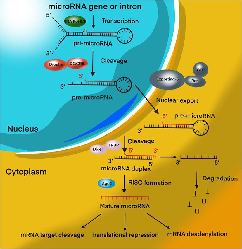
Gareev et al.                                                                                                              miRNAs and Tumor Resistance

  FIGURE 1 | MiRNA biogenesis pathway. Overview schematic representation of canonical miRNA biogenesis pathway.

Smits et al. (2012) showed there is significant low-expression of                between the main histological types of meningioma, for which
miR-125b in ECs co-cultured with U87 glioblastoma line cells.                    miR-21 expression showed a significant increase in World Health
Moreover, the authors demonstrated that miRNA-125b inhibits                      Organization (WHO) grade 2 and 3 lesions as compared to WHO
angiogenic processes by directly regulating Myc-associated zinc                  grade 1 lesions (Katar et al., 2017).
finger protein (MAZ)/VEGF signaling pathway expression. It                          Currently, there is an active search for new miRNAs and their
is known that, MAZ-binding sites are located in the promoter                     target genes involved in other important processes associated
regions of angiogenic factor VEGF.                                               with oncogenesis (and not only), for example, the control
   Xiao et al. (2016) demonstrated that miR-566 was                              of the balance of self-renewal and differentiation of stem
overexpressed in glioblastoma in vitro and in vivo, and inhibition               cells, epithelial-mesenchymal transition (EMT), regulation of
of miR-566 was able to suppress the invasion and migration of                    the immune response, the relationship of the tumor with the
glioblastoma cells, and angiogenesis via the VEGF/Von Hippel–                    microenvironment, etc.
Lindau tumor suppressor (VHL) pathway. This suggests that                           It should also be borne in mind that the reason for the change
miR-566 may function as an oncogene, and therefore, miR-566                      in the expression of miRNA may be a violation of the expression
may be considered a novel therapeutic target of glioblastoma                     of proteins involved in miRNA biogenesis. It is known that the
(Xiao et al., 2016).                                                             loss or insufficient expression of Drosha, Dicer, and TRBP can
   There is evidence that some miRNAs can participate in the                     lead to the development of a tumor process (Olejniczak et al.,
processes of malignant transformation in benign brain tumors.                    2018). Several groups showed impaired expression of Drosha and
Brain tumors of different histology are characterized by specific                Dicer in glioblastoma, pineoblastoma, and neuroblastoma, all of
miRNAs expression profiles associated with the clinical and                      which correlated with a poor prognosis of survival (Lin et al.,
pathological properties of the tumor (Van Roosbroeck and Calin,                  2010; Mansouri et al., 2016; de Kock et al., 2020). Decreased
2017). For instance, miR-21 makes it possible to distinguish                     expression of these proteins can be mediated by mutations or

Frontiers in Cell and Developmental Biology | www.frontiersin.org            3                                    October 2021 | Volume 9 | Article 740303
Gareev et al. miRNAs and Tumor Resistance
epigenetic inactivation of their genes. In addition, mutations in protein kinase (MAPK)/ERK; and 2) violation of the mechanism
Dicer can lead to impaired recognition of miRNA precursors of cell death under the influence of chemotherapy and
and a change in the balance of strands. Argonaute 2 (Ago2) ionizing radiation (Cao et al., 2019; Kapoor et al., 2020).
and GW proteins that act as direct partners of miRNAs are This mechanism includes the blocking of apoptosis with p53
often susceptible to somatic mutations in glioblastoma, which mutations, overexpression of B-cell lymphoma 2 (Bcl-2), a
are accompanied by a high level of instability of microsatellite decrease in the expression of cluster of differentiation 95 (CD95)
tumor DNA (Li S. et al., 2014; Li Z. et al., 2019). Disruption of (Kapoor et al., 2020). It is now known that miRNAs can control
the transport of pre-miRNAs into the cytoplasm can also lead to the regulation of target genes or signaling pathways involved in
a decrease in their expression. A mutation in the exportin-5 gene, malignant tumor resistance to therapy, including MPBTs. Among
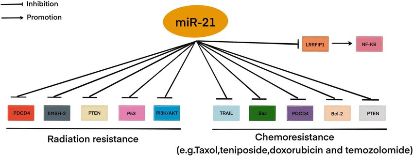
leading to the synthesis of a truncated protein that is unable to these miRNAs, miR-21 is the most studied in glioblastoma. MiR-
recognize pre-miRNAs, causes a decrease in the level of mature 21 is one of the important miRNAs involved in glioblastoma
miRNAs in a number of tumors (Wu et al., 2018). oncogenesis. A large number of studies indicated that miR-21
A number of oncogenic proteins can directly interfere with could affect a variety of cellular and molecular pathways. It has
miRNA biogenesis in tumors. Thus, wild and mutant forms of been showing that deregulation of miR-21 could be associated
p53 are involved in the biogenesis of a number of miRNAs, with resistance to radio- and chemotherapy of glioblastoma
primarily miR-34 (Zhang et al., 2019). It is known that mutant (Figure 2; Lan et al., 2015; Masoudi et al., 2018; Buruiană et al.,
forms of p53 can inhibit Drosha activity and prevent the 2020). In this article, we will consider current knowledge about
formation of pre-miRNA (Garibaldi et al., 2016). Transforming the role of miRNAs in the mechanisms of resistance to therapy
growth factor beta (TGF-β) affects the processing of miRNA in MPBTs. In addition, a summary of the role of some miRNAs
through the binding of effector proteins to the microprocessor in therapy resistance by targeting their gene targets is shown in
complex and pri-miRNA. YES-associated protein 1 (YAP1), one Table 2 (Pannuru et al., 2014; Dénes et al., 2015; Abdelfattah et al.,
of the components of the Hippo pathway, regulates the activity 2018; Chen et al., 2018; Li J. et al., 2019; Bogner et al., 2020; Hu
of the microprocessor complex depending on the density of cells et al., 2020; Sun et al., 2020; Zhao C. et al., 2020; Cardoso et al.,
in culture (Ruan et al., 2016). Tumor suppressor protein BRCA1 2021) and Table 3 (Lee et al., 2014; Li W. et al., 2014; Huynh et al.,
also stimulates the activity of the microprocessor complex (Lee 2015; Wu et al., 2015; Yang F. et al., 2017; Zhang Q. et al., 2020).
and Muller, 2010). Under hypoxic conditions, activated EGFR
can phosphorylate Ago2, inhibiting its interaction with Dicer and Glioblastoma
decreasing the level of activity of miRNA effector systems (Shen Glioblastoma is the most aggressive primary brain tumor and
et al., 2013). In addition, the interaction of p53 with Ago2 leads to usually has a poor prognosis. Thus, the median survival rate of
a change in the spectrum of miRNAs associated with it, leading to patients with glioblastoma after surgical resection and standard
the formation of complexes carrying tumor suppressor miRNAs, radio- and chemotherapy is no more than 12–15 months,
for example, let-7 (Krell et al., 2016). while the 2-year survival rate for this group of patients varies
Thus, miRNAs are one of the key factors in the development from 26 to 33% (Wirsching et al., 2016; Schiff and Alyahya,
of malignant forms of brain tumors, both as drivers of malignant 2020). Angiogenesis is the most important pathophysiological
transformation and as a victim of the deregulation of cellular mechanism for the growth and progression of glioblastoma
regulatory systems. The close relationship of miRNAs with due to the active development of the microvascular network.
MPBTs has led to the fact that they are currently being actively The accelerated development of the microvascular network in
studied (Table 1; Grunder et al., 2011; Kliese et al., 2013; Asuthkar glioblastoma occurs due to the synthesis of a large number
et al., 2014; Wang et al., 2015; Wei et al., 2015; Yu et al., 2016; of growth factors by tumor cells, including the VEGF family,
Zheng et al., 2017; Xu et al., 2018; Xue et al., 2019; Hou et al., placental growth factor (PLGF), platelet-derived growth factor
2020; Muñoz-Hidalgo et al., 2020; Negroni et al., 2020; Song (PDGF), and fibroblast growth factor (FGF) (Le Rhun et al.,
et al., 2020), including with the aim of creating diagnostic and 2019). It should be noted that the microvascular network of
therapeutic systems designed to increase the effectiveness of glioblastoma is characterized by a high degree of tortuosity,
treatment of this disease. increased permeability, as well as an increased diameter of the
vascular lumen, and a thickened basement membrane (Ahir et al.,
2020). It is believed that these features of the microvascular
MICRORNAs IN RESISTANCE TO network of glioblastoma increase the hypoxia of the tumor tissue,
CHEMORADIOTHERAPY AND thereby reducing the effectiveness of the use of cytotoxic drugs. It
PHARMACOLOGICAL TREATMENT is for this reason that the development and use of anti-angiogenic
drugs seem to be one of the most promising methods of targeted
Resistance to therapy of brain tumors is an important problem in treatment of glioblastoma patients. The effectiveness of the use of
modern neurosurgery. There are two main mechanisms for the anti-angiogenic drugs in the treatment of glioblastoma has been
emergence of radio- and chemotherapy resistance: (1) activation clearly demonstrated in a number of clinical studies (Sousa et al.,
of specific signaling pathways responsible for the “neutralization” 2019; Schulte et al., 2020). However, the widespread use of anti-
of the chemotherapy drug and ionizing radiation in the tumor angiogenic drugs in clinical practice has led to the development
cell. Such these signaling pathways include phosphoinositide of glioblastoma resistance to drugs of this group. The formation
3-kinases/protein kinase B (PI3K/AKT) è mitogen-activated of drug resistance of glioblastoma to anti-angiogenic drugs is
Frontiers in Cell and Developmental Biology | www.frontiersin.org 4 October 2021 | Volume 9 | Article 740303Gareev et al. miRNAs and Tumor Resistance
TABLE 1 | The most relevant studies to study the role of miRNAs in the oncogenesis of malignant primary brain tumors (MPBTs).
Tumor type miRNA Gene-target Biological function Regulation Phenotype References
Glioblastoma miR-191 NDST1 Promote tumor growth Up OncomiR Xue et al., 2019
and cells migration
Glioblastoma miR-200c ZEB1 Inhibit tumor growth Down Tumor Muñoz-Hidalgo
and cells migration suppressor et al., 2020
Glioblastoma miR-449b-5p WNT2B/Wnt/β-catenin Inhibits tumor cells Down Tumor Hou et al.,
proliferation, invasion, suppressor 2020
and migration
Atypical and anaplastic miR-145 COL5A1 Inhibit motility and Down Tumor Kliese et al.,
meningiomas proliferation of tumor suppressor 2013
cells
Anaplastic meningioma miR-195 FASN Inhibit proliferation, Down Tumor Song et al.,
migration, and invasion suppressor 2020
Atypical and anaplastic miR-497∼195 cluster GATA-4 Decreases tumor cell Down Tumor Negroni et al.,
meningiomas viability suppressor 2020
Atypical and anaplastic miR-224 ERG2 Promote tumor growth Up OncomiR Wang et al.,
meningiomas and reduce apoptosis 2015
of cells. Associated
with poor prognosis
MB miR-21 PDCD4 Decrease the motility of Up OncomiR Grunder et al.,
tumor cells and reduce 2011
their migration
MB miR-211 PI3K/AKT and mTOR Inhibit growth, Down Tumor Xu et al., 2018
migration and invasion suppressor
MB miR-494 MMP-9 and SDC1 Reduce tumor growth Down Tumor Asuthkar et al.,
and angiogenesis suppressor 2014
Invasive PA (NFA, GH, miR-106b PTEN-PI3K/AKT/MMP- In tumor cells induces Up OncomiR Zheng et al.,
ACTH, PRL) 9 invasive properties 2017
Invasive PA (NFA, GH, miR-26a PLAG1 In tumor cells induces Up OncomiR Yu et al., 2016
ACTH, PRL) invasive properties.
Associated with poor
prognosis
Pituitary carcinoma miR-20a, miR-106b PTEN and TIMP2 Activation of invasive Up OncomiR Wei et al., 2015
and miR-17-5p properties and
migration in a tumor
cells. Carcinoma
metastasis
miR, microRNA; MB, medulloblastoma; PA, pituitary adenoma; miR, microRNA; NFA, non-functioning adenoma; GH, growth hormone-secreting adenoma; FSH, follicle-
stimulating hormone adenoma; LH, luteinizing hormone-secreting adenoma; ACTH, adrenocorticotropic hormone-secreting adenoma; PRL, prolactin-secreting adenoma;
NDST1, Bifunctional heparan sulfate N-deacetylase/N-sulfotransferase 1; ZEB1, E-box-binding homeobox 1; WNT2B, Wnt family member 2B; COL5A1, collagen type
V alpha 1 chain; FASN, fatty acid synthase; GATA-4, GATA binding protein 4; Early growth response 2; MMP-9, matrix metallopeptidase 9; PDCD4, programmed cell
death 4; AKT, protein kinase B; mTOR, mammalian target of rapamycin; SDC1, Syndecan 1; PTEN, tensin homolog deleted on chromosome 10; PI3K, phosphoinositide
3-kinases; PLAG1, pleomorphic adenoma gene 1; TIMP2, tissue inhibitor of metalloproteinases 2.
associated with molecular and cellular features of the behavior of leads to the preservation of intracellular structures and the
tumor cells, including with the participation of certain miRNAs leveling of the secondary effects of anti-angiogenic drugs. It is
(Zeng et al., 2018b). assumed that in glioblastoma, drug resistance to bevacizumab
Autophagy is one of the main mechanisms of tumor is associated with non-selective hypoxia-induced factor (HIF)-
resistance. It should be noted that autophagy is the main dependent autophagy-mediated through the activity of the
cellular defense mechanism in the development of hypoxic protein-interacting protein 3 (BNIP 3) and hypoxia-inducible
conditions, which does not require tissue and extracellular factor 1-alpha (HIF1-α) protein (Hu et al., 2012). It is known
matrix remodeling (Kimmelman and White, 2017). As is already that HIF-1α can cause cell cycle arrest, initiating angiogenesis,
known, the use combination of anti-angiogenic therapy with and regulating cellular metabolism (Gabriely et al., 2017; Huang
radiotherapy leads to the development of tumor tissue hypoxia et al., 2019). Huang et al. identified that the expression of
due to the disturbance of the angiogenesis process necessary HIF-1α was increased in glioblastoma in vitro and in vivo
for the growth and progression of the tumor (Le Rhun et al., under hypoxia (Cardoso et al., 2021). Moreover, the longer the
2019). It is generally accepted that the process of autophagy duration of hypoxia, the higher was the expression of HIF-1α.
in tumor cells is aimed at the destruction of proteins and However, the expression of miR-224-3p was decreased under
signaling molecules formed during hypoxic conditions, which hypoxia conditions in a time-dependent manner. Their data
Frontiers in Cell and Developmental Biology | www.frontiersin.org 5 October 2021 | Volume 9 | Article 740303Gareev et al. miRNAs and Tumor Resistance
FIGURE 2 | MiR-21 involved in radio- and chemoresistance in glioblastoma. This figure shows through which signaling pathways miR-21 may be involved in
resistance to therapy in glioblastoma.
TABLE 2 | MiRNAs involved in the regulation of chemotherapeutic and pharmacological drug treatment resistance in malignant primary brain tumors (MPBTs).
miRNA Type of tumor Type of drug Drug Gene-target Mechanism References
miR-128-3p Glioblastoma DNA-targeted TMZ c-Met/EMT (c-Met, Reduces the Zhao C. et al.,
drugs PDGFRα, Notch1, and proliferation, invasion, 2020
Slug) and migration
miR-186 Glioblastoma DNA-targeted Cisplatin YY1 Inhibits the formation of Li J. et al., 2019
drugs the GIC phenotype
miR-302a Glioblastoma Tyrosine kinase Cediranib PGK1 Decrease glycolysis, Cardoso et al.,
inhibitors cell growth, migration, 2021
and invasion
miR-let-7f-1 MB DNA-targeted Cisplatin HMGB1 Inhibit autophagy Pannuru et al.,
drugs 2014
miR-29c-3p MB DNA-targeted Cisplatin Bcl-2/Wnt2 Increase apoptosis Sun et al., 2020
drugs
miR-584-5p MB Tubulin inhibitors Vincristine HDAC1/eIF4E3 Cause cell cycle arrest, Abdelfattah
DNA damage, and et al., 2018
spindle defects
miR-31 MB DNA-targeted Ginsenoside Rh2 Wnt/β-catenin Inhibit the proliferation Chen et al.,
drugs and migration, and 2018
induce apoptosis
miR-197 Meningioma DNA-targeted Quercetin Bcl-2/Bax Reduce tumor cell Hu et al., 2020
drugs proliferation and
increase apoptosis
miR-34a Aggressive SSAs Octreotide cAMP Antiproliferative effect Bogner et al.,
somatotropinoma 2020
miR-1299 Aggressive DA Bromocriptine FOXO1 Promotes the synthesis Dénes et al.,
prolactinoma and secretion of 2015
prolactin
miR, MicroRNA; MB, medulloblastoma; SSAs, somatostatin analogs; DA, dopamine antagonist; TMZ, Temozolomide; EMT, epithelial-mesenchymal transition; PDGFRα,
platelet-derived growth factor receptor A; YY1, Yin Yang 1; PGK1, phosphoglycerate kinase 1; HMGB1, high-mobility group protein B1; Bcl-2, B-cell lymphoma-2; Wnt2,
wingless-type MMTV integration site family, member 2; HDAC1, histone deacetylase 1; IF4E3, eukaryotic translation initiation factor 4E type 3.
showed that the miR-224-3p mimic significantly suppressed the experiments are needed to understand the complex link between
expression HIF-1α and inhibited cell mobility while increased miR-203 and HIF-1α expression.
chemosensitivity to Temozolomide (TMZ) of glioblastoma. In
addition, the miR-224-3p mimic suppressed the expression Medulloblastoma
of VEGF with an increased cell apoptosis rate. In another Medulloblastoma is a malignant primary tumor of the posterior
study, miR-203 could be a useful target for overcoming fossa (WHO grade 4), mainly manifested in children. MB arises
the radioresistance of glioblastoma by suppressing HIF-1α in the posterior fossa, usually from the cerebellar vermis and
expression in vitro (Chang et al., 2016). However, further in the roof of the fourth ventricle (Schiff and Alyahya, 2020).
Frontiers in Cell and Developmental Biology | www.frontiersin.org 6 October 2021 | Volume 9 | Article 740303Gareev et al. miRNAs and Tumor Resistance
TABLE 3 | Role miRNAs in regulating malignant primary brain tumors (MPBTs) radiosensitivity.
miRNA Type of tumor Gene-target Regulation Mechanism Response References
miR-221/222 Glioblastoma Akt Down Inhibit tumor growth Increase Li W. et al.,
radiosensitive 2014
miR-181d Glioblastoma NF-κB Up Suppress tumor cell proliferation, Increase Yang F. et al.,
colony formation and radiosensitive 2017
anchor-independent growth, as well
as migration, invasion and tube
formation
miR-124-3p Glioblastoma mTOR, MAPK, Up Inhibit tumor growth and promote Increase Wu et al., 2015
TGFbeta, and PI3K-Akt apoptosis radiosensitive
miR-205 Glioblastoma GRP78, Up Decreases tumor-sphere-formation Increase Huynh et al.,
c-Myc,β-catenin and and colony-forming abilities, inhibit radiosensitive 2015
vimentin migration and invasion
miR142-3p MB Sox2 and ADCY9 Down Elevate the expression of miRNA Increase Lee et al., 2014
decreases cancer stem-like radioresistance
characteristics and stemness
miR-584-5p MB HDAC1/eIF4E3 Up Cause cell cycle arrest, DNA Increase Abdelfattah
damage, and spindle defects radiosensitive et al., 2018
miR-221/222 Meningioma PTEN Down Inhibit tumor cell proliferation, Increase Zhang Q. et al.,
invasive and colony formation, and radiosensitive 2020
promote apoptosis
miR, MicroRNA; MB, medulloblastoma; Akt, protein kinase B alpha; NF-κB, nuclear factor kappa-light-chain-enhancer of activated B cells; mTOR, mammalian target of
rapamycin; MAPK, mitogen-activated protein kinase; TGF-beta, transforming growth factor beta; PI3K, phosphoinositide 3-kinases; GRP78, glucose regulatory protein
78; Sox2, SRY-Box transcription factor 2; ADCY9, adenylate cyclase 9; HDAC1, histone deacetylase 1; eIF4E3, eukaryotic translation initiation factor 4E family member
3; PTEN, phosphatase and tensin homolog deleted on chromosome 10.
MBs tend to metastasize along with the cerebrospinal fluid There is evidence that phosphatase and tensin homolog
(CSF) pathways, which is detected in 35% of cases at the time deleted on chromosome 10 (PTEN) dysfunction plays a crucial
of diagnosis. MBs are the most common malignant neoplasms role in the development and progression MB. PTEN plays
of the brain in childhood and account for 15 to 30% of all important roles in many cellular processes, including cell-cycle
primary CNS tumors in children, and about 70% of all cases progression and apoptosis (Tolonen et al., 2020). Li et al. (2015)
are diagnosed in children under 15 years of age (Quinlan and reported the upregulation of miR-106b in MB. In their study, the
Rizzolo, 2017). The age peak of diagnosis is between 3 and suppression of miR-106b inhibited cell proliferation, migration
5 years, and only 25% are patients between the ages of 20–44 and invasion, and anchorage-independent growth, tumorsphere
(Millard and De Braganca, 2016; Quinlan and Rizzolo, 2017). formation. In addition, downregulation of miR-106b suppressed
Patients with MB have a poor outcome despite surgical, radio- the tumor growth by promoting G1 arrest and apoptosis. Besides,
and chemotherapy. However, the molecular mechanisms that PTEN can be modulated by miR-106 family in various human
confer sensitivity or resistance of MB to chemoradiation therapy cancers. For instance, miR-106b caused cell radio resistance
are still unclear. in colorectal cancer via the PTEN/PI3K/AKT pathways (Zheng
Recent evidence has implicated miRNAs in modulating et al., 2015). MiR-106a induced cisplatin resistance via the
chemo- and radiosensitivity in MBs (Joshi et al., 2019). It has PTEN/AKT pathway in gastric cancer cells (Fang et al., 2013).
been shown that in MB therapy there is a balance between cell However, the role of miR-106b in therapy resistance of MB is still
cycle arrest and cell death (Kasuga et al., 2008). Melanoma- largely unknown. Nevertheless, this is an excellent opportunity to
associated antigen-A (MAGE-A) family acts as a cell cycle continue research on the role of miR-106b directly targeted PTEN
regulatory protein and plays a key role in the oncogenesis and in therapy resistance of MB.
therapy resistance in MB. Kasuga et al. (2008) demonstrate
that knockdown of MAGE-A increases apoptosis and sensitizes
MB cells to chemotherapeutic agents such as cisplatin and Pituitary Adenomas With Aggressive
etoposide. This finding supports the hypothesis that knockdown Behavior
of MAGE-A genes increases the susceptibility of MB cells to Among the tumors of the chiasmatic-sellar region, the most
cisplatin and etoposide, potentially by accumulating cells in the common are PAs, accounting for about 18% of all tumors
S phase. In contrast, Sheamal et al. showed that miR-34a directly of this localization. In the overwhelming majority of cases,
targets the MAGE-A family (MAGE-A2, MAGE-A3, MAGE- these are benign neoplasms, characterized by slow growth rates
A6, and MAGE-A12), disengaging p53 from MAGE-A–mediated and progression (Schiff and Alyahya, 2020). However, among
repression (Weeraratne et al., 2011). Moreover, an important them, there are PAs with aggressive behavior, which exhibits
consequence of this is a positive feedback loop that sensitizes MB the properties of resistance to traditional treatment methods
cells to cisplatin and etoposide via delayed G2/M progression and (Lake et al., 2020). Among PAs, the first place is occupied by
increased tumor cells apoptosis. tumors accompanied by the syndrome of hyperprolactinemia –
Frontiers in Cell and Developmental Biology | www.frontiersin.org 7 October 2021 | Volume 9 | Article 740303Gareev et al. miRNAs and Tumor Resistance
prolactinomas, as well as non-functioning (hormonally inactive) resistance to DA, or with a persistent increase in tumor size
PAs, each approximately 40%. The next most frequent is with the development of neuro-ophthalmic symptoms, or if
somatotropinomas, about 13–15%, accompanied by symptoms there is a rapid loss of vision or cranial nerve paralysis due to
of acromegaly. Gonadotropin-secreting PAs, ACTH-secreting intratumoral hemorrhage (Panigrahi et al., 2020). However,
PAs, GH-secreting PAs, mixed forms are less common. In the complete surgical removal of a giant tumor is rarely performed
age range, PAs occupy a period from 30 to 50 years, which due to the technical complexity and the greater risk of side
is the working age (Elsarrag et al., 2020; Lake et al., 2020). effects. Radio- and chemotherapy have a limited role in the
In connection with all of the above, PAs, their diagnosis, and treatment of giant prolactinomas; on the one hand, because
especially, treatment are important medical and social problems. of its dubious chemotherapy effectiveness and, on the other
The main methods of treatment for PAs are surgical hand, because of the complications that appear during tumor
removal of the tumor and pharmacological treatment and their irradiation (Iglesias et al., 2018). Therefore, a thorough and
combinations. Radio- and chemotherapy are used, as a rule, when deeper understanding of the molecular mechanisms underlying
it is impossible to perform a surgical intervention or when it is drug resistance of PAs with aggressive behavior like aggressive
at high risk, and when the tumor is highly aggressive (Fleseriu prolactinomas are urgently needed to find potential new targets
and Popovic, 2020). Since there is no clear definition and for improving therapeutic efficacy. For instance, Jian et al. (2019)
availability of reliable prognostic markers, PAs with aggressive demonstrated that miR-145-5p was greatly downregulated in
behavior are difficult to identify at initial presentation, and bromocriptine-resistant prolactinoma cell lines and tissues
therefore the primary therapeutic approach is no different from in vitro and in vivo. In addition, transfer miR-145 mimic into
other PAs depending on the type of tumor (Mete and Lopes, tumor cells and revealed that overexpression of miR-145-5p
2017). Resistance to drugs presenting as escalating hormone increased sensitivity for bromocriptine markedly. In conclusion,
levels and/or tumor growth where can be an early indicator of identification of tumor protein, translationally controlled 1
aggressiveness. There are many studies examining changes in (TPT1) as a direct target gene of miR-145-5p. In another study,
miRNA expression in PAs. Among them are studies on their role miR-93-5p was related to fibrosis and was involved in the
in drug resistance in various types of PAs (Ciato and Albani, bromocriptine -resistance mechanisms in prolactinoma by
2020). However, there is no evidence base on their potential role regulating the transforming growth factor beta 1/mothers against
in resistance to chemo- and radiotherapy in patients with PAs decapentaplegic homolog 3 (TGF-β1/Smad3) signaling pathway
with aggressive behavior and pituitary carcinomas. It is possible (Hu et al., 2019). Interesting that previous studies showed that
to suggest from previous studies which miRNAs and through TGF-β1 promotes the synthesis and secretion of collagen fibers
which signaling pathways can participate in the mechanisms in fibroblasts and that the TGF-β1/Smad3 signaling pathway
of resistance to chemo- and radiotherapy. For instance, in a is involved in the drug-resistance mechanism of prolactinoma
recent study, Wang Z. et al. (2019) successfully identified one key by increasing fibrosis through interactions with fibroblasts
target gene, EGFR, and two crucial miRNAs, miR-489 and miR- (Hu et al., 2018).
520b, associated with aggressiveness of prolactinomas based on
bioinformatics analysis. It is also known that EGFR is one of the High-Grade Meningiomas
most frequently altered oncogenes in tumors, which important Meningiomas are common tumors of the CNS, originating from
role in therapy resistance and is often associated with a negative the meninges of the brain or spinal cord. Most meningiomas
prognosis (Lee and Muller, 2010). are benign tumors characterized by slow growth and are
Prolactinoma is the most commonly seen secretory histologically WHO Grade 1 (Schiff and Alyahya, 2020).
tumor of pituitary glands. More than 90% of prolactinomas However, high-grade meningiomas [atypical (WHO Grade
are microprolactinomas (Gareev et al. miRNAs and Tumor Resistance
high-grade meningiomas and have suggested many important Exosomes are a new form of intercellular communication
molecular targets for the development of new drugs for the (Pegtel and Gould, 2019). These are small membrane vesicles of
treatment of high-grade meningiomas that are resistant to endosomal origin with a diameter of 30 to 100 nm, which are
radio- and chemotherapy. In particular, growth factors, such as secreted by various types of cells, normal or abnormal (Pegtel
PDGF, epidermal growth factor (EGF) and their receptors, and and Gould, 2019). Tumor cells play a particularly important role
cytokines, such as TGF-β, serve as major factors in high-grade in the production of exosomes (Kalluri and LeBleu, 2020). In
meningiomas leading to therapeutic resistance (Birzu et al., 2020; addition to miRNAs, exosomes contain a diverse set of molecules
Shao et al., 2020). such as DNA, proteins, other non-coding RNAs, translation
Unfortunately, studies on the role of miRNAs in the factors, metabolic enzymes, etc. There is evidence that exosomal
mechanisms of resistance to therapy in meningiomas are limited. miRNAs are actively involved in the oncogenesis of MPBTs,
For instance, microarray analysis, using the atypical meningioma including resistance to therapy (Zhang and Yu, 2019). The
tissue samples of 55 patients (43 from the radiosensitive and 12 presence of surface protein markers and adhesion molecules
from the radioresistant group), indicated that 14 miRNAs were allows exosomes to bind to cells (including tumor cells) that
significantly dysregulated in tumor tissue (Zhang X. et al., 2020). exhibit the corresponding receptors, via micropinocytosis or
Among them 7 significantly upregulated miRNAs (miR-4286, endocytosis, to be transported into these cells. All this suggests
miR-4695-5p, miR-6732-5p, miR-6855-5p, miR-7977, miR-6765- that miRNAs carried by exosomes can enter recipient cells and
3p, miR-6787-5p) and 7 significantly downregulated miRNAs regulate the expression of their target genes (Figure 4; Pegtel and
(miR-1275, miR-30c-1-3p, miR-4449, miR-4539, miR-4684-3p, Gould, 2019; Kalluri and LeBleu, 2020).
miR-6129, miR-6891-5p) in patients resistant to radiotherapy.
Furthermore, in order to investigate the signaling pathways Glioblastoma
affected by the differentially expressed 14 miRNAs between If we talk about the role of EVs in the formation of a resistant
radiosensitive and radioresistant atypical meningioma, the phenotype of tumor cells, then, of course, one of the most
authors used the DIANA-miRPath software and found three studied areas is the participation of exosomes in the development
enriched pathways: two pathways were fatty acid biosynthesis of multiple drug resistance (MDR) (Zhang and Yu, 2019).
and metabolism, and TGF-β signaling pathway. The role of Further studies made it possible to significantly expand the
TGF-β and these miRNAs in the oncogenesis, particularly list of biomolecules that are part of resistant tumor cell EVs
radiosensitivity, of meningiomas remains to be established. and are capable of inducing MDR in recipient cells. This
primarily refers to miRNAs that regulate the expression level
of a number of genes. For instance, Munoz et al. demonstrated
EXTRACELLULAR MICRORNAs IN that anti-miR-9 delivered via exosomes from MSCs to the TMZ-
TUMOR RESISTANCE resistant glioblastoma cells was able to reduce the endogenous
upregulation of miR-9 in response to TMZ (Koritzinsky et al.,
The main part of miRNAs is localized inside the cell. However, 2017). Moreover, to determine whether multidrug resistance
a certain proportion of miRNAs are present outside the cells gene 1(MDR1) expression is miR-9 dependent for sensitivity to
and they are called extracellular or circulating miRNAs. A series TMZ, the authors knocked down MDR1 with short hairpin RNA
of studies are devoted to the detection of extracellular miRNAs (shRNA), and then examined whether this affects the sensitivity
in various human fluids including whole blood, plasma/serum, of glioblastoma cells to TMZ. The results indicated that anti-miR-
saliva, urine, cerebrospinal fluid (Valihrach et al., 2020). In 9 increased active caspase by decreased the expression of MDR1
these biological fluids, the total concentration of miRNAs and and concomitantly caused enhanced glioblastoma cell death in
their ratio varies considerably, which may be due to the response to TMZ treatment. At the same time, in the protocol
peculiarities of the pathological or physiological status of the using manumycin A, which prevented the release of vesicles, it
organism. The discovery of significant changes in the expression was indicated that the transfer of anti-miR-9 occurs by vesicular
level of extracellular miRNAs in various diseases promoted transfer, particularly via exosomes.
the positioning of these molecules as promising non-invasive It should be noted that not only miRNAs but also mRNAs
biomarkers (Sohel, 2020). MiRNAs can be secreted by the cell as could be transported by EVs. Therefore, exosomes secreted
part of extracellular vesicles (EVs) (exosomes and microvesicles) by glioblastoma cells are enriched in mRNA, which enzymes
or apoptotic bodies; they can be found in the form of high- of DNA repair as methylation of the O(6)-Methylguanine-
density lipoprotein (HDL) bound and mostly in the form of DNA methyltransferase (MGMT) promoter and alkylpurine–
Argonaute2-containing ribonucleoprotein complexes (miRNA- DNA–N-glycosylase (APNG), and the transfer of these mRNAs
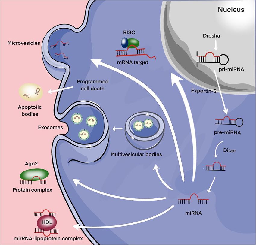
Ago2) (Figure 3; Valihrach et al., 2020). Then, regardless of into recipient cells can significantly increase the level of
the forms, miRNAs pass from the extracellular space into the chemoresistance (Shao et al., 2015).
biological fluid (for example, the general blood flow). While the participation of EVs in the formation of
Moreover, extracellular miRNAs, as exosomal miRNAs, chemoresistance of tumor cells is not in doubt today, the role
play an important role in intracellular communication and of EVs in the regulation of the response of cells to irradiation
the signaling system of cells. Over the past two decades, a has been studied to a much lesser extent. Only a few studies are
broad evidence base has been obtained regarding the role known in which the participation of EVs in the development
of extracellular miRNAs in maintaining cellular homeostasis. of the tumor response to radiation has been demonstrated, and
Frontiers in Cell and Developmental Biology | www.frontiersin.org 9 October 2021 | Volume 9 | Article 740303Gareev et al. miRNAs and Tumor Resistance FIGURE 3 | Secretion of miRNAs into the extracellular environment. Primary microRNA (pri-miRNAs) are transcribed by RNA polymerase II and then processed by Drosha into precursor-miRNAs (pre-miRNAs). Exportin5 transfers these pre-miRNAs from the nucleus to the cytoplasm, where Dicer converts them into mature miRNAs. Mature miRNAs can be selectively incorporated into extracellular vesicles (EVs) (exosomes and microvesicles) or linked to the Argonaute 2 (Ago2) protein and released into the extracellular environment. Alternatively, they can be attached to high-density lipoprotein (HDL) or contained in apoptotic bodies and then released into the extracellular environment. the results of these studies are rather contradictory. This is particular, in experiments on glioblastoma cells, the ability of primarily due to the bystander effect, a well-known biological exosomes from irradiated cells has been described to promotes process, radio-induced changes transmitted from irradiated cells a migratory phenotype of non-irradiated cells, as the authors to non-irradiated ones (Li and Nabet, 2019). It is assumed that believe, as a result of the active accumulation of DNA repair this effect is based on the transmission of induced or radiation- enzymes in the exosomes of irradiated cells (Arscott et al., 2013). modified biometabolites (reactive oxygen species, cytokines, growth factors, nucleic acid fragments, etc.) non-irradiated cells either by paracrine pathway or through intercellular gap Pituitary Adenomas With Aggressive junctions (Ni et al., 2019). In recent years, studies have appeared Behavior in which, in on both normal and tumor cells, direct evidence has Hormone therapy is one of the most common types of treatment been obtained for the participation of EVs of irradiated cells in for hormone-dependent malignant neoplasms, primarily PAs the induction of radiation changes (primarily, genetic instability, with aggressive behavior. Hormone therapy is based on the telomere contraction) in non-irradiated cells. At the same time, principle of creating an artificial deficiency of hormones EVs of irradiated cells can also have protective properties, in necessary for the growth of hormone-dependent tumors, which Frontiers in Cell and Developmental Biology | www.frontiersin.org 10 October 2021 | Volume 9 | Article 740303
Gareev et al.                                                                                                                            miRNAs and Tumor Resistance

  FIGURE 4 | Biogenesis of exosomes. The exosome membrane is formed as a result of the invagination of the early endosome into the membrane. Proteins, lipids,
  RNA, DNA enter the exosome from the cell cytoplasm. The fate of an endosome depends on the marking of its membrane with certain lipids: if it is labeled with
  lysobisphosphatidic acid, then its contents will be destroyed, and if ceramides, it will be pushed out of the cell. These processes are controlled by the GTPases of
  the Rab (G-protein) family, whose various members perform different functions: Rab5 directs the formation of the endosome, Rab7 organizes the degradation of the
  contents of the multivesicular body (late endosome) in the lysosome, and Rab11, Rab27, and Rab35 are necessary for the secretion of exosomes into the
  extracellular space. It has been shown that exosomes contain about 4,000 different proteins, more than 1,500 different miRNAs and mRNAs, as well as DNA.
  Bottom right – enlarged “generalized” exosome.

is achieved mainly in two ways: (1) by reducing the concentration                       EMT has been shown to contribute to drug resistance in tumors
of endogenous hormones and (2) by suppressing their synthesis                           (Du and Shim, 2016; Aleksakhina et al., 2019). Therefore, we can
or replacing hormones with their inactive analogs (Iglesias et al.,                     suggest that these miRNAs can be involved in the oncogenesis of
2020; van Bunderen and Olsson, 2021). Despite the unconditional                         PAs, and in particular, be responsible for drug resistance through
effectiveness, the use of hormonal therapy is limited by the                            intercellular communications. In another pilot study to discover
development of tumor resistance to hormones. The mechanism                              that the expression levels of circulating miR-200a in plasma of
of hormonal resistance is well understood. Much less is known                           patients with invasive PAs were significantly higher than that
about the role of intercellular interactions in the development                         in plasma of patients with non-invasive PAs. Moreover, invasive
of hormonal resistance of tumors and, in particular, PAs. For                           PA patients with residual after surgery had lower expression
instance, Zhao et al. (2021) demonstrated that 20 differentially                        levels of circulating miR-200a. Therefore, miR-200 was a potential
expressed miRNAs were identified in human invasive and non-                             influencing factor for invasiveness in PAs patients. However,
invasive PAs tissue, and rat PA cells, where among them, the                            further research is required for exploring the relationships
expression level of miR-99a-3p and mir-149 was significantly                            between circulating miR-200a expression and tumor size, clinical
reduced. Furthermore, it was shown that overexpression of miR-                          characteristics, and molecular mechanism for packaging and
149 and miR-99a-3p inhibits the growth and metastasis of PA                             secretion to biofluids of miR-200a (Beylerli et al., 2021).
cells and the formation of EC tubes. Interestingly, delivery of
miR-149 mimic and miR-99a-3p mimic via exosomes showed
similar suppressive effects on cell viability, metastasis, tube                         High-Grade Meningiomas and
formation ability, tumor growth in vivo, and expression of                              Medulloblastoma
markers associated with angiogenesis as VEGF. In additional, the                        Aberrant expression of extracellular miRNA circulating in
authors showed that NOVA1, denticleless E3 ubiquitin protein                            biofluids of certain brain tumor patients has recently been
ligase homolog (DTL), and RAB27B were targeted by miR-99a-                              reported to be non-invasive biomarkers and potential regulators
3p. It is known that this group of genes is directly promoted                           of the disease (Sohel, 2020; Valihrach et al., 2020). However,
EMT in various human tumors (Aleksakhina et al., 2019). Finally,                        the existence and role of miRNAs in MB and malignant

Frontiers in Cell and Developmental Biology | www.frontiersin.org                  11                                          October 2021 | Volume 9 | Article 740303
Gareev et al.                                                                                                                           miRNAs and Tumor Resistance

meningioma extracellular environment are unknown. Therefore,                            grade 1). However, receiver operating characteristic (ROC) curve
better understanding of extracellular miRNA secretion and                               analysis showed that exosomal miR-497 has better sensitivity
function in MB and malignant meningioma seems crucial for the                           and specificity than exosomal miR-195 in distinguishing between
development of novel insights for its regulation of oncogenesis                         low-grade (WHO grade 1) and higher-grade (WHO grade
including resistance to therapy. For instance, Choi et al. (2020)                       2 and 3) meningioma patients, where area under the curve
investigated whether secreted exosomal miR-135b and miR-135a                            (AUC) was 0.89 and 0.78, respectively. Furthermore, the authors
function at the microenvironment level by possibly impacting the                        demonstrated that the transcription factor GATA binding
stemness of brain tumor spheroid-forming cells (BTSCs). The                             protein 4 (GATA-4) is overexpressed in malignant meningioma,
authors suggest that the inhibition of miR-135b or miR-135a                             which in turn regulates Cyclin D1, and it negatively regulates
can suppress the self-renewal capacity and expression of stem                           the miR-497 expression with an increase in cell viability in vitro.
cell-related markers of BTSCs. In additional, they demonstrated                         Importantly, that a cell cycle protein cyclin D1 is an established
that miR-135b, miR-135a targeted angiomotin-like2 (AMOTL2),                             cancer-driving protein (Montalto and De Amicis, 2020). In
and the expression of AMOTL2 can be increased through miR-                              recent years, studies have reported that the high expression
135b and miR-135a inhibition. This result might be a clue that                          of Cyclin D1 is involved in drug resistance processes such as
exosomal miR-135b and miR-135a derived from BTSCs may                                   chemo- and radiation treatment, and targeted therapy in various
be able to regulate the Hippo pathway via AMOTL2, which                                 human tumors (Liu et al., 2020; Zuo et al., 2021). Therefore, the
plays a significant role in chemoresistance (Mohajan et al., 2021;                      clinical values of exosomal miR-497 in the therapy resistance of
Zeng and Dong, 2021).                                                                   high-grade meningioma must investigate in the future.
   Negroni et al. (2020) using reverse transcriptase real-
time quantitative polymerase chain reaction (qRT-PCR)                                   Discussion and Implications
assay demonstrated that exosomal miR-497 and miR-195 are                                The introduction of extracellular miRNAs into clinical practice
downregulated in serum of patients with high-grade meningioma                           is quite active. The search results of the clinical trial database
(WHO grade 2 and 3) compared to benign meningioma (WHO                                  clinicaltrials.gov for the keywords “extracellular,” “vesicle,”

TABLE 4 | MiRNAs in extracellular vesicles (EVs) and their signaling pathways through which they may be responsible for glioblastoma therapeutic resistance.

miRNAs              Drug/radiation          Vehicle                    Signaling pathways          Regulation      Effect                              References

miR-151a            TMZ                     Exosome                    XRCC4                           Up          Development of acquired             Zeng et al.,
                                                                                                                   resistance to TMZ                   2018a
miR-301a            Ionizing radiation      Exosome                    Wnt/b-catenin/TCEAL7            Up          Depresses radiation sensitivity     Yue et al., 2019
miR-221             TMZ                     Exosome                    DNM3                           Down         Inhibit cell proliferation,         Yang J. K.
                                                                                                                   migration, and TMZ resistance       et al., 2017
miR-34a             TMZ                     Exosome                    MYCN                            Up          Inhibit cell proliferation,         Wang B. et al.,
                                                                                                                   migration, invasion and TMZ         2019
                                                                                                                   resistance
miR-124             TMZ                     Exosome                    CDK6                            Up          Development of acquired             Sharif et al.,
                                                                                                                   resistance to TMZ and               2018
                                                                                                                   decreases the migration of
                                                                                                                   tumor cells
miR-603             Ionizing radiation      EV                         IGF1, IGF1R, and                Up          Promotes the CSC state and          Ramakrishnan
                                                                       MGMT                                        up-regulated DNA repair to          et al., 2020
                                                                                                                   promote acquired resistance.
                                                                                                                   Therapeutic platforms hold
                                                                                                                   translational potential in the
                                                                                                                   treatment of wtIDH/umMGMT
                                                                                                                   glioblastoma
miR-93 and          TMZ                     Exosome                    Cyclin D1                       Up          Decreases cell cycling              Munoz et al.,
miR-193                                                                                                            quiescence and contribute to        2019
                                                                                                                   TMZ resistance
miR-1238            TMZ                     Exosome                    CAV1/EGFR                      Down         Inhibit to TMZ resistance           Yin et al., 2019
miR-21-5p           Pacritinib + TMZ        Exosome                    STAT3/PDCD4                    Down         Inhibit to drug resistance          Chuang et al.,
                                                                                                                                                       2019
miR-27a-3p,         Ionizing radiation      EV                         CHD7                            Up          Promote PMT in GSCs. Inhibit        Zhang Z. et al.,
miR-22-3p and                                                                                                      radioresistance                     2020
miR-221-3p

XRCC4, X-ray repair cross-complementing protein 4; TCEAL7, transcription elongation factor A protein-like 7; DNM3, dynamin 3; CDK6, cyclin-dependent kinase 6; IGF1,
insulin-like growth factor 1; IGF1R, insulin-like growth factor 1 receptor; MGMT, O(6)-Methylguanine-DNA methyltransferase; CAV1, caveolin 1; EGFR, epidermal growth
factor receptor; STAT3, signal transducer and activator of transcription 3; PDCD4, programmed cell death protein 4; CHD7, chromodomain-helicase-DNA-binding protein
7; miR, microRNA; TMZ, Temozolomide; PMT, proneural-to-mesenchymal transition; GSCs, glioma stem cells.

Frontiers in Cell and Developmental Biology | www.frontiersin.org                  12                                        October 2021 | Volume 9 | Article 740303
Gareev et al. miRNAs and Tumor Resistance
“exosomes,” “miRNA,” and “tumor” contain more than 50 therapy. The discovery of EVs and, most importantly, their ability
projects, some of which are in the recruitment phase. A number to transfer biological material as miRNAs from cell to cell has
of projects have been launched to assess the effectiveness largely changed the understanding of the mechanism of tumor
of tumor therapy. In previous studies, it was shown that development and progression. First, this is the revealed ability of
metformin has cytotoxicity and decreases the viability of EVs to induce tumor transformation and/or induce a tumor-like
glioblastoma cells, a promising biguanide with pronounced phenotype in the cells of the surrounding tissue. Moreover, of
antitumor activity (Al Hassan et al., 2018). Furthermore, Soraya course, one of the most significant achievements in this reign –
et al. (2021) demonstrated that metformin significantly decreased the influence of EVs on the formation of a resistant phenotype of
the expression of miR-21, miR-155, and miR-182, indicating tumor cells. EVs can indeed provide the spread of resistance from
suppression of oncogenesis in glioblastoma cells. In addition, resistant to sensitive cells through various mechanisms based on
confirm metformin increased the exosome biogenesis and the transfer of specific regulatory molecules into cells including
secretion in glioblastoma cells. Their result also showed that the proteins, miRNAs, mRNA, etc. The induction of resistance to
expression level of Rab27a, Rab27b, and Rab11 upregulated in chemoradiotherapy and pharmacological treatment of MPBTs by
treated cells with metformin than that of control cells. In the miRNAs has been convincingly demonstrated in in vitro and
author’s opinion, the decreased expression levels of miR-21 and in vivo experiments. However, there are a number of questions.
miR-182 may be responsible for Rab genes upregulation, which And one of them in which To the extent that miRNAs are
may correlate with increased exosomes secretion. involved in the development of tumor resistance to antitumor
It turned out that not only exosomes of resistant cells, but therapy, can miRNAs actually participate in the development of
also exosomes produced by cells of the tumor stroma can induce resistance across the entire pool of tumor cells under in vivo
drug resistance in tumor cells. In experiments on head and neck conditions, and, most importantly, how important this process
cancer, it was found that exosomes produced by tumor-associated is in the development of acquired tumor resistance. Today these
fibroblasts are able to induce cisplatin resistance in nearby issues are being actively investigated, and, of course, their solution
tumor cells by transferring miR-196a and through targeting will make it possible to make significant progress in solving such
cyclin-dependent kinase inhibitor 1B (CDKN1B) and inhibitor of an important problem in neurosurgery as the resistance of MPBTs
growth protein 5 (ING5) (Qin et al., 2019). to antitumor therapy.
Another, no less important question is to what extent EVs can
affect the initial level of tumor radiosensitivity and, given their
protective properties, contribute to the spread of radioresistance AUTHOR CONTRIBUTIONS
to the brain tumors population. Research in this direction is just
beginning, and we can expect that soon it will be possible to get IG: conceptualization, writing – original draft, and
answers to this and other questions concerning the role of EVs in supervision. OB: writing – review and editing, investigation,
the tumor response to radiation. project administration, and resources. AA: formal analysis,
Thus, in recent years, extensive information has been methodology, and original draft. YL, HX, CL, XX, and CY: data
accumulated on the correlations of miRNA profiles of EV curation. GY: validation, visualization, and funding acquisition.
s and the development of a resistant phenotype of tumor All authors have read and agreed to the published version
cells. Despite significant advances in research on the role of of the manuscript.
extracellular miRNAs in therapeutic resistance in MPBTs, studies
besides glioblastoma with other tumors remains to be seen. In
conclusion, the most common miRNAs in EVs that have been
reported to be involved in glioblastoma therapeutic resistance are FUNDING
shown in Table 4 (Zeng et al., 2018a).
This work was supported by the National Natural Science
Foundation of China (81971135); Natural Science Foundation
CONCLUSION of Heilongjiang (YQ2020H014); “Chunhui Plan” of Ministry
of Education (HLJ2019009); Distinguished Young Foundations
The last decade has been accompanied by the emergence of of the First Affiliated Hospital of Harbin Medical University
a large number of studies devoted to the role of miRNAs in (HYD2020JQ0014); and the reported study was funded by RFBR
oncogenesis and the development of resistance to antitumor and NSFC, project number 21-515-53017.
REFERENCES Ahir, B. K., Engelhard, H. H., and Lakka, S. S. (2020). Tumor development and
angiogenesis in adult brain tumor: glioblastoma. Mol. Neurobiol. 57, 2461–2478.
Abdelfattah, N., Rajamanickam, S., Panneerdoss, S., Timilsina, S., Yadav, P., doi: 10.1007/s12035-020-01892-8
Onyeagucha, B. C., et al. (2018). MiR-584-5p potentiates vincristine Al Hassan, M., Fakhoury, I., El Masri, Z., Ghazale, N., Dennaoui, R., El Atat,
and radiation response by inducing spindle defects and DNA damage O., et al. (2018). Metformin treatment inhibits motility and invasion of
in medulloblastoma. Nat. Commun. 9:4541. doi: 10.1038/s41467-018- glioblastoma cancer cells. Anal. Cell. Pathol. (Amst.) 2018:5917470. doi: 10.
06808-8 1155/2018/5917470
Frontiers in Cell and Developmental Biology | www.frontiersin.org 13 October 2021 | Volume 9 | Article 740303You can also read