Beyond Immunity: Underappreciated Functions of Intestinal Macrophages
←
→
Page content transcription
If your browser does not render page correctly, please read the page content below
REVIEW
                                                                                                                               published: 28 September 2021
                                                                                                                             doi: 10.3389/fimmu.2021.749708

                                          Beyond Immunity: Underappreciated
                                          Functions of Intestinal Macrophages
                                          Pailin Chiaranunt , Siu Ling Tai , Louis Ngai and Arthur Mortha *

                                          Department of Immunology, University of Toronto, Toronto, ON, Canada

                                          The gastrointestinal tract hosts the largest compartment of macrophages in the body,
                                          where they serve as mediators of host defense and immunity. Seeded in the complex
                                          tissue-environment of the gut, an array of both hematopoietic and non-hematopoietic cells
                                          forms their immediate neighborhood. Emerging data demonstrate that the functional
                                          diversity of intestinal macrophages reaches beyond classical immunity and includes
                                          underappreciated non-immune functions. In this review, we discuss recent advances in
                                          research on intestinal macrophage heterogeneity, with a particular focus on how non-
                                          immune functions of macrophages impact tissue homeostasis and function. We delve into
                                          the strategic localization of distinct gut macrophage populations, describe the potential
                                          factors that regulate their identity and functional heterogeneity within these locations, and
                                          provide open questions that we hope will inspire research dedicated to elucidating a
                           Edited by:
                                          holistic view on macrophage-tissue cell interactions in the body’s largest mucosal organ.
                    Christoph Mueller,
       University of Bern, Switzerland    Keywords: macrophages, monocytes, niche, intestinal, homeostasis, mucosal, macrophage
                       Reviewed by:
                       Samuel Nobs,
 Weizmann Institute of Science, Israel
                       Andy Wullaert,     INTRODUCTION
          Ghent University, Belgium

                 *Correspondence:
                                          The gastrointestinal (GI) tract presents a unique environment for the local immune system. With its
                       Arthur Mortha      complex tissue structure and constant exposure to dietary and microbial antigens, it is not
          arthur.mortha@utoronto.ca       surprising that the GI tract houses the largest compartment of the immune system, particularly
                                          macrophages. Macrophages (MPs) are incredibly heterogenous and versatile members of the innate
                  Specialty section:      immune system, serving as mediators of tissue and immune homeostasis. Each organ in the body
        This article was submitted to     contains a functionally specialized population of resident MPs that is shaped by the local
                   Mucosal Immunity,      microenvironment (1). Named for their phagocytic capabilities, MPs have classically been
              a section of the journal    studied for their roles in clearing pathogens and dead cells through phagocytosis.
             Frontiers in Immunology
                                             MPs emerge as one of the earliest immune cells in the developing embryo, and the absence of MP
            Received: 29 July 2021        growth factors results in severe morphological defects, alluding to the importance of these cells
     Accepted: 03 September 2021          beyond that of immunity (2–4). In fact, this notion is not new – É lie Metchnikoff’s phagocytosis
     Published: 28 September 2021
                                          theory discussed the MP’s role not only in immunity but also in tissue development and
                            Citation:     embryogenesis (5). In addition to studies of the brain and bone, in recent years, researchers have
    Chiaranunt P, Tai SL, Ngai L and
                                          begun to appreciate these non-immune roles of MPs in other tissues, with particular focus on
  Mortha A (2021) Beyond Immunity:
      Underappreciated Functions of
                                          functions involved in vascularization, nerve growth, and wound healing. The use of genetic fate-
             Intestinal Macrophages.      tracking systems and high-dimensional transcriptome analysis have revealed how tissue-resident
         Front. Immunol. 12:749708.       MPs closely interact with stromal, neuronal, and endothelial cells in multiple organs to maintain
   doi: 10.3389/fimmu.2021.749708         structural integrity and homeostasis (6–11). Inspired by findings of MP heterogeneity in the liver,

Frontiers in Immunology | www.frontiersin.org                                  1                                 September 2021 | Volume 12 | Article 749708
Chiaranunt et al.                                                                                            Macrophage Functions Beyond Immunity

lung, and heart, similar non-immune functions are now being                tissues for the analysis of MPs may affect binding of antibodies to
elucidated for gut MPs as well (12–14). Here, we review recent             these additional markers.
advances in MP research in the intestinal tract with a particular              The mononuclear phagocyte system (MPS)—a classification
focus on murine studies. We summarize the heterogenous                     system coined by Van Furth et al. in the 1970s to delineate cells
ontogeny and functions of gut MPs, delve specifically into their            of the myeloid lineage—was believed for decades to encompass
non-immune roles, and propose new directions for the field                  cells that derive from and are constantly replaced by BM-derived
based on similarities of MP-tissue cell interactions in other              blood-circulating monocytic precursors (25, 26). However, novel
organs. Lastly, we discuss implications of these findings in                fate-mapping models and methods for transcriptomic analysis at
human intestinal disease.                                                  the single-cell level have allowed researchers to accurately
                                                                           identify the origin of distinct cell types within the MPS and
                                                                           dissect their ontogeny and function, revealing a refined model of
ONTOGENY AND HETEROGENEITY OF                                              MP development (27). Specifically, these studies identified three
INTESTINAL MPS                                                             distinct waves of hematopoiesis, yielding MPs that seed the
                                                                           developing organs during embryonic, pre-natal, and adult life.
MPs share many cell surface markers with other myeloid subsets             These MPs arise via (1): primitive hematopoiesis from yolk-sac-
(e.g. CD11b, CD11c or MHCII), thus historically hindering the              derived precursors (2), fetal liver-derived monocytes, and
accurate identification and isolation of these cells from each organ.       (3) adult definitive hematopoiesis via circulating blood
Recent studies however have allowed for a more complete and                monocytes (28, 29). An excellent in-depth review, summarizing
nuanced definition of MPs based on the identification of unifying            the ontogeny and developmental kinetics of tissue-resident
markers across all tissues, the demonstration of developmental             MPs across each organ, can be found elsewhere (30).
pathways with elegant fate-mapping experiments, and the                    Interestingly, yolk sac- and fetal liver-derived MPs were shown
discovery of MP subpopulations using single-cell high-                     to be long-lived and self-renewing, repopulating the local MP
throughput methods.                                                        pool during steady state.
    While intestinal MPs and some subsets of dendritic cells                   Intestinal MPs, unlike MPs in most other organs, were
(DCs) express CD11b, CD11c, and MHCII, the additional                      believed to lack these self-renewing capabilities and to be
markers CD64 and F4/80 can be used to reliably identify MPs                completely and constantly replaced by newly gut-infiltrating
in steady state tissues (14–16). However, these markers are not as         circulating blood monocytes (Figure 1A). The rate of
reliable under inflammatory conditions. For example, during                 replenishment of gut MPs throughout life is dependent on the
pulmonary viral infection, monocyte-derived DCs and                        presence of the microbiota, a key regulator of intestinal myeloid
inflammatory type 2 classical DCs (cDC2s) in the lung were                  cells (Figure 1B) (31, 32). Differentiation of monocytes to MPs in
found to express CD64 (17, 18). Another paper indicated that               the lamina propria (LP) follows the classic ‘monocyte waterfall’
CD64-expressing MPs can express the DC marker CD103 and                    pattern, whereby Ly6Chi blood monocytes infiltrate the gut,
CD11b in the steady state, although this apparent expression               downregulate the monocytic marker Ly6C, and upregulate
may be a misinterpretation of the data given the lack of mRNA              MHCII as they mature (16). Further heterogeneity within the
for CD103 (19). Recent work has demonstrated that intestinal               seemingly homogeneous MHCIIhi Ly6C-/lo MP population was
MPs can be specifically characterized by their high expression              reported in 2018. Shaw et al. identified a population, representing
levels of the chemokine receptor, C-X3-C motif chemokine                   approximately one third of the total mature LP MP pool, of long-
receptor 1 (CX3CR1). Through genetic ablation of CX3CR1                    lived, self-maintaining, primarily fetal liver-derived MPs in both
and its cognate ligand CX3CL1, Medina-Contreras et al.                     small intestine (SI) and colon that express CD4 and Tim-4 - the
established the receptor’s role in the local self-maintenance of           latter a marker also found on long-lived MP populations in other
MPs, inhibition of commensal encroachment, and prevention of               organs, such as the heart (Figure 1A) (33, 34). In contrast,
colitis (20). However, it is now clear that CD11c+ MHCII+ cells            Tim-4- CD4- and Tim-4-CD4+ MPs were rapidly replaced by
expressing intermediate levels of CX3CR1 represent several                 circulating monocytes throughout life in a microbiota-, CCR2-,
populations of mononuclear phagocytes, including                           and Nr4a1-dependent manner (Figure 1A) (33, 35, 36).
differentiating MPs and bona fide CD11b+ CD103- DCs (16,                        Confirming this analysis, fate-tracking of MPs using
21, 22). Meanwhile, CCR2, the chemokine receptor important                 Cx3cr1CreERT2 x Rosa26-LSL-YFP mice revealed YFP+ MPs in
for monocyte egress from the bone marrow (BM) and tissue                   the SI mucosa, submucosa, and muscularis layer even 35 weeks
homing in adult mice, is progressively lost over the course of MP          after tamoxifen administration, indicating the existence of long-
differentiation in the majority of MP populations, although it             lived fetal-derived MPs in all microanatomic regions of the gut
does appear to remain detectable in a subset of MPs, as well as on         (8). Interestingly, in these mice, almost half of the MPs within the
a subpopulation of CD103-CD11b+ DCs. In addition to CD64,                  ileal submucosa and muscularis layers were YFP+ at the time of
F4/80, CCR2, and CX3CR1, other, at times more important and                harvest, whereas only 10% of MPs in the ileal mucosa remained
appropriate markers for MP identity in the intestine include               YFP+, suggesting higher levels of monocyte replacement in the
MerTK and CSF1R (21, 23, 24). However, we would like to advise             latter region. Tracking of fetal liver-derived monocytes
the reader that careful validation of antibody stains is mandatory         demonstrated an elevated accumulation of pre-natal-emergent
when using these marker, as enzymatic-digestion of intestinal              MPs in the SI muscularis and submucosa, suggesting a role for

Frontiers in Immunology | www.frontiersin.org                          2                               September 2021 | Volume 12 | Article 749708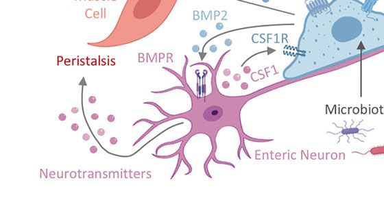
Chiaranunt et al.                                                                                                                Macrophage Functions Beyond Immunity

                         A

                         B

                         C

 FIGURE 1 | Intestinal MP heterogeneity is determined by distinct ontogeny, transcription profiles, and microanatomic locations. (A) Fetal liver monocytes and adult
 BM-derived monocytes seed the intestinal tract in two distinct waves, giving rise to at least three distinct MP populations in the gut: Tim-4-CD4- (DN) MPs, Tim-4-
 CD4+ (SP) MPs, and Tim-4+CD4+ (DP) MPs. Monocytes require CCR2 and NR4A1 for the egress from the BM and differentiation into subsets. Monocytes have
 been suggested to differentiate into DN MPs in a RUNX3-dependent manner and in an environment rich in CSF1, whereas DP MPs require CSF1 and TGF-b.
 CSF2 plays a role in gut MP development and impacts the functional and developmental profile of MP, possibly through the actions of IRF5. However, the exact
 differentiation pathways and plasticity between DN, SP, and DP MPs are yet to be elucidated. The color separation (blue: fetal, light brown: adult) in (A) indicates the
 developmental origin of MPs. Dashed lines indicate potential developmental relationships among gut MPs. It remains unclear if DN, SP and DP MPs constituted a
 developmental continuum or follow three distinctly developmental pathways. Plasticity between each MP population also remains to be addressed. After birth,
 infiltration of adult BM-derived monocytes into the intestinal tract requires signals from the microbiome. (B) Post infiltration of the gut, MPs accumulate at distinct
 microanatomic locations across the intestinal tract. Close proximity with characteristic structures allows for the classification of intestinal MPs into vasculature-
 associated MPs (vMPs, red), epithelium-associated MPs (eMPs, light brown), nerve-associated MPs (nMPs, light blue) and lymphoid tissue-associated MPs
 (lMPs, violet). Despite their shared monocytic origin, there is evidence of preferential anatomic localization of MPs that has been factored into our current working
 model for intestinal MP development. (C) We propose a working model for gut MP development, in which precursors of distinct developmental origins (blue: fetal,
 light brown: adult) differentiate into DN, SP, and DP MPs. Colors indicate their accumulation at microanatomic locations like the vasculature (red), the epithelium
 (light brown), neurons (light blue) or lymphoid tissues (violet). Each MP identity depends on ontogeny, their microanatomic location, and the environmental signals
 therein. Dashed lines indicate plasticity or direct developmental relationships between DN, SP, and DP MPs.

these anatomical locations in supporting intestinal MP                                  not express Tim-4 or CD4 at 2 weeks post-transfer (37, 38). An
heterogeneity (8). While LP Tim-4+ CD4+ MPs are capable of                              explanation for why self-renewing MPs were overlooked in the
self-renewing locally, subsequent studies using the monocyte-                           past could be the use of the experimental system or time of
specific fate-mapping model, Ms4a3Cre x Rosa26-LSL-tdTomato,                             analysis (37–39). While the selective expression of the diphtheria
paired with transcriptomic analyses of engrafted gut MPs after                          toxin receptor (DTR) on all CD11c+ or CX3CR1+ cells allows for
BM transfer, indicated that even the long-lived Tim-4+ CD4+ MP                          the removal of gut MPs, this depletion includes fetal-derived
population in both the SI and colon is replaced by monocytes                            long-lived Tim-4+ CD4+ MPs. Refinement of this system by
emerging through adult definitive hematopoiesis over time                                targeting the expression of DTR to gut MPs that arise during a
(Figure 1A) (8, 36). This is in contrast to studies examining                           distinct hematopoietic wave or by using shielded, instead of total
the engraftment of adoptively transferred monocytes, which do                           body, irradiation allows for the identification of these long-lived

Frontiers in Immunology | www.frontiersin.org                                       3                                     September 2021 | Volume 12 | Article 749708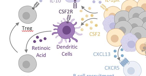
Chiaranunt et al.                                                                                            Macrophage Functions Beyond Immunity

self-sustaining cells (8). Importantly, De Schepper et al. and Liu         upon adoptive transfer into the lung (1). Interestingly, van de
et al. did not report complete monocyte replacement of all gut             Laar et al. refined this picture and demonstrated that precursors
MP populations. With 66-74% replacement after 6 weeks and                  from all three hematopoietic waves (embryonic, fetal, and adult)
close to 90% after 8 months, self-renewing cells may exist even            were capable of adopting the phenotype of an adult alveolar MP
within the Tim-4- CD4- and Tim-4- CD4+ MP populations                      when transferred into an empty niche. In contrast to Lavin et al.,
(8, 36, 40). An additional hypothesis that should be tested                they demonstrated that MPs isolated from the liver and colon
is whether monocytes can directly differentiate into Tim-4+                were unable to engraft into the lung. Even though engraftment of
CD4+ MPs, or whether there is gradual upregulation of Timd4                MPs from the peritoneum was observed in the lung, these cells
or Cd4 expression in the pool of long-lived gut-resident                   were incapable of fully adopting an alveolar MP phenotype.
MPs (Figure 1A).                                                           Notably, Lavin et al. performed adoptive transfer of peritoneal
    Notably, multiple populations exist within the pool of long-           MPs into conventional C57Bl/6 mice (already containing a local
lived Tim-4+ CD4+ MPs, as recently demonstrated by De                      pool of alveolar MPs), while van de Laar et al. transferred
Schepper et al. and Kang et al., alluding to location-specific              peritoneal MPs into Csf2rb-/- hosts (lacking all alveolar MPs).
adaptation of these cells (8, 31). Similarly, other markers, such          The results of these reports either suggest the presence of a
as CD206 or CD11c, have been used to delineate colonic MP                  peritoneal precursor population, an adaptable differentiation
populations based on other factors, such as their dependency               program specifically in peritoneal MPs, or the requirement of a
on the microbiota (31). This heterogeneity has made it                     pre-existing pool of alveolar MPs to initiate the reprogramming
extremely difficult to establish a standard nomenclature for                of mature infiltrating cells (42). A co-transfer of alveolar MPs or
intestinal MPs. In order to facilitate the classification of these          their precursors along with congenic peritoneal MPs into
cells, we have summarized a list of known surface markers,                 Csf2rb-/- hosts could help to validate these interpretations, at
transcription factors, and cytokines/chemokines based on the               least in parts. An intriguing study led by Li et al. revealed that
Tim-4/CD4 categorization of intestinal LP MPs. We advise                   fetal liver-derived monocytes have a superior capacity to replace
the reader that this list is by no means exhaustive and                    alveolar MPs in postnatal Csf2ra-/- hosts when compared to
should be independently validated (Table 1). Our table                     primitive, yolk-sac-derived MPs. An underlying transcriptional
alludes to differential localization even within each Tim-4/CD4            difference and increased metabolic fitness of fetal liver monocytes
population based on variable expression of Lyve1, Cd163, and               may possibly be the result of elevated CSF2RA expression on
Cd206. Our selection of markers further emphasizes the need for            these precursors. Collectively, these findings emphasize the need
additional study into the transcriptional regulation of each               to improve our understanding of both the necessary growth
MP identity.                                                               factors and the transcriptional regulators of MP identity across
    Given the anatomical and environmental differences between             organs and their microanatomic locations to uncover the true
the small intestine and the colon, it is also important to note that       complexity of MP heterogeneity.
MP heterogeneity exists across gut segments (41). For example,                 As with all MPs, intestinal MPs express a unique transcriptional
transcriptome analysis demonstrates variations between monocyte            program that dictates their core MP characteristics, such as
graft-derived colonic versus ileal MPs, particularly with regards to       phagocytosis and antigen sampling. For example, PU.1 has
genes associated with epithelial cell communication and cell               long been studied as a master regulator of MP lineage
metabolism (39). In correlation to the region-specific microbiota           specification and commitment (43, 44). Interferon regulatory
abundance, the colon also contains a higher frequency of LP MPs            factor 8 (IRF8), another pivotal transcriptional factor in adult
compared to the relatively microbe-scarce duodenum (15). It is             myeloid lineage commitment and inflammatory response,
important to keep these differences between small versus large             plays an additional role for the maturation of embryonically
intestinal MPs in mind, particularly when considering therapeutic          derived MPs (45–48). CCAAT/enhancer-binding protein b
approaches, as intestinal pathologies typically present in specific         (C/EBPb) has also been shown to be essential in the pro-
gut segments (39).                                                         inflammatory fate of MPs (49, 50). Importantly, PU.1 and
                                                                           C/EBPb induce the upregulation of colony stimulating factor 1
                                                                           receptor (CSF1R) via a two-step chromatin remodeling process
                                                                           in MP precursors as they differentiate (51). This mechanism
TRANSCRIPTIONAL REGULATION OF                                              results in a positive feedback loop that amplifies the cell’s
INTESTINAL MPS WITHIN TISSUE NICHES                                        responsiveness to CSF1 or IL-34 - growth factors required for
                                                                           MP differentiation and survival (52–54). Both CSF1 and IL-34
The vast heterogeneity of MPs across different organs and                  act through CSF1R, but display distinct patterns of expression.
microanatomic regions is governed by local tissue                          IL-34 has been reported to be secreted in the brain, eye, skin,
microenvironments that produce growth factors, chemokines,                 and peripheral nerves, while CSF1 shows a far broader
and cytokines to drive differential transcriptional programs within        expression across the body (52–54). Interestingly, CSF1
these MPs. Mature tissue-resident MPs can transcriptionally                has been shown to induce PU.1 expression in hematopoietic
adapt to changes in their environment, as illustrated in an                stem cells (HSCs), suggesting the possibility of a growth
elegant study by Lavin et al. Here, peritoneal MPs partially               factor-transcription factor feedback loop to further drive
adapt gene expression signatures characteristic of alveolar MPs            MP differentiation (54). The necessity of CSF1R in MP

Frontiers in Immunology | www.frontiersin.org                          4                               September 2021 | Volume 12 | Article 749708
Chiaranunt et al. Macrophage Functions Beyond Immunity
TABLE 1 | Heterogeneity of murine (A) and human (B) monocyte and MPs in the intestinal lamina propria.
A
Ly6Chi Mono Tim-4-CD4- MP Tim-4-CD4+ MP Tim-4+CD4+ MP
Surface markers Mertk – + + +
Adgre1 (F4/80) – + + +
Fcgr1 (CD64) – + + +
Cx3cr1 + ++ ++ ++
Mrc1 (CD206) – + + +
Itgax (CD11c) – + + +
Itgam (CD11b) ++ ++ ++ ++
Ccr2 ++ + – –
H2-Ab1 (MHCII) – ++ + +
Trem2 ++ + – –
Trpv4 – – + ++
Aif1 (Iba1) – + + ++
Adrb1 – – + ++
Lyz1 (Lysozyme) ++ + – –
P2ry2 – – + ++
Csf1r – + ++ ++
Csf2rb2 – + ++ ++
Cd209f – – + ++
Cd209g – – + ++
Traf1 – ++ + –
Adora2a ++ + – –
Adora2b ++ + – –
Tgfbr1/Tgfbr2 – + + ++
Il10rb – + + +
Vcam1 – + + ++
TF Id3 – + – –
Runx3 – ++ + +
Nr4a1 – ++ + +
Cytokines/ Ccl2 – ++ + +
Chemokines Il1b – ++ + +
Il10 – + + ++
Tnf – ++ + +
Vegfa/Vegfb – + + +
Wnt4/Wnt5b – – + ++
B
CD14+ Mono Mf1 Mf2 Mf3 Mf4
Surface markers CCR2 ++ + + – –
CX3CR1 ++ ++ ++ + +
CSF1R ++ + + + +
CD209 – + ++ ++ ++
MRC1 (CD206) – + + + -/+
HLA-DQ/HLA-DR – -/+ ++ ++ ++
LAMP2 + + – – –
IL1R1 – + – – –
CD163 ++ – – + +
CD172A + + + + +
ITGAM + + + – +
CD64 ++ ++ ++ + +
CD14 + ++ + + ++
CD1c – + ++ – –
ITGAX -/+ + + – –
TFs AHR – – + ++ ++
RXRA ++ + – – –
TCF7 – – – + +
Cytokines/ CCL18 – ++ + + –
Chemokines IL10 – – – + +
IL1B + ++ + + +
TNFA + + + – –
Frontiers in Immunology | www.frontiersin.org 5 September 2021 | Volume 12 | Article 749708
Chiaranunt et al.                                                                                               Macrophage Functions Beyond Immunity

development and survival has been established using genetic                   towards a Th1/Th17 phenotype and promoted inflammation in
mouse models and antibody-mediated neutralization of CSF1. In                 several autoimmune disorders (69). In line with these findings,
line with this, Csf1r-/- and Csf1op/op mice show significantly                 the absence of Irf5 in MPs ameliorated disease in two
reduced MP populations, with recognizable differences in                      microbiota-driven models of colitis due to a decrease in
microglia and Langerhans cell numbers in Csf1r-/- versus                      infiltrating Ly6Chi MHCII+ monocytes (70). Notably, Cx3cr1Cre
Csf1 op/op mice (3). Injections of neutralizing anti-CSF1                     x Irf5flox/flox mice revealed an apoptosis-independent defect in the
antibodies abrogate the development of tissue-resident MPs,                   colonic MP compartment even in the absence of inflammation,
with prolonged treatment resulting in an almost complete loss                 implicating the importance of this transcription factor during
of MPs even in the gut (3, 55, 56).                                           normal gut MP development (70).
    Similar to the brain and lung, an indispensable factor of                     Transcriptome analysis of tissue-resident MPs across organs
monocyte differentiation in the intestine is transforming growth              identified gut-specific expression of Runt-related transcription
factor-b (TGF-b) (24, 57–59). With regards to MPs, TGF-b has                  factor 3 (RUNX3) (Figure 1A) (1). RUNX3 expression has been
long been associated with an anti-inflammatory wound-healing                   shown to be driven by TGF-b in colonic MPs (24, 71). In line
phenotype as these cells have been shown to secrete the cytokine              with this, Runx3-/- mice show impaired differentiation of anti-
in models of chronic pulmonary fibrosis and skin wounds (60,                   inflammatory MPs and cDC2s and consequently develop
61). In the gut, TGF-b—particularly the isoform TGF-b1—plays                  spontaneous colitis (72). Recent work by Scott et al. identified
an extensive role in regulating both local adaptive and innate                the transcription factor, zinc finger E-box binding homeobox 2
immune responses to induce oral tolerance, and much of its                    (ZEB2), to be required for maintaining tissue-specific identities
prevalence in the tissue depends on an intact microbiota (62–64).             of MPs in the liver, lung, brain, spleen, and gut (73). Analogous
Given its ubiquitous role, it is likely that the multiple sources of          to RUNX3, TGF-b treatment enhances ZEB2 expression in MPs
TGF-b, including intestinal epithelial cells, DCs, and MPs                    in the context of human hepatocellular carcinoma, suggesting
themselves, may all affect gut-seeding monocytes during their                 possible co-regulation of both transcription factors through
differentiation process (62, 65, 66).                                         TGF-b-mediated signaling (74).
    As monocytes transition into MPs in the colon, genes                          Notably, transcriptional heterogeneity between monocyte- and
associated with the TGF-b receptor (TGF-bR) signaling                         fetal-derived intestinal MPs extends beyond their ontogeny and
pathway, such as Smad7 and Tgfbr1/2, were shown to be highly                  alludes to environmental influences. To study the effects of long-
upregulated (24). The same study demonstrated that the increased              term MP residency in the gut, De Schepper et al. labeled adult
expression of intestinal MP-specific genes, such as Cx3cr1, Il10,              intestinal MPs in Cx3cr1CreERT2 x Rosa26-LSL-YFP mice and
and Itgax, requires TGF-bR signaling (24). Notably, TGF-b                     compared the transcriptional signatures of YFP- monocyte-
signaling seems to induce differential gene expression profiles in             replaced MPs versus YFP+ self-maintaining MPs in the SI and
intestinal CD11c+ cells, containing DCs and MPs, versus those in              colonic LP 35 weeks later (8). Self-maintaining MPs were enriched
the brain or lung, alluding to the multiplicity of other extracellular        in genes involved in homeostatic tissue maintenance, including
factors in each organ that concurrently shape MP identity (24, 57, 59).       epithelial cell and neuronal differentiation and angiogenesis,
The necessity of TGF-b signaling on intestinal MPs has primarily              suggesting that self-maintaining, Tim4+CD4+ MPs contribute to
been studied in mouse strains with TGF-bR-truncated or                        tissue homeostasis during steady state (8). In the peritoneal cavity,
-dysfunctional CD11c+ cells. For example, mice containing MPs                 resident MPs were found to require the transcription factors KLF2
with truncated TGF-bR experience more severe DSS-induced                      and KLF4 for their Timd4 expression and consequent function of
colitis, with reduced IL-10 production and delayed goblet cell                apoptotic cell clearance—whether this applies to intestinal Tim-4+
regeneration, demonstrating the role of TGF-b in promoting MP-                MPs remains to be seen (75). On the contrary, MPs that were
mediated immunosuppression (67). As will be discussed shortly, a              replaced by adult BM-derived monocytes expressed genes known
unique characteristic of intestinal MPs is their hyporesponsiveness           to regulate innate immune responses and phagocytosis. Indeed,
to exogenous stimulation. However, this suppressive phenotype                 monocyte-derived Tim-4- CD4- MPs were found to express genes
does not seem to depend on TGF-b signaling but instead on IL-10               distinct from long-lived Tim-4+ CD4+ MPs, including those
signaling, with both pathways imprinting non-overlapping                      involved in cytokine production and phagocytosis (e.g. Stat4,
features of colonic MPs. Instead, mice with TGF-bR-deficient                   Slamf7, and Traf1) (33). Interestingly, these Tim-4- MPs also
CD11c + cells displayed elevated numbers of immature                          express inhibitor of DNA binding 3 (ID3), a TGF-b-responsive
Ly6ChiMHCIIlo colonic MPs and higher Ccl8 expression in                       transcriptional repressor that, by contrast, serves as a critical
mature MPs, suggesting that the TGF-b-TGF-bR axis may aid                     regulator of fetal-derived Kupffer cell development (7, 76). This
in monocyte recruitment and MP turnover (24).                                 not only reflects the TGF-b dependency of MP populations across
    Another interesting transcription factor that predominantly               organs, but also suggests that the same cytokine can induce either
regulates the differentiation program of monocytes and MPs                    monocyte- or fetal-derived MP identity depending on the
upon TLR engagement is interferon regulator factor 5 (IRF5).                  context (Figure 1A).
IRF5 is strongly upregulated upon LPS stimulation of human                        Interestingly, a small proportion of monocytes can
blood monocyte-derived MPs, resulting in elevated production                  differentiate into Tim-4+ self-maintaining MPs, both during
of TNFa, IL-1b, IL-12, and IL-23 while repressing IL-10 (68).                 steady state or upon MP depletion and gut inflammation
These activated MPs preferentially polarized CD4+ T cells                     (8, 32, 33, 36–38). These monocytes give rise to fully mature

Frontiers in Immunology | www.frontiersin.org                             6                               September 2021 | Volume 12 | Article 749708
Chiaranunt et al.                                                                                             Macrophage Functions Beyond Immunity

tissue-resident MPs that partially recapitulate the gene                     MPs, Luminal Microbes and Gut
expression profile of fetal-derived MPs (8, 39). Whether or not               Immune Homeostasis
their prolonged residence in the gut will enable these cells to              To detect and mediate responses to the microbiota, MPs are
eventually fully adopt the gene expression signature of fetal-               equipped with an array of sensory pattern-recognition receptors
derived long-lived MPs over time remains to be elucidated                    (PRR) (85). The mechanisms behind PRR regulation in intestinal
(Figure 1C). The determining factors of monocyte differentiation             MPs have been extensively studied and discussed over the past
into gut-resident MPs remain unknown, although a recent study                decade; we encourage our readers to seek reviews on this topic
on peritoneal MPs suggests that monocytes acquire a mature                   (86, 87). Inflammasomes, particularly NLRP3 and NLRC4, were
tissue-resident identity when entering an empty niche, wherein               found to play important roles in inducing IL-1b-dependent,
the original resident MPs have been ablated (77). Notably, the               monocyte and MP-driven type III immune responses following
presence of long-lived Tim-4+ MPs across other organs suggests               bacteria commensal and pathogen encounter (88–92). Similarly,
Timd4 expression may reflect the duration of residence within the             CX3CR1+ cells, including subsets of DCs, are implicated in
tissue and not necessarily MP ontogeny (78). Thus, in the context            initiating specific anti-fungal programs to promote pathogen
of intestinal MPs, we assert that these transcriptional complexities         clearance (93–96). Together, these data suggest that microbe-
call for careful characterization and definition of MP populations            initiated activation of inflammasomes in monocytes, MPs and
based more on their function and localization than merely on                 DCs can result in a context-dependent immune response. While
their ontogeny.                                                              there are multiple mechanisms of microbial uptake and
    Indeed, the heterogenous transcriptional signatures of                   recognition, the exact contributions of monocytes, MPs and
intestinal MPs reflects the differences in their local                        DCs remain controversial, and distinction of these cell types
microenvironments and functional specializations. For example,               should be considered when interpreting experiments centered
distinct MP populations can be defined in the gut based on their              around antigen sampling and microbial regulation (97–99).
expression of genes implicated in either epithelial, neuronal, or            Persson et al. provide a detailed discussion on mechanisms of
vascular homeostasis. The physical proximity to non-                         antigen sampling by DC and MPs in the gut (100).
hematopoietic cells across the gut suggest that MPs adapt these                  Cytokine secretion and activation of MPs by the microbiota
specialized transcriptional programs to support their neighboring            has been suggested and was found to promote the constitutive
cells (8). Guilliams et al. reported a similar regional specialization       secretion of IL-10 by SI and colonic MPs in support of oral
of MPs in the liver (7, 9). MP interactions with other long-lived,           tolerance and regional abrogation of inflammatory disease (98,
tissue-resident cells, like innate lymphoid cells (ILCs), also shape         101, 102). Some of the microbiota-dependent mechanisms
regional MP identity. For instance, lung ILCs and the lung                   proposed for gut MP secretion of IL-10 include the attachment
epithelium produce high levels of colony stimulating factor 2                of commensal microbes to the gut epithelium, the colonization
(CSF2), a growth factor involved in differentiation and                      with helminths, or the secretion of IL-4 and IL-5 by T helper type
homeostasis of alveolar MPs (79–81). Gut MP homeostasis                      2 (Th2) cells (103). Without a doubt, MP-derived IL-10
partially depends on CSF2, which is most abundant within                     contributes to several homeostatic and anti-inflammatory
intestinal cryptopatches (CPs) and isolated lymphoid follicles               processes, including the modulation of intestinal epithelial
(ILFs) (82). Since Csf2-/- mice display only a partial reduction in          healing and the host immune response promoting local
intestinal MPs, the aforementioned lymphoid structures, which                regulatory T (Treg) cell expansion (15, 101, 104–107). Varying
contain group 3 ILCs (ILC3s) and MPs, may serve as a niche for a             levels of IL-10 were observed in colonic, but not SI, MPs of mice
unique CSF2-dependent MP population (82–84). Refining the                     colonized with different microbiota, suggesting that the
map of myeloid growth factors across the intestinal tract will help          tolerogenic function of MPs relies on regional differences in
to identify niches that support the maintenance and                          environmental factors, including the microbiota (101, 106).
transcriptional adaptation of MPs in the gut.                                Collectively, these data suggest an instructive role for
                                                                             commensals and pathogens alike in shaping local MP
                                                                             development and function, leaving several questions on the
                                                                             involvement of non-bacterial commensal in the regulation of
MP RECOGNITION OF THE                                                        MP functions unexplored (108–110).
INTESTINAL MICROBIOME                                                            Although the microbiota is required for proper MP function
                                                                             and intestinal health, it also requires tight control by the host
As their name suggests, MPs are best known for their proficient               innate immune system. In fact, MPs, DCs and intestinal barrier
phagocytic abilities, particularly in the context of engulfing                dysfunction have been shown to impair oral tolerance and
microbes for innate host defense. Given that the intestinal tract            promote inflammation and disease, as implicated in the
hosts the most diverse microflora in the body, it may seem                    pathogenesis of Crohn’s disease (CD) (111). Genetic ablation
surprising that gut MPs are not constantly activated and                     of CX3CR1, CSF-2, or IL-10 in mice results in a loss of oral
secreting proinflammatory cytokines. Since MP-microbe                         tolerance towards luminal antigens, consequently increasing
interactions are a classic immune function of intestinal MPs,                susceptibility to intestinal inflammation (82, 101, 103, 112).
we will only briefly summarize how MPs respond to the                         For instance, mice deficient in IL-10 develop spontaneous
microbiota and contribute to immune homeostasis.                             colitis through an aberrant response to commensals (113, 114).

Frontiers in Immunology | www.frontiersin.org                            7                              September 2021 | Volume 12 | Article 749708
Chiaranunt et al.                                                                                         Macrophage Functions Beyond Immunity

This is supported by data in humans, where genetic mutations in          in the gut is an expanding area of research and we will therefore
the IL-10 receptor (IL-10R) were shown to be associated with the         focus on intestinal MP functions going beyond their traditional
development of severe pediatric IBD (115). Specifically, IL-10R           roles by summarizing recent progress on this new frontier of MP
signaling was reported to be critical for gut MPs to abrogate            non-hematopoietic cell interactions.
colitis through the activation of STAT3 (116, 117). A subsequent
study demonstrated that selective IL-10R-deficiency in MPs
drives intestinal inflammation through uncontrolled IL-22-                INTESTINAL MPS GO BEYOND IMMUNITY
dependent activation of IECs and the subsequent recruitment
of inflammatory neutrophils (118). Notably, Il10 deletion                 Damage to the gut during acute and chronic inflammation has
in colonic MPs does not perturb homeostasis, suggesting                  immediate and long-lasting effects on the organ’s physiology,
that the inflammatory phenotype seen in Il10 -/- mice is                  reaching beyond both transient and permanent activation of
independent of MP-derived IL-10. These findings further                   hematopoietic immune cells. The changes in the hematopoietic
confirm the requirement for Treg-derived IL-10 in dampening               compartment of an organ are only a fraction of the complete
inflammation at mucosal surfaces, suggesting a possible                   response during inflammation. The adaptations of non-
interaction between T regs and intestinal MPs (112, 119).                hematopoietic cells during inflammation and repair are
Zigmond et al. ’s study indicates that the MP responsiveness to          beginning to gain their well-deserved attention in assembling a
IL-10 is critical in preventing spontaneous colitis. Thus, these         holistic, organ-wide immune response (125–128). Tissue cells
data together allude to the necessity of the IL-10-IL-10R axis for       show significant and long-lasting changes during development,
both MPs and Tregs in maintaining local immune tolerance                 damage, inflammation, or repair, raising the possibility of tissue
(112, 119). While the IL-10-IL-10R interaction sustains Treg and         cell-MP interactions that may permanently alter intestinal
MP homeostasis, intriguing new data suggest that intestinal MPs          homeostasis (125). MPs collaborate with non-hematopoietic
may also contribute to the induction of immunomodulatory and             cells to actively remodel tissues under homeostatic and
barrier-supporting IgA through the production of the B cell-             inflammatory conditions and in turn, are nurtured by the
recruiting chemoattractant CXCL13 and the release of cytokines           growth and survival factors secreted by their non-hematopoietic
involved in class-switch recombination, suggesting a possible            neighbors. The environmental and anatomical complexity of the
role for MPs in humoral immunity at mucosal surfaces                     intestine, paired with a seemingly arbitrary distribution of MPs
(Figure 2A) (84, 103).                                                   across the gut’s microanatomic locations had hindered the
    Crosstalk between MPs and type 3 innate lymphoid cells               identification of obvious tissue cell-MP liaisons (Figure 1B).
(ILC3s) in ILFs offers yet another mechanism for tolerance               Paradigm-setting reports on tissue cell-MP interactions in the
induction and host defense against enteric infections. In the            skin, brain, and liver, however, have provided roadmaps on how to
steady state, microbiota-driven IL-1b production by colonic MPs          tackle this problem in the intestine. These studies identify unique
promotes CSF2 release by ILC3s, which in turn activates IL-10            and critical roles for specific MPs in sustaining and shaping local
and retinoic acid (RA) secretion in MPs and DCs (82). These              tissue cells via interactions that go beyond classical immunity
activated ILC3s also produce IL-22, which maintains barrier              (Figure 1B) (8, 24, 33). In the following sections, we summarize
integrity and induces antimicrobial peptide secretion by                 our current understanding of these non-immune functions of
epithelial cells (120). These interactions have been shown to be         intestinal MPs, highlight the importance of the local tissue
critical for the induction of oral tolerance and defense against         microenvironment in shaping these interactions, and share
attaching/effacing enteric pathogens such as Citrobacter                 outstanding questions. We then superimpose gut-specific MP-
rodentium (82, 120–123). Given the importance of CSF2 in                 tissue interactions with those reported in other organs to inspire
maintaining monocytes and MPs, it is likely that the MP-ILC3             an overarching understanding of MP-tissue cell interactions
crosstalk in colonic ILFs may also serve as a local site for the         (Figures 2 and 3).
growth and differentiation of monocyte-derived MPs (83).
Importantly, the combination of proinflammatory IL-1b-                    Intestinal MPs Fortify the Epithelial Barrier
secreting MPs and immunomodulatory IL-10-producing MPs                   Intestinal epithelial cells (IECs), forming a monolayer barrier
in this crosstalk further emphasizes the complexity and                  between the LP and lumen, are the first physical line of defense in
heterogeneity of intestinal MPs in facilitating intestinal               the protection against commensal and pathogenic microbes.
immune and tissue homeostasis (Figure 2A).                               MPs line the intestinal epithelium, and emerging data have
    Taken together, the development and function of MPs have             indicated that MPs aid these cells in their function and
profound consequences on intestinal health. It is evident that the       development. Steady state MPs constitutively express low levels
location of MPs within an organ is critical to their identity, as        of TNFa, which has been shown to regulate enterocyte growth,
microenvironmental cues from neighboring cells imprint                   maintain epithelial barrier integrity, and stimulate the
functional programs tailored to serve the local microanatomic            production of tissue remodeling proteins in intestinal
niche (Figure 1B) (124). Additional MP-immune interactions               mesenchymal cells (23, 129, 130). The secretion of soluble
and classical contribution to the regulation of intestinal immune        factors, such as the lipid mediator prostaglandin E2 (PGE2) by
homeostasis have been excellently reviewed elsewhere (86).               MPs, further supports the epithelial barrier (131). Given the vast
However, the interactions of MPs and non-hematopoietic cells             surface of the epithelium and the regional delineation of different

Frontiers in Immunology | www.frontiersin.org                        8                              September 2021 | Volume 12 | Article 749708
Chiaranunt et al. Macrophage Functions Beyond Immunity
A B
C D
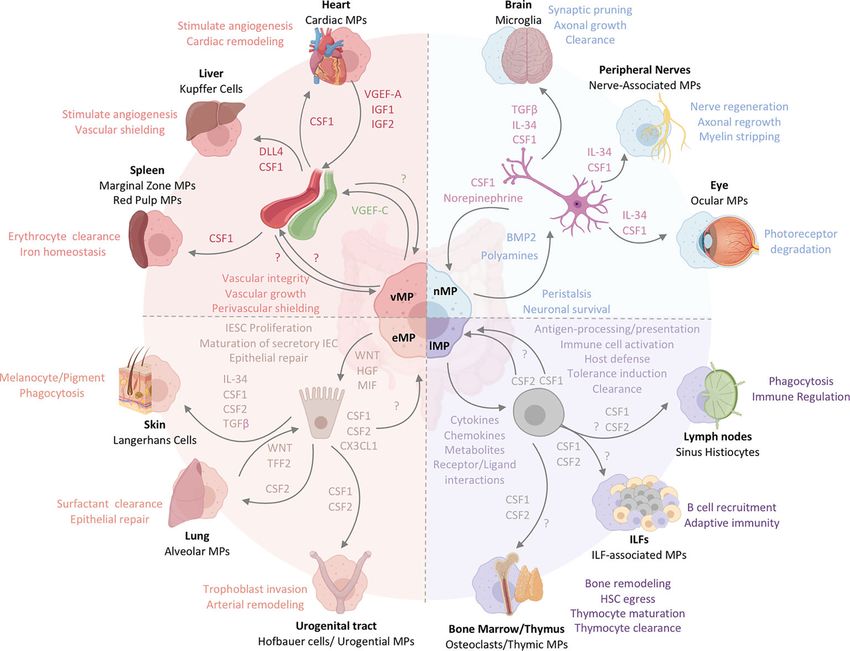
FIGURE 2 | Intestinal MPs play multiple non-immune roles to maintain tissue homeostasis. (A) Lymphoid tissue -associated MPs (lMPs) accumulate in PPs, ILFs
and CPs. These lMPs sense and sample microbes and surround the B cell follicles within PPs and ILFs. Microbial recognition by lMPs facilitates the release of IL-1b
and activates lymphoid tissue-resident group 3 innate lymphoid cells (ILC3s). ILC3s release CSF2 and IL-22 to engage CSF2R on myeloid cells or IL-22R on
epithelial cells. The latter is prominent in facilitating antimicrobial activity. The former (CSF2-CSF2R) acts on DCs and lMPs to induce the production of IL-10 and RA,
both critical in facilitating the conversion of naïve T cells in to regulatory T cells (Treg). Lymphoid tissue -associated MPs have also been shown to release the B cell-
recruiting chemokine CXCL13 and clear apoptotic B cells resulting from failed somatic hypermutation or class-switch recombination, thus contributing to the local IgA
response. (B) MPs lining the epithelium (eMPs) near intestinal crypts induce stem cell renewal by inducing WNT signaling in intestinal epithelial stem cells (IESC).
Epithelium-associated MPs adopt an alternative activation phenotype when stimulated with by IL-4 and IL-13, by upregulating TREM-2 and aiding in epithelial repair
and goblet cell proliferation. MPs in the subepithelial dome of the PPs have been shown to induce M cell maturation through the release of MIF. Hepatocyte growth
factor (HGF) is another protein that mediates epithelial repair and is possibly produced by eMPs post injury with likely varying locations around the crypt and the
villus. (C) Nerve-associated MPs (nMPs) in the intestinal muscularis layer are stimulated by the gut microbiota and regulate peristalsis via BMP2-mediated activation
of enteric neurons. Enteric neurons release neurotransmitters to induce smooth muscle cell contractions. Direct activation of smooth muscle cell contraction is
mediated by nMP-release of PGE2. In addition, nMPs release polyamines in response to signals from the microbiome, catecholamines, and other stress cues to
facilitate neuronal protection. In turn, enteric neurons secrete CSF1 to support nMP survival. (D) Across the lamina propria, vasculature-associated MPs (vMPs) wrap
around blood vessels to aid in angiogenesis, lipid transport, dead cell clearance, vessel integrity and elongation. In the presence of an intact microbiota, these MPs
are rapidly replaced by monocyte-derived cells to induce vascular repair while protecting against bacterial dissemination upon injury and infection. How the
endothelium in turn maintains MP survival is currently unknown. Vasculature-associated MPs in close proximity to small intestinal lacteals facilitate vessel elongation
and integrity through the microbiota-driven production of VGEF-C.
epithelial cell types, these MP-tissue cell interactions may serve affecting the transcytosis of luminal particles across the
different functions depending on location. For example, intestinal wall (55). Defining the interaction of MPs and IEC
interactions where MPs aid in epithelial stem cell repair may should therefore be seen in a multicontextual setting, accounting
most likely be observed at the crypts of the epithelium, while for the microanatomic localization of MPs, their functional
MPs targeted to support enterocytes or other differentiated specialization and effects on the epithelial layer
epithelial cells may execute their functions in the upper regions It is in cases of intestinal insult that these epithelium-associated
of the LP and the villus. The transient ablation of gut MPs MPs (eMPs), found along the crypts and villi of the LP, become
through anti-CSF1R antibody treatment for example disrupts the truly critical for repair. For example, following tissue damage, MPs
proliferation of intestinal stem cells, resulting in reduced around intestinal crypts were responsible for the proliferation and
expression of the WNT receptor Lgr5 on intestinal epithelial survival of epithelial progenitor cells in a MyD88-dependent
stem cells, and impair maturation of Paneth cells across the SI manner (132–134). In a murine model of acute colonic
(55). Depletion of MPs further indirectly blocks M cell epithelial injury, Seno et al. demonstrated increased infiltration
differentiation in favor of goblet cells, thereby potentially of triggering receptor expressed on myeloid cells 2 (TREM-2)+
Frontiers in Immunology | www.frontiersin.org 9 September 2021 | Volume 12 | Article 749708
Chiaranunt et al.                                                                                                                      Macrophage Functions Beyond Immunity

 FIGURE 3 | The functional heterogeneity of intestinal MPs mirrors the multifaceted roles of tissue-resident MPs across organs. Intestinal MPs maintain tissue
 homeostasis through their interactions with neurons, immune cells, epithelial cells, and endothelial cells. These functions are dependent upon the associations with
 both immune and non-hematopoietic cells within their respective microenvironments. Released factors and physiological consequences of vasculature-associated
 MPs (vMPs, red), epithelium-associated MPs (eMPs, light brown), nerve-associated MPs (nMPs, light blue) and lymphoid tissue-associated MPs (lMPs, violet) are
 indicated by their respective colors. Arrows indicate the source of a given regulator (i.e. produced by MPs perceived by neighboring cell). Nerve-associated MPs in
 the brain, peripheral nervous system or the eye have specialized functions tailored to their location. These cells depend on neuron-released TGF-b, IL-34 and CSF1.
 Whether intestinal nMPs require the full spectrum of growth factors is unknown. In addition, a role for maintaining sensory neurons, neuronal growth or synaptic
 pruning in the gut remains to be shown. Lymphoid tissue-associated MPs are vital in antigen sampling, processing and presentation and mediate dead cell clearance
 of leukocytes within gut-associated lymphoid tissues. They contribute a plethora of cytokines, chemokines, metabolites and receptor ligands which plays an essential
 role in host defense against microbes, the orchestration of tolerance induction, and immune cell activation. CSF2, produced by T cells and ILC3s, is essential in
 facilitating most of these processes in the intestinal tract. It remains to be shown whether CSF1 or other myeloid growth factors like IL-10 support their biological
 relevance in intestinal immune homeostasis. Peripheral lymph node- and ILF-associated MPs mirror the function of lMPs in the gut, likely due to the similarities in the
 development and architecture of these organs. The thymus and BM, primary lymphoid tissues, host MPs that mediate tissue remodeling and control the hematopoietic
 stem cell egress, while also facilitating T cell maturation and clearance. Whether lMPs in the gut are capable of these functions remains to be addressed. The interactions of
 epithelium and MPs in the gut support IESC proliferation, maturation of secretory IEC and the repair the of damaged epithelial monolayer. CX3CL1 controls the adaptation
 of an eMPs phenotype but it remains to be shown if CSF1 or CSF2 produced by IECs in the gut contribute to this identity. In contrast to the gut, Keratinocytes, alveolar
 type 2 cells and urogenital epithelial cells produce distinct combinations of myeloid growth factors that facilitate development and functional specialization of MPs in their
 respective environments. In the case of the lung, WNT-release by lung macrophages mirrors the mechanism used by intestinal eMPs to facilitate epithelial repair. Functions
 of eMPs collectively promote tissue homeostasis and tissue remodeling. While the small intestinal lymphatic system is supported by vMPs through the production of VGEF-
 C to mediate vessel integrity and elongation, molecular mechanisms allowing for a deeper understanding of vMP-blood vessel interactions are currently not known for the
 intestinal tract. Vasculature-associated MPs surrounding intestinal blood vessel in a symmetric pattern that shields the endothelium from the environment, serving as a
 cellular firewall for the prevention of microbial spread into neighboring organs. Filter and barrier functions have been assigned to MPs in the spleen and the liver, where these
 cells are supported by endothelium-derived growth and differentiation factors. Cardiac MPs in fact receive survival signals from the endothelium via the release of CSF1 and
 in turn support the release of insulin growth factor (IGF) 1 and 2 as well as VGEF-A for cardiac remodeling and angiogenesis.

MPs around injured areas. TREM-2 promotes repair genes in                                   motif-containing 33 (TRIM33) is a cell surface protein that
IL-4- and IL-13-stimulated, alternatively activated MPs by                                  plays a role in inducing an immunomodulatory MP phenotype.
dampening NFkB signaling and thus facilitates wound closure                                 For example, Trim33-/- mice contain monocytes that do not
and epithelial proliferation and repair (Figure 2B) (135). On the                           differentiate into colonic MPs and display impaired resolution of
other hand, colonic MP expression of TREM-1 is associated with                              colonic inflammation in DSS-induced colitis (136, 137).
colitis, and blocking TREM-1 abrogates local levels of monocyte                                 Another role for eMPs in epithelial repair was found by a
chemoattractants and pro-inflammatory cytokines. Tripartite                                  different group, who reported CD206+ MPs in close proximity to

Frontiers in Immunology | www.frontiersin.org                                          10                                      September 2021 | Volume 12 | Article 749708
Chiaranunt et al.                                                                                           Macrophage Functions Beyond Immunity

the damaged mucosa of ulcerative colitis (UC) patients. These             found to be enriched in SI-resident MPs when compared to
MPs expressed elevated levels of Wnt1 and Wnt3a compared to               monocytes, while BMP expression appeared highest in Tim-
pro-inflammatory MP populations and activated the reparative               4+CD4+ MPs, suggesting a functional specialization of MP
WNT-b-catenin pathway in IECs (Figure 2B). Co-culturing                   subsets (33). However, it remains unknown whether WNT and
IECs with alternatively activated murine MPs increased their              BMPs are co-produced by specific MP subsets or exclusively
levels of nuclear b-catenin through a STAT6-dependent pathway             produced by MPs within specific microanatomic regions.
from MPs (138, 139). Conversely, deletion of STAT6 in epithelial              WNT signaling regulates rapid intestinal epithelial renewal,
cells drastically impaired CD206+ MP numbers in the colonic LP,           with the entire epithelium being completely replaced every 4-5
altered epithelial repair, and exacerbated tissue damage,                 days. As apoptotic IECs are shed off at the tip of the villi, they are
suggesting a possible STAT6-dependent circuit for both                    phagocytosed by closely situated MPs and DCs. Removal of dead
epithelial cell proliferation and MP activation (140).                    IECs by eMPs supports barrier homeostasis under both steady
Hepatocyte growth factor (HGF) is growth factor that aids in              state and inflammatory conditions (19, 153). This engulfment of
the eMP-IEC interactions and was reported to be produced by               apoptotic cells has been shown to alter MP identity, presenting
human and murine MPs to promote the growth of epithelial cell             an intriguing aspect of how the epithelium can shape their
lines in vitro, independent of direct MP-epithelial cell contact.         associated MPs. These transcriptional changes, reported by
The epithelial intrinsic effects and actions of HGF were not              Cummings et al., revealed an upregulation of genes involved in
investigated in this report. However, MP-derived HGF may                  dead cell uptake (Timd4, Axl, Tryo3), phagosome maturation
utilize its receptor c-MET and its putative co-receptor CD44v,            (Pikfyve), lipid and amino acid catabolism, and a downregulation
both targets of WNT signaling and expressed on IESC, to                   of genes involved in proinflammatory processes (19). Deficiency
facilitate epithelial repair (141). Notably, c-MET deficient               in, or blockade of dead cell uptake conclusively impacted the
epithelial cells developed normally during steady state but               polarization of T cells by MPs and DCs and elevated
failed to regenerate at rates comparable to c-MET sufficient               susceptibility to intestinal autoinflammation (154–158). The
epithelial cells (141). Interestingly, depletion of Porcn, a gene         uptake of apoptotic cells induces transcriptional changes
essential for the synthesis and secretion of WNT ligands, in              within MPs and DCs that prevent the development of
intestinal MPs resulted in hypersensitivity to radiation-induced          inflammation but also facilitates their functions as gatekeepers
injury and delayed repair of intestinal stem cells, suggesting MP-        of immune cell entry into mucosal tissues. Their role in
derived WNT ligands are critical regulators of epithelial repair          regulating the entry of immune cells into the gut differs
(Figure 2B) (142). It remains to be shown whether MP-derived              between steady state and inflammation and relies on the ability
WNT secretion precedes the beneficial effects of HGF in                    to interpret different types of cell death and danger signals (159).
promoting epithelial repair (143).                                        An elegant study demonstrated that resident MPs are central in
    HGF is not the only factor regulating WNT’s actions on the            recognition of microbial infection and tissue damage to induce
intestinal epithelium. Concerted actions of WNT, bone                     the cooperation of resident and newly infiltrating myeloid cells in
morphogenetic protein (BMP) and Notch maintain the crypt-                 the bladder. In this study, resident macrophages triggered the
villus axis (144–146). Prompted by evidence that MPs in the               chemotactic recruitment of inflammatory monocytes that in turn
muscularis layer express BMP2 to aid in enteric neuron                    facilitated the trans-epithelial migration of infiltrating
maintenance, it remains possible that eMPs contribute to                  neutrophils across the urogenital epithelium. This process
regulating epithelial cell differentiation through BMP                    required the release of matrix metalloproteinase (MMP) 9 by
production (147). Within the epithelium, BMP2 and BMP4                    infiltrating monocytes (160).
make up the main ligands that drive intestinal BMP signaling.                 Cell migration into the intestine or across the gut epithelium
Opposing WNT signaling, BMPs promote the differentiation of               has been shown to be reliant on matrix metalloproteinases
crypt-residing IECs into the secretory lineage further evident by         (MMPs) and chemokines. Notably, high expression of MMP-2,
the development of polyposis in the stem cell zone upon                   MMP-9, MMP-13, and MMP-14 was uniformly reported in all
conditional deletion of the BMP receptor 1A (BMPR1A) in the               recently identified MP populations (8, 33). MMP-2 in particular
epithelium (148–150). To maintain a stem cell niche, crypt-               has been found to propagate the severity of intestinal
surrounding myofibroblasts and smooth muscle cells were                    inflammation by facilitating the accumulation of infiltrating
shown to produce BMP inhibitors, such as Noggin or                        leukocytes into the colon, while MMP-9 did not participate in
Chordin-like 1, generating opposing gradients of high WNT                 this process (161–163). In contrast to the gut, MMP-9 has been
and BMPs along the crypt-villus axis, respectively (149, 151,             demonstrated to facilitate the extravasation of neutrophils across
152). The contribution of MPs in shaping the WNT/BMP-                     the bladder epithelium, a behaviour also observed during acute
gradient remains an intriguing possibility and requires further           intestinal inflammation (160, 164). Whether MMP-9 secretion
research dissecting the MP-specific release of BMPs across                 by gut MPs could facilitate the extravasation of neutrophils
microanatomic regions of the intestine. Considering the                   across the gut epithelium remains an open question.
steady-state regulation of BMP production in muscularis                   Interestingly, MMP-8 expression was elevated in blood
macrophages through the TLR-Myd88 axis, BMP production by                 monocytes and infiltrating leukocytes in the inflamed intestine
MPs in the LP may follow a similar pathway and could possibly             of IBD patients, suggesting a possible contribution of MMP-8 in
counterbalance MP-produced WNT (147). WNT expression was                  the pathogenesis of this disease (144). These studies hint at a

Frontiers in Immunology | www.frontiersin.org                        11                               September 2021 | Volume 12 | Article 749708
You can also read