CHEK1 and circCHEK1_246aa evoke chromosomal instability and induce bone lesion formation in multiple myeloma
←
→
Page content transcription
If your browser does not render page correctly, please read the page content below
Gu et al. Molecular Cancer     (2021) 20:84
https://doi.org/10.1186/s12943-021-01380-0

 RESEARCH                                                                                                                                           Open Access

CHEK1 and circCHEK1_246aa evoke
chromosomal instability and induce bone
lesion formation in multiple myeloma
Chunyan Gu1,2†, Wang Wang2†, Xiaozhu Tang2†, Tingting Xu2, Yanxin Zhang2, Mengjie Guo1,2, Rongfang Wei2,
Yajun Wang2, Artur Jurczyszyn3, Siegfried Janz4, Meral Beksac5, Fenghuang Zhan6, Anja Seckinger7, Dirk Hose7,
Jingxuan Pan1,8* and Ye Yang1,2*

  Abstract
  Background: Multiple myeloma (MM) is still incurable and characterized by clonal expansion of plasma cells in the
  bone marrow (BM). Therefore, effective therapeutic interventions must target both myeloma cells and the BM
  niche.
  Methods: Cell proliferation, drug resistance, and chromosomal instability (CIN) induced by CHEK1 were confirmed
  by Giemsa staining, exon sequencing, immunofluorescence and xenograft model in vivo. Bone lesion was evaluated
  by Tartrate-resistant acid phosphatase (TRAP) staining. The existence of circCHEK1_246aa was evaluated by qPCR,
  Sanger sequencing and Mass Spectrometer.
  Results: We demonstrated that CHEK1 expression was significantly increased in human MM samples relative to
  normal plasma cells, and that in MM patients, high CHEK1 expression was associated with poor outcomes.
  Increased CHEK1 expression induced MM cellular proliferation and evoked drug-resistance in vitro and in vivo.
  CHEK1-mediated increases in cell proliferation and drug resistance were due in part to CHEK1-induced CIN. CHEK1
  activated CIN, partly by phosphorylating CEP170. Interestingly, CHEK1 promoted osteoclast differentiation by
  upregulating NFATc1 expression. Intriguingly, we discovered that MM cells expressed circCHEK1_246aa, a circular
  CHEK1 RNA, which encoded and was translated to the CHEK1 kinase catalytic center. Transfection of circCHEK1_
  246aa increased MM CIN and osteoclast differentiation similarly to CHEK1 overexpression, suggesting that MM cells
  could secrete circCHEK1_246aa in the BM niche to increase the invasive potential of MM cells and promote
  osteoclast differentiation.
  Conclusions: Our findings suggest that targeting the enzymatic catalytic center encoded by CHEK1 mRNA and
  circCHEK1_246aa is a promising therapeutic modality to target both MM cells and BM niche.
  Keywords: Multiple myeloma, CHEK1, circCHEK1_246aa, Proliferation, Drug resistance, Chromosomal instability

* Correspondence: panjx2@mail.sysu.edu.cn; yangye876@sina.com
†
 Chunyan Gu, Wang Wang and Xiaozhu Tang contributed equally to this
work.
1
 Nanjing Hospital of Chinese Medicine affiliated to Nanjing University of
Chinese Medicine, Nanjing, China
Full list of author information is available at the end of the article

                                        © The Author(s). 2021 Open Access This article is licensed under a Creative Commons Attribution 4.0 International License,
                                        which permits use, sharing, adaptation, distribution and reproduction in any medium or format, as long as you give
                                        appropriate credit to the original author(s) and the source, provide a link to the Creative Commons licence, and indicate if
                                        changes were made. The images or other third party material in this article are included in the article's Creative Commons
                                        licence, unless indicated otherwise in a credit line to the material. If material is not included in the article's Creative Commons
                                        licence and your intended use is not permitted by statutory regulation or exceeds the permitted use, you will need to obtain
                                        permission directly from the copyright holder. To view a copy of this licence, visit http://creativecommons.org/licenses/by/4.0/.
                                        The Creative Commons Public Domain Dedication waiver (http://creativecommons.org/publicdomain/zero/1.0/) applies to the
                                        data made available in this article, unless otherwise stated in a credit line to the data.
Gu et al. Molecular Cancer   (2021) 20:84                                                                   Page 2 of 14

Introduction                                                   downstream CHEK1 targets. These findings provide sig-
Multiple myeloma (MM) is a plasma cell malignancy              nificant insight into the underlying CHEK1-dependent
that originates in the bone marrow (BM), is character-         mechanisms of MM malignancy and bone lesion
ized by clonal heterogeneity and BM dependency, and            formation.
remains incurable, although novel interventions such as
proteasome inhibitors, immune modulators, and bio-             Methods
logical therapies have improved disease outcomes [1–3].        Gene expression profiling
Genetic and epigenetic aberrations, copy number alter-         Gene expression profiling (GEP) cohorts were collected
ations, clonal heterogeneity, and clonal evolution are         using the GEO database as described previously [22, 23].
well-known to contribute to MM proliferation, therapy          The Total therapy 2 (TT2) and TT3 patient cohorts, the
resistance, and relapse, although the mechanisms of MM         Dutch-Belgian Cooperative Trial Group for Hematology
remain incompletely understood, and no single mechan-          Oncology Group-65 (HOVON65) trial (GSE19784) pa-
ism of disease has been identified as a common regulator       tient cohort, and the Assessment of Proteasome Inhib-
of MM [2–5].                                                   ition for Extending Remission (APEX) patient cohort
   In addition, the BM microenvironment supports MM            (GSE9782) were included in analyses, which used pub-
cell survival and drug resistance. BM osteoclasts, macro-      licly available gene expression profile data for each of
phages [6, 7], and adipocytes [8] contribute to these          these patient cohorts [3].
pathologies through distinct mechanisms [9–11]. Osteo-
clasts in particular are thought to play a central role in     Antibodies and reagents
MM and have been intensely investigated in this context.       Antibodies used were as follows: CHEK1 (sc-8408; Santa
MM cells can survive over 10 weeks in co-culture with          Cruz Biotechnology, USA); rabbit IgG (a7016); mouse
osteoclasts alone [12], while MM cells adhering to osteo-      IgG (a7028; Beyotime Institute of Biotechnology, China);
clasts in vivo are quiescent and drug-resistant [13].          FLAG (F-4020; Merck KGaA, Germany); PARP (9542S),
Moreover, detection of focal lesions (FLs) in MM pa-           Caspase-3 (9662S), β-actin (4970S; Cell Signaling Tech-
tients using magnetic resonance imaging (MRI) revealed         nology, USA); MYC (16286–1-AP), CEP170 (18899–1-
that number of FLs was negatively correlated with MM           AP; ProteinTech Group, China); and α-Tubulin (ab7291;
outcome [14]. Due to the complex etiology of MM and            Abcam, UK).
pro-cancer effects mediated by the BM niche, effective           Doxycycline (DOX) was purchased from the Beyotime
targeted therapy requires drug combinations that target        Institute of Biotechnology. Puromycin was purchased
both MM cells and the BM niche.                                from Merck KGaA. Bortezomib (BTZ), Adriamycin
   RAS is the most commonly mutated gene in MM [4],            (ADR), dexamethasone (DEX), LY2603618, and other re-
and simultaneous inhibition of Checkpoint Kinase 1             agents were purchased from Selleck Chemicals (Hous-
(CHEK1) and MK2 MAPK Activated Protein Kinase 2                ton, TX). The rapid Giemsa staining kit was obtained
(MK2) has synergistic effects in suppressing KRAS-             from BBI Life Sciences (Shanghai, China).
mutant cancer [15]. Our group therefore began to evalu-
ate the therapeutic potential of MK2 and CHEK1 inhibi-         Cell lines and cell culture
tors in monotherapy, combined therapies, and dual              Human MM cell lines, including the BTZ-resistant cell
MK2/CHEK2 inhibitors. In our previous study, we dem-           lines ARP1, H929, ANBL6 wild-type (WT) and ANBL6/
onstrated that MK2 was elevated in high-risk MM pa-            BTZ-resistant, and the DEX-resistant cell lines MM1S
tients, and MK2 inhibition prolonged the survival in           and MM1R, were cultured in RPMI-1640 (Biological In-
MM patients and suppressed MM cell growth [5, 16].             dustries, Israel). HEK-293 cells were cultured in DMEM
Subsequently, we have sought to evaluate the role of           (Thermo Fisher Scientific, USA). All media were supple-
CHEK1 in MM. Although several prior pharmacologic              mented with 10% fetal bovine serum (Gibco, USA), 100
reports have assessed the therapeutic efficacy of CHEK1        U/mL penicillin, and 100 μg/mL streptomycin (Sigma
inhibitors in MM, the detailed molecular mechanism of          Aldrich, Germany). All cells were cultured at 37 °C in
CHECK1-mediated promotion of MM has not yet been               5% CO2.
elucidated [17–21]. The present study first identified the
contributing role of CHEK1 to MM cell growth and               Plasmids and transfection
drug resistance. Furthermore, we newly discovered cir-         Plasmids containing human CHEK1 cDNA and CHEK1
cCHEK1_246aa, a CHEK1 circular RNA, which encoded              shRNA cassettes were purchased from Generay Biotech
and translated the CHEK1 kinase catalytic center in MM         Co., China. The construct number of CHEK1 shRNA
cells and could potentially be secreted into the BM            that used in the functional assay was 1168–2. The
microenvironment, promoting both MM proliferation              CHEK1-coding sequence was cloned into a BTZ-
and osteoclast differentiation. Finally, we identified novel   resistant flag-tagged lentiviral vector, CD513B-1.
Gu et al. Molecular Cancer   (2021) 20:84                                                                     Page 3 of 14

CHEK1-targeting shRNA under the control of a DOX-             kit (Thermo       Scientific)   per   the   manufacturer’s
inducible promoter was cloned into the pTRIPZ vector.         instructions.
Lentiviruses were produced by co-transfection of the ex-
pression vector of interest with the packaging plasmids       Immunofluorescent staining and confocal microscopy
PLP1, PLP2, and VSVG into HEK293 cells using Lipo-            Cells were fixed with 4% paraformaldehyde, perme-
fectamine™2000 Transfection Reagent (Invitrogen, USA).        abilized with PBS containing 0.1% Triton X-100,
Virus supernatant was collected after 48 h. Transfected       quenched with 50 mM NH4Cl (xx min), and blocked
MM cells were selected by puromycin resistance. Trans-        with 1% BSA. After overnight incubation with primary
duction efficiency was determined by western blotting         antibodies at 4 °C, slides were incubated with corre-
(WB).                                                         sponding secondary antibodies. Images were captured
                                                              using a confocal microscope (TCS SP8; Leica, Germany).
MM xenografts
This study was conducted in accordance with the               Mass spectrometry analysis
Government-published recommendations for the Care             SDS-PAGE was used to separate proteins, and gel bands
and Use of Laboratory Animals, and were approved by           at the expected size were excised and digested with
the Institutional Ethics Review Boards of Nanjing Uni-        sequencing-grade trypsin (Promega, USA). The resulting
versity of Chinese Medicine (Ethics Registration no.          peptides were analyzed using a QExactive mass spec-
201905A003).                                                  trometer (Thermo Fisher Scientific). Fragment spectra
  WT and CHEK1-overexpressing cells (1 × 10⁶) were            were analyzed according to the National Center for Bio-
injected subcutaneously into the left and right abdom-        technology Information nonredundant protein database.
inal flanks, respectively, of 6–8-week-old SCID/NOD
mice, which were treated with intraperitoneal (IP) ad-        Statistical analyses
ministrations of BTZ (1 mg/kg) or ADR (1 mg/kg) twice         Statistical analyses were performed using SPSS version
weekly.                                                       22.0 or GraphPad Prism 6.01 software, and all values
  WT and CHEK1 knockdown (KD) cells (5 × 10⁶) were            were expressed as mean ± SD unless otherwise specified.
injected subcutaneously on the flanks of 6–8-week-old         A two-tailed Student’s t-test (2 groups) or one-way ana-
SCID/NOD mice. On day 3 after MM cell transfer, DOX           lysis of variance (ANOVA) with Tukey’s posthoc com-
(2 mg/mL) was added to the drinking water to induce           parison (≥3 groups) was utilized to evaluate statistical
CHEK1 shRNA expression.                                       significance. A Kaplan–Meier curve and Log-rank test
  Tumor diameter was measured 2–3 times weekly                were employed to determine MM patient survival. P <
using calipers. Once the tumor diameter reached 20 mm,        0.05 was considered statistically significant.
mice were sacrificed, and tumor tissues were collected,
weighed, and photographed.                                    Results
                                                              CHEK1 expression is associated with poor MM outcome
Cell proliferation, colony formation, and cell cycle assays   We first examined CHEK1 expression in MM GEP co-
Cell proliferation rate and viability were detected using a   horts. Intriguingly, CHECK1 mRNA was significantly in-
trypan blue exclusion assay, and counted using a              creased in MM cells compared with normal plasma (NP)
hemocytometer.                                                cells and monoclonal gammopathy of undetermined sig-
  For colony formation assays, clonogenic growth was          nificance (MGUS) cells (Fig. 1A). Further, higher CHEK1
determined by plating 1 × 10⁴ cells in 0.5 mL of 0.33%        expression was associated with poor outcome in the
agar/RPMI 1640 supplemented with 10% FBS. Medium              TT2 (Fig. 1B), HOVON65 (Fig. 1C), and GMMG-HD
was replaced twice weekly, and cells were cultured for        (Figure S1) patient cohorts, which included over 1200
around 14 days. Clusters of cells were considered to be a     MM patients. Taken together, these findings suggested
clonogenic colony if > 40 cells were present. Colonies        that increased CHEK1 expression was associated with
were imaged, and colony numbers were counted in               poor MM outcome [3, 5].
blinded images using ImageJ.
  For cell cycle assays, samples were washed with PBS         CHEK1 promotes MM cell proliferation and clonal
and treated with propidium iodide (PI) solution (Yeasen,      expansion
China) for 30 min. Samples were analyzed using flow cy-       The protein level of CHEK1 endogenously expressed in
tometry (Merck Millipore, Germany).                           commonly used MM cell lines was measured by WB
                                                              (Fig. 1D), revealing that all cell lines tested expressed
WB and co-immunoprecipitation (co-IP)                         CHEK1. To further determine if CHEK1 was a contrib-
WB was performed as previously described [24]. Co-IP          uting factor to MM rather than an artifact of other on-
was conducted using a Pierce Direct Magnetic IP/Co-IP         cogenes, CHEK1 was overexpressed (OE) in MM cells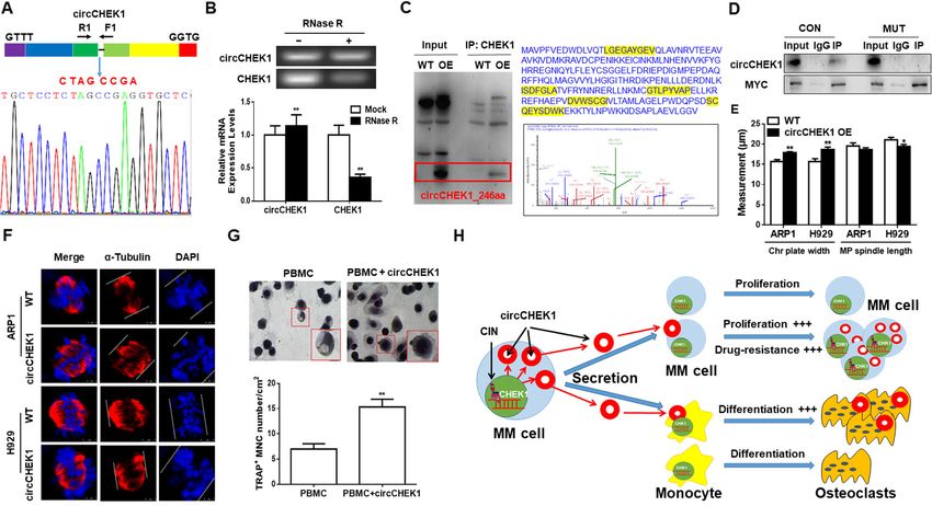
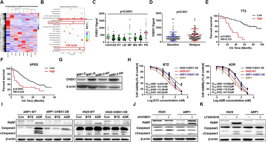
Gu et al. Molecular Cancer      (2021) 20:84                                                                                            Page 4 of 14

 Fig. 1 Elevated CHEK1 expression is associated with poor outcomes in MM patients and promotes MM cell proliferation in vitro. A CHEK1 mRNA
 levels were significantly increased in MM samples. The signal level of CHEK1 is shown on the y-axis. Patients were designated as being healthy
 donors with normal bone marrow plasma cells (NP, n = 22), monoclonal gammopathy of undetermined significance (MGUS, n = 44), or multiple
 myeloma (MM, n = 351), and are sorted on the x-axis. B Increased CHEK1 mRNA expression was associated with poor overall survival (OS) in MM
 patients from the TT2 patient cohort. C Increased CHEK1 mRNA expression was associated with poor OS in MM patients from the HOVON65
 cohort. D Western blot analysis revealed that CHEK1 was endogenously expressed in the specified MM cell lines. E Validation of CHEK1
 overexpression (OE) in CHEK1-OE ARP1 and H929 cells relative to vehicle-transfected control cells (WT). F Four-day cell growth curve, as detected
 by trypan blue staining and counting of WT, CHEK1-OE ARP1, and H929 cells. G Confirmation of CHEK1 protein knockdown (KD) in ARP1 and
 H929 cells after transfection with three independent CHEK1-targeting shRNAs. H Four-day cell growth curve in WT, CHEK1-KD ARP1, and H929
 cells. I Images of representative soft agar plates, revealing increased clonogenic growth of CHEK1-OE cells and decreased clonogenic growth in
 CHEK1-KD cells relative to WT. J Cell cycle analysis revealed that the proportion of G2/M phase cells significantly increased in CHEK1-OE cells
 relative to WT. K Cell cycle analysis revealed that the proportion of G2/M phase cells significantly decreased in CHEK1-KD cells

using a lentiviral system, which was validated by WB                        CHEK1 is a high-risk MM marker and induces drug
(Fig. 1E). Interestingly, proliferation was increased in                    resistance
CHEK1-OE cells relative to WT in both ARP1 and H929                         We further employed RNA-sequencing (RNA-seq) to as-
cells, as demonstrated by a trypan blue dye exclusion                       sess activation of CHEK1-related signaling pathways, re-
assay (Fig. 1F), suggesting that CHEK1 promoted MM                          vealing activation of two pathways related to CHEK1
proliferation. Conversely, CHEK1 was knocked down                           and MM progression, cell cycle regulation and osteoclast
(KD) by three distinct CHEK1-targeting shRNAs, which                        differentiation (Fig. 2A–B). Because high-risk MM is
were all validated by WB in both ARP1 and H929 cells                        characterized by aggressive proliferation, we measured
(Fig. 1G). Cell proliferation was decreased by CHEK1                        CHEK1 mRNA expression in MM subgroups, and found
KD in both ARP1 and H929 cells (Fig. 1H). Moreover, a                       that CHEK1 expression was highest in the PR subgroup,
clonal formation assay revealed that CHEK1 OE in-                           considered the highest-risk MM subgroup (Fig. 2C) [22].
creased clonal formation, while CHEK1 KD inhibited                          MM patients in the PR group are characterized by high
clonal formation in both ARP1 and H929 cells (Fig. 1I).                     MM proliferation rate and poor clinical outcomes, and
Consistently, flow cytometric cell cycle analysis demon-                    increased CHEK1 mRNA levels in this subgroup sug-
strated that in CHEK1-OE ARP1 and H929 cells, an in-                        gested that CHEK1 could be a biomarker for high-risk
creased proportion of cells were in the G2/M phase                          MM. Furthermore, CHEK1 expression was increased in
relative to WT cells (Fig. 1J), with a decreased propor-                    MM relapse samples relative to first-diagnosis MM sam-
tion of G2/M phase cells with CHEK1 KD in both cell                         ples in 88 paired patient samples (Fig. 2D). In patients
lines (Fig. 1K). Taken together, these findings suggested                   who experienced relapse, increased CHEK1 expression
that CHEK1 promoted MM proliferation and clonal                             was significantly associated with decreased overall sur-
expansion.                                                                  vival (OS) relative to patients with lower CHEK1
Gu et al. Molecular Cancer     (2021) 20:84                                                                                          Page 5 of 14

 Fig. 2 CHEK1 is a marker for high-risk MM and induces drug resistance. A Heatmap of RNA-seq data showing significantly differentiated genes
 before and after doxycycline-induced CHEK1 OE. B Pathway enrichment analysis of RNA-seq data revealed enrichment of two pathways, which
 were related to cell cycle regulation and osteoclast differentiation. C Box plot representing CHEK1 expression in eight MM risk subgroups from
 the TT2 patient cohort. D In paired patient MM samples collected at first diagnosis and relapse, CHEK1 mRNA expression was increased in the
 relapsed samples relative to the corresponding samples from first diagnosis. E–F Increased CHEK1 expression was correlated with decreased OS in
 relapsed patients from the (E) TT2 and (F) APEX cohorts. G Western blotting confirmed that CHEK1 protein levels were significantly increased in
 MM1.R (dexamethasone-resistant) and ANBL6 DR (Bortezomib-resistant) cells. H Effects of Bortezomib and Adriamycin on the cell viability of H929
 and ARP1 cells with or without CHEK1 OE. I Western blots demonstrated that CHEK1 OE induced resistance to Adriamycin and Bortezomib in
 ARP1 and H929 cells, as indicated by cleavage of the apoptotic regulators PARP and Caspase 3. J–K Pro-apoptotic effects of (J) CHEK1 shRNA
 silencing and the (K) CHEK1 selective inhibitor LY2603618 in H929 and ARP1 cells, as demonstrated by increased cleavage of PARP and Caspase 3

expression in two independent cohorts, the TT2 (Fig.                      CHEK1 inhibitor LY2603618 (Fig. 2K) increased apop-
2E) and APEX (Fig. 2F) cohorts [25].                                      tosis in ADR- and BTZ-treated cells. Taken together,
  Because high-risk MM is generally associated with drug                  these findings suggested that CHEK1 was a high-risk
resistance, we measured CHEK1 expression in two pairs                     MM marker associated with relapse and drug resistance
of drug-susceptible and -resistant cell lines, the MM1.S                  in MM patients, and induced drug resistance in cultured
and MM1.R lines, which are susceptible and resistant to                   MM cells.
dexamethasone, respectively, and ANBL6 WT and BTZ-
resistant cells. WB analysis revealed that CHEK1 protein                  CHEK1 evokes chromosomal instability (CIN) in MM
levels were increased in both drug-resistant cell lines com-              We next sought to investigate the mechanisms by which
pared with paired drug-susceptible controls, suggesting an                CHEK1 promoted MM proliferation, malignancy, and
association between CHEK1 upregulation and multiple                       drug resistance. Our prior study reported that CHEK1
drug resistance (Fig. 2G).                                                was included in the chromosomal instability gene list for
  To determine if CHEK1 induced drug resistance, we                       cancer cells [26, 27]. We therefore explored whether
performed MTT and WB assays on CHEK1 WT and OE                            CHEK1 prompted MM CIN, resulting in MM prolifera-
cells to measure the IC50s of ADR and BTZ, as well as                     tion and drug resistance.
the protein levels of apoptotic markers in drug-treated                     Gimsa staining revealed that CHEK1 OE increased the
cells. The IC50s for both ADR and BTZ were signifi-                       separation error rate and numbers of multiple nuclear
cantly higher in CHEK1-OE cells relative to WT cells                      cells, two key features of CIN [28, 29], in ARP1 and
(Fig. 2H), while cleavage of the apoptotic markers PARP                   H929 cells (Fig. 3A–B). Immunofluorescent (IF) staining
and Caspase 3 was decreased in drug-treated CHEK1-                        for α-Tubulin and DAPI was next used to further evalu-
OE cells relative to WT (Fig. 2I). Contrastingly, treat-                  ate the extent of CHEK1-induced CIN. In both cell lines,
ment with either CHEK1 shRNA (Fig. 2J) or the selective                   CHEK1 OE increased chromosomal plate width and
Gu et al. Molecular Cancer (2021) 20:84 Page 6 of 14
Fig. 3 CHEK1 evokes chromosomal instability (CIN) in MM. A–B Giemsa staining revealed that CHEK1 OE increased the separation error rate and
number of multi-nuclear cells in (A) ARP1 and (B) H929 cells. C–D Increased chromosomal plate width and decreased mitotic bipolar spindle
length in CHEK1-OE ARP1 and H929 cells relative to WT, as demonstrated by immunofluorescent (IF) staining for α-tubulin and DAPI. E A
comparative genomic hybridization (CGH) array revealed significant gains and losses of multiple chromosomal segments in CHEK1-OE ARP1 and
H929 cells relative to WT. F In WT and CHEK1-OE cells treated with vehicle or Borbezomib, chromosomal plate width was highest and mitotic
spindle length lowest in the Borbezomib-treated CHEK1-OE group
decreased mitotic bipolar spindle length, two additional spectrometry (MS) to determine which proteins inter-
indicators of CIN in MM cells [28, 30–32] (Fig. 3C–D). acted with CHEK1. Hundreds of proteins were identified
We subsequently performed a comparative genomic by MS, and these candidate proteins were screened
hybridization (CGH) array to directly assess the effect of against the CIN-related gene list, and genes associated
CHEK1 on MM chromosomal composition [26], which with poor outcome in the TT2 cohort (Fig. 4A). Centro-
identified significant gains and losses of multiple somal Protein 170 (CEP170) was identified as a candi-
chromosomal segments in CHEK1-OE ARP1 and H929 date CIN gene that could potentially interact with
cells relative to the corresponding WT cells (Fig. 3E). CHEK1 (Fig. 4A–B), and high expression of CEP170
Taken together, these data suggested that increased mRNA was significantly correlated with decreased OS in
CHEK1 expression promoted CIN in MM cells. the TT2 MM cohort (Fig. 4C). Physical interaction be-
CIN contributes to drug resistance in multiple types of tween CHEK1 and CEP170 was identified using a Co-IP
cancer. We therefore determined if CHEK1 OE could assay in CHEK1-OE ARP1 and H929 cells (Fig. 4D).
overcome BTZ sensitivity by inducing CIN. CHEK1-OE CEP170 is a centrosomal component, and is required for
ARP1 and H929 cells were resistant to BTZ treatment centriole appendage assembly [33]. IF staining revealed
(Fig. 2H–I). IF staining for α-Tubulin and DAPI revealed that CEP170 OE significantly increased chromosomal
that chromosomal plate width increased and mitotic plate width and decreased mitotic bipolar spindle length
spindle length decreased in BTZ-treated CHEK1-OE in ARP1 and H929 MM cells, suggesting that CEP170
ARP1 and H929 cells relative to both vehicle-treated evoked MM CIN (Fig. 4E–F).
CHEK1-OE cells and WT cells, suggesting that CHEK1- Our findings suggested that CHEK1 induced MM CIN
induced CIN was an important contributor to MM drug by directly interacting with CEP170. CHEK1 belongs to
resistance (Fig. 3F). the kinase family, and we hypothesized that CHEK1
could phosphorylate CEP170. Consistent with this no-
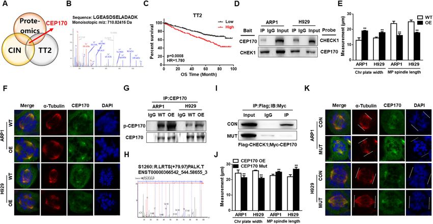
CHEK1 promotes MM CIN through CEP170 activation tion, a Co-IP assay revealed that the phosphorylated
To further determine how CHEK1 promoted MM CIN, form of CEP170, as detected by an anti-phospho-serine
we performed a Co-IP assay followed by mass antibody, was increased in CHEK1-OE cells relative to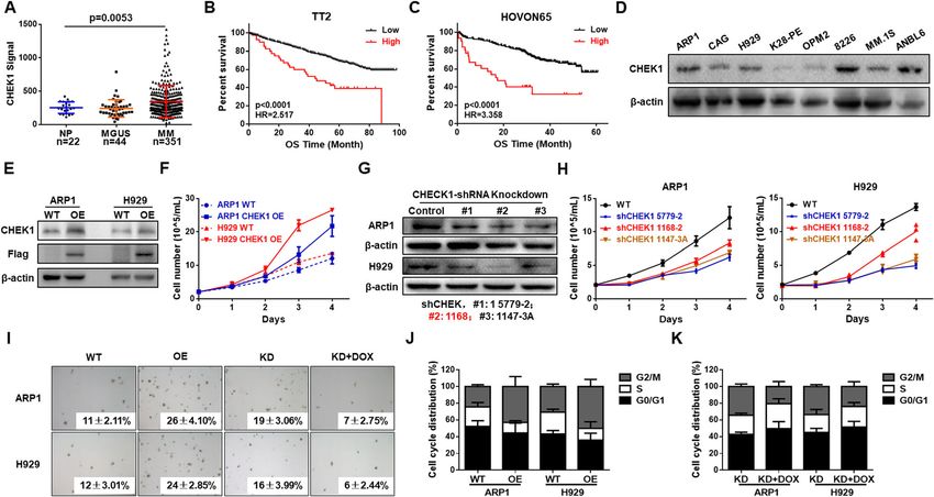
Gu et al. Molecular Cancer     (2021) 20:84                                                                                        Page 7 of 14

 Fig. 4 CHEK1 promotes CIN through CEP170 activation in MM. A–B Centrosomal Protein 170 (CEP170) was selected among candidate genes of
 the CIN-related gene list and genes associated with poor outcome in the TT2 MM patient cohort. C Increased CEP170 expression was associated
 with decreased OS in the TT2 patient cohort. D A Co-IP assay revealed that CHEK1 directly interacted with CEP170 in CHEK1-OE ARP1 and H929
 cells. E–F CEP170 OE significantly increased chromosomal plate width and decreased mitotic bipolar spindle length in ARP1 and H929 cells. G A
 Co-IP assay confirmed that CHEK1 physically interacted with and phosphorylated CEP170 in CHEK1-OE cells compared with WT cells, as detected
 by total anti-phospho-serine antibody. H Mass spectrometry (MS) was used to determine the CHEK1 phosphorylation site of CEP170, Ser1260. I A
 Myc-tagged CEP170 Ser1260Ala mutant, containing a defective CHEK1 phosphorylation site, exhibited dramatically decreased interaction with
 flag-tagged CHEK1, as demonstrated by Co-IP followed by western blotting. J–K OE of mutated CEP170 Ser1260Ala decreased chromosomal plate
 width and increased mitotic bipolar spindle length in (J) ARP1 and (K) H929 cells

WT cells in both cell lines (Fig. 4G). The CEP170                         (Fig. 5A). To evaluate the potential mechanism for
CHEK1 phosphorylation site was Ser1260, as identified                     CHEK1-promoted bone lesion formation, we overex-
by Thermo Q-Exactive (MS) (Fig. 4H). To further con-                      pressed murine Chek1 cDNA in cultured murine
firm that CEP170 Ser1260 was the CHEK1 phosphoryl-                        RAW264.7 macrophages. Tartrate-resistant acid phos-
ation site, we mutated Ser1260 to Ser1260Ala. A Co-IP                     phatase (TRAP) staining revealed that increased Chek1
assay confirmed that the interaction between mutant                       expression promoted osteoclast differentiation in macro-
Ser1260Ala CEP170 and CHEK1 protein, linked with                          phages treated with RANKL (50 ng/mL) or M-CSF (15
Myc and Flag, respectively, was attenuated dramatically                   ng/mL) for 10 days (Fig. 5B–C). When the concentra-
in CHEK1-OE cells compared with WT cells (Fig. 4I).                       tions of RANKL and M-CSF were decreased, exogenous
Further, Ser1260Ala mutant CEP170 OE decreased CIN                        m-Chek1 cDNA expression was still able to prompt
markers, as indicated by decreased chromosomal plate                      osteoclast differentiation in a RANKL and M-CSF dose-
width and increased mitotic spindle length (Fig. 4J–K).                   dependent manner (Fig. 5D–E), indicating that CHEK1
Collectively, these data demonstrated that CHEK1 in-                      was an important activator of osteoclast differentiation.
duced MM CIN by phosphorylating CEP170 at the                             This finding was verified in human primary peripheral
Ser1260 site.                                                             blood mononuclear cells (PBMCs). PBMCs transfected
                                                                          with human CHEK1 cDNA developed significantly more
CHEK1 induces osteoclast by upregulating NFATc1                           osteoclasts than vehicle-transfected control cells (Fig.
expression                                                                5F–G). Inversely, the CHEK1 inhibitor LY2603618 pre-
Because RNA-seq analysis revealed that CHEK1 expres-                      vented RAW264.7 cells from differentiating into osteo-
sion was correlated with osteoclast differentiation (Fig.                 clasts in a dose-dependent manner, and decreased
2A–B), we evaluated MRI data from MM patients of the                      expression of NFATc1, which is the key factor for osteo-
TT2 cohort and found that CHEK1 expression was                            clast differentiation (Fig. 5H–I). We then performed a
higher in MM patients with bone lesions than in MM                        Co-IP assay in m-Chek1-OE RAW264.7 cells to deter-
patients without bone lesions, as detected by MRI                         mine if CHEK1 directly interacted with NFATc1 (Fig.
Gu et al. Molecular Cancer (2021) 20:84 Page 8 of 14
Fig. 5 CHEK1 induces macrophage osteoclast by upregulating NFATc1 expression. A Magnetic resonance imaging (MRI) revealed that increased
CHEK1 expression was positively correlated with bone lesion formation in TT2 cohort MM patients. B–C TRAP staining revealed that Chek1 OE
promoted osteoclast differentiation in RAW 264.7 mouse macrophages co-treated with RANKL (50 ng/mL) and M-CSF (15 ng/mL) in a time-
dependent manner. D–E TRAP staining confirmed that Chek1 OE prompted osteoclast differentiation in RAW 264.7 cells treated with varying
doses of RANKL and M-CSF in a manner dependent on RANKL and M-CSF dosages. F–G TRAP staining revealed that human primary peripheral
blood mononuclear cells (PBMCs) transfected with human CHEK1 cDNA developed significant more osteoclasts than non-transfected control cells.
H–I Western blotting and TRAP staining confirmed that the CHEK1 inhibitor LY2603618 decreased NFATc1 expression and suppressed osteoclast
differentiation in RAW 264.7 cells. J Co-IP revealed that CHEK1 interacted with NFATc1 in RAW 264.7 cells. K Western blotting confirmed that the
expression of NFATc1 was increased in Chek1-OE RAW264.7 cells relative to WT cells. L CHEK1 knockdown prevented myeloma-associated bone
loss in 5TMM3VT model. Micro-CT analysis of 5TMM3VT-involved tibia bone performed at 4 weeks confirmed the presence of osteolytic lesions
and demonstrated decreased trabecular bone volume (BV/TV) compared with CHEK1 gene knockdown
5J). Further, the expression of NFATc1 was increased in primers to detect linear mRNA and circular RNA, re-
m-Chek1-OE RAW264.7 cells relative to WT cells (Fig. spectively. RNase R treatment significantly diminished
5K) indicating CHEK1 promotes osteoclasts formation linear CHEK1 mRNA, while circCHEK1 was resistant to
through upregulating NFATc1 expression. 5TMM3VT RNase R digestion (Fig. 6B), indicating that circCHEK1
model eventually confirmed this in vivo and demon- was more stable than its linear counterpart.
strated that 5TMM3VT-KD cells induced less bone Emerging studies have identified the presence of cir-
damage characterized by increased bone volume and tra- cRNAs with protein-coding capacity [34]; we therefore
becular numbers (data not shown) compared to the con- analyzed the putative open reading frame of circCHEK1.
trol group by microCT (Fig. 5L). Bioinformatics analysis revealed that circCHEK1 con-
tained a putative internal ribosome entry site (IRES) se-
MM cells secrete circCHEK1_246aa, inducing MM CIN and quence that encoded a novel CHEK1 isoform with 246
promoting osteoclast differentiation in the BM amino acids, termed “circCHEK1_246aa” in the present
microenvironment study. The predicted size of this isoform was 28.1 kDa,
To explore how MM cells disrupted cells of the normal so we adopted the mass spectrometer to confirm the
BM microenvironment, genomic structure analysis was presence of this novel isoform in MM cells. We first
performed, revealing the presence of a secreted cir- used a CHEK1 antibody that specifically recognizes the
cCHEK1 circular RNA fragment (738 bp) containing six CHEK1 N-terminus to conduct a Co-IP experiment that
exons (Supplementary Figure 2). Use of a divergent pri- enriched CHEK1 protein isoforms containing the N-
mer in cDNA samples and Sanger sequencing confirmed terminus sequence. WB analysis confirmed that the
that back-splicing occurred in the CHEK1 exons CHEK1 antibody recognized circCHEK1_246aa at the
(Fig. 6A). We then designed convergent and divergent expected size (Fig. 6C). The enriched protein wasGu et al. Molecular Cancer (2021) 20:84 Page 9 of 14
Fig. 6 MM cells secrete circCHEK1_246aa circular RNA to induce MM CIN and promote osteoclast differentiation in the bone marrow
microenvironment. A The number of exons and exact circCHEK1 sequences produced from CHEK1 were validated by Sanger sequencing. The
blue arrow represents the “head-to-tail” splicing sites of circCHEK1. B mRNA levels of circCHEK1 and linear CHEK1 ± RNase R were determined by
RT-PCR and qRT-PCR. C After pull-down using a CHEK1 antibody, protein samples at the expected size were excised and subjected to mass
spectrometry (MS) analysis, and specific peptides from circCHEK1_246aa were identified. D A Co-IP assay revealed that circCHEK1_246aa more
robustly interacted with native CEP170 than mutated CEP170. E–F circCHEK1 OE increased chromosomal plate width and decreased mitotic
bipolar spindle length in ARP1 and H929 cells. G TRAP staining revealed that circCHEK1-OE human primary PBMCs developed into significantly
more osteoclasts relative to vehicle-transfected control cells. H Graphic illustrating that CHEK1 and circCHEK1_246aa promote multiple myeloma
malignancy by evoking CIN and bone lesion formation
separated by SDS-PAGE, excised from the gel, and sub- then divided into three groups (n = 8 mice/group), in-
jected to MS to detect circCHEK1_246aa. The specific cluding untreated control, ADR-treated, and BTZ-
peptide fragments from circCHEK1_246aa were success- treated. After 28 days, we visually observed that tumors
fully identified by MS analysis, as marked in yellow (Fig. derived from CHEK1-OE cells grew faster than tumors
6C), confirming the expression of circCHEK1_246aa in derived from WT cells (Fig. 7A & C), with significantly
MM cells. To further examine the kinase function of cir- increased tumor volume and weight (Fig. 7B & D). In
cCHEK1_246aa, a Co-IP assay was conducted, revealing addition, tumors derived from CHEK1-OE cells were re-
that circCHEK1_246aa strongly interacted with native sistant to both ADR and BTZ, whereas WT cells were
CEP170, which was significantly diminished in cells ex- sensitive to the treatment (Fig. 7A–D), suggesting that
pressing mutant CEP170 (Fig. 6D). In addition, cir- CHEK1 induced MM drug resistance in vivo. Con-
cChek1_246aa expression induced features of CIN in versely, targeting CHEK1 by doxycycline-inducible h-
MM cells (Fig. 6E–F), and promoted osteoclast differen- CHEK1 shRNA significantly inhibited tumor growth
tiation in PBMCs (Fig. 6G). Together, these findings in- when NOD-SCID mice were administered doxycycline
dicated that the newly identified circular RNA through drinking water to induce h-CHEK1 shRNA ex-
circCHEK1_246aa exacerbated MM by evoking CIN and pression (Fig. 7E–H). Collectively, these data suggested
inducing bone lesion formation (Fig. 6H). that targeting CHEK1 had therapeutic effects in an
in vivo MM murine xenograft model.
CHEK1 inhibition alleviates MM progression in an in vivo
MM murine xenograft model Discussion
We next evaluated the effect of CHEK1 on MM progres- MM remains an incurable disease due to clonal hetero-
sion and dug resistance in vivo. ARP1 CHEK1 WT or geneity and BM dependency. Therefore, therapeutic
OE cells were injected subcutaneously into the right or strategies able to target both MM cell survival and
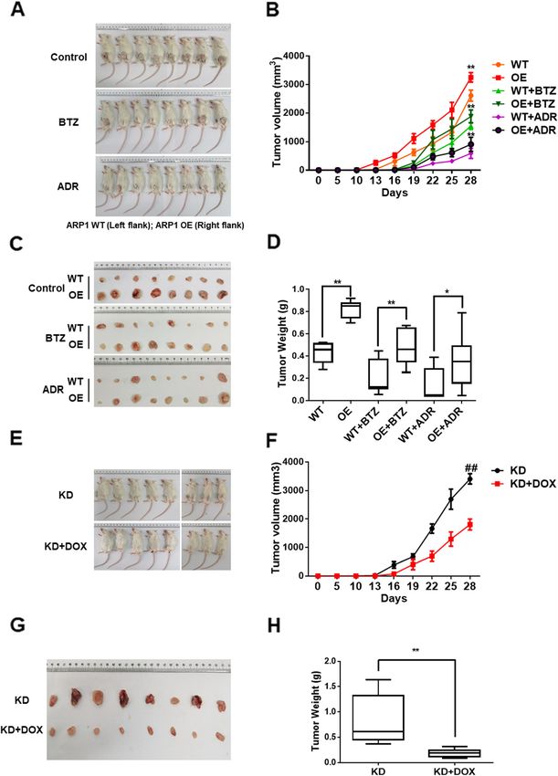
left flanks of NOD-SCID mice, respectively. Mice were modulation of the BM niche represent a significantGu et al. Molecular Cancer (2021) 20:84 Page 10 of 14 Fig. 7 (See legend on next page.)
Gu et al. Molecular Cancer    (2021) 20:84                                                                                      Page 11 of 14

 (See figure on previous page.)
 Fig. 7 CHEK1 promotes MM growth in vivo and is a potential therapeutic target. A Photographic images of xenograft-bearing mice from each
 group were taken at day 28. B Time course of tumor growth in NOD-SCID mice treated with vehicle, BTZ, or ADR. C Photographic images of
 xenografts from NOD-SCID mice of the specified groups on day 28. D Mean tumor weights in the six experimental groups at day 28 after
 implantation of the specified MM cells. E Photographic images of xenograft-bearing mice from the KD and KD + DOX groups were collected at
 day 28. F Time course of tumor growth in the NOD-SCID mice of the specified groups. G Xenografts from the NOD-SCID mice of the specified
 groups were collected at day 28. H Mean tumor weights in the specified two experimental groups at day 28 after implantation of MM cells

unmet clinical need. The present study demonstrated                      important CA regulator [39, 40], was identified by high-
that CHEK1 promoted both MM proliferation and                            throughput screening of MS and MM patient cohorts.
macrophage osteoclast differentiation, and could there-                  CEP170 plays an important role in microtubule
fore be a novel therapeutic strategy for MM.                             organization and microtubule stability, and aberrant
  CHEK1 expression in MM patient samples was associ-                     microtubule stability triggers defects in mitosis, leading
ated with MM proliferation, bone lesion formation, and                   to CIN in cancer cells [41]. Our findings demonstrated
poorer OS in four independent MM cohorts with over                       that CHEK1 directly bound with and phosphorylated
1000 patient samples. Mechanistic studies in in vitro and                CEP170, and that CEP170 overexpression in MM cells
in vivo MM models directly demonstrated that CHEK1                       induced features of CIN, such as increased chromosomal
OE induced MM cell proliferation, MM cell drug resist-                   plate width and decreased mitotic bipolar spindle length.
ance, and macrophage osteoclast differentiation, whereas                 Mutation of the Ser1260 residue of CEP170, the phos-
CHEK1 KD had converse effects.                                           phorylation site of CHEK1, abolished the CIN features
  Intriguingly, we newly identified the expression of cir-               induced by CEP170 overexpression. Therefore, the
cCHEK1_246aa, a CHEK1 circular RNA, which encoded                        present study identified CEP170 as a novel target of
and translated the CHEK1 kinase catalytic center in MM                   CHEK1-induced MM CIN.
cells. Circular RNA is a relatively newly discovered                        In addition, we identified that CHEK1 activated NEK2
means of intercellular communication and can be deliv-                   (data not shown), an established MM CIN marker re-
ered by MM cells to the BM microenvironment [35–37].                     ported in our previous study [26], while NEK2 stimu-
Our study found that MM cells secreted circCHEK1_                        lated CIN in cancer cells by regulating CEP250, a core
246aa into the BM niche, while transfection with cir-                    centrosomal protein essential for centriole–centriole co-
cCHEK1_246aa induced CIN in MM cells and promoted                        hesion [42, 43]. In MM, CIN is accompanied by replica-
osteoclast differentiation in macrophages. Collectively,                 tion errors, leading to impaired DNA repair
the sequence of the CHEK1 kinase catalytic center is a                   characterized by increased expression of DNA repair
promising therapeutic target for MM. Inhibiting this                     genes, including ATM, ATR, RAD51, and others [44].
catalytic center not only inhibited MM cell proliferation                Our unpublished data revealed that in MM cells,
and macrophage osteoclast differentiation, but also sup-                 CHEK1-OE upregulated RAD51, indicating the add-
pressed the interaction between MM cells and BM niche                    itional involvement of CHEK1 in DNA repair signaling.
cells.                                                                   Consequently, CHEK1 induces CIN in MM, activating
  The present study demonstrated that CHEK1 OE in                        multiple key centrosomal mediators and DNA repair sig-
MM cells increased multi-nuclear cells, as demonstrated                  naling, including NEK2, CEP170, RAD51, and others.
by Giemsa pathological staining. Increased chromosomal                      To assess the role of CHECK1 in vivo, we evaluated
plate width and decreased mitotic bipolar spindle length,                the role of CHEK1 in MM cell proliferation and drug re-
typical features of CIN, were also observed in CHEK1-                    sistance using an MM xenograft model. CHEK1 overex-
OE MM cells, as demonstrated by α-Tubulin and DAPI                       pression in MM cells not only promoted tumor growth,
IF. In addition, a CGH array study identified significant                but also conferred partial resistance to the chemothera-
gains and losses of multiple chromosomal segments in                     peutic drugs BTZ and ADR. By contrast, targeting
CHEK1-OE ARP1 and H929 cells relative to their WT                        CHEK1 by shRNA KD significantly inhibited MM tumor
counterparts. As identified in our prior studies, CIN is                 growth relative to WT controls. Together, these in vivo
an independent predictor of poor MM prognosis, and                       findings suggested that CHEK1 is a promising thera-
induces MM proliferation and drug resistance [26, 38].                   peutic target for MM.
These studies, combined with the present findings, sug-                     Several selective CHEK1 inhibitors, including Prexa-
gest that CHEK1 induces MM proliferation and drug re-                    sertib, SRA737, and others, have been developed, and
sistance by promoting MM CIN.                                            early-phase clinical trials have identified the potential
  Abnormal centrosome amplification (CA) resulting in                    therapeutic effects of these modalities in MM [45–47].
more than two centrosomes contributes to genomic in-                     However, at present, no CHEK1 inhibitors have been ap-
stability in MM. In the present study, CEP170, as an                     proved in Phase 3 clinical trials, due in part to
Gu et al. Molecular Cancer          (2021) 20:84                                                                                                   Page 12 of 14

cumulative normal tissue toxicities, off-target effects of                        Funding
simultaneous CHEK2 inhibition, and inefficient drug de-                           This work was supported by National Key R&D Program of China (No.
                                                                                  2020YFA0509400), National Natural Science Foundation of China 81970196
livery in cancer patients [46, 48]. More specific CHEK1                           (to CG) and 82073885 (to YY) and Natural Science Foundation of Jiangsu
inhibitors in combination therapy with other drugs, such                          Province BK20200097 (to CG); The Priority Academic Program Development
as p38 inhibitors, have recently been developed, and                              of Jiangsu Higher Education Institutions for Chinese Medicine; A Project
                                                                                  Funded by the Priority Academic Program Development of Jiangsu Higher
early-phase clinical trials have identified promising                             Education Institutions (Integration of Chinese and Western Medicine).
therapeutic effects for this modality. We also proposed
that co-inhibition of both CHEK1 and MK2 could have                               Availability of data and materials
                                                                                  All data that support the findings of this study are available from the
a synergistic effect in MM, as we identified in prior stud-                       corresponding authors upon reasonable request.
ies that single inhibition of each kinase had potential
therapeutic effects in MM [5, 15].                                                Declarations
                                                                                  Ethics approval and consent to participate
Conclusion                                                                        All animal experiments were conducted in accordance with the
In conclusion, our findings demonstrated that both                                Government-published recommendations for the Care and Use of Laboratory
                                                                                  Animals and approved by the Institutional Ethics Review Boards of Nanjing
CHEK1 and circCHEK1_246aa evoke MM CIN, partially                                 University of Chinese Medicine (Ethics Registration no. 201905A003).
through activation of CEP170. Further, CHEK1 and cir-
cCHEK1_246aa induce MM cell proliferation, drug re-                               Consent for publication
sistance, and bone lesion formation. Selectively targeting                        All authors of this article have directly participated in the planning and
                                                                                  drafting and all authors listed have read and approved the final version
the catalytic center encoded by CHEK1 mRNA and cir-                               including details and images.
cCHEK1_246aa could effectively target MM cell growth,
bone lesion formation, and pathologic changes in the                              Competing interests
                                                                                  The authors declare they have no competing interests.
BM niche such as osteoclast differentiation.
                                                                                  Author details
Abbreviations                                                                     1
                                                                                   Nanjing Hospital of Chinese Medicine affiliated to Nanjing University of
GEO: Gene expression omnibus; GEP: Gene expression profiling; MS: Mass            Chinese Medicine, Nanjing, China. 2School of Medicine & Holistic Integrative
spectrometry; IF: Immunofluorescence; OS: Overall survival; MM: Multiple          Medicine, Nanjing University of Chinese Medicine, 138 Xianlin Road, Nanjing
myeloma; MGUS: Monoclonal gammopathy of undetermined significance;                210023, China. 3Department of Hematology, Jagiellonian University Medical
CIN: Chromosomal instability; BM: Bone marrow; TT2: Total therapy 2;              College, Cracow, Poland. 4Division of Hematology and Oncology, Medical
CHEK1: Checkpoint Kinase 1; CEP170: Centrosomal Protein 170; OS: Overall          College of Wisconsin, Milwaukee, USA. 5Department of Hematology, School
survival; PBMCs: Peripheral blood mononuclear cells; CA: Centrosome               of Medicine, Ankara University, Ankara, Turkey. 6Myeloma Center, University
amplification; IRES: Internal ribosome entry site                                 of Arkansas for Medical Sciences, Little Rock, USA. 7Laboratory of Hematology
                                                                                  and Immunology & Labor für Myelomforschung, Vrije Universiteit Brussel
                                                                                  (VUB), Jette, Belgium. 8State Key Laboratory of Ophthalmology, Guangdong
Supplementary Information                                                         Provincial Key Laboratory of Ophthalmology and Visual Science, Zhongshan
The online version contains supplementary material available at https://doi.
                                                                                  Ophthalmic Center, Sun Yat-sen University, 54 South Xianlie Road,
org/10.1186/s12943-021-01380-0.
                                                                                  Guangzhou 510060, China.

 Additional file 1 Figure S1. Elevation of CHEK1 expression is                    Received: 4 January 2021 Accepted: 27 May 2021
 significantly associated with poor outcome in both event free survival (A)
 and overall survival (B) in GEP database of GMMG-HD4 cohort. Figure
 S2. Illustration of the annotated genomic region of CHEK1, the putative          References
 different RNA splicing forms, and the validation strategy for circular exon      1. Bianchi G, Anderson KC. Understanding biology to tackle the disease:
 1 to 7 (circCHEK1).                                                                  multiple myeloma from bench to bedside, and back. CA Cancer J Clin. 2014;
 Additional file 2. Materials and Methods.                                            64(6):422–44. https://doi.org/10.3322/caac.21252.
                                                                                  2. Pawlyn C, Morgan GJ. Evolutionary biology of high-risk multiple myeloma.
 Additional file 3. Supplementary Table 1 - RNA seq analysis of
                                                                                      Nat Rev Cancer. 2017;17(9):543–56. https://doi.org/10.1038/nrc.2017.63.
 differentially expressed genes in osteoclast differentiation.
                                                                                  3. Gu C, Lu T, Wang W, Shao M, Wei R, Guo M, et al. RFWD2 induces cellular
 Additional file 4. Supplementary Table 2 - A list of interacting                     proliferation and selective proteasome inhibitor resistance by mediating P27
 proteins for CHEK1.                                                                  ubiquitination in multiple myeloma. Leukemia. 2020. https://doi.org/10.103
 Additional file 5. Supplementary Table 3 - Statistics of Pathway                     8/s41375-020-01033-z.
 Enrichment.                                                                      4. Lohr JG, Stojanov P, Carter SL, Cruz-Gordillo P, Lawrence MS, Auclair D, et al.
                                                                                      Widespread genetic heterogeneity in multiple myeloma: implications for
                                                                                      targeted therapy. Cancer Cell. 2014;25(1):91–101. https://doi.org/10.1016/j.
Acknowledgments                                                                       ccr.2013.12.015.
The authors acknowledge the participants who generously gave their help           5. Gu C, Cheng H, Yang H, Bian Y, Wang Y, Zhang Y, et al. MK2 is a therapeutic
on the study. Especially, thank Dr. Shengfeng Lu and Bin Xu for the support           target for high-risk multiple myeloma. Haematologica. 2018;1:1. https://doi.
on microCT.                                                                           org/10.3324/haematol.2017.182121.
                                                                                  6. Zheng Y, Cai Z, Wang S, Zhang X, Qian J, Hong S, et al. Macrophages are an
Authors’ contributions                                                                abundant component of myeloma microenvironment and protect
C.G., J.P. and Y.Y. designed the projects, analyzed the experimental data and         myeloma cells from chemotherapy drug-induced apoptosis. Blood. 2009;
drafted the manuscript. W.W., X.T., T.X., M.G., R.W., Y.W. and C.G. performed         114(17):3625–8. https://doi.org/10.1182/blood-2009-05-220285.
the experimental work. F.Z., A.S. and D.H. offered the GEP cohorts. A.J., S.J.,   7. Zheng Y, Wang Q, Li T, Qian J, Lu Y, Li Y, et al. Role of myeloma-derived MIF
M.B., F. Z, A.S. and D.H. supervised the project and revised the manuscript.          in myeloma cell adhesion to bone marrow and chemotherapy response. J
The author(s) read and approved the final manuscript.                                 Natl Cancer Inst. 2016;108(11):djw131. https://doi.org/10.1093/jnci/djw131.
Gu et al. Molecular Cancer (2021) 20:84 Page 13 of 14
8. Liu H, He J, Koh SP. Reprogrammed marrow adipocytes contribute to 28. Canovas B, Igea A, Sartori AA, Gomis RR, Paull TT, Isoda M, et al. Targeting
myeloma-induced bone disease, vol. 11; 2019. p38alpha Increases DNA Damage, Chromosome Instability, and the Anti-
9. Bianchi G, Munshi NC. Pathogenesis beyond the cancer clone(s) in multiple tumoral Response to Taxanes in Breast Cancer Cells. Cancer Cell. 2018;33:
myeloma. Blood. 2015;125(20):3049–58. https://doi.org/10.1182/blood-2 1094–1110.e1098.
014-11-568881. 29. Liu G, Ye CJ, Chowdhury SK, Abdallah BY, Horne SD, Nichols D, et al.
10. Ghobrial IM, Detappe A, Anderson KC, Steensma DP. The bone-marrow Detecting chromosome condensation defects in gulf war illness patients.
niche in MDS and MGUS: implications for AML and MM. Nat Rev Clin Oncol. Curr Genomics. 2018;19(3):200–6. https://doi.org/10.2174/13892029186661
2018;15(4):219–33. https://doi.org/10.1038/nrclinonc.2017.197. 70705150819.
11. Gu C, Holman C, Sompallae R, Jing X, Tomasson M, Hose D, et al. 30. Hara Y, Kimura A. An allometric relationship between mitotic spindle width,
Upregulation of FOXM1 in a subset of relapsed myeloma results in poor spindle length, and ploidy in Caenorhabditis elegans embryos. Mol Biol Cell.
outcome. Blood Cancer J. 2018;8(2):22. https://doi.org/10.1038/s41408-018- 2013;24(9):1411–9. https://doi.org/10.1091/mbc.e12-07-0528.
0060-0. 31. Ganapathi Sankaran D, Stemm-Wolf AJ, Pearson CG. CEP135 isoform
12. Chen L, Wang S, Zhou Y, Wu X, Entin I, Epstein J, et al. Identification of early dysregulation promotes centrosome amplification in breast cancer cells.
growth response protein 1 (EGR-1) as a novel target for JUN-induced Mol Biol Cell. 2019;30(10):1230–44. https://doi.org/10.1091/mbc.E18-10-0674.
apoptosis in multiple myeloma. Blood. 2010;115(1):61–70. https://doi.org/1 32. Gulluni F, Martini M, De Santis MC, Campa CC, Ghigo A, Margaria JP, et al.
0.1182/blood-2009-03-210526. Mitotic Spindle Assembly and Genomic Stability in Breast Cancer Require
13. Lawson MA, McDonald MM, Kovacic N, Hua Khoo W, Terry RL, Down J, et al. PI3K-C2alpha Scaffolding Function. Cancer Cell. 2017;32:444–459.e447.
Osteoclasts control reactivation of dormant myeloma cells by remodelling 33. Huang N, Xia Y, Zhang D, Wang S, Bao Y, He R, et al. Hierarchical assembly
the endosteal niche. Nat Commun. 2015;6(1):8983. https://doi.org/10.1038/ of centriole subdistal appendages via centrosome binding proteins
ncomms9983. CCDC120 and CCDC68. Nat Commun. 2017;8(1):15057. https://doi.org/10.1
14. Dimopoulos MA, Hillengass J, Usmani S, Zamagni E, Lentzsch S, Davies FE, 038/ncomms15057.
et al. Role of magnetic resonance imaging in the management of patients 34. Yang Y, Gao X, Zhang M, Yan S, Sun C, Xiao F, et al. Novel role of FBXW7
with multiple myeloma: a consensus statement. J Clin Oncol. 2015;33(6): circular RNA in repressing glioma tumorigenesis. J Natl Cancer Inst. 2018;
657–64. https://doi.org/10.1200/JCO.2014.57.9961. 110(3):304–15. https://doi.org/10.1093/jnci/djx166.
15. Dietlein F, Kalb B, Jokic M, Noll EM, Strong A, Tharun L, et al. A synergistic 35. Dou Y, Kawaler EA, Cui Zhou D, Gritsenko MA, Huang C, Blumenberg L,
interaction between Chk1- and MK2 inhibitors in KRAS-mutant Cancer. Cell. et al. Proteogenomic Characterization of Endometrial Carcinoma. Cell. 2020;
2015;162(1):146–59. https://doi.org/10.1016/j.cell.2015.05.053. 180:729–748.e726.
16. Guo M, Sun D, Fan Z, Yuan Y, Shao M, Hou J, et al. Targeting MK2 is a novel 36. Zhou F, Wang D, Wei W, Chen H, Shi H, Zhou N, et al. Comprehensive
approach to interfere in multiple myeloma. Front Oncol. 2019;9:722. https:// profiling of circular RNA expressions reveals potential diagnostic and
doi.org/10.3389/fonc.2019.00722. prognostic biomarkers in multiple myeloma. BMC Cancer. 2020;20(1):40.
17. de Boussac H, Bruyer A, Jourdan M, Maes A, Robert N, Gourzones C, et al. https://doi.org/10.1186/s12885-020-6515-2.
Kinome expression profiling to target new therapeutic avenues in multiple 37. Feng Y, Zhang L, Wu J, Khadka B, Fang Z, Gu J, et al. CircRNA circ_0000190
myeloma. Haematologica. 2020;105(3):784–95. https://doi.org/10.3324/ha inhibits the progression of multiple myeloma through modulating miR-767-
ematol.2018.208306. 5p/MAPK4 pathway. J Exp Clin Cancer Res. 2019;38(1):54. https://doi.org/1
18. Pei XY, Dai Y, Felthousen J, Chen S, Takabatake Y, Zhou L, et al. 0.1186/s13046-019-1071-9.
Circumvention of Mcl-1-dependent drug resistance by simultaneous Chk1 38. Zhou W, Yang Y, Gu Z, Wang H, Xia J, Wu X, et al. ALDH1 activity identifies
and MEK1/2 inhibition in human multiple myeloma cells. PLoS One. 2014; tumor-initiating cells and links to chromosomal instability signatures in
9(3):e89064. https://doi.org/10.1371/journal.pone.0089064. multiple myeloma. Leukemia. 2014;28(5):1155–8. https://doi.org/10.1038/
19. Landau HJ, McNeely SC, Nair JS, Comenzo RL, Asai T, Friedman H, et al. The leu.2013.383.
checkpoint kinase inhibitor AZD7762 potentiates chemotherapy-induced 39. Denu RA, Shabbir M, Nihal M, Singh CK, Longley BJ, Burkard ME, et al.
apoptosis of p53-mutated multiple myeloma cells. Mol Cancer Ther. 2012; Centriole Overduplication is the predominant mechanism leading to
11(8):1781–8. https://doi.org/10.1158/1535-7163.MCT-11-0949. centrosome amplification in melanoma. Mol Cancer Res. 2018;16(3):517–27.
20. Pei XY, Dai Y, Youssefian LE, Chen S, Bodie WW, Takabatake Y, et al. https://doi.org/10.1158/1541-7786.MCR-17-0197.
Cytokinetically quiescent (G0/G1) human multiple myeloma cells are 40. Guarguaglini G, Duncan PI, Stierhof YD, Holmstrom T, Duensing S, Nigg EA.
susceptible to simultaneous inhibition of Chk1 and MEK1/2. Blood. 2011; The forkhead-associated domain protein Cep170 interacts with polo-like
118(19):5189–200. https://doi.org/10.1182/blood-2011-02-339432. kinase 1 and serves as a marker for mature centrioles. Mol Biol Cell. 2005;
21. Sun D, Tao W, Zhang F, Shen W, Tan J, Li L, et al. Trifolirhizin induces 16(3):1095–107. https://doi.org/10.1091/mbc.e04-10-0939.
autophagy-dependent apoptosis in colon cancer via AMPK/mTOR signaling. 41. Pillai S, Nguyen J, Johnson J, Haura E, Coppola D, Chellappan S. Tank
Signal Transduct Target Ther. 2020;5(1):174. https://doi.org/10.1038/s41392- binding kinase 1 is a centrosome-associated kinase necessary for
020-00281-w. microtubule dynamics and mitosis. Nat Commun. 2015;6(1):10072. https://
22. Zhan F, Huang Y, Colla S, Stewart JP, Hanamura I, Gupta S, et al. The doi.org/10.1038/ncomms10072.
molecular classification of multiple myeloma. Blood. 2006;108(6):2020–8. 42. Panic M, Hata S, Neuner A, Schiebel E. The centrosomal linker and
https://doi.org/10.1182/blood-2005-11-013458. microtubules provide dual levels of spatial coordination of centrosomes.
23. Broyl A, Hose D, Lokhorst H, de Knegt Y, Peeters J, Jauch A, et al. Gene PLoS Genet. 2015;11(5):e1005243. https://doi.org/10.1371/journal.pgen.1
expression profiling for molecular classification of multiple myeloma in 005243.
newly diagnosed patients. Blood. 2010;116(14):2543–53. https://doi.org/1 43. Cervenka I, Valnohova J, Bernatik O, Harnos J, Radsetoulal M, Sedova K, et al.
0.1182/blood-2009-12-261032. Dishevelled is a NEK2 kinase substrate controlling dynamics of centrosomal
24. Yang Y, Zhou W, Xia J, Gu Z, Wendlandt E, Zhan X, et al. NEK2 mediates linker proteins. Proc Natl Acad Sci U S A. 2016;113(33):9304–9. https://doi.
ALDH1A1-dependent drug resistance in multiple myeloma. Oncotarget. org/10.1073/pnas.1608783113.
2014;5(23):11986–97. https://doi.org/10.18632/oncotarget.2388. 44. Beksac M, Balli S, Akcora Yildiz D. Drug targeting of genomic instability in
25. Mulligan G, Mitsiades C, Bryant B, Zhan F, Chng WJ, Roels S, et al. Gene multiple myeloma. Front Genet. 2020;11:228. https://doi.org/10.3389/fgene.2
expression profiling and correlation with outcome in clinical trials of the 020.00228.
proteasome inhibitor bortezomib. Blood. 2007;109(8):3177–88. https://doi. 45. Plummer ER, Kristeleit RS, Cojocaru E, Haris NM, Carter L, Jones RH, et al. A
org/10.1182/blood-2006-09-044974. first-in-human phase I/II trial of SRA737 (a Chk1 inhibitor) in subjects with
26. Zhou W, Yang Y, Xia J, Wang H, Salama ME, Xiong W, et al. NEK2 induces advanced cancer. J Clin Oncol. 2019;37(15_suppl):3094. https://doi.org/10.12
drug resistance mainly through activation of efflux drug pumps and is 00/JCO.2019.37.15_suppl.3094.
associated with poor prognosis in myeloma and other cancers. Cancer Cell. 46. Dent P. Investigational CHK1 inhibitors in early phase clinical trials for the
2013;23(1):48–62. https://doi.org/10.1016/j.ccr.2012.12.001. treatment of cancer. Expert Opin Investig Drugs. 2019;28(12):1095–100.
27. Wang W, Zhang Y, Chen R, Tian Z, Zhai Y, Janz S, et al. Chromosomal https://doi.org/10.1080/13543784.2019.1694661.
instability and acquired drug resistance in multiple myeloma. Oncotarget. 47. Bendell JC, Bischoff HG, Hwang J, Reinhardt HC, Zander T, Wang X, et al. A
2017;8(44):78234–44. https://doi.org/10.18632/oncotarget.20829. phase 1 dose-escalation study of checkpoint kinase 1 (CHK1) inhibitorGu et al. Molecular Cancer (2021) 20:84 Page 14 of 14
prexasertib in combination with p38 mitogen-activated protein kinase (p38
MAPK) inhibitor ralimetinib in patients with advanced or metastatic cancer.
Investig New Drugs. 2019;38:1145.
48. Warren NJH, Eastman A. Comparison of the different mechanisms of
cytotoxicity induced by checkpoint kinase I inhibitors when used as single
agents or in combination with DNA damage. Oncogene. 2020;39(7):1389–
401. https://doi.org/10.1038/s41388-019-1079-9.
Publisher’s Note
Springer Nature remains neutral with regard to jurisdictional claims in
published maps and institutional affiliations.You can also read