Leukocytospermia induces intraepithelial recruitment of dendritic cells and increases SIV replication in colorectal tissue explants - Nature
←
→
Page content transcription
If your browser does not render page correctly, please read the page content below
ARTICLE
                  https://doi.org/10.1038/s42003-021-02383-9                OPEN

                  Leukocytospermia induces intraepithelial
                  recruitment of dendritic cells and increases
                  SIV replication in colorectal tissue explants
                  Mariangela Cavarelli 1 ✉, Stéphane Hua1, Naima Hantour1, Sabine Tricot1, Nicolas Tchitchek1,4,
                  Céline Gommet1,5, Hakim Hocini2,3, Catherine Chapon1, Nathalie Dereuddre-Bosquet 1 & Roger Le Grand                                                 1

                  Mucosal exposure to infected semen accounts for the majority of HIV-1 transmission events,
                  with rectal intercourse being the route with the highest estimated risk of transmission. Yet,
1234567890():,;

                  the impact of semen inflammation on colorectal HIV-1 transmission has never been
                  addressed. Here we use cynomolgus macaques colorectal tissue explants to explore the
                  effect of leukocytospermia, indicative of male genital tract inflammation, on SIVmac251
                  infection. We show that leukocytospermic seminal plasma (LSP) has significantly higher
                  concentration of a number of pro-inflammatory molecules compared to normal seminal
                  plasma (NSP). In virus-exposed explants, LSP enhance SIV infection more efficiently than
                  NSP, being the increased viral replication linked to the level of inflammatory and immuno-
                  modulatory cytokines. Moreover, LSP induce leukocyte accumulation on the apical side of the
                  colorectal lamina propria and the recruitment of a higher number of intraepithelial dendritic
                  cells than with NSP. These results suggest that the outcome of mucosal HIV-1 infection is
                  influenced by the inflammatory state of the semen donor, and provide further insights into
                  mucosal SIV/HIV-1 pathogenesis.

                  1 Université
                             Paris-Saclay, Inserm, CEA, Center for Immunology of Viral, Auto-immune, Hematological and Bacterial diseases (IMVA-HB/IDMIT), Fontenay-
                  aux-Roses & Le Kremlin-Bicêtre, France. 2 Vaccine Research Institute - VRI, Hôpital Henri Mondor, Cret́ eil, France. 3 Équipe 16 Physiopathologie et
                  Immunotheŕ apies dans l’Infection VIH, Institut Mondor de Recherche Biomed ́ icale - INSERM U955, Cret́ eil, France. 4Present address: UMRS 959
                  Immunology-Immunopathology-Immunotherapy (i3), Sorbonne University and Inserm, Paris, France. 5Present address: Sanofi R&D, Translational In vivo
                  Models/In Vivo Research Center France/Veterinary Services, Centre de Recherche de Vitry/Alfortville, Vitry-sur-Seine, France. ✉email: mariangela.
                  cavarelli@cea.fr

                  COMMUNICATIONS BIOLOGY | (2021)4:861 | https://doi.org/10.1038/s42003-021-02383-9 | www.nature.com/commsbio                                         1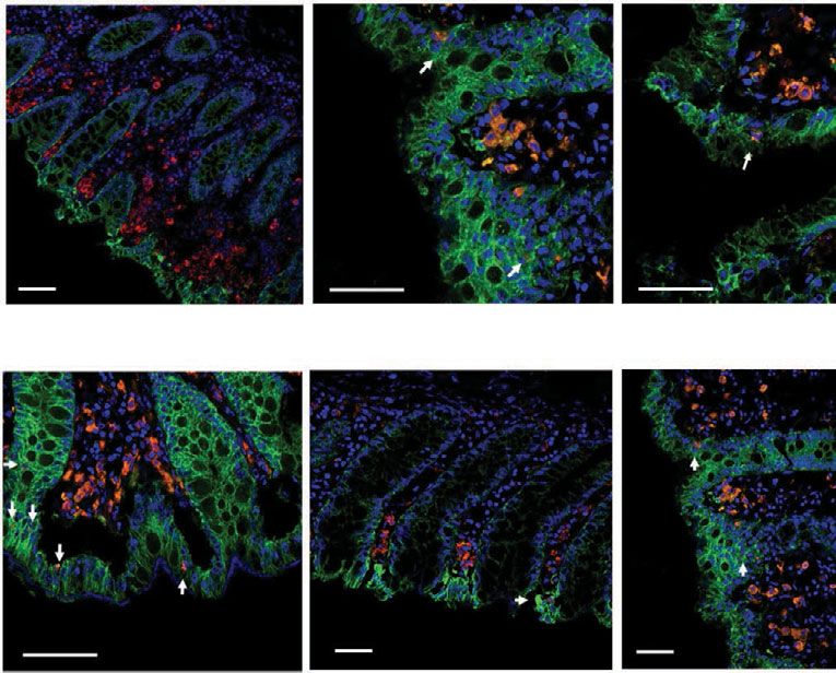
ARTICLE COMMUNICATIONS BIOLOGY | https://doi.org/10.1038/s42003-021-02383-9
H
IV-1 transmission in men who have sex with men most content in leukocytospermic than normal animals was reflected
commonly occurs via colorectal exposure to infected by a significantly higher count of CD3+ T cells (p = 0.0025),
semen. Receptive anal intercourse is also an under- including both CD4+ (p = 0.0051) and CD8+ (p = 0.0025) cell
estimated contributor to heterosexual infections1,2. Tissue mor- subsets, macrophages (p = 0.0025), and granulocytes (p = 0.0013)
phology, integrity, and the inflammatory state, as well as the (Wilcoxon rank-sum test, Fig. 1e–i).
distribution of several cell types within the mucosa, greatly There was a significantly higher concentration of a number of
influence viral transmission. CD4+ T cells in the gastrointestinal pro-inflammatory (IL-6, IL-8, IL-12/23, IL-13, IL-17a, IL-18, G-
tract are predominantly activated and well-differentiated to CSF, MIP-1ß, MCP-1, sCD40L, TGF-α, TNF-α, TGF-ß1, and
express a clear memory phenotype and constitute the main target VEGF) and immunoregulatory (IL-2, IL-10, IL-15) molecules in
for HIV-1 and SIV replication3,4. Moreover, an extensive network LSP than NSP samples (Wilcoxon rank-sum test, Supplementary
of resident innate immune cells with antigen-presenting function, Fig. 1). Conversely, there were no significant differences in the
such as dendritic cells (DCs) and macrophages, are potential levels of IL-1ß, IL1RA, IL-4, IL-5, IFN-γ, MIP-1α, TGF-ß2, or
targets for the incoming virus5. Colonic and rectal CCR5+, DC- TGF-ß3 between NSP and LSP. Principal component analysis
SIGN+ DCs are subepithelial and found throughout the thickness (PCA) clearly distinguished the NSP from LSP samples (Fig. 2a).
of the mucosa6,7. We previously showed in vitro and ex vivo that Furthermore, specific signatures were unraveled by hierarchical
human colonic CD11c+, DC-SIGN+, CCR5+ DCs can extend clustering analysis of the relative cytokine levels in the LSP and
dendrites containing HIV-1 to epithelial cells, as well as retract NSP samples (Fig. 2b). Due to the low amount of semen collected
them, and thus transfer infection to CD4+ T cells: a clear proof of from each animal, a pool of NSP (n = 4) and LSP (n = 7) was
principle of an HIV-1-DC uptake mechanism in the gut8. During made for further studies. Pooled seminal plasma samples were
anal intercourse, semen is delivered up to 60 cm up the colorectal representative of high vs low inflammatory cytokine profiles
tract9 and thus extends the possible contribution of DCs to HIV-1 (Fig. 2b and Supplementary Fig. 1).
transmission.
It is well established that semen is more than merely a vehicle
Seminal plasma is not toxic for the sigmoid tissue. The potential
of HIV-1 particles, as it is a complex mixture of cells and bio-
cytotoxic effect of LSP on colorectal tissue was excluded by his-
logical factors, including cytokines and chemokines, that may
tological examination, with the explants exposed to LSP showing a
affect HIV-1/SIV transmission and further influence host
similar morphological structure as control tissues (Fig. 3a). Treated
immune responses and susceptibility to infection10–16. The ability
explants presented an intact epithelium (Fig. 3b, c and Supple-
of seminal plasma, the acellular semen fraction, to enhance HIV-
mentary Fig. 2a) and mononuclear cells distributed evenly
1 transmission has been proven by its capacity to recruit HIV-1
throughout the lamina propria. The evaluation of the permeability
target cells, including macrophages, DCs, and T cells, to the
to FITC-dextran (FD4) in the presence or absence of LSP further
female reproductive tract (FRT)12,17–21 and to increase infectivity,
substantiated the tightness between the cells after 2 or 4 h of
even at low viral titers22. Moreover, seminal plasma levels of
treatment (mean 2.98 ± 0.21, 3.20 ± 0.18, and 3.15 ± 0.23% in
inflammatory cytokines have been shown to affect the activation
medium, 25% LSP, and 50% LSP, respectively) (Fig. 3d). Exposure
state of the recipients’ cells in the FRT mucosa, thus enhancing
to LSP also did not significantly affect tissue viability (0.86 ± 0.06%
transmission23. However, the structure and environment of the
and 0.93 ± 0.02% after 2 h and 0.936 ± 0.09% and 0.99 ± 0.16%
gastrointestinal tract are different from that of the FRT, and the
after 4 h of treatment with 25% or 50% LSP versus treatment with
effect of semen deposition onto the colorectal mucosa has been
medium, respectively) (Fig. 3e). Similar results were obtained using
less investigated. Moreover, none of the previous studies ana-
an in vitro polarized colonic Caco-2 epithelial cells monolayer
lyzing HIV-1 colorectal transmission took into consideration the
(Supplementary Fig. 2b–d).
level of inflammatory cytokines present in seminal plasma and
did not assess the presence of an underlying immune response
induced by seminal plasma treatment24,25. Enhancement of SIVmac251 replication in colorectal tissue
Inflammatory cytokines, including IL-8, IL-6, IL-1ß, are enri- explants is influenced by the cytokine profile of seminal
ched in seminal fluids from leukocytospermic individuals26–28 plasma. We first optimized the explant culture conditions (Sup-
and we reported an increase in inflammatory molecules in semen plementary Fig. 3) and the amount of virus needed to infect 100%
associated with leukocytospermia and SIV infection in non- of the explants while avoiding excessive viral replication and
human primates (NHPs)29,30. Here, we investigated the effect of cytotoxicity (Supplementary Results and Supplementary Fig. 4a, b).
increased inflammatory molecule concentrations in seminal The tissue explants were incubated with SIVmac251, with or
plasma due to leukocytospermia on SIV infection of macaque without 25% NSP or LSP, to test the hypothesis that a different
sigmoid colonic tissue ex vivo. We found that seminal plasma inflammatory state of the seminal plasma may affect the
facilitates SIV transmission and replication by recruiting leuko- susceptibility of the colorectal mucosa to SIV infection. SIVmac251
cytes to the subepithelial level of the lamina propria and replication was significantly higher in explants (n = 3) treated with
attracting CD11c+ DCs inside the intact intestinal epithelium. LSP from a single macaque (Supplementary Fig. 4c) and the result
There was significantly greater enhancement of SIV infection and was confirmed using seminal plasma pools on tissues obtained
DC recruitment in the presence of elevated cytokine concentra- from two additional donors (Fig. 4). Indeed, we observed
tions when explants were exposed to leukocytospermic seminal significant differences in SIVmac251 replication at 9 and 12 days
plasma (LSP) compared to normal seminal plasma (NSP). post-infection (dpi) for tissues incubated with SIVmac251 and LSP
(p = 0.0197 and 0.011, respectively, Friedman test) versus that of
controls without LSP, whereas there was only a trend in the
Results presence of SIVmac251 with NSP at 12 dpi (p = 0.0583 Friedman
Upregulation of inflammatory cytokines in leukocytospermic test, Fig. 4a). An analysis of the area under the curve (AUC)
seminal plasma of uninfected cynomolgus macaques. We confirmed these differences between conditions (p = 0.0003,
assessed leukocytospermia in the semen of SIV-negative cyno- 0.0585, and 0.0419 in SIV + LSP vs SIV, SIV + NSP vs SIV, and
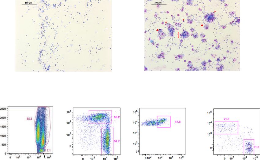
molgus macaques by immunocytochemistry (Fig. 1a, b) and flow SIV + LSP vs SIV + NSP, respectively, Friedman test). Treatment
cytometry (Fig. 1c–i). The leukocytospermic threshold was set to with LSP or NSP resulted in 3.8- or 1.6-fold (median) greater
10,000 CD45+ events acquired (Fig. 1d). A higher total leukocyte cumulative viral production at 12 dpi, respectively, than in
2 COMMUNICATIONS BIOLOGY | (2021)4:861 | https://doi.org/10.1038/s42003-021-02383-9 | www.nature.com/commsbio
COMMUNICATIONS BIOLOGY | https://doi.org/10.1038/s42003-021-02383-9                                                                       ARTICLE

Fig. 1 Characterization of normal and leukocytospermic semen of uninfected macaques. a, b Representative immunocytochemical staining of cells from a
normal semen (NS) and b leukocytospermic semen (LS). Numerous spermatozoa (black arrowhead) were present in NS. LS was rich in white blood cells,
including lymphocytes (black *), macrophages (red arrowhead) and neutrophils (red *), frequently forming aggregates ([) with spermatozoa. c Gating
strategy for semen leukocyte characterization. After the exclusion of doublets, cell debris, and dead cells, leukocytes are identified with the pan-leukocyte
marker CD45. The SSC-A versus CD45 gate distinguishes lymphocytes from macrophages and polymorphonuclear cells on the basis of morphology.
CD11b and HLA-DR distinguish HLA-DRbright CD11bmid-to-bright antigen-presenting cells (APC) from CD11bbright HLA-DRneg-to-low polymorphonuclear
(PMN) cells. CD3+ T cells are gated against CD45 and CD4+ T cells are separated from CD8+ T cells. d–i Number of events acquired by flow cytometry
per sample, as all collected semen cells were analyzed for NS (n = 7) and LS (n = 5): d CD45+ cells, e CD3+ lymphocytes, f CD4+ T cells, g CD8+ T cells,
h APC, and i PMN. The dotted line in panels (d) represents the leukocytospermia threshold (10,000 CD45+ events acquired). The mean is shown.
Statistical significance between groups was tested using Wilcoxon rank-sum tests, **p < 0.01.

SIVmac251 infected non-treated explants (p = 0.0022 and 0.0476,                positively correlated with a number of cytokines and chemokines.
respectively, Wilcoxon rank-sum test, Fig. 4b). In addition,                   The strongest correlation (p < 0.001 and r > 0.6) was observed with
explants treated with seminal plasma showed an accumulation of                 IL-2, IL-5, IL-10, IL12/23, sCD40L, and VEGF (Spearman
SIV DNA (median number of SIV DNA copies = 257,500 with                        correlation, Table 1). Overall, these results show that seminal
LSP, 199,000 with NSP, and 90,229 for SIVmac251 control                        plasma favors ex vivo SIV infection of the colonic mucosa and that
cultures) (Fig. 4c), confirming that higher virus production is                 the enhancement of infection is influenced by the inflammatory
associated with a higher number of infected cells. To dissect                  factors present in seminal fluids.
whether the increased SIV replication was linked to higher level of
seminal plasma inflammatory and immunoregulatory cytokines we                   Exposure to seminal plasma activates lamina propria lym-
analyzed the association between cytokine concentration in                     phocytes. In order to identify the SIV target cells in the colonic
seminal plasma and explants infection. Viral replication was                   explants and evaluate the effect that seminal plasma exposure

COMMUNICATIONS BIOLOGY | (2021)4:861 | https://doi.org/10.1038/s42003-021-02383-9 | www.nature.com/commsbio                                               3

ARTICLE                                                               COMMUNICATIONS BIOLOGY | https://doi.org/10.1038/s42003-021-02383-9

Fig. 2 Influence of leukocytospermia on cytokine levels in the seminal plasma of uninfected macaques. a Principal component analysis (PCA) showing
that normal (NSP, pink dots) and leukocytospermic seminal plasma (LSP, blue dots) samples cluster separately. b Heatmap from Luminex data of 26
cytokines and chemokines in seminal plasma from 13 normal (NSP) and 15 leukocytospermic (LSP) uninfected macaques (with the exception of TGF-ß 1-2-
3, for which 5 NSP and 10 LSP were evaluated) and comparison with the seminal plasma pools (NSP and LSP) shown at the bottom of the heatmap. Boxed
codes represent individual NSP and LSP used for ex vivo infection experiments. Data are shown as relative molecule concentration compared to the mean
value. Upregulated molecules are shown in red and downregulated molecules in blue.

4                          COMMUNICATIONS BIOLOGY | (2021)4:861 | https://doi.org/10.1038/s42003-021-02383-9 | www.nature.com/commsbio

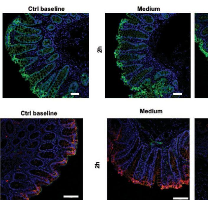
COMMUNICATIONS BIOLOGY | https://doi.org/10.1038/s42003-021-02383-9                                                                       ARTICLE

Fig. 3 Ex vivo evaluation of seminal plasma toxicity. a Representative histology (H&E staining) of the explant before and after 2 and 4 h of culture with or
without 50% LSP, showing intact epithelium. Magnification ×20. Scale bar = 50 µm. b, c Immunofluorescence staining for the human epithelial antigen
(HEA-FITC, green), E-cadherin (red), and the junctional adhesion protein (JAM, green) of explants before and after treatment for 2 h with culture medium
(negative control), SIVmac251 with or without 50% LSP, and 50% LSP. Scale bar = 50 µm in (b) and 100 µm in (c). d Integrity of the epithelial barrier
measured by the addition of Dextran-FITC (FD4, 4 kDa, 250 µg/ml) to the apical side of the explant with or without 25% or 50% LSP for 2 and 4 h. Results
are shown as the percentage of positive control (dye added to the basal medium at the beginning of the experiment). EDTA (100 mM) was included as a
control of barrier disruption. The statistical significance between conditions was tested using Wilcoxon signed-rank tests, ***p < 0.0005. e Viability of the
explants exposed, or not, for 2 and 4 h to 25% or 50% LSP measured by the MTT assay. A solution of 10% bleach was included as a control for death.
Results are shown as the mean ± SD of triplicates from a representative experiment of three.

might have on their phenotype, lamina propria mononuclear cells                strategy is shown in Supplementary Fig. 5a. Most colonic CD4+
(LPMCs) were isolated from the sigmoid tissue and exposed                      T cells and CD8+ T cells were central memory cells (CD28+
during 2 h to either complete medium, LSP or NSP. Immuno-                      /CD95+, 49.4 ± 7.8% and 40.3 ± 2.9%, respectively) (Fig. 5a). A
phenotyping was performed after 72 h of culture. The gating                    higher proportion of effector memory cells (CD28−/CD95+

COMMUNICATIONS BIOLOGY | (2021)4:861 | https://doi.org/10.1038/s42003-021-02383-9 | www.nature.com/commsbio                                               5
ARTICLE                                                                  COMMUNICATIONS BIOLOGY | https://doi.org/10.1038/s42003-021-02383-9

Fig. 4 Increased SIVmac251 replication in sigmoid tissue explants is associated with seminal plasma cytokine content. Sigmoid colon explants were
infected with SIVmac251 with or without a pooled NSP or LSP. a Kinetics of SIVmac251 replication in explants treated with virus alone (yellow line), virus
plus 25% LSP (red line), or virus plus 25% NSP (gray line). Culture medium was used as negative control. Virus replication was evaluated in the basal
supernatant as viral RNA copies/ml. b Fold change increase in cumulative viral production by each (n = 6) biopsy measured by RT-PCR compared to the
mean positive control value (explant exposed to SIVmac251 alone, mean of 6 values). c SIV DNA quantification in explants infected in the presence or
absence of 25% LSP or 25% NSP, harvested at the end of the culture (day 12). Results shown in panels (a–c) are the mean ± SEM of triplicates from two
independent experiments. Statistical significance between the different conditions was tested using Friedman tests with post hoc Benjamini, Krieger, and
Yekutieli tests, except for panel (c), for which Wilcoxon rank-sum tests were used.

                                                                              NSP in CD8 T cells, respectively, Friedman tests, Fig. 5b, e),
 Table 1 Correlations between the level of cytokines and                      whereas a significant increase of CD69+ cells was observed only
 chemokines in seminal plasma and viral replication at day                    among CD4+ T cells (p = 0.0153 and p = 0.0213 for LSP and
 12 p.i.                                                                      NSP, respectively, Friedman test, Fig. 5c, f). Interestingly, highly
                                                                              activated CD38+HLA-DR+CD69+CD4+ T cells were induced by
 Molecules                        p-value                         r-value     both treatments (p = 0.0015 and p = 0.0292 for LSP and NSP in
 IL-1ß
COMMUNICATIONS BIOLOGY | https://doi.org/10.1038/s42003-021-02383-9                                                                    ARTICLE

Fig. 5 Phenotype and activation status of leukocyte populations present in the sigmoid colon before and after exposure to seminal plasma. Lamina
propria mononuclear cells isolated from six different donors were incubated 2 h with medium, 25% NSP or 25% LSP, washed, and immunophenotyped 72 h
later. a The graph shows the proportion of naive, central memory (CM), effector memory (EM), and effector memory RA (TEMRA) cells among colonic
 CD3+CD4+ and CD3+CD8+ T cells. Each symbol represents one animal and the mean ± SD is shown. b–g Expression of the activation markers HLA-DR
(b, e), CD69 (c, f) and highly activated CD38+HLA-DR+CD69+ cells (d, g) by CD3+CD4+ (b–d) and CD3+CD8+ T cells (e–g) from the colon. Each symbol
represents one animal. p < 0.05 denotes significant difference between conditions (Friedman tests with post hoc Benjamini, Krieger, and Yekutieli tests).

Intraepithelial cells were not detected in medium-treated explants.            concentration of CCR5-binding molecules found in the former
Quantification of intraepithelial HLA-DR+ and CD11c+ cells                      one (Fig. 2b). LPMC were isolated from the sigmoid colon and
revealed a further increase in DC migration relative to that in the            exposed ex vivo during 2 h to both LSP and NSP and rested for
presence of the virus alone when LSP was mixed with SIVmac251                  another 4 h. DCs were identified among live CD45+ cells as
(p = 0.0044 and 0.0012 for HLA-DR+ and CD11c+ cells,                           lineage-HLA-DR+CD11c+CD64−CD123−CD14−CD16− cells
respectively, Kruskal–Wallis test), whereas an additive effect was             (Supplementary Fig. 5b). While some variation was observed
not observed in presence of NSP (Fig. 7c, d). These results were               between donors (n = 6), compared to medium-treated cells, a
confirmed using an in vitro dual-chamber Caco-2/DC co-culture                   tendency toward an upregulation of CCR5+ cells was observed
model (Supplementary Fig. 7).                                                  following exposure to NSP (1.7 mean fold), whereas a down-
   We then asked whether the differential DC migration observed                regulation was induced by LSP treatment (0.6 mean fold). A
in LSP versus NSP-treated explants could be due to the higher                  statistical significative difference was observed when comparing

COMMUNICATIONS BIOLOGY | (2021)4:861 | https://doi.org/10.1038/s42003-021-02383-9 | www.nature.com/commsbio                                           7

ARTICLE                                                               COMMUNICATIONS BIOLOGY | https://doi.org/10.1038/s42003-021-02383-9

Fig. 6 Imaging of leukocyte recruitment by seminal plasma by time-lapse microscopy. a Three-dimensional rendering of representative fields obtained
using NIS-Elements AR Analysis 5.02.0 (NIKON) and ImageJ software. Mouse anti-human CD45 antibody shows the leukocytes (red) and NucBlue dye
label the nuclei (blue). Explants were either exposed to medium (without LSP), SIVmac251, or 25% LSP. An explant incubated with an isotype control
antibody and exposed to LSP is shown as a negative control. A representative image obtained after 600 min of acquisition is shown. Scale bar = 25 µm.
Experiments were repeated three times. b Number/mm2 of CD45+ cells imaged during 14 h of acquisition in medium, LSP, and SIV-treated explants. Each
dot represents the number of cells/mm2 imaged in the z-stack. Mean and SEM of three experiments are shown. Statistical analysis was performed using
Wilcoxon signed-rank tests. c Number/mm2 of CD45+ cells imaged every 29 min in the presence (red symbols) or absence (blue symbols) of seminal
plasma as well as SIVmac251 (yellow symbols). The mean and SEM of three experiments are shown.

LSP vs NSP exposed cells (p = 0.0039, Friedman test, Fig. 7e).               inflammation35–37. Thus, inflammatory semen contains high
Conversely, a modulation of CCR5 by CD4+ T cells exposed to                  concentrations of immune mediators that are involved in the
NSP and LSP was not observed (Fig. 7f). These results suggest                recruitment, maturation, and survival of immune cells. Under
that increased concentration of CCR5-binding chemokines in                   conditions of infection, these mediators may be involved in local
LSP may account for increased CD11c+ DC recruitment inside                   viral replication and the associated increased risk of viral trans-
the intact colonic epithelium, a mechanism that might facilitates            mission. Specifically, the pro-inflammatory cytokine G-CSF
SIV transmission.                                                            attracts and promotes the survival of neutrophils, influences T-
                                                                             cell function, and activates DCs35,38. IL-8 may promote HIV-1
                                                                             infection39. IL-6 and IL-8 can directly upregulate HIV-1 gene
Discussion                                                                   transcription and increase the activation and life-span of HIV-
We took advantage of the SIV model and explant culture con-                  infected or bystander target cells40,41. Moreover, transient
ditions, which recapitulate the main features of in vivo exposure            induction of IL-8 and IL-1ß by cervical cells following exposure to
of the colorectal mucosa to semen, to investigate whether the                semen or seminal plasma was suggested to have implications for
levels of inflammatory cytokines present in seminal plasma                    HIV risk in women12,42. Future NHP studies or cohort studies of
influence the likelihood of SIV/HIV infection. Semen cytokine                 HIV-1 patients evaluating the effect of infected semen are also
levels may show heterogeneity, irrespective of HIV status, pos-              warranted, since LSP from uninfected macaques might not be
sibly linked to genital inflammation11,26. Previous studies con-              fully representative of SIV-infected seminal plasma.
sidered the cytokine/chemokine content of semen to be                           A strength of our ex vivo transmission model is that it closely
homogeneous and did not evaluate the influence of these seminal               mimics the biology of HIV/SIV sexual transmission and the
plasma factors on the local response of the colorectal mucosa24,25.          natural route and dynamics of viral entry into the mucosa. The
Here, we explored the contribution of inflammatory and                        use of a non-polarized explant system probably explains why no
immune-modulatory cytokines present in normal and leukocy-                   effect of seminal plasma on HIV-1 colorectal transmission was
tospermic semen to mucosal SIV infection.                                    observed in a previous study24. In addition, cytotoxicity, a
   As described in human semen, the cytokine profile of macaque               laboratory artifact representing one of the major limitations of
LSP was distinct from that of NSP samples, as shown by the                   working with seminal plasma, was not observed under our
strong upregulation of several molecules (IL-1ß, IL-6, IL-8, IL-15,          experimental conditions. In macaques, SIV has been shown to
G-CSF, GM-CSF, and MCP-1)32,33. Among them, MCP-1 is a                       infect rectal target cells within 1–4 h post-exposure43,44. Thus, we
chemoattractant for monocytes, memory T cells, DCs, and NK                   focused on the relevant time-frame of 2–4 h to study the impact
cells34 and has been shown to play a role in recruiting immune               of seminal plasma on viral transmission. In accordance with the
cells to the FRT following ejaculation20. IL-8, GM-CSF, and IL-15            results of previous ex vivo24 and in vitro45 studies, we did not
are involved in the recruitment, maturation, and proliferation of            observe any impact of seminal plasma on the integrity of the
monocytes, T and B cells, DCs, and NK cells at potential sites of            epithelial cell barrier in either of our models.

8                          COMMUNICATIONS BIOLOGY | (2021)4:861 | https://doi.org/10.1038/s42003-021-02383-9 | www.nature.com/commsbio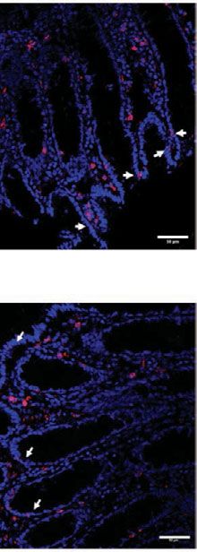
COMMUNICATIONS BIOLOGY | https://doi.org/10.1038/s42003-021-02383-9                                                                        ARTICLE

Fig. 7 Seminal plasma favors dendritic cell migration inside the colonic epithelial barrier. a, b Immunofluorescence staining for the human epithelial
antigen (HEA-FITC, green), HLADR+ cells (red, a), and CD11c+ cells (red, b) after 1.5 h of treatment of the macaque colonic explant with medium,
SIVmac251, 25% LSP, SIVmac251 + 25% LSP, 25% NSP, or SIVmac251 + 25% NSP. Nuclei are stained with DAPI. Arrows point to intraepithelial HLA-DR+
or CD11c+ cells. Scale bar = 50 µm. c, d Quantification of the number of HLA-DR+ cells (c) and CD11c+ cells (d) present inside the intestinal epithelium
following exposure to the different stimuli. Bars represent the mean ± SD of three randomly chosen fields from three different experiments. Statistical
significance between the different conditions was tested using Kruskal–Wallis tests with post hoc Benjamini, Krieger, and Yekutieli tests. e, f Lamina propria
mononuclear cells isolated from 6 different donors were incubated 2 h with medium, 25% NSP or 25% LSP, washed, and immunophenotyped 6 h later.
Percentage of CCR5+HLA-DR+CD11c+ DC (e) and CCR5+CD4+ T cells (f) exposed to NSP and LSP is expressed as fold change compared to the control
(medium). p < 0.05 denotes significant difference between conditions (Friedman tests with post hoc Benjamini, Krieger, and Yekutieli tests).

   By analogy with other studies using human cervico-vaginal and                viral treatment. Although degeneration could affect the quality of
colorectal explant cultures12,24,46,47, long-term culture led to                the living, infectable cells, the presence of infected cells within
alteration of the colonic tissue, irrespective of seminal plasma or             explants at the end of the culture was confirmed by SIV DNA

COMMUNICATIONS BIOLOGY | (2021)4:861 | https://doi.org/10.1038/s42003-021-02383-9 | www.nature.com/commsbio                                                9

ARTICLE                                                              COMMUNICATIONS BIOLOGY | https://doi.org/10.1038/s42003-021-02383-9

quantification, suggesting that the fraction of SIV target cells             addition, the higher number of migrating cells detected when
residing in the mucosa are sufficient to support viral replication           LSP, but not NSP, was mixed with SIV, is supporting the
and that modulation by seminal plasma occurred.                             enhancing role of LSP. Because a higher number of CCR5-
   A major finding of our study is that LSP enhances SIVmac251               binding molecules have been found in LSP than NSP, it is possible
infection, whereas NSP is less efficient, as shown by differences in         that these mediators are responsible for the differential DC
culture supernatant viral load. As described by others, we                  migration also observed under these in vitro and ex vivo condi-
observed cells emigrating from tissues as soon as 24 h after                tions. Accordingly, CCR5 downregulation was observed in LSP vs
culture48. We do not exclude the possibility that a higher number           NSP-treated lamina propria DCs, as indirect evidence of cognate
of SIV-infected cells might have exited explants upon treatment             chemokine binding to the receptor. Ex vivo assessment of CCR5
with LSP compared to NSP, thus explaining the non-significant                downregulation in seminal plasma-treated explants will be an
difference in the number of infected cells at 12 days p.i. in the two       important consideration for future studies, as to substantiate our
treatment conditions. We accounted for intra-donor variation of             observation. Moreover, our findings support further studies to
seminal plasma cytokine content by performing independent                   evaluate the use of CCR5 inhibitors for the prevention of rectal
experiments using either NSP and LSP from an individual                     HIV-1/SIV transmission, a strategy known to be effective in
macaque or a pool of NSP and LSP. In the absence of breaches in             preventing vaginal transmission of SHIV in Rhesus macaques54,55
the mucosa, the early mechanisms involved in crossing of the                   In conclusion, we provide a comprehensive study demon-
intestinal epithelium by HIV/SIV imply the recruitment of                   strating that the inflammatory and chemotactic profile of seminal
immune cells that possess migratory properties, including lym-              plasma may influence SIV uptake and replication ex vivo in the
phocytes, macrophages, and DCs. They can be considered as the               intestinal mucosa. Further validation and in-depth mechanistic
first target cells. Due to their proximity to the luminal environ-           studies on these pathways through SIV/NHP in vivo models,
ment and constant exposure to a myriad of food and microbial                should provide further basic insights on mucosal HIV-1 patho-
antigens, mucosal CD4+ T cells in the gastrointestinal tract have           genesis. Such knowledge could help to reveal novel therapeutic
been described to be predominantly activated and expressing a               targets to counteract HIV-1 infection and inflammation in the
memory phenotype49. In agreement with these observations, we                gastrointestinal tract.
found most CD4+ T and CD8+ T cells of the macaque sigmoid
mucosa to be central memory cells. These cells are prone to                 Methods
infection with HIV and, once depleted, do not repopulate the                Ethical statement and animal care. Cynomolgus macaques (Macaca fascicularis),
lamina propria, as shown in both untreated and treated patients             weighing 5–11 kg, were imported from Mauritius and housed according to Eur-
and SIV-infected macaques with progressive disease3,4,31,50–52.             opean guidelines for animal care (Official Journal of European Union, L 358,
                                                                            December 18, 1986 and new directive 63/2010). All work related to animals was
Direct exposure of LPMCs to both NSP and LSP, did not change                conducted in compliance with institutional guidelines and protocols approved by
the proportion of CD4 and CD8 T-cell subsets, and induced an                the local ethics committee (Comité d’Ethique en Experimentation Animale de la
activated memory phenotype of CD4+ T cells. Of note, our                    Direction des Sciences du Vivant au CEA under numbers: 2015032511332650
immunophenotype analysis was restricted to 72 h post culture.               (APAFIS#373); 10-062; 12-103). The sigmoid colon was collected at necropsy.
                                                                                Healthy human peripheral blood mononuclear cells (PBMCs) were obtained
Accordingly, a significative difference in viral replication was not         from buffy coats provided by the Établissement Français du sang (EFS). The
observed at 3 dpi in explant exposed to NSP and LSP, which                  human donors signed an informed consent form. All procedures were performed
instead became evident at 9 dpi. It is conceivable that the complex         in accordance with the ethical standards of the institutional and the regional ethical
mucosal environment of the explants responding to NSP vs LSP                committee.
could differentially affect expression of CD4+ T cells activation
markers at this later time point, and further experiments are               Semen collection, seminal plasma, and cell preparation. All cynomolgus
                                                                            macaques tested negative for antibody responses to SIV, simian retrovirus type D
required to substantiate this hypothesis.                                   (SRV), and simian T-cell lymphotropic virus (STLV) at the beginning of the study.
   It is also well established that both sperm and seminal plasma           Semen was collected from sedated animals following 5 mg/kg intra-muscular
elicit transient infiltration of the cervix mucosa by                        injection of Zoletil®100 (Virbac, Carros, France). Electroejaculation was performed
leukocytes12,19,53, suggesting a role for seminal plasma in the             by intrarectal electrostimulation of the prostate using a probe (12.7 mm diameter)
recruitment of immune cells. We primarily evaluated the effect of           lubricated with a conductor gel and an AC-1 electro-ejaculator (Beltron Instru-
                                                                            ment, Longmont, USA)29. Sequential stimulations were performed, with a pattern
LSP on the mucosal barrier and cell chemotactic properties,                 of 6 cycles, each cycle consisting of nine 2-s stimulations followed by a tenth
independently of virions. We confirmed and extended these                    stimulation lasting 10 s. The voltage was increased every two cycles (1–3 V for the
published data, established for the FRT, by assessing the increased         first two cycles, 2–4 V volts for the third and fourth cycles, and 6–8 V for the last
number of total leukocytes in the lamina propria of LSP-treated             two cycles). If a complete ejaculate had not been obtained after six cycles of
                                                                            stimulation, a 7th cycle of stimulation at 7–10 V was performed. Total ejaculate was
explants versus controls by confocal time-lapse microscopy,                 centrifuged 15 min at 775 × g immediately after collection to separate the acellular
which allowed us to measure cell migration within a 3D phy-                 fraction (seminal plasma) from the cellular fraction (semen cells). The cells were
siological context. Specifically, our results show an accumulation           maintained at room temperature for a maximum of 1 h, centrifuged 10 min at
of CD45+ cells toward the apical side of the lamina propria. The            1500 × g, filtered through a 70-µM sieve, and washed with 5 ml PBS supplemented
                                                                            with 10% FCS. Seminal plasma was filtered, aliquoted and stored at −80° until use.
accumulation of monocytes and neutrophils following FRT                     For the constitution of the pools, 500 µl of seminal plasma from seven leukocy-
exposure to seminal plasma has been reported, in accordance                 tospermic and four normal animals were mixed. pH of the seminal plasma pools
with our findings12. Although leukocytes migration by NSP was                ranged between 7.2 and 7.4.
not assessed in this experimental setting, differential chemotaxis
of HLA-DR+, CD11c+ DC, one of the first cell types to contact                Phenotypic characterization of semen leukocytes and identification of leu-
HIV-1 and playing an active role in capturing the virus8, was               kocytospermic animals. Seminal cell staining was performed after saturation of Fc
observed when analyzing explants treated with LSP vs NSP. DC                receptors by healthy macaque serum (in-house production) for 1 h at 4 °C. Live/
                                                                            dead® Fixable Blue amine-reactive dye (Life Technologies) was used to assess cell
migration across the tight intestinal epithelium is triggered by the        viability and exclude dead cells from the analysis. To identify T lymphocytes,
R5 viral envelope, which engages DCs via the CCR5 molecule.                 macrophages, and granulocytes, cells were stained with monoclonal antibodies for
Here, we show that DCs respond to SIV treatment in a similar                30 min at 4 °C, washed in PBS/10% FCS, and fixed in CellFIXTM (BD Biosciences).
way. Interestingly, the absence of migration in Caco-2/DC cul-              The antibodies were anti-CD45 PerCp (clone B058-1283), anti-CD3 V500 (clone
                                                                            SP34-2), anti-CD4 PE-Cy7 (clone L200), anti-CD8 V450 (clone BW138/80; Mil-
tures exposed to the supernatant of CEMx174 cells confirmed                  tenyi Biotec), anti-CD11b Alexa Fluor 700 (clone ICRF44), and anti-HLA-DR
that DC chemotaxis occurs in response to the virus itself and not           APC-H7 (clone G46-6) (all from BD Biosciences). Acquisition was performed on a
the cytokines/chemokines present in the viral inoculum. In                  BD LSRII and analyzed using FlowJo 9.8.3 (Tree Star, Ashland, OR). According to

10                        COMMUNICATIONS BIOLOGY | (2021)4:861 | https://doi.org/10.1038/s42003-021-02383-9 | www.nature.com/commsbio
COMMUNICATIONS BIOLOGY | https://doi.org/10.1038/s42003-021-02383-9 ARTICLE
the WHO, leukocytospermia was defined as a leukocyte concentration in the eja- substrate (0.5 mg/mL, Sigma-Aldrich) for 3 h at 37 °C. After removal of the MTT
culate ≥1 × 10⁶/ml56. Semen samples were considered to be leukocytospermic if a solution, tissues were incubated in methanol overnight at room temperature in the
minimum of 50,000 CD45+ events/ml were acquired by flow cytometry (consisting dark to dissolve non-specific precipitates. Absorbance of the MTT-formazan
of 10,000 events per ejaculate)30. This cut-off value was defined as the minimal product was measured at 570 nm (corrected at 630 nm) using a Spark plate reader
number of acquired CD45+ events required to perform statistics on the various (TECAN SPARK 10 M). Tissue viability was determined by dividing the absor-
leukocyte populations. bance by the dry weight of the explant. The effect of seminal plasma on tissue
For immunocytochemical analysis by microscopy, semen was centrifugated as viability was calculated as the ratio between seminal plasma-treated explants and
described above and 150 µl of a 1/100 dilution of the pellet was deposited onto a negative controls.
microscope slide using a cytospin (7 min at 500 × g). Staining was performed using A suspension of fluorescein isothiocyanate-dextran (250 µg/ml FD4, 4 kDa;
the RAL 555 rapid May-Grunwald Giemsa staining kit (Ral Diagnostics, France) Sigma-Aldrich) was added to the apical surface of the sealed polarized explant and
and images acquired with a Nikon Eclipse 80i microscope. a tight Caco-2 monolayer treated, or not, with seminal plasma (25%, 50% in
complete medium). After 2 or 4 h of incubation at 37 °C, the basolateral medium
was sampled and FD4 quantified at 520 nm using a Spark plate reader (TECAN
Cytokine quantification in semen. Inflammatory cytokines/chemokines and TGFβ SPARK 10 M). The optical density was expressed as the percentage of the positive
isoforms were measured in 25 µL seminal plasma using the Milliplex® Map Non- control (FD4 added to the basal medium at the beginning of the experiment).
Human Primate Cytokine Magnetic Bead Panel - Premixed 23-plex and TGFβ 1,2,3
Magnetic Bead Kit (Merck Millipore). Immunoassays were performed according to
the manufacturer’s instructions. Data were acquired using a Bio-Plex 200 instru- (3) Histopathology analysis, immunofluorescence markers, and confocal microscopy.
ment and analyzed using Bio-Plex Manager Software, version 6.1 (Bio-Rad). Tissues were fixed in 4% PFA for 4 h, cryoprotected in 10% sucrose overnight,
embedded in optimal cutting temperature embedding medium (OCT, VWR) in
plastic cryomolds (Corning), and snap-frozen. One explant was fixed immediately
Cells, virus, and reagents. Immature DCs were obtained from CD14+ cells iso- at baseline to serve as the baseline control. Tissue sections were stained with
lated from human PBMCs and cultured for 5 to 6 days in RPMI 1640 with 1% FCS, Hematoxylin/Eosin and the morphology evaluated under a light microscope
50 ng/ml granulocyte–monocyte colony-stimulating factor (GM-CSF), and 20 ng/ml (Nikon Eclipse 80i). For confocal microscopy analysis of colonic tissues, at least 5
Interleukin-4 (Peprotech Inc.). DCs were characterized as CD1a+, CD11c+, CD4+, to 6 serially cut 10-µm thick sections, sampled 100 µm apart, were obtained. A list
CCR5+, DC-SING+, CD14−, CXCR4− cells by flow cytometry. Caco-2 cells (clone of the antibodies used for confocal microscopy analysis of colonic tissues is pro-
E, kindly provided by Dr. Maria Rescigno, Humanitas Institute, Italy) were grown in vided in Supplementary Table 1. Primary antibodies were used at a final con-
Dulbecco’s modified Eagle’s medium (DMEM) with 10% FCS, 1% non-essential centration of 10 µg/ml. Tissues were stained with secondary antibody alone (anti-
amino acids (NEAA), and penicillin (100 U/ml)/streptomycin (100 mg/ml). All mouse IgG Alexa Flour conjugates, Invitrogen) as a negative control. Single-
reagents were purchased from the Lonza Group (Switzerland) unless otherwise channel images from z-series were collected from at least three representative fields
indicated. with a Leica TCS SP8 confocal microscope (Leica Microsystems GmbH, Wetzlar
SIVmac251 virus used for infection of colorectal explants was kindly provided Germany) and the images processed using ImageJ software, as described above.
by Dr. M. Muller Trutwin, Institute Pasteur, and the viral stock was produced in
CEMx174 cells grown in RPMI medium. Cells (30 × 106) were incubated at a 10−2
Quantification of viral load in explant culture supernatants and real-time PCR
multiplicity of infection (MOI) for 2 h at 37 °C and the volume adjusted to 1 × 10⁶
cells/ml. Cultures were maintained for 21 days, changing half of the cells every 3 to for SIV DNA quantification. The SIV copy number in culture supernatants was
determined in 100-fold diluted culture supernatants by quantitative real-time RT-
4 days. Viral replication was monitored by measuring p27 levels in culture
supernatants by ELISA (RETRO-TEK SIV p27 Ag ELISA kit, Helvetica Health PCR57, using SIV gag primers F 5′-GCAGAGGAGG AAATTACCCAGTAC-3′, R
Care). Cell-free viral stock was passed through a 0.2-μm pore-size filter, aliquoted, 5′-CAATTTTACCCAGGCATTTAATGTT-3′, probe FAM-5′-TGTCCACCTGC-
and stored at −80 °C. CATTAAGCCCGA-3′-BHQ1, Superscript III platinum One-Step qRT-PCR sys-
tem (Invitrogen), and CFX96 thermocycler (Bio-Rad). Reverse transcription was
done at +56 °C for 30 min and followed by 5 min of denaturation at +95 °C and by
Polarized explant treatment. Macaque sigmoid colon was harvested at necropsy, 50 cycles of 15 s at 95 °C and 30 s at 60.3 °C. Calibrated SIVmac251 virus was used
placed in cold PBS containing 100 U penicillin/ml, 100 mg streptomycin/ml, and 50 to generate a standard curve and the SIVmac251 gag cDNA sequence, ligated into
mg/ml gentamicin and processed within 30 min. After abundant washes in the same the pCR4-TOPO (Invitrogen) plasmid and purified with the HiSpeed Maxiprep kit
solution, specimens were transferred into RPMI supplemented with 10% FCS, 100 U/ (Invitrogen), was used as a positive control. For each qRT-PCR run, standard
ml penicillin/streptomycin, 1% glutamine, 1% NEAA, 1% Na-pyruvate, 1% 1 M curve, positive and negative controls, and samples were run in duplicate. Copy
HEPES buffer (complete medium), and the epithelial surface exposed. A polarized numbers were calculated by interpolating CT of samples in the standard curve. The
ex vivo culture model was established, slightly modifying our previously published limit of detection was 1000 copies/mL in this setting where 100-fold diluted
protocol8. Briefly, small tissue explants (4.0 mm in diameter), including the epithe- samples were used. For determination of SIV DNA copy numbers58, total DNA
lium and submucosa, were cut with a biopsy punch (Stiefel, Laboratories, Inc.) and was extracted from explants frozen after 12 days of culture. Quantitative real-time
placed on a 24-well hanging insert (3 µm PET, Millicell cell culture insert, Millipore), PCR was performed in duplicate on 500 ng of DNA using SIV gag primers and
with the submucosa facing the filter. The explant was surrounded with 3% agarose gel probe as described above and on 50 ng of DNA for GAPDH gene using primers F
prepared in RPMI and a polystyrene cylinder (I.D. × H 4.7 mm × 8 mm, Sigma- 5′-ATGACCCCTTCATTGGCCTC-3′, R 5′-TCCACGACATACTCAGTGCC-3′,
Aldrich) was immediately inserted in the liquid agarose gel to seal the tissue and avoid probe FAM-5′-CGAGCTTCCCGTTCTCAGCC-3′-BHQ1. The GAPDH gene was
leakage. The basal chamber was filled with 1 ml complete medium. Mucosal explant used to normalize results per million cells using a standard curve of DNA con-
cultures were treated apically with viral culture supernatants (100 µl of sidering that 1 µg of DNA corresponds to 131,300 cells. SIV gag standard curve was
SIVmac251 stock) or complete medium (negative control) with or without seminal generated by dilution of pCR4-TOPO-SIVmac251 gag cDNA in DNA of lymph
plasma at 1:1 ratio for 2 h (unless otherwise specified). At the end of stimulation, the nodes of SIV-negative macaques. SIV DNA copy numbers were calculated by
cylinder was removed, the tissue gently collected, while avoiding damage to the interpolating CT of samples in gag standard curve and were normalized using
mucosa, and abundantly washed in RPMI. Explants were further subjected to either GAPDH gene data to be expressed per million cells. The limit of detection is 10
(1) ex vivo culture, (2) a viability and tightness check, or (3) immunohistochemistry copies per million cells.
(IHC)/immunofluorescence (IF) as further described. A schematic representation of
the experimental protocol is shown in Supplementary Fig. 3.
Time-lapse confocal microscopy analysis. Monoclonal antibody anti-CD45
(clone DO58-123) and its isotype control (IgG1-MOPC-03, both from BD Phar-
(1) Ex vivo tissue culture. Explants were cultured in pairs, with the epithelium mingen) were covalently conjugated to fluorochromes with AlexaFluor-647, using a
uppermost, supported on a presoaked support (Gelfoam(R) rafts, Pfizer) at the microscale protein labeling kit (Life Technology), and the protein and fluor-
air–media interface in 24-well plates containing 500 μl media. Experiments were ochrome concentrations of the conjugated antibodies measured using a Nanodrop
performed two or three times in triplicate. Culture supernatant was replaced with microvolume spectrometer system (Thermo Scientific, Waltham, USA). NucBlue
fresh complete medium every three days for up to 12 days of culture, collected, and Live Cell Stain, ReadyProbes (Hoechts33342) (Invitrogen) was used to label cell
frozen at −80 °C until use. Supernatants were used to measure viral production by nuclei. A schematic representation of explant treatment is shown in Supplementary
RT-qPCR and quantify inflammatory cytokines by Luminex. The basolateral Fig. 6. Explants were incubated for 1 h, either with 25 µg/ml of the conjugated
medium was also sampled 24 h after treatment for specific experiments and the CD45 antibody or isotype control, and one drop of the NucBlue nuclear dye in a
cells that migrated from tissues were collected and co-cultured with uninfected final volume of 200 µl and then washed in 1X PBS. They were then incubated for 1
CEMx174, as described in Supplementary Information. Explants were collected at h with either complete medium, SIVmac251, or 25% LSP in complete medium in a
the end of the culture, rinsed in PBS, and frozen at −80 °C. polarized manner. After extensive washing, the explants were transferred to a six-
well plate with complete medium and submitted for video-microscopy. Three
(2) Tissue explant viability by 1-(4,5-dimethylthiazol-2-yl)-3,5-diphenylformazan different regions of each explant were randomly acquired with a Plan Fluor 20x
(MTT) assay and fluorescein isothiocyanate-dextran leakage assay. Explants were DIC objective (NA: 0.45) on a Nikon A1R confocal fast-laser scanning system
exposed in triplicate for 2 or 4 h to complete medium (negative control) or seminal (Nikon Corporation, Japan) equipped with a thermostatic chamber (37 °C, 5%
plasma (25%, 50% in complete medium), washed, weighed, and incubated in RPMI CO2). Images were recorded with a high-speed resonant scanner at 29 min intervals
containing 3-(4,5-dimethyl-2-thiazolyl)-2,5-diphenyltetrazolium bromide (MTT) for up to 14 h. Z-series of 54 µm were acquired every 3 µm.
COMMUNICATIONS BIOLOGY | (2021)4:861 | https://doi.org/10.1038/s42003-021-02383-9 | www.nature.com/commsbio 11ARTICLE COMMUNICATIONS BIOLOGY | https://doi.org/10.1038/s42003-021-02383-9
NIS-Elements AR Analysis 5.02.0 (Nikon) and ImageJ software were used to is presented in Supplementary Data 1. All other data are available from the
analyze and quantify CD45+ cells. An automatic threshold algorithm was applied corresponding author upon reasonable request.
to filter the background signal using ImageJ software. The number of fluorescent
objects, with a size >4.5 μm, was quantified after binary transformation of each
selected frame. Results were represented and analyzed by GraphPad Prism
Received: 27 November 2020; Accepted: 21 June 2021;
v8.0.2 software.
Purification of lamina propria mononuclear cells, exposure to seminal plasma
and immunophenotyping. To isolate lamina propria mononuclear cells (LPMCs),
colonic sigmoid tissue from uninfected animals was cut into small pieces and
References
1. Misegades, L., Page-Shafer, K., Halperin, D. & McFarland, W. Anal
incubated for 1 h at 37 °C in HBSS (Fisher Scientific) containing collagenase IV
intercourse among young low-income women in California: an overlooked
(0.3 mg/ml, Sigma-Aldrich), FCS (5%, Fisher Scientific), and DNase I (5 U/ml,
risk factor for HIV? AIDS 15, 534–535 (2001).
Sigma-Aldrich). Thawed cells from six different donors (1 million/condition) were
incubated during 2 h with complete medium (control), 25% NSP pool or 25% LSP 2. Baldwin, J. I. & Baldwin, J. D. Heterosexual anal intercourse: an understudied,
pool, then extensively washed and cultured in 96-well plate. Seven hours and 72 h high-risk sexual behavior. Arch. Sex. Behav. 29, 357–373 (2000).
later, DC and lymphocyte phenotyping was performed, respectively. Cells were 3. Li, Q. et al. Peak SIV replication in resting memory CD4+ T cells depletes gut
incubated with the antibodies listed in Supplementary Table 2. Acquisition was lamina propria CD4+ T cells. Nature 434, 1148–1152 (2005).
performed on a BD Fortessa and analyzed using FlowJo 9.8.3 (Tristar, USA) 4. Veazey, R. S. et al. Gastrointestinal tract as a major site of CD4+ T cell
software. At least 500 events were recorded for rare cell populations (i.e., pDC). depletion and viral replication in SIV infection. Science 280, 427–431 (1998).
5. Cavarelli, M. & Scarlatti, G. HIV-1 infection: the role of the gastrointestinal
tract. Am. J. Reprod. Immunol. 71, 537–542 (2014).
Co-culture of migrating cells with CEMx174. Cells migrating from colonic 6. Preza, G. C. et al. Antigen-presenting cell candidates for HIV-1 transmission
explants after 24 h of culture on a Gelfoam(R) raft (see above) were centrifuged and in human distal colonic mucosa defined by CD207 dendritic cells and CD209
transferred to a U-bottomed 96-well plate. Then, uninfected CEMx174 cells were
macrophages. AIDS Res. Hum. Retroviruses 30, 241–249 (2014).
added (ratio 3:1 vs. migrated cells) in complete medium. A minimum of 50,000
7. Jameson, B. et al. Expression of DC-SIGN by dendritic cells of intestinal and
CEMx174 cells/well was added if the number of migrating cells wasCOMMUNICATIONS BIOLOGY | https://doi.org/10.1038/s42003-021-02383-9 ARTICLE
27. Bezold, G. et al. Prevalence of sexually transmissible pathogens in semen from 54. Lederman, M. M. et al. Prevention of vaginal SHIV transmission in rhesus
asymptomatic male infertility patients with and without leukocytospermia. macaques through inhibition of CCR5. Science 306, 485–487 (2004).
Fertil. Steril. 87, 1087–1097 (2007). 55. Veazey, R. S. et al. Protection of macaques from vaginal SHIV challenge
28. Havrylyuk, A., Chopyak, V., Boyko, Y., Kril, I. & Kurpisz, M. Cytokines in the by vaginally delivered inhibitors of virus-cell fusion. Nature 438, 99–102
blood and semen of infertile patients. Cent. Eur. J. Immunol. 40, 337–344 (2005).
(2015). 56. World Health Organization. WHO Laboratory Manual for the Examination
29. Suphaphiphat, K. et al. Innate and adaptive anti-SIV responses in Macaque and Processing of Human Semen (World Health Organization, 2010).
semen: implications for infectivity and risk of transmission. Front. Immunol. 57. Karlsson, I. et al. Dynamics of T-cell responses and memory T cells during
11, 850 (2020). primary simian immunodeficiency virus infection in cynomolgus macaques. J.
30. Bernard-Stoecklin, S. et al. Semen CD4+ T cells and macrophages are Virol. 81, 13456–13468 (2007).
productively infected at all stages of SIV infection in Macaques. PLoS Pathog. 58. Mannioui, A. et al. Dynamics of viral replication in blood and lymphoid
9, 1–13 (2013). tissues during SIVmac251 infection of macaques. Retrovirology 6, 1–15 (2009).
31. Veazey, R. S. et al. Identifying the target cell in primary simian immunodeficiency
virus (SIV) infection: highly activated memory CD4+ T cells are rapidly
eliminated in early SIV infection in vivo. J. Virol. 74, 57–64 (2000). Acknowledgements
32. Rametse, C. L. et al. Role of semen in altering the balance between We thank all members of the FlowCyTech, Animal Science and Welfare, and L2I and L3I
inflammation and tolerance in the female genital tract: does it contribute to core facilities of the IDMIT infrastructure for their excellent expertise and outstanding
HIV risk? Viral Immunol. 27, 200–206 (2014). contribution. The SIVmac251 viral strain was kindly provided by Dr. Micaela Muller
33. Hagan, S. et al. Differential expression of novel biomarkers (TLR-2, TLR-4, Trutwin and Dr. Beatrice Jacqueline, Institute Pasteur. This work was funded by the
COX-2, and Nrf-2) of inflammation and oxidative stress in semen of French Agence Nationale de Recherches sur le Sida et les Hépatites Virales (ANRS,
leukocytospermia patients. Andrology 3, 848–855 (2015). decision n° 14415/15516). M.C. was a beneficiary of a Marie Curie Individual fellowship
34. Deshmane, S. L., Kremlev, S., Amini, S. & Sawaya, B. E. Monocyte (grant agreement n° 658277 for the project DCmucoHIV). N.T. was supported by fel-
chemoattractant protein-1 (MCP-1): an overview. J. Interferon Cytokine Res. lowships from the ANRS. This work was also supported by the “Programme Inves-
29, 313–325 (2009). tissements d’Avenir” (PIA) managed by the ANR under reference ANR-11-INBS-0008,
35. Turner, M. D., Nedjai, B., Hurst, T. & Pennington, D. J. Cytokines and funding the Infectious Disease Models and Innovative Therapies (IDMIT, Fontenay-aux-
chemokines: at the crossroads of cell signalling and inflammatory disease. Roses, France) infrastructure, and ANR-10-EQPX-02-01, funding the FlowCyTech
Biochimica et. Biophysica Acta - Mol. Cell Res. 1843, 2563–2582 (2014). facility (IDMIT, Fontenay-aux-Roses, France). The funders had no role in study design,
36. Mueller, Y. M. & Katsikis, P. D. IL-15 in HIV infection: pathogenic or data collection or interpretation, or the decision to submit the work for publication.
therapeutic potential? Eur. Cytokine Netw. 21, 219–221 (2010).
37. Chahroudi, A. & Silvestri, G. Interleukin-7 in HIV pathogenesis and therapy. Author contributions
Eur. Cytokine Netw. 21, 202–207 (2010).
38. Roberts, A. W. G-CSF: A key regulator of neutrophil production, but that’s Study conception and design: M.C. and R.L.G. Data acquisition: M.C., N.H., S.T., and
not all! Growth Factors 23, 33–41 (2005). C.G. Analysis and interpretation of the data: M.C., S.H., N.H., S.T., C.G., N.T., H.H.,
39. Lane, B. R. et al. Interleukin-8 stimulates human immunodeficiency virus type C.C., and N.D.B. Draft of the manuscript: M.C. Critical revisions: M.C., S.H., N.T., and
1 replication and is a potential new target for antiretroviral therapy. J. Virol. R.L.G. All authors have read and approved the final version of the manuscript.
75, 8195–8202 (2001).
40. Asin, S. N., Eszterhas, S. K., Rollenhagen, C., Heimberg, A. M. & Howell, A. L.
HIV type 1 infection in women: increased transcription of HIV type 1 in Competing interests
ectocervical tissue explants. J. Infect. Dis. 200, 965–972 (2009). The authors declare no competing interests.
41. Narimatsu, R., Wolday, D. & Patterson, B. K. IL-8 increases transmission of
HIV type 1 in cervical explant tissue. AIDS Res. Hum. Retroviruses 21,
228–233 (2005). Additional information
42. Denison, F. C., Grant, V. E., Calder, A. A. & Kelly, R. W. Seminal plasma Supplementary information The online version contains supplementary material
components stimulate interleukin-8 and interleukin-10 release. Mol. Hum. available at https://doi.org/10.1038/s42003-021-02383-9.
Reprod. 5, 220–226 (1999).
43. Ribeiro dos Santos, P. et al. Rapid dissemination of SIV follows multisite entry Correspondence and requests for materials should be addressed to M.C.
after rectal inoculation. PLoS ONE 6, e19493 (2011).
44. Hu, J., Gardner, M. B. & Miller, C. J. Simian immunodeficiency virus rapidly Peer review information Communications Biology thanks the anonymous reviewers for
penetrates the cervicovaginal mucosa after intravaginal inoculation and infects their contribution to the peer review of this work. Primary Handling Editor: George
intraepithelial dendritic cells. J. Virol. 74, 6087–6095 (2000). Inglis. Peer reviewer reports are available.
45. Mullin, J. M. et al. Zinc reduces epithelial barrier compromise induced by
human seminal plasma. PLoS ONE 12, e0170306 (2017). Reprints and permission information is available at http://www.nature.com/reprints
46. Fletcher, P. S. et al. Ex vivo culture of human colorectal tissue for the
evaluation of candidate microbicides. AIDS 20, 1237–1245 (2006). Publisher’s note Springer Nature remains neutral with regard to jurisdictional claims in
47. Abner, S. R. et al. A human colorectal explant culture to evaluate topical published maps and institutional affiliations.
microbicides for the prevention of HIV infection. J. Infect. Dis. 192,
1545–1556 (2005).
48. Hu, Q. et al. Blockade of attachment and fusion receptors inhibits HIV-1 Open Access This article is licensed under a Creative Commons
infection of human cervical tissue. J. Exp. Med. 199, 1065–1075 (2004). Attribution 4.0 International License, which permits use, sharing,
49. Schieferdecker, H. L., Ullrich, R., Hirseland, H. & Zeitz, M. T cell adaptation, distribution and reproduction in any medium or format, as long as you give
differentiation antigens on lymphocytes in the human intestinal lamina appropriate credit to the original author(s) and the source, provide a link to the Creative
propria. J. Immunol. 149, 2816–2822 (1992). Commons license, and indicate if changes were made. The images or other third party
50. Mattapallil, J. J. et al. Massive infection and loss of memory CD4+ T cells in material in this article are included in the article’s Creative Commons license, unless
multiple tissues during acute SIV infection. Nature 434, 1093–1097 (2005). indicated otherwise in a credit line to the material. If material is not included in the
51. Julg, B. et al. Broadly neutralizing antibodies targeting the HIV-1 envelope V2
article’s Creative Commons license and your intended use is not permitted by statutory
apex confer protection against a clade C SHIV challenge. Sci. Transl. Med. 9,
regulation or exceeds the permitted use, you will need to obtain permission directly from
eaal1321 (2017).
the copyright holder. To view a copy of this license, visit http://creativecommons.org/
52. Gordon, S. N. et al. Disruption of intestinal CD4+ T cell homeostasis is a key
licenses/by/4.0/.
marker of systemic CD4+ T cell activation in HIV-infected individuals. J.
Immunol. 185, 5169–5179 (2010).
53. Pandya, I. J. & Cohen, J. The leukocytic reaction of the human uterine cervix © The Author(s) 2021
to spermatozoa. Fertil. Steril. 43, 417–421 (1985).
COMMUNICATIONS BIOLOGY | (2021)4:861 | https://doi.org/10.1038/s42003-021-02383-9 | www.nature.com/commsbio 13You can also read