CURRENT UNDERSTANDING OF THE INNATE CONTROL OF TOLL-LIKE RECEPTORS IN RESPONSE TO SARS-COV-2 INFECTION - MDPI
←
→
Page content transcription
If your browser does not render page correctly, please read the page content below
viruses
Review
Current Understanding of the Innate Control of Toll-like
Receptors in Response to SARS-CoV-2 Infection
Hi Eun Jung            and Heung Kyu Lee *

                                          Graduate School of Medical Science and Engineering, Korea Advanced Institute of Science and
                                          Technology (KAIST), Daejeon 34141, Korea; euphoric@kaist.ac.kr
                                          * Correspondence: heungkyu.lee@kaist.ac.kr; Tel.: +82-42-350-4281

                                          Abstract: The global coronavirus disease 2019 (COVID-19) pandemic, caused by severe acute respi-
                                          ratory syndrome-coronavirus-2 (SARS-CoV-2) infection, threatens the entire world. It has affected
                                          every aspect of life and increased the burden on both healthcare and socioeconomic systems. Current
                                          studies have revealed that excessive inflammatory immune responses are responsible for the severity
                                          of COVID-19, which suggests that anti-inflammatory drugs may be promising therapeutic treatments.
                                          However, there are currently a limited number of approved therapeutics for COVID-19. Toll-like
                                          receptors (TLRs), which recognize microbial components derived from invading pathogens, are
                                          involved in both the initiation of innate responses against SARS-CoV-2 infection and the hyperinflam-
                                          matory phenotype of COVID-19. In this review, we provide current knowledge on the pivotal role of
                                          TLRs in immune responses against SARS-CoV-2 infection and demonstrate the potential effectiveness
                                          of TLR-targeting drugs on the control of hyperinflammation in patients with COVID-19.

                                          Keywords: SARS-CoV-2; COVID-19; Toll-like receptor (TLR); cytokine storm; hyperinflammation


Citation: Jung, H.E.; Lee, H.K.
Current Understanding of the Innate       1. Introduction
Control of Toll-like Receptors in
Response to SARS-CoV-2 Infection.
                                               The coronavirus disease 2019 (COVID-19) pandemic remains a threat to human life.
Viruses 2021, 13, 2132. https://
                                          Since the first case of infection was reported in China in December 2019, severe acute
doi.org/10.3390/v13112132
                                          respiratory syndrome-coronavirus-2 (SARS-CoV-2), which causes COVID-19, has been
                                          rapidly transmitted from person to person worldwide. Confirmed cases of global COVID-
Academic Editor: William M. Mitchell      19 have surpassed 238 million, and more than 4.85 million people have died from the
                                          disease according to researchers at Johns Hopkins University. Patients with COVID-19
Received: 18 September 2021               have reported a wide range of symptoms, ranging from mild to severe illness, with several
Accepted: 19 October 2021                 studies suggesting that robust expression of proinflammatory cytokines is involved in
Published: 22 October 2021                the pathogenesis of the most severe cases [1–3]. The dysregulated release of cytokines,
                                          including tumor necrosis factor (TNF)-α, interferon (IFN)-γ, interleukin (IL)-1β, and IL-6,
Publisher’s Note: MDPI stays neutral      are related to a poor prognosis in patients with COVID-19 [4–8].
with regard to jurisdictional claims in        SARS-CoV-2, which belongs to the Betacoronavirus genus, is an enveloped, positive-
published maps and institutional affil-   sense, single-stranded RNA (ssRNA) virus. In March 2020, the World Health Organization
iations.                                  declared the outbreak of COVID-19 a pandemic. Before the outbreak, two members of
                                          the Betacoronavirus genus, SARS-CoV and Middle East respiratory syndrome-coronavirus
                                          (MERS-CoV), had previously been documented to have caused epidemics: SARS-CoV
                                          infected 8422 people and killed 916 in 2003 according to the WHO, and MERS-CoV caused
Copyright: © 2021 by the authors.         2574 confirmed cases with 886 deaths from 2012 until June 2021 [9,10]. These zoonotic
Licensee MDPI, Basel, Switzerland.        coronaviruses circulated in bats, jumping to humans via intermediate hosts, and resulted in
This article is an open access article    public health emergencies. Compared with these two epidemics, the current COVID-19 out-
distributed under the terms and           break has led to an unprecedented burden on both healthcare and socioeconomic systems.
conditions of the Creative Commons             While the mechanism by which SARS-CoV-2 triggers immune responses has not
Attribution (CC BY) license (https://     been fully elucidated, much research has been devoted to investigating the virological
creativecommons.org/licenses/by/          characteristics of SARS-CoV-2 and host immune responses. In this review, we summarize
4.0/).

Viruses 2021, 13, 2132. https://doi.org/10.3390/v13112132                                                   https://www.mdpi.com/journal/viruses
Viruses 2021, 13, 2132                                                                                              2 of 11

                         the current knowledge of the mechanisms in which host Toll-like receptors (TLRs) recognize
                         SARS-CoV-2.

                         2. Virological Features of SARS-CoV-2
                               Betacoronavirus is one of four genera (Alphacoronavirus, Betacoronavirus, Deltacoronavirus,
                         and Gammacoronavirus) of the Coronavirinae subfamily, which belongs to Coronaviridae
                         family. Among these, Alphacoronavirus (HCoV-229E and HCoV-NL63) and Betacoronavirus
                         (HCoV-OC43, HCoV-HKU1, SARS-CoV, MERS-CoV, and SARS-CoV-2) can infect humans.
                         Prior to the emergence of SARS-CoV, human coronaviruses were most commonly responsi-
                         ble for the common cold [11] and mild upper respiratory tract infections. However, highly
                         pathogenic coronaviruses (SARS-CoV, MERS-CoV, and SARS-CoV-2) have emerged in
                         human populations, resulting in serious health problems and even death.
                               The intact SARS-CoV-2 virion is surrounded by a lipid envelope that contains the
                         envelope protein (E), membrane protein (M), and spike glycoprotein (S). The genome
                         of SARS-CoV-2 consists of large, single-stranded positive RNA (from 29.8 to 29.9 kb)
                         that contains 14 open-reading frames (ORFs) encoding 27 proteins [12–15]. The genome
                         sequence of SARS-CoV-2 displays 79.0% homology with SARS-CoV and 51.8% with MERS-
                         CoV [14]. Nucleocapsid (N) proteins form complexes with genomic RNA for genome
                         packaging [16,17].
                               During viral entry into host cells, the surface trimeric S glycoprotein mediates receptor
                         recognition and viral-host cell membrane fusion. The host protease furin cleaves the S
                         protein into S1 and S2 subunits for preactivation, and the receptor-binding domain (RBD)
                         of S1 binds to angiotensin-converting enzyme 2 (ACE2) expressed on the surfaces of
                         host cells [18–21]. Then, SARS-CoV-2 enters host cells by either direct fusion [22,23] or
                         endocytosis [24,25]. The transmembrane protease serine subtype 2 (TMPRSS2) on host
                         cells leads to a conformational change in the S protein by cleaving the S20 site to initiate
                         membrane fusion [25,26]; additionally, the endosomal cysteine proteases cathepsins B
                         and L promote the fusion of viral and endosomal membranes [25,27,28]. Following viral
                         entry into host cells, the viral RNA genome is released into the host cell cytoplasm and is
                         translated into the viral proteins required for viral replication.
                               SARS-CoV-2 replicates in the host cell cytoplasm [29]. Initially, viral polymerase
                         proteins are directly translated from the RNA genome, which the polymerases use as a
                         template [30]. Two major ORFs, ORF1a and ORF1b, encode pp1a and pp1b polyproteins
                         that are proteolytically cleaved into 16 nonstructural proteins (nsps) [15,31]. The nsps com-
                         pose the viral replication and transcription complex (RTC), and nsp12, the RNA-dependent
                         RNA polymerase, synthesizes viral RNAs, including genomic RNA and subgenomic (sg)
                         RNA, in double-membrane vesicles (DMVs) in the perinuclear region [29,32–34]. The other
                         ORFs encode structural proteins S, E, M, and N, as well as accessory proteins [31]. sgRNAs
                         are translated into viral proteins, and newly synthesized viral RNAs and proteins are
                         translocated to single-membrane vesicles (SMVs) where viral assembly occurs, with new
                         virions released from infected cells by exocytosis [33,35] (Figure 1).
es 2021, 13,Viruses PEER13,
x FOR 2021, 2132
REVIEW 3 of 11 3 of 11
Figure 1. replication
Figure 1. SARS-CoV-2 SARS-CoV-2 replication
cycle. cycle.
Viral entry ofViral entry of SARS-CoV-2
SARS-CoV-2 is initiated byisthe
initiated by theofrecognition
recognition of receptor
the host cell
the host cell receptor ACE2 via the RBD of the S glycoprotein. After binding to host cell receptors,
ACE2 via the RBD of the S glycoprotein. After binding to host cell receptors, SARS-CoV-2 enters cells by endocytosis or
SARS-CoV-2 enters cells by endocytosis or direct fusion with the plasma membrane. The host pro-
direct fusion with the plasma membrane. The host proteases TMPRSS2 and cathepsins B and L mediate the proteolytic
teases TMPRSS2 and cathepsins B and L mediate the proteolytic cleavage of the S protein, triggering
cleavage of the S protein, triggering membrane fusion and viral genome release into the cytoplasm. The RTC carries out
membrane fusion and viral genome release into the cytoplasm. The RTC carries out viral RNA syn-
viral RNA synthesis
thesis ininDMVs,
DMVs,and andnewly
newlyproduced
producedviral
viralRNAs
RNAsandandproteins
proteinsare
aredelivered
deliveredtotoSMVs
SMVsfor forassembly
assembly of new
viruses. Finally, virions are secreted by exocytosis. This figure was created by BioRender.com accessed
of new viruses. Finally, virions are secreted by exocytosis. This figure was created by BioRender.com on 10 September
2021 (BioRender, Toronto, ON, Canada).
accessed on 10 September 2021 (BioRender, Toronto, ON, Canada).
3. TLRs Are Involved in SARS-CoV-2 Recognition
3. TLRs Are Involved in SARS-CoV-2 Recognition
During an infection, the immune system works to protect the host from foreign in-
During an infection, the immune system works to protect the host from foreign
vaders. Innate immune responses are the first line of defense against pathogens entering
invaders. Innate immune responses are the first line of defense against pathogens entering
the body and are responsible for the priming of adaptive immune responses. Innate im-
the body and are responsible for the priming of adaptive immune responses. Innate
mune cells express
immunepattern-recognition receptors, such
cells express pattern-recognition as TLRs,such
receptors, retinoic acid-inducible
as TLRs, retinoic acid-inducible
gene-I-like receptors,
gene-I-likenucleotide-binding oligomerization
receptors, nucleotide-binding domain-like domain-like
oligomerization receptors, C-type
receptors, C-type
lectin receptors, and
lectin absent in
receptors, andmelanoma-2-like receptors, receptors,
absent in melanoma-2-like to recognize pathogen-associ-
to recognize pathogen-associated
ated molecular patternspatterns
molecular (PAMPs) on pathogens
(PAMPs) [36]. Among
on pathogens them, them,
[36]. Among TLRs play
TLRsaplay
crucial
a crucial role in
role in the activation of innate immune responses against various pathogens
the activation of innate immune responses against various pathogens (Table (Table 1). 1). TLRs are
TLRs are expressed
expressed in immune cells, fibroblasts, and epithelial cells, including type IIIIpneumocytes
in immune cells, fibroblasts, and epithelial cells, including type
pneumocyteswhichwhichhighly
highlyexpress
expressACE2
ACE2ininthe theairways
airways[37–39].
[37–39]. The
The activation
activation ofof TLRs
TLRs initiates the
initiates the recruitment
recruitment of adaptor molecules, such as MyD88 and TRIF, that lead to the
of adaptor molecules, such as MyD88 and TRIF, that lead to the subsequent
subsequent production
production of of type
typeI IIFNs
IFNsand
and inflammatory
inflammatory cytokines
cytokines via via the activation
the activation of
of nuclear factor-κB
nuclear factor-κB (NF-κB) and IFN-regulatory
(NF-κB) and IFN-regulatory factors (IRFs). factors (IRFs).
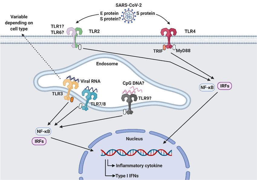
According toAccording
the latest research [40–45],
to the latest several
research TLRsseveral
[40–45], are involved
TLRs arein the sensing
involved inof
the sensing of
PAMPs fromPAMPs SARS-CoV-2. It has been suggested
from SARS-CoV-2. It has beenthat TLR2, TLR3,
suggested TLR4,TLR3,
that TLR2, TLR7/8, andTLR7/8, and
TLR4,
TLR9 contribute
TLR9 tocontribute
antiviral responses against
to antiviral SARS-CoV-2
responses infection (Figure
against SARS-CoV-2 2). (Figure 2).
infection
Table 1. Toll-like receptors (TLRs) in humans.
Primary Adaptor Signaling
TLRs Ligands Refs.
Localization molecules Characteristics
Heterodimerization
TLR1 Triacyl lipopeptides Cell surface MyD88 [46]
with TLR2
TLR2 Lipoproteins, Zymosan, etc Cell surface MyD88 [47,48]Viruses 2021, 13, 2132 4 of 11
Table 1. Toll-like receptors (TLRs) in humans.
Primary Adaptor Signaling
TLRs Ligands Refs.
Localization Molecules Characteristics
Triacyl Heterodimerization
TLR1 Cell surface MyD88 [46]
2021, 13, x FOR PEER REVIEW lipopeptides with TLR2 4 of 11
Lipoproteins,
TLR2 Cell surface MyD88 [47,48]
Zymosan, etc
TLR3 dsRNA Intracellular TRIF [49]
R3 dsRNA LPS, Viral Intracellular TRIF [49]
R4 LPS, ViralTLR4 envelopeetc
envelope glycoproteins, Cell surface
Cell surface MyD88/TRIF
MyD88/TRIF [50–53] [50–53]
glycoproteins, etc
R5 Flagellin Cell surface MyD88 [54,55]
TLR5 Flagellin Cell surface MyD88 [54,55]
Heterodimerization
R6 Diacyl lipopeptidesDiacyl
TLR6 CellCell
surface
surface MyD88 MyD88
Heterodimerization
[48] [48]
lipopeptides with TLR2
with TLR2
R7 TLR7 ssRNA ssRNA Intracellular
Intracellular MyD88 MyD88 [56,57] [56,57]
R8 TLR8 ssRNA ssRNA Intracellular
Intracellular MyD88 MyD88 [58] [58]
Unmethylated
Unmethylated
TLR9 CpG-rich DNA frag-
CpG-rich DNA Intracellular MyD88 [59,60]
R9 Intracellular MyD88 [59,60]
ment, mtDNA fragment, mtDNA
R10 TLR10Undefined Undefined CellCell surface
surface MyD88
MyD88 [61–63] [61–63]
Figure 2. SARS-CoV-2
Figure 2. SARS-CoV-2 recognitionrecognition
by Toll-like by Toll-like
receptors receptors
(TLRs). TLRs(TLRs). TLRs are for
are responsible responsible for recogniz-
recognizing pathogen-associated
ing pathogen-associated molecular patterns (PAMPs) derived from invading pathogens.
molecular patterns (PAMPs) derived from invading pathogens. Surface TLR2 and TLR4 and intracellular Surface
TLR3, TLR7/8,
TLR2 and TLR4 and intracellular TLR3, TLR7/8, and TLR9 are thought to be involved in the sensing
and TLR9 are thought to be involved in the sensing of SARS-CoV-2 infection. Activated TLRs initiate downstream
of SARS-CoV-2 infection. Activated TLRs initiate downstream signaling pathways by recruiting
signaling pathways by recruiting adaptor molecules, such as MyD88 and TRIF, which results in the subsequent production
adaptor molecules, such as MyD88 and TRIF, which results in the subsequent production of inflam-
of inflammatory
matorycytokines
cytokinesand
andtype
typeI IIFNs
IFNsthrough
throughtranscription
transcriptionfactors
factorsNF-κβ
NF-κβand
andIRFs.
IRFs. This
This figure
figure was
was created by
BioRender.com accessed
created on 10 September
by BioRender.com 2021 (BioRender,
accessed Toronto,
on 10 September 2021ON, Canada).Toronto, ON, Canada).
(BioRender,
3.1. Cell Surface TLRs
3.1. Cell Surface TLRs
Currently, 10 members of the TLR family have been identified in humans. TLRs
Currently, 10 members of the TLR family have been identified in humans. TLRs are
are currently classified into two categories based on their cellular localization [64]: TLR1,
currently classified into two categories based on their cellular localization [64]: TLR1,
TLR2, TLR4, TLR5, TLR6, and TLR10 belong to cell surface TLRs that recognize microbial
TLR2, TLR4, TLR5, TLR6, and TLR10 belong to cell surface TLRs that recognize microbial
components including proteins and lipids derived from invading pathogens, whereas
TLR3, TLR7, TLR8, and TLR9 are intracellular TLRs that sense nucleic acid ligands [65].
TLR2 is a surface receptor that recognizes diverse ligands derived from viruses, bac-
teria, fungi, and parasites [66]. TLR2 forms heterodimers with TLR1 and TLR6, utilizing
MyD88 for signal transduction. While the involvement of TLR2 in immune responsesViruses 2021, 13, 2132 5 of 11
components including proteins and lipids derived from invading pathogens, whereas
TLR3, TLR7, TLR8, and TLR9 are intracellular TLRs that sense nucleic acid ligands [65].
TLR2 is a surface receptor that recognizes diverse ligands derived from viruses,
bacteria, fungi, and parasites [66]. TLR2 forms heterodimers with TLR1 and TLR6, utilizing
MyD88 for signal transduction. While the involvement of TLR2 in immune responses
against coronavirus infections has not been elucidated, a recent study revealed that the
SARS-CoV-2 E protein is sensed by TLR2 [40,67]. Zheng and colleagues reanalyzed the
expression of MyD88 and TLRs in patients with different severity grades of COVID-19
using a public dataset and found that the expression of MyD88, TLR1, TLR2, TLR4, TLR5,
TLR8, and TLR9 was increased in patients with severe to critical illness. To clarify which
TLRs were essential for the sensing of Betacoronavirus, they infected bone marrow-derived
macrophages deficient in TLR2, TLR4, TLR7, or TLR9 with mouse hepatitis virus, which
belongs to the Betacoronavirus genus, and found that TLR2 deficiency resulted in the
abrogated expression of inflammatory cytokine genes. They then investigated the role of
TLR2 in SARS-CoV-2 infection using human peripheral blood mononuclear cells treated
with a TLR2 inhibitor. They performed experiments using heat-inactivated SARS-CoV-2
to identify the viral components responsible for TLR2 activation, and structural proteins
were identified as promising targets. Among four viral structural proteins identified, the
E protein activated the TLR2 signaling pathway. In addition, the authors found that the
SARS-CoV-2 E protein induced TLR2-dependent inflammation in mice and that the TLR2
inhibitor protected mice from lethal SARS-CoV-2 infection, indicating that the SARS-CoV-2
E protein is a novel ligand for TLR2 activation.
However, a separate group identified the SARS-CoV-2 S protein as a TLR2 ligand
in non-peer reviewed preprints [68]. They observed that recombinant S protein induced
inflammatory mediators in macrophages, monocytes, and human lung epithelial A549
cells via the activation of the TLR2-mediated NF-κB pathway. Indeed, intraperitoneal
injection of recombinant S protein triggered TLR2-mediated proinflammatory cytokine
production in mice. While further studies are required, these results offer valuable insight
into TLR2-dependent immune responses against SARS-CoV-2 infection.
TLR4 is well known to recognize lipopolysaccharides produced by Gram-negative
bacteria. Early in the COVID-19 pandemic, an in silico study suggested a possible interac-
tion between the SARS-CoV-2 S protein and TLR4 [69]. Investigators used a computational
approach to study the interaction between the S protein and host receptors. Interestingly,
they found that the SARS-CoV-2 S protein strongly bound to TLR4, suggesting a poten-
tial role for TLR4 in SARS-CoV-2 recognition. This hypothesis has been supported by
other evidence demonstrating that TLR4 and its downstream signaling molecules are
significantly upregulated in patients with severe COVID-19 compared to those with mild
illness [40,70]. Recently, two groups confirmed that the S protein leads to proinflammatory
cytokine production in monocytes and macrophages in a TLR4-dependent manner [43,44].
However, the identity of TLR4-binding sites in the S protein remains unclear. Shirato
and Kizaki showed that the S1 subunit (residues 16–671) induced the activation of the
NF-κB and mitogen-activated protein kinase pathways as well as subsequent proinflamma-
tory cytokine production in macrophages [44]. S1-induced proinflammatory responses in
macrophages were abrogated following treatment with a TLR4 antagonist or by transfec-
tion with TLR4 siRNA. On the other hand, Zhao and colleagues demonstrated that only
the trimeric S protein, rather than its N-terminal domain (NTD) (residues 1–307) or RBD
(residues 319–541), activates immune responses in macrophages [43]. They suggested that
a conformational binding site composed of the RBD and NTD of the S protein was likely
to interact with TLR4. Further studies are required to elucidate the relationship between
TLR4 and the S protein.
3.2. Intracellular TLRs
Nucleic acid-sensing TLRs (TLR3, TLR7, TLR8, and TLR9) are localized in endo-
somes to prevent the recognition of self-DNA or -RNA. As SARS-CoV-2 is an ssRNAViruses 2021, 13, 2132 6 of 11
virus and produces double-stranded RNA (dsRNA) during replication in host cells [34,71],
intracellular RNA sensors are thought to be involved in the recognition of SARS-CoV-2
infection. TLR3 senses dsRNA in endosomes, and TLR3 stimulation leads to the production
of proinflammatory cytokines and type I IFNs via the activation of the TRIF signaling
pathway. In contrast, ssRNA is recognized by TLR7 and TLR8, which use MyD88 as a
downstream adapter protein. Recently, the roles of TLR3 and TLR7 in antiviral responses
following SARS-CoV-2 infection were identified using three-dimensional lung multicellular
spheroids [41]. The relative expression levels of TLR3 and TLR7, as well as the production
of proinflammatory cytokines and type I IFNs, were elevated in SARS-CoV-2-infected
multicellular spheroids, and both IRF3 and NF-κB appeared to participate in the signaling
pathway downstream of TLR3 and TLR7. Furthermore, another group demonstrated that
an ssRNA fragment of SARS-CoV-2 genomic RNA was responsible for the activation of
the TLR7/8-dependent MyD88 pathway in human DCs [45]. As TLR7/8 is activated by
guanosine (G)- and uridine (U)-rich ssRNA [72], this group scanned putative TLR7/8 lig-
ands within the SARS-CoV-2 genome and selected two GU-rich ssRNA sequences—termed
SCV2-RNA—to test their hypothesis. Using human monocyte-derived DCs (MoDCs), the
authors found that SCV2-RNA treatment induced the expression of pro-inflammatory
cytokines, including TNF-α, IL-6, and IL-12, as well as the secretion of the T cell-recruiting
chemokine CXCL9. SCV2-RNA-stimulated MoDCs exhibited a mature form and triggered
cocultured CD4 and CD8 T cells to produce IFN-γ, suggesting that SCV2-RNA can mediate
DC activation. Similar results were observed in pDCs. SCV2-RNA induced the upregula-
tion of CD86 expression and the production of IFN-α and TNF-α in pDCs. The authors
suggested that such ssRNA-induced activation is mediated via the TLR8/MyD88/NF-κB
pathway in MoDCs and by TLR7 in pDCs. Taken together, these results indicate that
ssRNA and dsRNA produced by SARS-CoV-2 are recognized by endosomal RNA sensors.
TLR9 recognizes CpG-rich DNA fragments derived from bacteria, viruses, and mito-
chondrial DNA (mtDNA) [60]. While the relationship between TLR9 and the recognition
of SARS-CoV-2 infection is not clear, the coding region of the E protein and ORF10 in
the SARS-CoV-2 genome was shown to be enriched with CpG [73]. Recently, TLR9 was
suspected of inducing severe COVID-19 [42]. This TLR9-COVID-19 hypothesis, presented
by Bezemer and Garssen, proposes that CpG islands in SARS-CoV-2 or mtDNA released
from damaged host cells could trigger TLR9 activation and subsequently elicit pathogenic
hyperinflammatory responses. As it is still unclear whether TLR9 directly detects viral
components derived from SARS-CoV-2, future studies are needed to address this issue.
While it is well known that intracellular TLRs are physically separated from the cell
surface to prevent autoimmune responses following receptor activation by host nucleic
acids [74], interestingly, several studies have suggested the cell surface positioning of endo-
somal TLRs in certain cell types [75–78]. Cell surface expression of TLR3 was observed in
human fibroblast cell lines [79], mouse splenic CD8+ dendritic cells, and marginal zone B
cells [80]. Mouse bone marrow (BM)-derived macrophages, BM-conventional dendritic
cells, BM-plasmacytoid DCs and B cells expressed TLR7 on the cell surface [77]. TLR9
was expressed on the surface of mouse splenic DCs [78], monocytes [81], and human
neutrophils [82]. These findings suggest that both the cell surface and intracellular expres-
sion of nucleic acid-sensing TLRs may participate in the activation of immune responses.
Interestingly, human airway epithelial cells expressed TLR3, TLR7 and TLR9 on the apical
cell membrane [76], indicating that nucleic acid-sensing TLRs on the epithelial cell surface
may play a crucial role in innate responses against inhaled pathogens such as SARS-CoV-2.
However, further studies are required to prove this hypothesis.
4. Conclusions
TLRs participate in the first line of defense against invading pathogens. They rec-
ognize a broad range of PAMPs derived from microorganisms, and the main function of
TLRs is the ability to activate innate immune responses, including cytokine production.
At present, TLR2, TLR4, TLR3, TLR7, TLR8, and TLR9 have been suggested as possibleViruses 2021, 13, 2132 7 of 11
receptors capable of recognizing SARS-CoV-2 infection. Viral components including the
S protein, E protein, ssRNA, and dsRNA seem to serve as ligands for these TLRs. TLR-
mediated immune responses are essential for host protection; however, uncontrolled and
exacerbated inflammation can result in pathologic responses such as tissue damage. In
patients with COVID-19, robust innate immune responses and hyperinflammation were
observed in severe cases [3,4,7], resulting in septic shock [83], acute lung injury [84], and
acute respiratory distress syndrome [85]. Therefore, immunomodulatory therapeutic strate-
gies, including the use of anti-inflammatory drugs, have been suggested as promising
treatments for severe COVID-19. Many approaches targeting TLR signaling pathways
have been tested for their ability to attenuate hyperinflammation following SARS-CoV-2
infection. For example, the U.S. FDA has approved an investigation into the efficacy of a
PUL-042 inhalation solution that blocks TLR2/6/9 to reduce the infection rate, progression,
and disease severity of COVID-19 (NCT04312997, NCT04313023) [42,86]. Additionally,
a clinical study to evaluate the inhibition of TLR3-mediated inflammatory responses
by Famotidine [87] to improve outcomes in patients with COVID-19 has been conducted
(NCT04389567, NCT04504240, NCT04724720, NCT04370262) [88,89]. Various TLR4 modula-
tors, such as EB05 (NCT04401475), Eritoran (NCT02735707), Naltrexone [90] (NCT04604704,
NCT04604678), Curcumin [91] (NCT04382040), and Berberine [92–94] (NCT04479202) are
undergoing clinical trials for COVID-19. Although current studies have not produced
sufficient encouraging results, based on the evidence presented in this review, targeting
TLRs could be an effective treatment for COVID-19.
Author Contributions: Writing and original draft preparation, H.E.J. and H.K.L.; writing, reviewing,
and editing, H.E.J. and H.K.L.; supervision, H.K.L.; funding acquisition, H.K.L. All authors have
read and agreed to the published version of the manuscript.
Funding: This study was supported by the National Research Foundation of Korea (NRF-2021M3A9D
3026428 and NRF-2021M3A9H3015688) funded by the Ministry of Science and ICT of Korea. This
work was also supported by Mobile Clinic Module Project funded by KAIST and the 2020 Joint
Research Project of Institutes of Science and Technology. H.E. Jung was supported by Basic Science Re-
search Program through the NRF funded by the Ministry of Education (NRF-2021R1I1A1A01059948).
Institutional Review Board Statement: This review was completed with no requirement for institu-
tional review.
Informed Consent Statement: Not applicable.
Data Availability Statement: No new data were generated.
Acknowledgments: The authors would like to thank the members of the Laboratory of Host Defenses
for helpful advice.
Conflicts of Interest: The authors declare no conflict of interest.
References
1. Zhang, W.; Zhao, Y.; Zhang, F.; Wang, Q.; Li, T.; Liu, Z.; Wang, J.; Qin, Y.; Zhang, X.; Yan, X.; et al. The use of anti-inflammatory
drugs in the treatment of people with severe coronavirus disease 2019 (COVID-19): The Perspectives of clinical immunologists
from China. Clin. Immunol. 2020, 214, 108393. [CrossRef]
2. Park, J.H.; Lee, H.K. Re-analysis of Single Cell Transcriptome Reveals That the NR3C1-CXCL8-Neutrophil Axis Determines the
Severity of COVID-19. Front. Immunol. 2020, 11, 2145. [CrossRef]
3. Yang, L.; Xie, X.; Tu, Z.; Fu, J.; Xu, D.; Zhou, Y. The signal pathways and treatment of cytokine storm in COVID-19. Signal.
Transduct Target. Ther. 2021, 6, 255. [CrossRef]
4. Del Valle, D.M.; Kim-Schulze, S.; Huang, H.H.; Beckmann, N.D.; Nirenberg, S.; Wang, B.; Lavin, Y.; Swartz, T.H.; Madduri, D.;
Stock, A.; et al. An inflammatory cytokine signature predicts COVID-19 severity and survival. Nat. Med. 2020, 26, 1636–1643.
[CrossRef]
5. Mazzoni, A.; Salvati, L.; Maggi, L.; Capone, M.; Vanni, A.; Spinicci, M.; Mencarini, J.; Caporale, R.; Peruzzi, B.; Antonelli, A.; et al.
Impaired immune cell cytotoxicity in severe COVID-19 is IL-6 dependent. J. Clin. Investig. 2020, 130, 4694–4703. [CrossRef]
6. Investigators, R.-C.; Gordon, A.C.; Mouncey, P.R.; Al-Beidh, F.; Rowan, K.M.; Nichol, A.D.; Arabi, Y.M.; Annane, D.; Beane, A.;
van Bentum-Puijk, W.; et al. Interleukin-6 Receptor Antagonists in Critically Ill Patients with COVID-19. N. Engl. J. Med. 2021,
384, 1491–1502. [CrossRef]Viruses 2021, 13, 2132 8 of 11
7. Huang, C.; Wang, Y.; Li, X.; Ren, L.; Zhao, J.; Hu, Y.; Zhang, L.; Fan, G.; Xu, J.; Gu, X.; et al. Clinical features of patients infected
with 2019 novel coronavirus in Wuhan, China. Lancet 2020, 395, 497–506. [CrossRef]
8. Karki, R.; Sharma, B.R.; Tuladhar, S.; Williams, E.P.; Zalduondo, L.; Samir, P.; Zheng, M.; Sundaram, B.; Banoth, B.; Malireddi,
R.K.S.; et al. Synergism of TNF-alpha and IFN-gamma Triggers Inflammatory Cell Death, Tissue Damage, and Mortality in
SARS-CoV-2 Infection and Cytokine Shock Syndromes. Cell 2021, 184, 149–168. [CrossRef] [PubMed]
9. Cherry, J.D.; Krogstad, P. SARS: The first pandemic of the 21st century. Pediatr. Res. 2004, 56, 1–5. [CrossRef] [PubMed]
10. Al-Tawfiq, J.A.; Petersen, E.; Memish, Z.A.; Perlman, S.; Zumla, A. Middle East respiratory syndrome coronavirus—The need for
global proactive surveillance, sequencing and modeling. Travel Med. Infect. Dis. 2021, 43, 102118. [CrossRef]
11. Mesel-Lemoine, M.; Millet, J.; Vidalain, P.O.; Law, H.; Vabret, A.; Lorin, V.; Escriou, N.; Albert, M.L.; Nal, B.; Tangy, F. A human
coronavirus responsible for the common cold massively kills dendritic cells but not monocytes. J. Virol. 2012, 86, 7577–7587.
[CrossRef]
12. Wu, A.; Peng, Y.; Huang, B.; Ding, X.; Wang, X.; Niu, P.; Meng, J.; Zhu, Z.; Zhang, Z.; Wang, J.; et al. Genome Composition and
Divergence of the Novel Coronavirus (2019-nCoV) Originating in China. Cell Host Microbe 2020, 27, 325–328. [CrossRef]
13. Lu, R.; Zhao, X.; Li, J.; Niu, P.; Yang, B.; Wu, H.; Wang, W.; Song, H.; Huang, B.; Zhu, N.; et al. Genomic characterisation and
epidemiology of 2019 novel coronavirus: Implications for virus origins and receptor binding. Lancet 2020, 395, 565–574. [CrossRef]
14. Ren, L.L.; Wang, Y.M.; Wu, Z.Q.; Xiang, Z.C.; Guo, L.; Xu, T.; Jiang, Y.Z.; Xiong, Y.; Li, Y.J.; Li, X.W.; et al. Identification of a novel
coronavirus causing severe pneumonia in human: A descriptive study. Chin. Med. J. 2020, 133, 1015–1024. [CrossRef]
15. Chan, J.F.; Kok, K.H.; Zhu, Z.; Chu, H.; To, K.K.; Yuan, S.; Yuen, K.Y. Genomic characterization of the 2019 novel human-
pathogenic coronavirus isolated from a patient with atypical pneumonia after visiting Wuhan. Emerg. Microbes Infect. 2020, 9,
221–236. [CrossRef]
16. Cubuk, J.; Alston, J.J.; Incicco, J.J.; Singh, S.; Stuchell-Brereton, M.D.; Ward, M.D.; Zimmerman, M.I.; Vithani, N.; Griffith, D.;
Wagoner, J.A.; et al. The SARS-CoV-2 nucleocapsid protein is dynamic, disordered, and phase separates with RNA. Nat. Commun.
2021, 12, 1936. [CrossRef] [PubMed]
17. Kang, S.; Yang, M.; Hong, Z.; Zhang, L.; Huang, Z.; Chen, X.; He, S.; Zhou, Z.; Zhou, Z.; Chen, Q.; et al. Crystal structure of
SARS-CoV-2 nucleocapsid protein RNA binding domain reveals potential unique drug targeting sites. Acta Pharm. Sin. B 2020,
10, 1228–1238. [CrossRef]
18. Walls, A.C.; Park, Y.J.; Tortorici, M.A.; Wall, A.; McGuire, A.T.; Veesler, D. Structure, Function, and Antigenicity of the SARS-CoV-2
Spike Glycoprotein. Cell 2020, 181, 281–292. [CrossRef] [PubMed]
19. Shang, J.; Wan, Y.; Luo, C.; Ye, G.; Geng, Q.; Auerbach, A.; Li, F. Cell entry mechanisms of SARS-CoV-2. Proc. Natl. Acad. Sci. USA
2020, 117, 11727–11734. [CrossRef] [PubMed]
20. Hoffmann, M.; Kleine-Weber, H.; Pohlmann, S. A Multibasic Cleavage Site in the Spike Protein of SARS-CoV-2 Is Essential for
Infection of Human Lung Cells. Mol. Cell 2020, 78, 779–784. [CrossRef]
21. Yan, R.; Zhang, Y.; Li, Y.; Xia, L.; Guo, Y.; Zhou, Q. Structural basis for the recognition of SARS-CoV-2 by full-length human ACE2.
Science 2020, 367, 1444–1448. [CrossRef] [PubMed]
22. Zhang, Q.; Xiang, R.; Huo, S.; Zhou, Y.; Jiang, S.; Wang, Q.; Yu, F. Molecular mechanism of interaction between SARS-CoV-2 and
host cells and interventional therapy. Signal Transduct. Target. Ther. 2021, 6, 233. [CrossRef] [PubMed]
23. Murgolo, N.; Therien, A.G.; Howell, B.; Klein, D.; Koeplinger, K.; Lieberman, L.A.; Adam, G.C.; Flynn, J.; McKenna, P.;
Swaminathan, G.; et al. SARS-CoV-2 tropism, entry, replication, and propagation: Considerations for drug discovery and
development. PLoS Pathog. 2021, 17, e1009225. [CrossRef] [PubMed]
24. Bayati, A.; Kumar, R.; Francis, V.; McPherson, P.S. SARS-CoV-2 infects cells after viral entry via clathrin-mediated endocytosis. J.
Biol. Chem. 2021, 296, 100306. [CrossRef] [PubMed]
25. Ou, X.; Liu, Y.; Lei, X.; Li, P.; Mi, D.; Ren, L.; Guo, L.; Guo, R.; Chen, T.; Hu, J.; et al. Characterization of spike glycoprotein of
SARS-CoV-2 on virus entry and its immune cross-reactivity with SARS-CoV. Nat. Commun. 2020, 11, 1620. [CrossRef] [PubMed]
26. Hoffmann, M.; Kleine-Weber, H.; Schroeder, S.; Kruger, N.; Herrler, T.; Erichsen, S.; Schiergens, T.S.; Herrler, G.; Wu, N.H.; Nitsche,
A.; et al. SARS-CoV-2 Cell Entry Depends on ACE2 and TMPRSS2 and Is Blocked by a Clinically Proven Protease Inhibitor. Cell
2020, 181, 271–280. [CrossRef] [PubMed]
27. Zhao, M.M.; Yang, W.L.; Yang, F.Y.; Zhang, L.; Huang, W.J.; Hou, W.; Fan, C.F.; Jin, R.H.; Feng, Y.M.; Wang, Y.C.; et al. Cathepsin L
plays a key role in SARS-CoV-2 infection in humans and humanized mice and is a promising target for new drug development.
Signal Transduct. Target. Ther. 2021, 6, 134. [CrossRef] [PubMed]
28. Padmanabhan, P.; Desikan, R.; Dixit, N.M. Targeting TMPRSS2 and Cathepsin B/L together may be synergistic against SARS-
CoV-2 infection. PLoS Comput. Biol. 2020, 16, e1008461. [CrossRef] [PubMed]
29. Klein, S.; Cortese, M.; Winter, S.L.; Wachsmuth-Melm, M.; Neufeldt, C.J.; Cerikan, B.; Stanifer, M.L.; Boulant, S.; Bartenschlager,
R.; Chlanda, P. SARS-CoV-2 structure and replication characterized by in situ cryo-electron tomography. Nat. Commun. 2020, 11,
5885. [CrossRef] [PubMed]
30. Harrison, A.G.; Lin, T.; Wang, P. Mechanisms of SARS-CoV-2 Transmission and Pathogenesis. Trends Immunol. 2020, 41, 1100–1115.
[CrossRef] [PubMed]
31. Finkel, Y.; Mizrahi, O.; Nachshon, A.; Weingarten-Gabbay, S.; Morgenstern, D.; Yahalom-Ronen, Y.; Tamir, H.; Achdout, H.; Stein,
D.; Israeli, O.; et al. The coding capacity of SARS-CoV-2. Nature 2021, 589, 125–130. [CrossRef]Viruses 2021, 13, 2132 9 of 11
32. Hillen, H.S.; Kokic, G.; Farnung, L.; Dienemann, C.; Tegunov, D.; Cramer, P. Structure of replicating SARS-CoV-2 polymerase.
Nature 2020, 584, 154–156. [CrossRef]
33. Mendonca, L.; Howe, A.; Gilchrist, J.B.; Sheng, Y.; Sun, D.; Knight, M.L.; Zanetti-Domingues, L.C.; Bateman, B.; Krebs, A.S.; Chen,
L.; et al. Correlative multi-scale cryo-imaging unveils SARS-CoV-2 assembly and egress. Nat. Commun. 2021, 12, 4629. [CrossRef]
[PubMed]
34. Horova, V.; Landova, B.; Hodek, J.; Chalupsky, K.; Krafcikova, P.; Chalupska, D.; Duchoslav, V.; Weber, J.; Boura, E.; Klima,
M. Localization of SARS-CoV-2 Capping Enzymes Revealed by an Antibody against the nsp10 Subunit. Viruses 2021, 13, 1487.
[CrossRef] [PubMed]
35. Ghosh, S.; Dellibovi-Ragheb, T.A.; Kerviel, A.; Pak, E.; Qiu, Q.; Fisher, M.; Takvorian, P.M.; Bleck, C.; Hsu, V.W.; Fehr, A.R.; et al.
β-Coronaviruses Use Lysosomes for Egress Instead of the Biosynthetic Secretory Pathway. Cell 2020, 183, 1520–1535. [CrossRef]
36. Li, D.; Wu, M. Pattern recognition receptors in health and diseases. Signal Transduct. Target. Ther. 2021, 6, 291. [CrossRef]
[PubMed]
37. Ziegler, C.G.K.; Allon, S.J.; Nyquist, S.K.; Mbano, I.M.; Miao, V.N.; Tzouanas, C.N.; Cao, Y.; Yousif, A.S.; Bals, J.; Hauser, B.M.;
et al. SARS-CoV-2 Receptor ACE2 Is an Interferon-Stimulated Gene in Human Airway Epithelial Cells and Is Detected in Specific
Cell Subsets across Tissues. Cell 2020, 181, 1016–1035. [CrossRef] [PubMed]
38. Zou, X.; Chen, K.; Zou, J.; Han, P.; Hao, J.; Han, Z. Single-cell RNA-seq data analysis on the receptor ACE2 expression reveals the
potential risk of different human organs vulnerable to 2019-nCoV infection. Front. Med. 2020, 14, 185–192. [CrossRef] [PubMed]
39. Qi, F.; Qian, S.; Zhang, S.; Zhang, Z. Single cell RNA sequencing of 13 human tissues identify cell types and receptors of human
coronaviruses. Biochem. Biophys. Res. Commun. 2020, 526, 135–140. [CrossRef] [PubMed]
40. Zheng, M.; Karki, R.; Williams, E.P.; Yang, D.; Fitzpatrick, E.; Vogel, P.; Jonsson, C.B.; Kanneganti, T.D. TLR2 senses the
SARS-CoV-2 envelope protein to produce inflammatory cytokines. Nat. Immunol. 2021, 22, 829–838. [CrossRef] [PubMed]
41. Bortolotti, D.; Gentili, V.; Rizzo, S.; Schiuma, G.; Beltrami, S.; Strazzabosco, G.; Fernandez, M.; Caccuri, F.; Caruso, A.; Rizzo,
R.J.M. TLR3 and TLR7 RNA Sensor Activation during SARS-COV-2 Infection. Microorganisms 2021, 9, 1820. [CrossRef] [PubMed]
42. Bezemer, G.F.G.; Garssen, J. TLR9 and COVID-19: A Multidisciplinary Theory of a Multifaceted Therapeutic Target. Front. Pharm.
2020, 11, 601685. [CrossRef]
43. Zhao, Y.; Kuang, M.; Li, J.; Zhu, L.; Jia, Z.; Guo, X.; Hu, Y.; Kong, J.; Yin, H.; Wang, X.; et al. SARS-CoV-2 spike protein interacts
with and activates TLR41. Cell Res. 2021, 31, 818–820. [CrossRef]
44. Shirato, K.; Kizaki, T. SARS-CoV-2 spike protein S1 subunit induces pro-inflammatory responses via toll-like receptor 4 signaling
in murine and human macrophages. Heliyon 2021, 7, e06187. [CrossRef] [PubMed]
45. Salvi, V.; Nguyen, H.O.; Sozio, F.; Schioppa, T.; Gaudenzi, C.; Laffranchi, M.; Scapini, P.; Passari, M.; Barbazza, I.; Tiberio, L.; et al.
SARS-CoV-2-associated ssRNAs activate inflammation and immunity via TLR7/8. JCI Insight 2021, 6, 150542. [CrossRef]
46. Takeuchi, O.; Sato, S.; Horiuchi, T.; Hoshino, K.; Takeda, K.; Dong, Z.; Modlin, R.L.; Akira, S. Cutting edge: Role of Toll-like
receptor 1 in mediating immune response to microbial lipoproteins. J. Immunol. 2002, 169, 10–14. [CrossRef] [PubMed]
47. Takeuchi, O.; Hoshino, K.; Kawai, T.; Sanjo, H.; Takada, H.; Ogawa, T.; Takeda, K.; Akira, S. Differential roles of TLR2 and TLR4
in recognition of gram-negative and gram-positive bacterial cell wall components. Immunity 1999, 11, 443–451. [CrossRef]
48. Kang, J.Y.; Nan, X.; Jin, M.S.; Youn, S.J.; Ryu, Y.H.; Mah, S.; Han, S.H.; Lee, H.; Paik, S.G.; Lee, J.O. Recognition of lipopeptide
patterns by Toll-like receptor 2-Toll-like receptor 6 heterodimer. Immunity 2009, 31, 873–884. [CrossRef]
49. Alexopoulou, L.; Holt, A.C.; Medzhitov, R.; Flavell, R.A. Recognition of double-stranded RNA and activation of NF-κB by
Toll-like receptor 3. Nature 2001, 413, 732–738. [CrossRef]
50. Poltorak, A.; He, X.; Smirnova, I.; Liu, M.Y.; Van Huffel, C.; Du, X.; Birdwell, D.; Alejos, E.; Silva, M.; Galanos, C.; et al. Defective
LPS signaling in C3H/HeJ and C57BL/10ScCr mice: Mutations in Tlr4 gene. Science 1998, 282, 2085–2088. [CrossRef]
51. Georgel, P.; Jiang, Z.; Kunz, S.; Janssen, E.; Mols, J.; Hoebe, K.; Bahram, S.; Oldstone, M.B.; Beutler, B. Vesicular stomatitis virus
glycoprotein G activates a specific antiviral Toll-like receptor 4-dependent pathway. Virology 2007, 362, 304–313. [CrossRef]
52. Haynes, L.M.; Moore, D.D.; Kurt-Jones, E.A.; Finberg, R.W.; Anderson, L.J.; Tripp, R.A. Involvement of toll-like receptor 4 in
innate immunity to respiratory syncytial virus. J. Virol. 2001, 75, 10730–10737. [CrossRef]
53. Kurt-Jones, E.A.; Popova, L.; Kwinn, L.; Haynes, L.M.; Jones, L.P.; Tripp, R.A.; Walsh, E.E.; Freeman, M.W.; Golenbock, D.T.;
Anderson, L.J.; et al. Pattern recognition receptors TLR4 and CD14 mediate response to respiratory syncytial virus. Nat. Immunol.
2000, 1, 398–401. [CrossRef] [PubMed]
54. Hayashi, F.; Smith, K.D.; Ozinsky, A.; Hawn, T.R.; Yi, E.C.; Goodlett, D.R.; Eng, J.K.; Akira, S.; Underhill, D.M.; Aderem, A. The
innate immune response to bacterial flagellin is mediated by Toll-like receptor 5. Nature 2001, 410, 1099–1103. [CrossRef]
55. Gewirtz, A.T.; Navas, T.A.; Lyons, S.; Godowski, P.J.; Madara, J.L. Cutting edge: Bacterial flagellin activates basolaterally
expressed TLR5 to induce epithelial proinflammatory gene expression. J. Immunol. 2001, 167, 1882–1885. [CrossRef]
56. Lund, J.M.; Alexopoulou, L.; Sato, A.; Karow, M.; Adams, N.C.; Gale, N.W.; Iwasaki, A.; Flavell, R.A. Recognition of single-
stranded RNA viruses by Toll-like receptor 7. Proc. Natl. Acad. Sci. USA 2004, 101, 5598–5603. [CrossRef]
57. Diebold, S.S.; Kaisho, T.; Hemmi, H.; Akira, S.; Reis e Sousa, C. Innate antiviral responses by means of TLR7-mediated recognition
of single-stranded RNA. Science 2004, 303, 1529–1531. [CrossRef] [PubMed]
58. Heil, F.; Hemmi, H.; Hochrein, H.; Ampenberger, F.; Kirschning, C.; Akira, S.; Lipford, G.; Wagner, H.; Bauer, S. Species-specific
recognition of single-stranded RNA via toll-like receptor 7 and 8. Science 2004, 303, 1526–1529. [CrossRef] [PubMed]Viruses 2021, 13, 2132 10 of 11
59. Hemmi, H.; Takeuchi, O.; Kawai, T.; Kaisho, T.; Sato, S.; Sanjo, H.; Matsumoto, M.; Hoshino, K.; Wagner, H.; Takeda, K.; et al. A
Toll-like receptor recognizes bacterial DNA. Nature 2000, 408, 740–745. [CrossRef] [PubMed]
60. Zhang, Q.; Raoof, M.; Chen, Y.; Sumi, Y.; Sursal, T.; Junger, W.; Brohi, K.; Itagaki, K.; Hauser, C.J. Circulating mitochondrial
DAMPs cause inflammatory responses to injury. Nature 2010, 464, 104–107. [CrossRef]
61. Hasan, U.; Chaffois, C.; Gaillard, C.; Saulnier, V.; Merck, E.; Tancredi, S.; Guiet, C.; Briere, F.; Vlach, J.; Lebecque, S.; et al. Human
TLR10 is a functional receptor, expressed by B cells and plasmacytoid dendritic cells, which activates gene transcription through
MyD88. J. Immunol. 2005, 174, 2942–2950. [CrossRef] [PubMed]
62. Hess, N.J.; Jiang, S.; Li, X.; Guan, Y.; Tapping, R.I. TLR10 Is a B Cell Intrinsic Suppressor of Adaptive Immune Responses. J.
Immunol. 2017, 198, 699–707. [CrossRef] [PubMed]
63. Fore, F.; Indriputri, C.; Mamutse, J.; Nugraha, J. TLR10 and Its Unique Anti-Inflammatory Properties and Potential Use as a
Target in Therapeutics. Immune Netw. 2020, 20, e21. [CrossRef] [PubMed]
64. Marks, K.E.; Cho, K.; Stickling, C.; Reynolds, J.M. Toll-like Receptor 2 in Autoimmune Inflammation. Immune Netw. 2021, 21, e18.
[CrossRef] [PubMed]
65. Kawasaki, T.; Kawai, T. Toll-like receptor signaling pathways. Front. Immunol. 2014, 5, 461. [CrossRef] [PubMed]
66. Oliveira-Nascimento, L.; Massari, P.; Wetzler, L.M. The Role of TLR2 in Infection and Immunity. Front. Immunol. 2012, 3, 79.
[CrossRef] [PubMed]
67. Sariol, A.; Perlman, S. SARS-CoV-2 takes its Toll. Nat. Immunol. 2021, 22, 801–802. [CrossRef] [PubMed]
68. Khan, S.; Shafiei, M.S.; Longoria, C.; Schoggins, J.; Savani, R.C.; Zaki, H. SARS-CoV-2 spike protein induces inflammation via
TLR2-dependent activation of the NF-kappaB pathway. bioRxiv 2021. [CrossRef]
69. Choudhury, A.; Mukherjee, S. In silico studies on the comparative characterization of the interactions of SARS-CoV-2 spike
glycoprotein with ACE-2 receptor homologs and human TLRs. J. Med. Virol. 2020, 92, 2105–2113. [CrossRef] [PubMed]
70. Sohn, K.M.; Lee, S.G.; Kim, H.J.; Cheon, S.; Jeong, H.; Lee, J.; Kim, I.S.; Silwal, P.; Kim, Y.J.; Paik, S.; et al. COVID-19 Patients
Upregulate Toll-like Receptor 4-mediated Inflammatory Signaling That Mimics Bacterial Sepsis. J. Korean Med. Sci. 2020, 35, e343.
[CrossRef] [PubMed]
71. Li, Y.; Renner, D.M.; Comar, C.E.; Whelan, J.N.; Reyes, H.M.; Cardenas-Diaz, F.L.; Truitt, R.; Tan, L.H.; Dong, B.; Alysandratos,
K.D.; et al. SARS-CoV-2 induces double-stranded RNA-mediated innate immune responses in respiratory epithelial-derived cells
and cardiomyocytes. Proc. Natl. Acad. Sci. USA 2021, 118, e2022643118. [CrossRef] [PubMed]
72. Li, Y.; Chen, M.; Cao, H.; Zhu, Y.; Zheng, J.; Zhou, H. Extraordinary GU-rich single-strand RNA identified from SARS coronavirus
contributes an excessive innate immune response. Microbes Infect. 2013, 15, 88–95. [CrossRef]
73. Digard, P.; Lee, H.M.; Sharp, C.; Grey, F.; Gaunt, E. Intra-genome variability in the dinucleotide composition of SARS-CoV-2.
Virus Evol. 2020, 6, veaa057. [CrossRef]
74. Blasius, A.L.; Beutler, B. Intracellular toll-like receptors. Immunity 2010, 32, 305–315. [CrossRef] [PubMed]
75. Mielcarska, M.B.; Bossowska-Nowicka, M.; Toka, F.N. Cell Surface Expression of Endosomal Toll-Like Receptors-A Necessity or a
Superfluous Duplication? Front. Immunol. 2020, 11, 620972. [CrossRef] [PubMed]
76. Ioannidis, I.; Ye, F.; McNally, B.; Willette, M.; Flano, E. Toll-like receptor expression and induction of type I and type III interferons
in primary airway epithelial cells. J. Virol. 2013, 87, 3261–3270. [CrossRef]
77. Kanno, A.; Tanimura, N.; Ishizaki, M.; Ohko, K.; Motoi, Y.; Onji, M.; Fukui, R.; Shimozato, T.; Yamamoto, K.; Shibata, T.; et al.
Targeting cell surface TLR7 for therapeutic intervention in autoimmune diseases. Nat. Commun. 2015, 6, 6119. [CrossRef]
78. Onji, M.; Kanno, A.; Saitoh, S.; Fukui, R.; Motoi, Y.; Shibata, T.; Matsumoto, F.; Lamichhane, A.; Sato, S.; Kiyono, H.; et al.
An essential role for the N-terminal fragment of Toll-like receptor 9 in DNA sensing. Nat. Commun. 2013, 4, 1949. [CrossRef]
[PubMed]
79. Matsumoto, M.; Kikkawa, S.; Kohase, M.; Miyake, K.; Seya, T. Establishment of a monoclonal antibody against human Toll-like
receptor 3 that blocks double-stranded RNA-mediated signaling. Biochem. Biophys. Res. Commun. 2002, 293, 1364–1369. [CrossRef]
80. Murakami, Y.; Fukui, R.; Motoi, Y.; Kanno, A.; Shibata, T.; Tanimura, N.; Saitoh, S.; Miyake, K. Roles of the cleaved N-terminal
TLR3 fragment and cell surface TLR3 in double-stranded RNA sensing. J. Immunol. 2014, 193, 5208–5217. [CrossRef] [PubMed]
81. Murakami, Y.; Fukui, R.; Motoi, Y.; Shibata, T.; Saitoh, S.I.; Sato, R.; Miyake, K. The protective effect of the anti-Toll-like receptor 9
antibody against acute cytokine storm caused by immunostimulatory DNA. Sci. Rep. 2017, 7, 44042. [CrossRef] [PubMed]
82. Lindau, D.; Mussard, J.; Wagner, B.J.; Ribon, M.; Ronnefarth, V.M.; Quettier, M.; Jelcic, I.; Boissier, M.C.; Rammensee, H.G.; Decker,
P. Primary blood neutrophils express a functional cell surface Toll-like receptor 9. Eur. J. Immunol. 2013, 43, 2101–2113. [CrossRef]
[PubMed]
83. Guan, W.J.; Ni, Z.Y.; Hu, Y.; Liang, W.H.; Ou, C.Q.; He, J.X.; Liu, L.; Shan, H.; Lei, C.L.; Hui, D.S.C.; et al. Clinical Characteristics
of Coronavirus Disease 2019 in China. N. Engl. J. Med. 2020, 382, 1708–1720. [CrossRef] [PubMed]
84. Russell, C.D.; Millar, J.E.; Baillie, J.K. Clinical evidence does not support corticosteroid treatment for 2019-nCoV lung injury.
Lancet 2020, 395, 473–475. [CrossRef]
85. Chen, N.; Zhou, M.; Dong, X.; Qu, J.; Gong, F.; Han, Y.; Qiu, Y.; Wang, J.; Liu, Y.; Wei, Y.; et al. Epidemiological and clinical
characteristics of 99 cases of 2019 novel coronavirus pneumonia in Wuhan, China: A descriptive study. Lancet 2020, 395, 507–513.
[CrossRef]
86. Schijns, V.; Lavelle, E.C. Prevention and treatment of COVID-19 disease by controlled modulation of innate immunity. Eur J.
Immunol. 2020, 50, 932–938. [CrossRef] [PubMed]Viruses 2021, 13, 2132 11 of 11
87. Mukherjee, R.; Bhattacharya, A.; Bojkova, D.; Mehdipour, A.R.; Shin, D.; Khan, K.S.; Hei-Yin Cheung, H.; Wong, K.B.; Ng, W.L.;
Cinatl, J.; et al. Famotidine inhibits toll-like receptor 3-mediated inflammatory signaling in SARS-CoV-2 infection. J. Biol. Chem.
2021, 297, 100925. [CrossRef] [PubMed]
88. Samimagham, H.R.; Hassani Azad, M.; Haddad, M.; Arabi, M.; Hooshyar, D.; KazemiJahromi, M. The Efficacy of Famotidine
in improvement of outcomes in Hospitalized COVID-19 Patients: A structured summary of a study protocol for a randomised
controlled trial. Trials 2020, 21, 848. [CrossRef] [PubMed]
89. Malone, R.W.; Tisdall, P.; Fremont-Smith, P.; Liu, Y.; Huang, X.P.; White, K.M.; Miorin, L.; Moreno, E.; Alon, A.; Delaforge, E.; et al.
COVID-19: Famotidine, Histamine, Mast Cells, and Mechanisms. Front. Pharm. 2021, 12, 633680. [CrossRef] [PubMed]
90. Wang, X.; Zhang, Y.; Peng, Y.; Hutchinson, M.R.; Rice, K.C.; Yin, H.; Watkins, L.R. Pharmacological characterization of the opioid
inactive isomers (+)-naltrexone and (+)-naloxone as antagonists of toll-like receptor 4. Br. J. Pharm. 2016, 173, 856–869. [CrossRef]
[PubMed]
91. Youn, H.S.; Saitoh, S.I.; Miyake, K.; Hwang, D.H. Inhibition of homodimerization of Toll-like receptor 4 by curcumin. Biochem.
Pharm. 2006, 72, 62–69. [CrossRef] [PubMed]
92. Varghese, F.S.; van Woudenbergh, E.; Overheul, G.J.; Eleveld, M.J.; Kurver, L.; van Heerbeek, N.; van Laarhoven, A.; Miesen, P.;
den Hartog, G.; de Jonge, M.I.; et al. Berberine and Obatoclax Inhibit SARS-Cov-2 Replication in Primary Human Nasal Epithelial
Cells In Vitro. Viruses 2021, 13, 282. [CrossRef] [PubMed]
93. Zhang, B.Y.; Chen, M.; Chen, X.C.; Cao, K.; You, Y.; Qian, Y.J.; Yu, W.K. Berberine reduces circulating inflammatory mediators in
patients with severe COVID-19. Br. J. Surg. 2021, 108, e9–e11. [CrossRef] [PubMed]
94. Chu, M.; Ding, R.; Chu, Z.Y.; Zhang, M.B.; Liu, X.Y.; Xie, S.H.; Zhai, Y.J.; Wang, Y.D. Role of berberine in anti-bacterial as a
high-affinity LPS antagonist binding to TLR4/MD-2 receptor. BMC Complement. Altern. Med. 2014, 14, 89. [CrossRef] [PubMed]You can also read