EDGE-STRAND OF BEPA INTERACTS WITH IMMATURE LPTD ON THE B-BARREL ASSEMBLY MACHINE TO DIRECT IT TO ON- AND OFF-PATHWAYS
←
→
Page content transcription
If your browser does not render page correctly, please read the page content below
RESEARCH ARTICLE

                                     Edge-strand of BepA interacts with
                                     immature LptD on the b-barrel assembly
                                     machine to direct it to on- and off-
                                     pathways
                                     Ryoji Miyazaki†, Tetsuro Watanabe, Kohei Yoshitani, Yoshinori Akiyama*

                                     Institute for Frontier Life and Medical Sciences, Kyoto University, Kyoto, Japan

                                     Abstract The outer membrane (OM) of Gram-negative bacteria functions as a selective
                                     permeability barrier. Escherichia coli periplasmic Zn-metallopeptidase BepA contributes to the
                                     maintenance of OM integrity through its involvement in the biogenesis and degradation of LptD, a
                                     b-barrel protein component of the lipopolysaccharide translocon. BepA either promotes the
                                     maturation of LptD when it is on the normal assembly pathway (on-pathway) or degrades it when
                                     its assembly is compromised (off-pathway). BepA performs these functions probably on the
                                     b-barrel assembly machinery (BAM) complex. However, how BepA recognizes and directs an
                                     immature LptD to different pathways remains unclear. Here, we explored the interactions among
                                     BepA, LptD, and the BAM complex. We found that the interaction of the BepA edge-strand located
                                     adjacent to the active site with LptD was crucial not only for proteolysis but also, unexpectedly, for
                                     assembly promotion of LptD. Site-directed crosslinking analyses indicated that the unstructured
                                     N-terminal half of the b-barrel-forming domain of an immature LptD contacts with the BepA edge-
*For correspondence:                 strand. Furthermore, the C-terminal region of the b-barrel-forming domain of the BepA-bound
yakiyama@infront.kyoto-u.ac.jp       LptD intermediate interacted with a ‘seam’ strand of BamA, suggesting that BepA recognized LptD
Present address: †Division of        assembling on the BAM complex. Our findings provide important insights into the functional
Biological Science, Nara Institute   mechanism of BepA.
of Science and Technology,
Ikoma, Japan
Competing interests: The
authors declare that no              Introduction
competing interests exist.
                                     The cell envelope of diderm bacteria is composed of two membranes, namely the inner (cytoplasmic)
Funding: See page 18                 membrane (IM) and the outer membrane (OM). The intermembrane space, known as periplasmic
Preprinted: 28 March 2021            space, contains a peptidoglycan layer. The OM is the outermost layer of a cell directly facing the
Received: 20 May 2021                external milieu and acts as a selective permeability barrier that prevents the penetration of toxic
Accepted: 26 August 2021             compounds including antibiotics (Nikaido, 2003). The cell surface localization as well as the func-
Published: 31 August 2021            tional importance of the OM make its components suitable drug targets.
                                         Outer membrane proteins (OMPs), generally exhibiting a b-barrel fold formed by more than eight
Reviewing editor: Heedeok
Hong, Michigan State University,
                                     b-strands, play important roles in maintaining the structural and functional integrity of the OM
United States                        (Konovalova et al., 2017). Therefore, irregularities in OMP biogenesis result in elevated cellular sen-
                                     sitivity to toxic compounds (Hart et al., 2019; Oh et al., 2011). After synthesis in the cytoplasm and
   Copyright Miyazaki et al. This
                                     following translocation across the IM to the periplasm through the SecYEG translocon, OMPs are
article is distributed under the
                                     delivered to the OM by periplasmic chaperones such as DegP, Skp, and SurA, and are finally inte-
terms of the Creative Commons
Attribution License, which           grated into the OM (Konovalova et al., 2017; Plummer and Fleming, 2016; Ricci and Silhavy,
permits unrestricted use and         2019). The OM assembly of OMPs is mediated by the b-barrel assembly machinery (BAM) complex
redistribution provided that the     consisting of a b-barrel OMP, BamA, and four lipoproteins, namely BamB, BamC, BamD, and BamE
original author and source are       (Plummer and Fleming, 2016; Ricci and Silhavy, 2019; Tomasek and Kahne, 2021). Among the
credited.                            BAM complex subunits, BamA and BamD are the only essential components, although recent studies

Miyazaki et al. eLife 2021;10:e70541. DOI: https://doi.org/10.7554/eLife.70541                                                       1 of 21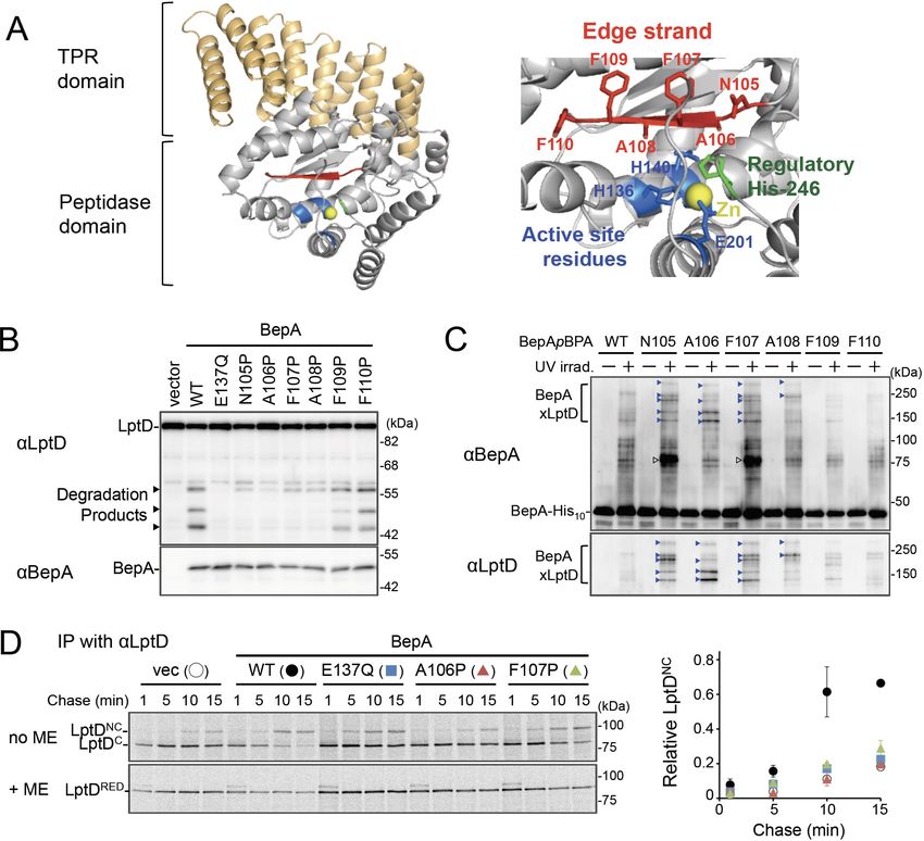
Research article Biochemistry and Chemical Biology Cell Biology
have shown that certain BamA mutations render all other BAM subunits dispensable (Hart et al.,
2020). The BAM complex has a silk-hat-like structure (Bakelar et al., 2016; Gu et al., 2016;
Han et al., 2016; Iadanza et al., 2016; Tomasek et al., 2020); the OM-embedded C-terminal b-bar-
rel domain of BamA forms the ‘crown’ and the N-terminal periplasmic polypeptide transport-associ-
ated (POTRA) domains of BamA form the ‘brim’ together with the BamB/C/D/E lipoproteins.
Lipopolysaccharide (LPS), another major OM constituent localized in the outer leaflet of the OM,
is also important for the maintenance of the structure and function of the OM (Sperandeo et al.,
2017). LPS is synthesized on the cytoplasmic side of the IM and flipped across the IM to the peri-
plasm. After maturation, it is transported to the OM by the LPS transport (Lpt) proteins
(Sperandeo et al., 2017). A heterodimer of LptD, a b-barrel OMP, and LptE, a lipoprotein, plays
roles in the final step to insert LPS into the OM (Dong et al., 2014; Qiao et al., 2014; Wu et al.,
2006). The OM assembly process of LptD is unique in that it is accompanied by the rearrangement
of intramolecular disulfide bonds. The mature form of LptD possesses two disulfide bonds formed
by non-consecutive pairs of Cys residues (C31–C724 and C173–C725) (Ruiz et al., 2010). It has been
shown, however, that an assembly intermediate of LptD having disulfide bonds formed by consecu-
tive pairs of the Cys residues (C31–C173 and C724–C725) (LptDC; LptD with Consecutive disulfide
bods) is first generated and isomerized to LptDNC (LptD with Non-Consecutive disulfide bonds) dur-
ing its assembly/maturation, which is triggered by the association of LptD with LptE (Chng et al.,
2012; Narita et al., 2013). The LptDC to LptDNC conversion should occur at a later step in the OM
assembly because LptD presumably associates with LptE on the BAM complex
(Chimalakonda et al., 2011; Chng et al., 2012; Lee et al., 2016; Narita et al., 2013).
BepA (formally called YfgC), a bi-functional periplasmic protein that plays an important role in
maintaining OM integrity (Narita et al., 2013), belongs to the M48 family zinc-metallopeptidases
that include prokaryotic and eukaryotic proteases (such as Ste24, Oma1, and HptX) involved in mem-
brane quality control (Rawlings et al., 2018). We have previously shown that BepA is involved in the
biogenesis and quality control of LptD. While BepA promotes the LptDC to LptDNC conversion
(chaperone-like function) (Narita et al., 2013), it also degrades the stalled or misassembled LptDC
molecules that are generated due to an lptD mutation (lptD4213) or decreased availability of or
weakened interaction with LptE (protease function) (Narita et al., 2013; Soltes et al., 2017). BepA
also degrades BamA whose assembly/folding has been impaired in the absence of a periplasmic
chaperone, SurA (Daimon et al., 2017), suggesting that BepA can also act in quality control of some
other OM proteins. The BepA protein consists of an N-terminal M48 metallopeptidase domain and a
C-terminal tetratricopeptide repeat (TPR) domain that are associated closely to form a compact
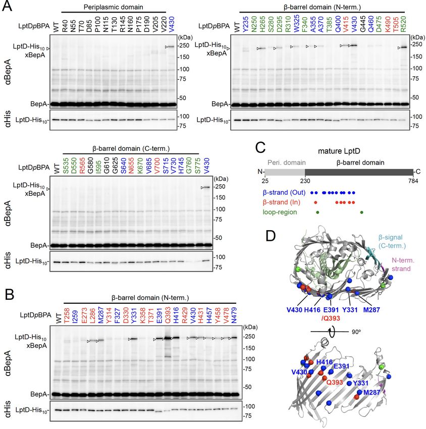
structure (Figure 1A; Bryant et al., 2020; Shahrizal et al., 2019). Our previous study suggested
that the TPR domain of BepA directly interacts with LptD and with the BAM complex with its TPR
domain inserted into the interior of the periplasmic part (brim) of the BAM complex (Daimon et al.,
2017; Narita et al., 2013). A mutational study has suggested that these interactions are important
for BepA functions (Daimon et al., 2017). Recent studies have also shown that the His-246 residue
of BepA that coordinates the zinc ion at the proteolytic active site acts as an ON/OFF switch (His
switch) for the proteolytic activity of BepA (Bryant et al., 2020; Daimon et al., 2020). The dual func-
tions of BepA should be appropriately regulated because the unregulated expression of the proteo-
lytic activity of BepA caused by an H246 mutation leads to the degradation of the normally
assembling LptD intermediate (Daimon et al., 2020). However, information on the molecular mecha-
nism of this regulation and the modes of the BepA–LptD interaction in each BepA function remains
elusive.
Here, we investigated the mechanism by which BepA established interaction with LptD in
the promotion of its assembly and degradation. Our results showed that a conserved b-strand
(edge-strand) located adjacent to the BepA active site directly contacts with LptD and plays impor-
tant roles in substrate proteolysis, like many other proteases. In addition, we unexpectedly found
that the edge-strand-mediated interaction with a substrate is also required for the chaperone-like
function of BepA, which should be enabled by the His switch-mediated repression of the proteolytic
activity. Crosslinking experiments demonstrated that BepA could interact with an LptD molecule
assembling on the BAM complex. Based on these observations, we propose a model explaining the
edge-strand and His switch-mediated functional regulation of BepA in LptD assembly/degradation.
Miyazaki et al. eLife 2021;10:e70541. DOI: https://doi.org/10.7554/eLife.70541 2 of 21
Research article                                                                           Biochemistry and Chemical Biology Cell Biology

Figure 1. The edge-strand of BepA is crucial for functional interaction with LptD. (A) Crystal structure of BepA (PDB code: 6AIT). The peptidase and the
TPR domains of BepA are shown in gray and orange, respectively. The edge-strand, the proteolytic active site (the HExxH motif and the third zinc
ligand, Glu-201), and the regulatory His-246 residue (His switch) in the peptidase domain are shown in red, blue, and green, respectively, and the
coordinated zinc atom is shown in yellow. An enlarged view of the active site region is shown in right. (B) Protease activities of the BepA edge-strand
mutants. Cells of SN56 (DbepA) carrying pTWV228-lptD-his10 and either pSTD689 or pSTD689-bepA plasmids were grown at 30˚C in L-medium until
early log phase and induced with 1 mM IPTG for 1 hr. Total cellular proteins were acid-precipitated and analyzed by 7.5 or 10% Laemmli SDS-PAGE
and immunoblotting with the indicated antibodies. (C) In vivo photo-crosslinking analysis of the BepA edge-strand. Cells of SN56 carrying pEVOL-pBpF
and pUC18-bepA(E137Q, amb)-his10 plasmids were grown at 30˚C in L-medium containing 0.02% arabinose and 0.5 mM pBPA until early log phase,
and induced with 1 mM IPTG for 1 hr to express the indicated BepA(pBPA) variants. The cultures were divided into two portions, each of which was
treated with or without UV-irradiation for 10 min at 4˚C. Proteins of the total membrane fractions were subjected to pull-down with Ni-NTA agarose.
Purified proteins were analyzed by 7.5% Laemmli SDS-PAGE and immunoblotting with the indicated antibodies. Open triangles indicate unknown
crosslinked products. (D) Chaperone-like activities of the BepA edge-strand mutants. Cells of SN56 carrying pSTD689 or a pSTD689-bepA plasmid were
grown at 30˚C in M9-based medium until early log phase, induced with 1 mM IPTG for 15 min, pulse-labeled with 35S-Met for 1 min and chased for the
indicated periods. At each time point, total cellular proteins were acid-precipitated, subjected to IP with an anti-LptD antibody, and analyzed by 7.5%
Laemmli SDS-PAGE followed by phosphorimaging. The ratio of the band intensities of LptDNC at each time point to that of total LptD (LptDC+LptDNC)
at 5 min was quantitated and the mean values were plotted with S.D. (n=2). The result shown is a representative of two independent experiments that
were conducted using the same transformants (i.e., two technical replicates). See Figure 1—source data 1 for gel images and quantitated band
intensities data for (D).
The online version of this article includes the following source data and figure supplement(s) for figure 1:
Source data 1. A Zip file containing gel images.
Figure supplement 1. Sequence alignment of BepA homologs and Escherichia coli M48 family peptidases.
Figure supplement 2. Effects of the BepA edge-strand Pro mutations on the BamA degradation and the BepA self-cleavage.
Figure supplement 2—source data 1. A Zip file containing gel images (A–C) for the immunoblotting experiments using the anti-BepA and anti-LptD
antibodies.
Figure supplement 3. Functionality of the BepA derivatives having pBPA in the edge-strand.
Figure 1 continued on next page

Miyazaki et al. eLife 2021;10:e70541. DOI: https://doi.org/10.7554/eLife.70541                                                                   3 of 21
Research article                                                                     Biochemistry and Chemical Biology Cell Biology

Figure 1 continued
Figure supplement 3—source data 1. A Zip file containing plate images (A) and gel images (B) for the immunoblotting experiments using the anti-
BepA antibody.
Figure supplement 4. The chaperone-like and proteolytic activities of the BepA edge-strand mutants.
Figure supplement 4—source data 1. A Zip file containing gel images (A, B) for the immunoblotting experiments using the anti-BepA and anti-BamA
antibodies.

                                    Results
                                    Interaction of the BepA edge-strand with LptD is crucial not only for
                                    proteolysis but also for assembly promotion of LptD by BepA
                                    Zinc-metallopeptidases usually possess a b-strand, called edge-strand, located close to their proteo-
                                    lytic active sites (Akiyama et al., 2015; López-Pelegrı́n et al., 2013; Stöcker and Bode, 1995).
                                    While the edge-strand is known to play a critical role in substrate proteolysis by directly interacting
                                    with a substrate polypeptide by the strand addition mechanism and converts it into an extended
                                    conformation for its presentation to the active site and proteolysis, it has not been
                                    well characterized in M48 proteases. The solved structures of Escherichia coli BepA (Bryant et al.,
                                    2020; Shahrizal et al., 2019) show that it has a b-strand (b2) that is conserved among the M48-pep-
                                    tidases and is located adjacent to the active site (Figure 1A and Figure 1—figure supplement 1),
                                    suggesting that this strand presumably acts as an edge-strand. To examine the role of the b2-strand
                                    in BepA functions, we constructed BepA mutants by introducing Pro at each position in b2 (from
                                    Asn-105 to Phe-110; Figure 1A). We then investigated the effects of b2 mutations on the proteolytic
                                    activity of BepA against overproduced LptD. When LptD is overproduced from a multi-copy plasmid,
                                    it mainly accumulates in the form of LptDC possibly due to the limited availability of its partner pro-
                                    tein, LptE (Daimon et al., 2020; Daimon et al., 2017). This species probably represents a ‘normal’
                                    assembly intermediate as it is associated with the BAM complex (see below) and can be converted
                                    to the mature form (LptDNC) when LptE is co-expressed (Miyazaki et al., 2018). As reported previ-
                                    ously (Daimon et al., 2020; Daimon et al., 2017), overproduced LptD was degraded by co-
                                    expressed wild-type BepA to generate discrete degradation products (Figure 1B). To examine the
                                    possible roles of the b2-strand in the function of BepA, we introduced a Pro substitution into the b2-
                                    strand, as a Pro residue would affect the secondary structure, and thus the function, of this strand.
                                    We found that the expression of a few BepA mutants (N105P, A106P, F107P, and A108P) led to a
                                    significantly decreased generation of the LptD degradation products (Figure 1B). Furthermore,
                                    some of these mutations compromised the degradation of BamA in a DsurA strain (Daimon et al.,
                                    2017) and the self-cleavage of BepA-His10 (BepA possessing a C-terminal His10-tag) within the His10-
                                    tag (Narita et al., 2013; Figure 1—figure supplement 2A and B). These results strongly suggest
                                    that the b2-strand is important for the proteolytic activity of BepA.
                                        We next analyzed whether the b2-strand directly interacts with a substrate by using a site-
                                    directed in vivo photo-crosslinking approach (Chin and Schultz, 2002; Miyazaki et al., 2020a). We
                                    expressed derivatives of BepA-His10 harboring a photoreactive amino acid analog, p-benzoyl-L-phe-
                                    nylalanine (pBPA), at each position in the b2-strand in a DbepA strain and examined their comple-
                                    mentation activity regarding erythromycin (EM) sensitivity of the cell and self-cleavage of BepA-
                                    His10. The results showed that certain mutants (F107pBPA, F109pBPA, and F110pBPA) were as func-
                                    tional as the wild-type, but others exhibited neither significant complementation activity nor self-
                                    cleavage (Figure 1—figure supplement 3A and B). For the photo-crosslinking experiments con-
                                    ducted in this study, we used BepA variants harboring the E137Q mutation in the H136ExxH motif
                                    for two reasons. First, this mutation would repress the possible degradation of LptD by the BepA
                                    derivatives. Second, our previous study has shown that this mutation stabilizes the interaction of
                                    BepA with LptD (Daimon et al., 2017). Following UV-irradiation of cells expressing each of the
                                    BepA mutants, BepA-His10 and its crosslinked products were purified from the membrane fractions
                                    by affinity isolation using Ni-NTA agarose and subjected to immunoblotting analysis (Figure 1C).
                                    The N105pBPA, A106pBPA, F107pBPA, and A108pBPA derivatives of BepA generated, in a UV-
                                    dependent manner, evident crosslinked products that reacted with both anti-BepA and anti-LptD
                                    antibodies. Taken together, these results imply that the b2-strand of BepA directly interacts with
                                    LptD and plays an important role in their degradation. The structure, intramolecular disposition, as

Miyazaki et al. eLife 2021;10:e70541. DOI: https://doi.org/10.7554/eLife.70541                                                          4 of 21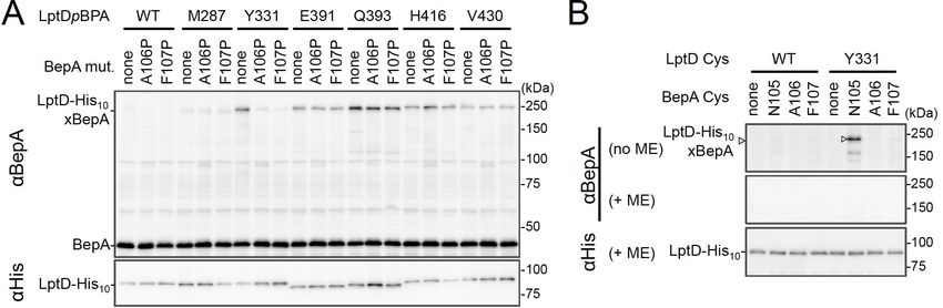
Research article                                                                  Biochemistry and Chemical Biology Cell Biology

                                    well as involvement in substrate interaction and proteolysis of the b2-strand strongly supports the
                                    hypothesis that it indeed acts as the edge-strand to recognize and present a substrate to the active
                                    site. We have thus referred to the b2-strand as the ‘edge-strand’ hereafter. BepA N105pBPA and
                                    F107pBPA derivatives also generated the unidentified crosslinked products (Figure 1C, open arrow-
                                    heads), suggesting that the edge-strand would interact with some other substrates. The differential
                                    effects of the mutations on the degradation of LptD and BamA and the self-cleavage of the C-termi-
                                    nal tag (Figure 1B and Figure 1—figure supplement 2A and B) might reflect their different interac-
                                    tion properties and/or affinities for the BepA edge-strand. Our previous study also suggested
                                    substrate-specific interaction of an edge-strand for another metallopeptidase, RseP (Akiyama et al.,
                                    2015).
                                        BepA not only degrades the stalled or misassembled LptD (when LptD is on the off-pathway) but
                                    also promotes the maturation of LptD through facilitating its assembly with the partner protein LptE
                                    (chaperone-like activity) (on the normal (on-) pathway) (Narita et al., 2013). Therefore, we examined
                                    the effects of the edge-strand Pro mutations on the chaperone-like activity of BepA. LptD possesses
                                    two intramolecular disulfide bonds. While the mature (fully assembled) LptD has non-consecutive
                                    disulfide bonds, an assembly intermediate form of LptD (LptDC) that accumulates in the absence of
                                    functional BepA possesses consecutive disulfide bonds. Consistent with our previous results
                                    (Narita et al., 2013), the expression of the wild-type BepA in a DbepA strain markedly decreased
                                    the accumulation of LptDC (Figure 1—figure supplement 4A). On the contrary, only a partial
                                    decrease in accumulation was observed with the expression of the E137Q mutant. We found that
                                    the A106P mutant was defective in reducing LptDC accumulation, suggesting that a mutation in the
                                    edge-strand could affect the chaperone-like function (Figure 1—figure supplement 4A). Subse-
                                    quently, we examined the effects of two edge-strand mutations—A106P and F107P—that markedly
                                    compromised LptD degradation on the chaperone-like function of BepA using pulse-chase experi-
                                    ments. Pulse-chase analysis showed that LptD was largely stable, but received a slight degradation
                                    during the chase period under the condition used (Figure 1D, left panel, +ME). It is likely that some
                                    of LptDC was degraded by BepA during the chase period, because LptDNC is largely BepA-resistant
                                    (Daimon et al., 2020). Thus, the ratio of LptDNC to the total LptD (LptDC+LptDNC) at each chase
                                    point does not exactly reflect the efficiency of the LptDC-to-LptDNC conversion of pulse-labeled
                                    LptD. To examine only the chaperone-like activities (promotion of the LptDNC generation) without
                                    the possible effect of the LptD degradation by the residual proteolytic activities of the A106P and
                                    the F107P, the relative amounts of LptDNC at each chase point to the total LptD (LptDC+LptDNC) at
                                    an early chase point (5 min) were calculated and plotted as quantified data (Figure 1D, right panel).
                                    The expression of the wild-type BepA in DbepA cells significantly accelerated the conversion of
                                    LptDC to LptDNC, whereas the expression of the protease-dead E137Q mutant demonstrated weak-
                                    ened conversion (Figure 1D). The acceleration of the LptDC to LptDNC conversion by the F107P and
                                    the A106P mutants was much weaker than that by the wild-type BepA (almost the same as com-
                                    pared with the E137Q mutant) (Figure 1D). These results unexpectedly suggested that the edge-
                                    strand was also important for the chaperone-like activity of BepA.
                                        We constructed BepA derivatives with a Cys substitution (note that BepA intrinsically possesses
                                    no Cys residue) at the position of Asn-105, Ala-106, or Phe-107 in the edge-strand to examine
                                    whether a specific residue in this strand is required for the BepA’s functions. We chose a Cys substi-
                                    tution, because (i) the Cys mutants can be used in the disulfide crosslinking experiments described
                                    below, and (ii) a previous study strongly suggested that a Cys mutation does not affect the second-
                                    ary structure of an edge-strand in another protease RseP (Akiyama et al., 2015). These BepA Cys
                                    constructs exhibited almost normal chaperone-like and proteolytic functions (Figure 1—figure sup-
                                    plement 4B and C). This supports the idea that the secondary structure of the edge-strand is more
                                    important than the individual amino acid residues for its function, although some contribution of the
                                    side-chains of the amino acids to the functions of the edge-strand cannot be excluded. Note that,
                                    although it is possible that a Pro mutation also affects the structures of the overall BepA protein
                                    and/or the active site around the edge-strand, the edge-strand Pro mutants (other than F107P) still
                                    exhibited significant self-cleavage of the probably unstructured-terminal tag (Figure 1—figure sup-
                                    plement 2B). In addition, the Pro mutants (other than A106P) degraded mis- or un-folded BamA at
                                    a detectable level (Figure 1—figure supplement 2B). Together with the result that these mutants
                                    accumulated at a level comparable to that of wild-type BepA, the above observations suggest that

Miyazaki et al. eLife 2021;10:e70541. DOI: https://doi.org/10.7554/eLife.70541                                                      5 of 21
Research article Biochemistry and Chemical Biology Cell Biology
most of the Pro mutations specifically affected the edge-strand structure, but not drastically altered
the active site or the protein’s overall structures.
BepA interacts with the N-terminal half of the b-barrel-forming domain
of the LptD assembly intermediate
While BepA interacts with LptD to promote either its proper OM assembly or proteolytic elimination
depending on the situation (Narita et al., 2013), the details of the BepA–LptD interaction, including
the region(s) in LptD to which BepA binds, remain largely unknown. Thus, we performed a system-
atic photo-crosslinking analysis to identify the BepA-contact region in the LptD assembly intermedi-
ate LptDC. We performed photo-crosslinking experiments in cells ectopically co-expressing an LptD
derivative containing a photo-reactive amino acid analog pBPA [LptD(pBPA)] and a protease-dead
variant of BepA, BepA(E137Q). We first introduced pBPA at each of the 50 positions (approximately
every 15 residues) in the mature part of LptD and performed photo-crosslinking analysis. Cells
expressing LptD(pBPA)-His10 and BepA(E137Q) were grown and UV-irradiated, and the total cellular
proteins were analyzed by immunoblotting with anti-BepA and anti-His antibodies. Under this condi-
tion, expressed LptD-His10 was considerably accumulated as LptDC (Figure 2—figure supplement
1) irrespective of co-expression of BepA(E137Q). We detected clear crosslinking with BepA mainly in
the N-terminal half of the LptD b-barrel-forming domain (Figure 2A). We then performed a detailed
photo-crosslinking analysis for the 20 additional sites in the N-terminal half of the LptD b-barrel-
forming domain (Figure 2B) and found that BepA was crosslinked at several of these sites. The resi-
dues at the BepA-cross-linkable sites were oriented both inward and outward in the mature LptD b-
barrel (Figure 2C and D). Moreover, the residue Gln-393 at which the strongest crosslinking was
observed was oriented inward. These results suggest that LptD while interacting with BepA would
not assume a higher-order structure like closed b-barrel (see Discussion). We selected a few LptD
(pBPA) derivatives that had been crosslinked with BepA as representatives and examined their func-
tionality. They supported the growth of LptD-depleted cells when expressed from a plasmid, indicat-
ing that they were functional (Figure 2—figure supplement 2). The above-mentioned crosslinking
results thus likely reflect a functional interaction of LptD with BepA in the normal assembly pathway.
BepA edge-strand directly interacts with the Tyr-331 residue in the b7
strand of the LptD b-barrel domain
We investigated further to identify the region of LptD that interacts with the BepA edge-strand.
First, we examined the effects of the BepA edge-strand Pro mutations (F107P and A106P) on the
LptD(pBPA)–BepA crosslinking. The F107P mutation significantly decreased the efficiency of the
crosslinking of BepA(N105pBPA) and BepA(A106pBPA) with LptD (Figure 3—figure supplement 1).
Additionally, the A106P mutation exhibited a similar effect on the crosslinking of BepA(N105pBPA)
with LptD. Based on these effects on crosslinking, we inferred that these mutations affected the
interaction of the edge-strand with LptD. Subsequently, we selected several LptD(pBPA) derivatives
that showed relatively strong crosslinking with BepA and examined the effect of F107P and A106P
mutations in the BepA edge-strand on the crosslinking of LptD(pBPA) with BepA. We found that
these mutations altered LptD–BepA crosslinking in a site-specific manner. Further, the amount of
crosslinked products markedly decreased for LptD(Y331pBPA), but not for other mutants
(Figure 3A). These results strongly suggest that the region around Tyr-331 in the b7 strand of the
LptD b-barrel domain (Figure 2D) is crosslinked with the edge-strand of BepA. Note that, while we
detected LptD-BepA crosslinked products ranging from 150 to 250 kDa with BepA derivatives hav-
ing pBPA in the edge-strand (Figure 1C), we did not observe similar multiple crosslinked products
with LptD(Y331pBPA) (Figure 2). The exact reason for this is unknown, but it might be ascribed to
the crosslinking of pBPA in the BepA edge-strand to different LptD positions that are close spatially
but distant in the primary sequence, which could generate crosslinked products with different mobil-
ity (i.e., different apparent sizes).
To further confirm the direct interaction of the BepA edge-strand with the LptD b7 strand, we
conducted site-specific disulfide crosslinking experiments. For this analysis, we used the above-
described single Cys derivatives of BepA harboring a Cys residue at the position of Asn-105, Ala-
106, or Phe-107, and derivatives of LptD having a Cys substitution at either of the six positions,
including Tyr-331 at which introduction of pBPA showed clear crosslinking with BepA (Figure 3A).
Miyazaki et al. eLife 2021;10:e70541. DOI: https://doi.org/10.7554/eLife.70541 6 of 21Research article Biochemistry and Chemical Biology Cell Biology Figure 2. Photo-crosslinking of the b-barrel forming domain of LptD with BepA. (A, B) In vivo photo-crosslinking between LptD and BepA. Cells of RM2243 (bepA(E137Q)) carrying pEVOL-pBpF, pMW118-bepA(E137Q), and pRM294-lptD(amb)-his10 plasmids were grown at 30˚C in L-medium containing 0.02% and 0.5 mM pBPA until early log phase and induced with 1 mM IPTG for 3 hr to express the indicated LptD(pBPA) variants. The cultures were then divided into two portions, each of which was UV-irradiated for 10 min at 4˚C. Total cellular proteins were acid-precipitated and analyzed by 7.5% Laemmli SDS-PAGE and immunoblotting with the indicated antibodies. Most of the LptD mutants were accumulated in comparable amounts. LptD-His10xBepA crosslinked products were not detectable with an anti-His antibody due to its low reactivity to LptD-His10 in this and the following experiments. Amino acid residues shown in red and blue indicate the ones whose side chain is pointing inward and outward, respectively. Amino acid residues shown in green indicate the ones located in the loop regions. The result shown is a representative of two technical replicates. (C) Summary of the BepA crosslinked positions in LptD. Positions where the crosslinking with BepA was clearly and reproducibly detected are indicated by colored dots. (D) Mapping of the BepA crosslinked positions on the barrel domain of LptD in the Escherichia coli LptD–LptE structure (PDB code: 4RHB). LptD and LptE are shown in gray and light green, respectively. The N-terminal strand and the b-signal (C-terminal region) in the LptD b-barrel domain are shown in magenta and light blue, respectively. The top view of the LptD/E structure from extracellular space (upper) and the side view of the N-terminal region of LptD b-domain (lower) are shown. The positions where the crosslinking with BepA was observed were indicated by spheres colored as above. See Figure 2—source data 1 for gel images for (A, B). The online version of this article includes the following source data and figure supplement(s) for figure 2: Source data 1. A Zip file containing gel images (A, B) for the immunoblotting experiments using the anti-BepA and anti-His tag antibodies. Figure supplement 1. Overexpressed LptD molecules mainly accumulate as LptDC. Figure supplement 1—source data 1. A Zip file containing gel images for the immunoblotting experiments using the anti-LptD, anti-BepA, and anti- BamA antibodies. Figure supplement 2. Complementation activity of the LptD(pBPA) derivatives. Figure supplement 2—source data 1. A Zip file containing plate images for the complementation assays for the LptD(pBPA) derivatives. Miyazaki et al. eLife 2021;10:e70541. DOI: https://doi.org/10.7554/eLife.70541 7 of 21
Research article                                                                         Biochemistry and Chemical Biology Cell Biology

Figure 3. BepA edge-strand directly contacts with the Tyr-331 residue in the N-terminal half region of the LptD b-barrel forming domain. (A) Effect of
the BepA edge-strand mutations on the crosslinking between BepA and the LptD derivatives having pBPA in the N-terminal half region of the LptD b-
barrel-forming domain. Cells of SN56 (DbepA) carrying pEVOL-pBpF, pMW118-bepA(E137Q, mut), and pRM294-lptD(amb)-his10 were grown, induced
to express a BepA and a LptDpBPA derivative, and subjected to photo-crosslinking analysis as described in Figure 2. (B) Disulfide crosslinking between
the Cys residues in the edge-strand of BepA and the N-terminal half region of the LptD b-barrel-forming domain. Cells of SN56 (DbepA) carrying a
combination of plasmids encoding WT or a Cys-introduced mutant of BepA and LptD-His10 as indicated were grown in L-medium and induced with 1
mM IPTG for 3 hr to express BepA(Cys) and LptD(Cys)-His10. Total cellular proteins were acid-precipitated, solubilized with SDS buffer containing NEM
(for blocking free thiol groups), and subjected to pull-down with Ni-NTA agarose. The purified proteins were treated with or without 2-mercaptoethanol
(ME) and analyzed by 7.5% Laemmli SDS-PAGE and immunoblotting with the indicated antibodies. The result shown is a representative of two technical
replicates. See Figure 3—source data 1 for gel images for (A, B).
The online version of this article includes the following source data and figure supplement(s) for figure 3:
Source data 1. A Zip file containing gel images (A, B) for the immunoblotting experiments using the anti-BepA and anti-His tag antibodies.
Figure supplement 1. Effects of the BepA edge-strand mutations on the crosslinking of the BepA edge-strand to LptD.
Figure supplement 1—source data 1. A Zip file containing gel images for the immunoblotting experiments using the anti-LptD, anti-BepA, and anti-
BamA antibodies.
Figure supplement 2. Disulfide crosslinking between BepA and LptD.
Figure supplement 2—source data 1. A Zip file containing plate images (A) for the complementation assays for the LptD(Cys) derivatives and gel
images (B) for the immunoblotting experiments using the anti-BepA and anti-His antibodies.

                                    The wild-type LptD protein harbors intrinsic four Cys residues that form two disulfide bonds essential
                                    for the LptD function; therefore, each of these Cys-substituted LptD mutants possessed five Cys resi-
                                    dues in total. We confirmed that these Cys-substituted LptD derivatives accumulated normally and
                                    retained their function (Figure 3—figure supplement 2). Cells expressing a combination of BepA
                                    Cys mutants and LptD Cys mutants were grown, and total proteins were acid-denatured and dis-
                                    solved in SDS containing N-ethylmaleimide (NEM; NEM was included to block free Cys residues).
                                    Then, LptD-His10 and its crosslinked products were affinity-isolated using the C-terminal His10-tag,
                                    treated with or without 2-mercaptoethanol (ME), and analyzed by SDS-PAGE and anti-BepA immu-
                                    noblotting. We observed that certain combinations of BepA and LptD derivatives showed a high-
                                    molecular-mass band in electrophoresis results. Among them, the combination of BepA(N105C) and
                                    LptD(Y331C) showed the most intense band that exhibited reaction with the anti-BepA antibody
                                    (Figure 3B and Figure 3—figure supplement 2B, no ME). These high-molecular-mass bands were
                                    not observed with the wild-type LptD (no additional Cys) and disappeared upon treatment with ME,
                                    suggesting that they were disulfide-crosslinked products (Figure 3B and Figure 3—figure supple-
                                    ment 2B, + ME). These results are consistent with the photo-crosslinking experiments (Figure 3A)
                                    and indicate that the edge-strand of BepA can directly bind to several regions in the N-terminal half
                                    of the LptD b-barrel-forming domain, which includes the b7 strand containing Tyr-331.

Miyazaki et al. eLife 2021;10:e70541. DOI: https://doi.org/10.7554/eLife.70541                                                                 8 of 21
Research article Biochemistry and Chemical Biology Cell Biology
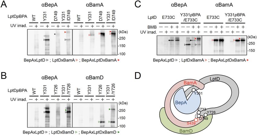
BepA interacts with an LptD intermediate associating with the seam
region of BamA on the BAM complex
We further investigated the mode of the interaction of the BepA-associated LptD with the BAM
complex. It has been recently shown that LptD4213, a mutant form of LptD that has a short (23
amino acids) deletion in an extracellular loop (eL4) and is stalled on the BAM complex mimicking a
late assembly complex (Lee et al., 2016), interacts with the seam region formed by the N- and
C-terminal b-strands (b1 and b16, respectively) in the BamA barrel domain and forms a hetero-com-
plex, in which the C-terminal b-signal of the LptD4213 was associated with the b1 strand of the
BamA seam (Lee et al., 2019). It has been suggested that the interaction of the b-signal with the
BamA b1 strand generally facilitates the folding of the b-barrel domain of a substrate OMP (see
Figure 4D and 5B; Tomasek and Kahne, 2021). We first examined the interaction of the BepA-asso-
ciated LptD intermediate with BamA and BamD by conducting in vivo photo-crosslinking experi-
ments using the LptD derivatives with pBPA at the position of Glu-749 or Tyr-726 in addition to the
position of Tyr-331. pBPA at Glu-749 and Tyr-726 residue, both of which are located near the b-sig-
nal of LptD, have been reported to be crosslinked with BamA and BamD, respectively, during the
LptD assembly (Figure 4D; Lee et al., 2019; Lee et al., 2018). Complementation assay results
showed that either of the LptD derivatives containing one or two pBPA at Glu-749 and Tyr-726 were
functional (Figure 4—figure supplement 1). After UV-irradiation of the cells expressing LptD-His10
and the pBPA derivatives, LptD-His10 and its crosslinked products were affinity-isolated from the
membrane fractions and analyzed by immunoblotting. The single pBPA derivatives indeed gener-
ated crosslinked products with the expected factors as: LptD(Y331pBPA) with BepA, LptD
(D749pBPA) with BamA, and LptD(Y726pBPA) with BamD (Figure 4A and B). With the double pBPA
derivatives LptD(Y331/D749pBPA) and LptD(Y331/Y726pBPA), new crosslinked products with higher
molecular sizes were observed in addition to the ones observed with single pBPA mutants
(Figure 4A and B). These results showed that the higher molecular-sized products represented
BepA–LptD(Y331/D749pBPA)–BamA and BepA–LptD(Y331/Y726pBPA)–BamD crosslinked products
and that BepA interacts with an assembly intermediate of LptD on the BAM complex.
We further conducted photo-crosslinking experiments using LptD locked on the BAM complex by
using an SH-crosslinker, 1,4-bismaleimidobutane (BMB). It has been previously shown (Lee et al.,
2019) that a cysteine placed near the N-terminal b-strand of the BamA b-barrel (S439C) was cross-
linked with a cysteine introduced near the b-signal of LptD (E733C) via BMB treatment (Figure 4D;
Figure 4—figure supplement 2A). We introduced a Y331pBPA mutation into LptD(E733C) and con-
firmed that the resultant mutant was functional (Figure 4—figure supplement 2B). When the LptD
(Y331pBPA/E733C) mutant was expressed in a strain having a chromosomal bamA(S439C) mutant
gene, LptD–BepA crosslinked products were detected upon UV-irradiation, whereas LptD–BamA
crosslinked products was detected upon BMB treatment. When cells were first treated with BMB
and then UV-irradiated, a higher mass product that reacted with both anti-BepA and anti-BamA anti-
bodies was generated. The generation of this product depended on both BMB-treatment and UV-
irradiation (Figure 4C). These results are fully consistent with the above photo-crosslinking results
and further demonstrated that BepA could interact with an LptD assembly intermediate associating
with the seam site of BamA on the BAM complex.
Discussion
The involvement of BepA in the maintenance of structural and functional integrity of the OM was
first suggested on the basis of increased sensitivities of the DbepA(yfgC) strain to several antibiotics
and chemicals, which was similar to the characteristics of strains with a disruption of genes encoding
proteins engaged in outer membrane biogenesis (Narita et al., 2013; Ruiz et al., 2005;
Tamae et al., 2008). We have previously shown that BepA is involved in the biogenesis and quality
control of LptD, probably on the BAM complex (Daimon et al., 2020; Daimon et al., 2017;
Narita et al., 2013). However, the mechanism by which BepA recognizes and interacts with LptD
remains elusive. This information is important to understand the mechanism of BepA to distinguish
between the normal (on-) and off-pathway intermediates of LptD that are either assembled into the
OM or degraded, respectively.
To gain insight into the BepA–LptD interaction, we examined the role of the conserved edge-
strand of BepA in its function. Our results showed that the BepA edge-strand participates not only
Miyazaki et al. eLife 2021;10:e70541. DOI: https://doi.org/10.7554/eLife.70541 9 of 21Research article Biochemistry and Chemical Biology Cell Biology Figure 4. BepA interacts with an LptD intermediate assembling on the BAM complex. (A, B) In vivo photo-crosslinking of an LptD mutant having pBPA at two positions with BepA. Cells of RM2243 (bepA(E137Q)) carrying pEVOL-pBpF, pMW118-bepA(E137Q) and pRM294-lptD(amb)-his10 plasmids were grown at 30˚C in L-medium containing 0.5 mM pBPA until early log phase and induced with 1 mM IPTG for 3 hr to express the indicated LptD(pBPA) variants. The cultures were divided into two portions, each of which was treated with or without UV-irradiation for 30 min at 4˚C. Proteins of the total membrane fractions were subjected to pull-down with Ni-NTA agarose. Purified proteins were analyzed by 7.5% Laemmli SDS-PAGE by immunoblotting with the indicated antibodies. Asterisk in the anti-BepA blots possibly indicates an LptD-BamA crosslinked product that was detected due to the apparent cross-reactivity of the anti-BepA antibody with the LptDxBamA crosslinked product (see below). (C) Simultaneous crosslinking of LptD having Y331pBPA and E733C with the BepA edge-strand and the seam region of BamA(S439C). Cells of RM3655 (bamA(S439C), DbepA)/pEVOL- pBpF/pMW118-bepA(E137Q) carrying pRM294-lptD(E733C)-his10, or pRM294-lptD(Y331amb, E733C)-his10 were grown and induced as in (A). After treatment with or without BMB and the following quenching of BMB by addition of excess cysteine, the cultures were divided into two portions, each of which was treated with or without UV-irradiation for 30 min at 4˚C. Total cellular proteins were acid-precipitated, solubilized with SDS buffer containing NEM, and subjected to pull-down with Ni-NTA agarose. Purified proteins were analyzed by 7.5% Laemmli SDS-PAGE and immunoblotting with the indicated antibodies. The anti-BamA immunoblotting showed that the amount of the BepAxLptDxBamA crosslinked product was much lower than that of the LptDxBamA crosslinked product. Although the anti-BepA antibodies apparently cross-reacted weakly with the LptDxBamA crosslinked products (closed circles), the higher signal intensity of the BepAxLptDxBamA crosslinked product band as compared with the intensity of the LptDxBamA band (closed circles) indicate that the detection of the former band with the anti-BepA antibodies cannot be ascribed to this cross-reactivity. The identities of the bands marked by open circles in (A, B) are unclear; they might represent BepA-LptD crosslinked products or BepA-BamA crosslinked products (detected due to the cross-reactivity of anti-BepA antibodies with LptDxBamA crosslinked products as described above). In (A–C), we confirmed that the amounts of the isolated non-crosslinked LptD-His10 derivatives were roughly equal by CBB staining or anti-His immunoblotting (Figure 4—figure supplement 3). The result shown is a representative of two technical replicates. (D) A schematic cartoon of the interaction of the LptD assembly intermediate with BepA and BamA/D on the BAM complex. See Figure 4—source data 1 for gel images for (A–C). The online version of this article includes the following source data and figure supplement(s) for figure 4: Source data 1. A Zip file containing gel images (A–C) for the immunoblotting experiments using the anti-BepA, anti-BamA, and anti-BamD antibodies. Figure supplement 1. Complementation activity of the LptD derivatives having pBPA at one or two positions. Figure supplement 1—source data 1. A Zip file containing plate images for the complementation assays for the LptD(pBPA) derivatives. Figure supplement 2. BMB crosslinking between LptD(Y331pBPA/E733C) and BamA(S439C). Figure supplement 2—source data 1. A Zip file containing gel images (A) for the immunoblotting experiments using the anti-BamA and anti-His anti- bodies and plate images (B) for the complementation assays for the LptD(pBPA/Cys) derivatives. Figure supplement 3. The amount of affinity purified LptD-His10 derivatives. Figure supplement 3—source data 1. A Zip file containing stained gel images. Miyazaki et al. eLife 2021;10:e70541. DOI: https://doi.org/10.7554/eLife.70541 10 of 21
Research article                                                                          Biochemistry and Chemical Biology Cell Biology

Figure 5. Model for the substrate recognition and discrimination by BepA. (A) A schematic cartoon of the substrate recognition by BepA at its active
site. See the text for details. (B) An overview of the proposed LptD assembly process and BepA-mediated discrimination of the assembling and stalled
LptD species. See the text for details. Association of BepA with the assembly intermediate form of LptD on the BAM complex could transiently stabilize
the LptD assembly intermediate and facilitate the association of LptE with LptD.
The online version of this article includes the following figure supplement(s) for figure 5:
Figure supplement 1. a6- and a9-loop regions shield the intramolecular active site and edge-strand of BepA.

                                    in the proteolytic activity but also, unexpectedly, in the chaperone-like activity through its direct
                                    interaction with LptD (Figure 1). The results of the photo- and disulfide-crosslinking analyses indi-
                                    cated that the N-terminal half of the LptD b-barrel-forming domain interacts with the BepA edge-
                                    strand (Figure 2 and Figure 3). Moreover, we showed that BepA demonstrated interaction with an
                                    LptD assembly intermediate whose C-terminal region was associated with the seam strand of BamA
                                    on the BAM complex (Figure 4). A ternary complex formation among an assembly intermediate of
                                    an OM protein (EspP, an autotransporter), a periplasmic chaperone (SurA/Skp), and the BAM com-
                                    plex has been suggested form biochemical studies including crosslinking, although the ternary com-
                                    plex was not directly detected (Ieva et al., 2011). We here experimentally demonstrated the ternary
                                    complex formation for LptD, BepA, and the BAM complex. A similar client-chaperone-BAM ternary
                                    complex might be formed in the assembly of other OM proteins. These observations provide useful
                                    insights into the BepA functions involved in the biogenesis and quality control of LptD, and also the
                                    assembly mechanism of other OM proteins.

Miyazaki et al. eLife 2021;10:e70541. DOI: https://doi.org/10.7554/eLife.70541                                                                11 of 21
Research article Biochemistry and Chemical Biology Cell Biology
Interaction between the edge-strand of BepA and an LptD assembly
intermediate
Our previous results have shown that BepA interacts with the BAM complex via its C-terminal TPR
domain partly inserted into the periplasmic ring-like structure of the BAM complex (Daimon et al.,
2017; Shahrizal et al., 2019). In this study, we observed that BepA showed crosslinking with the
N-terminal half of the LptD b-barrel-forming domain but not with the C-terminal half. This observa-
tion was consistent with the localization of LptD on the BAM complex; the N-terminal region was
localized at/near the periplasmic surface of the BAM complex, and the C-terminal region was
inserted deep into the BAM complex’s interior. The results of the disulfide-crosslinking experiments
showed that several positions, including Tyr-331, in the N-terminal half of the LptD b-barrel-forming
domain interacted with the edge-strand. The recently solved structures of BepA (Bryant et al.,
2020; Shahrizal et al., 2019) showed that the active site region including the edge-strand was
located inside the BepA molecule, leading to the suggestion that structural changes in BepA includ-
ing the movement of the a6- and a9-loops covering the active site/edge-strand were necessary to
enable access of a substrate to the active site/edge-strand region (Figure 5—figure supplement 1).
However, even after such structural changes, the edge-strand should be located at a recessed posi-
tion. This suggests that the regions around the BepA-crosslinked positions in the LptD assembly
intermediate do not form an extensive b-sheet structure as found in the mature LptD to gain access
to the edge-strand of BepA (Figure 5—figure supplement 1). Furthermore, pBPA at positions of
both the inward-pointing and outward-pointing residues in the mature b-barrel domain of LptD was
crosslinked with BepA.
Collectively, these results support the hypothesis that the BepA-interacting region of the LptD
intermediate is largely unstructured. The unstructured nature of these proteins is in fact helpful to
accommodate in or around the narrow space inside the BAM complex. The TPR domain of BepA has
also been shown to contact with LptD to promote its biogenesis and degradation (Daimon et al.,
2017). Currently, we have no information available on the part of LptD that interacts with the TPR
domain. The TPR domain may act either together with the edge-strand at the same step or indepen-
dently at other steps during LptD assembly and degradation.
The edge-strand and His switch mediate functional regulation of BepA
in the assembly promotion and proteolytic quality control of LptD
Further, our results suggest that the proper interaction of LptD with the edge-strand of BepA is
important for the promotion of its assembly as well as degradation by BepA (Figure 1 and
Figure 5A). This finding was unexpected as it raised a question of how the degradation of the nor-
mally assembling LptD intermediate can be avoided despite its interaction with the edge-strand
near the protease active site. In the BepA structures, the conserved His-246 residue is coordinated
to a zinc ion in the active site to block the activation of a water molecule necessary for the catalysis
of the proteolytic reaction (Bryant et al., 2020; Shahrizal et al., 2019). We have recently reported
that His-246 acts as a switch to regulate the proteolytic activity of BepA (Daimon et al., 2020). This
His-switch-mediated repression of the protease activity would enable the interaction of LptD inter-
mediates at the protease active site of BepA without degradation. Indeed, the derepressed BepA
(H246A) mutant degrades an LptD intermediate on the normal assembly pathway, instead of pro-
moting its biogenesis (Daimon et al., 2020).
While the exact role of the BepA’s edge-strand in the chaperone-like activity remains unclear, our
results raise the possibility that the edge-strand-mediated binding of a substrate is involved in the
discrimination of LptD intermediates by BepA for assembly or degradation. One possible model
would be as follows (Figure 5A). In both assembly and degradation pathways of LptD, an unknown
signal(s), such as a specific interaction of BepA with the substrate and/or the BAM complex, induces
a structural change in BepA (dislocation of the a6-loop covering the active site) to expose its active
site and to enable the interaction between the LptD polypeptide and the edge-strand, whereas His-
246 continues to repress the degradation of LptD by BepA. This interaction is necessary for both
proper assembly and degradation of LptD. The transient interaction of normally assembling LptD
with the ‘protease activity-repressed’ state of BepA would provide sufficient time for the progression
of the maturation processes of LptD (including association with LptE), resulting in the final release of
mature LptD from BamA. In contrast, during the prolonged stay of stalled or misassembled LptD
Miyazaki et al. eLife 2021;10:e70541. DOI: https://doi.org/10.7554/eLife.70541 12 of 21Research article Biochemistry and Chemical Biology Cell Biology
molecules, BepA undergoes a further structural change including the movement of the a9-loop, sto-
chastically or induced by a specific signal(s) that was transmitted from the BAM complex and/or
LptD through their interaction with BepA, to remove His-246 from the active site zinc, leading to the
degradation of the molecules that cannot be subjected to maturation successfully. Our previous
results of pulse-chase experiments showed that, although the conversion of LptDC to LptDNC was
relatively slow, it started immediately after the synthesis of LptD and proceeded rather constantly
(Narita et al., 2013). In contrast, degradation of stalled LptD in an LptE-depleted strain apparently
occurred in a biphasic manner; little degradation of stalled LptD was observed for several minutes
after synthesis (Narita et al., 2013), indicating the presence of a lag period for degradation of
stalled LptD to occur. These observations are consistent with the above model.
Notably, it has been reported that BepA expression can result in the degradation of a mutant
form of LptD (LptD4213) that probably mimics a late-state assembly intermediate of LptD
(Lee et al., 2019; Lee et al., 2016) that possesses a substantial degree of a higher order (b-barrel-
like) structure and interacts with the Bam components (BamA and BamD), and LptE. The mechanism
by which LptD4213 is degraded by BepA remains unclear, but its N-terminal region may interact
with BepA.
A proposed function of BepA in the promotion of the assembly of an
immature LptD and LptE
BepA possibly facilitates the association of LptD with LptE on the BAM complex as the phenotypes
caused by the disruption of BepA, including drug sensitivity and retarded disulfide rearrangement in
LptD, can be suppressed by the overproduction of LptE (Narita et al., 2013). We have previously
detected two species of LptDC; one is not associated with LptE and observed only in an extremely
early phase of its membrane assembly, and the other is associated with LptE and formed at a later
phase (Miyazaki et al., 2018). It is likely that the LptDC molecule that was simultaneously crosslinked
with BepA and BamA was not associated with LptE, as it was accumulated in the LptE-limiting condi-
tion; therefore, it would represent the former species of LptDC mentioned above. We assume that
this LptD intermediate may possess a partially folded structure in which the BamA-associating C-ter-
minal region of the barrel domain has a certain degree of a higher order (b-sheet) structure, but the
BepA-associating N-terminal region is largely unstructured. Such a state of LptD may be favorable
for the association of LptE with LptD (Figure 4D).
Interestingly, Tyr-331 of LptD that contacts with the edge-strand of BepA is located at the end of
the LptE-surrounding region of the LptD b-barrel in the mature LptD/E complex (Figure 2D). The
interaction with the edge-strand of BepA may pin the partially folded structure of LptD transiently
and facilitate the association between LptD and LptE. It might also assist the formation of the b-
sheet structure in the b-barrel-forming domain of LptD. Although it is possible that BepA actively
promotes the LptD–LptE association by interacting with both LptD and LptE, it is also possible that
BepA plays a passive role by maintaining an appropriate structure of LptD for association with LptE
without showing direct interaction with LptE.
A model of the BepA-assisted biogenesis process of LptD
The biogenesis of LptD has been well studied. LptD has been a focus of OM protein research
because it provides important information on its essential cellular function and it can be used as a
model for OM protein insertion into the OM by the BAM complex. Based on the results obtained in
the previous and current studies, we propose a model of the BepA-assisted biogenesis of LptD
(Figure 5B): (i) After synthesis in the cytoplasm, LptD is translocated to the periplasm through the
SecYEG translocon, during or just after which the Cys residues are oxidized by DsbA to form LptDC
(Chng et al., 2012; Narita et al., 2013).
It is then targeted to the BAM complex with the aid of periplasmic chaperones including SurA
(Schwalm et al., 2013; Vertommen et al., 2009). (ii) The C-terminal b-signal region of LptD is
inserted into the BAM complex possibly through the periplasmic ring-like structure formed by the
BamA POTRA domains and the lipoprotein subunits of the BAM complex, and it establishes interac-
tion with BamD and BamA (Lee et al., 2019). (iii) Although details of the exact juncture at which
interaction occurs are unknown, BepA with its protease activity repressed by His-246 residue inter-
acts with the largely unstructured N-terminal half of the LptD b-barrel-forming domain whose
Miyazaki et al. eLife 2021;10:e70541. DOI: https://doi.org/10.7554/eLife.70541 13 of 21Research article Biochemistry and Chemical Biology Cell Biology
C-terminal b-signal region is associated with the seam region of BamA. This stabilizes the partially
unfolded assembly intermediate of LptD on the BAM complex to help the association of LptE with
LptD. (iv) Then, the folding (b-sheet formation) of the unstructured N-terminal region of the LptD b-
barrel domain occurs to form a premature form of LptD with a substantially folded b-barrel domain
and LptE within it, like LptD4213 (Lee et al., 2019; Lee et al., 2016). (v) The b-barrel domain of
LptD b-barrel is finally released from BamA and closed to form the mature LptD/E complex.
The isomerization of two disulfide bonds in LptD (LptDC to LptDNC) should occur at a later step
after the association of LptD and LptE (Lee et al., 2016; Miyazaki et al., 2018). The LptD intermedi-
ates that are stalled at certain steps in the above-mentioned processes as a result of misfolding are
eliminated by the action of several peptidases including DegP (in the periplasm) and BepA /YcaL (on
the BAM complex) (Soltes et al., 2017).
Future perspectives of BepA study
Our study reports several new findings on the interaction of BepA with LptD and BamA on the BAM
complex where BepA plays a crucial role in the biogenesis and degradation of LptD. Further, we
proposed a model explaining the role of BepA in these processes. Nonetheless, there are many
questions that warrant further investigation. It would be especially important to elucidate the mecha-
nism by which the substrate gains access to the active site buried inside the BepA molecule and the
manner in which the switching of BepA from the state with chaperone-like function to that with pro-
tease function occurs. The possible movement of the a6- and a9-loops should be directly examined.
It is possible that there are signals that arise from the BAM complex and/or from (stalled) LptD to
induce this structural/functional conversion of BepA. Further study, including structural and biochem-
ical analysis of the BepA–LptD–BAM complex, is necessary to substantiate our model and to eluci-
date the molecular details of BepA functions. It remains an open question whether BepA also acts in
the assembly and quality control of some other OM proteins, like in the case of LptD. Systematic
identification and analysis of the additional BepA substrate OM proteins will be needed to know
how generally BepA acts in biogenesis/degradation of OM proteins. Cell surface proteins are suit-
able drug targets as they are more easily accessible from the external milieu than cytoplasmic pro-
teins. These studies can provide a basis for the development of new drugs targeted to BepA/LptD/
BAM.
Materials and methods
Key resources table
Reagent type
(species) or Source Additional
resource Designation or reference Identifiers information
strain, strain E. coli strains This study N/A Supplementary file 1
background
(Escherichia coli)
strain, strain P1vir Laboratory stock CGSC12133
background
(P1 bacteriophage)
recombinant Plasmids This study N/A Supplementary file 2
DNA reagent
sequence- PCR primers This study N/A described in
based reagent the below
antibody Penta-His HRP QIAGEN 34460 (1:2000 or
conjugate(mouse monoclonal) 1:3000 dilution)
antibody Anti-BepA Narita et al., 2013 N/A (1:10000 dilution)
(rabbit polyclonal)
antibody Anti-LptD Narita et al., 2013 N/A (1:50000 dilution)
(rabbit polyclonal)
antibody Anti-BamA (rabbit polyclonal) Gunasinghe et al., N/A (1:20000 dilution)
2018
Continued on next page
Miyazaki et al. eLife 2021;10:e70541. DOI: https://doi.org/10.7554/eLife.70541 14 of 21You can also read