GLUT5 (SLC2A5) enables fructose-mediated proliferation independent of ketohexokinase
←
→
Page content transcription
If your browser does not render page correctly, please read the page content below
Liang et al. Cancer & Metabolism     (2021) 9:12
https://doi.org/10.1186/s40170-021-00246-9

 RESEARCH                                                                                                                                         Open Access

GLUT5 (SLC2A5) enables fructose-mediated
proliferation independent of
ketohexokinase
Roger J. Liang1,2, Samuel Taylor1,2,3,4, Navid Nahiyaan5, Junho Song2, Charles J. Murphy6,7, Ezequiel Dantas1,
Shuyuan Cheng3, Ting-Wei Hsu3, Shakti Ramsamooj1,2, Rahul Grover8, Seo-Kyoung Hwang1,2, Bryan Ngo2,3,
Lewis C. Cantley2, Kyu Y. Rhee5 and Marcus D. Goncalves1,2*

  Abstract
  Background: Fructose is an abundant source of carbon and energy for cells to use for metabolism, but only certain
  cell types use fructose to proliferate. Tumor cells that acquire the ability to metabolize fructose have a fitness
  advantage over their neighboring cells, but the proteins that mediate fructose metabolism in this context are
  unknown. Here, we investigated the determinants of fructose-mediated cell proliferation.
  Methods: Live cell imaging and crystal violet assays were used to characterize the ability of several cell lines (RKO,
  H508, HepG2, Huh7, HEK293T (293T), A172, U118-MG, U87, MCF-7, MDA-MB-468, PC3, DLD1 HCT116, and 22RV1) to
  proliferate in fructose (i.e., the fructolytic ability). Fructose metabolism gene expression was determined by RT-qPCR
  and western blot for each cell line. A positive selection approach was used to “train” non-fructolytic PC3 cells to
  utilize fructose for proliferation. RNA-seq was performed on parental and trained PC3 cells to find key transcripts
  associated with fructolytic ability. A CRISPR-cas9 plasmid containing KHK-specific sgRNA was transfected in 293T
  cells to generate KHK-/- cells. Lentiviral transduction was used to overexpress empty vector, KHK, or GLUT5 in cells.
  Metabolic profiling was done with seahorse metabolic flux analysis as well as LC/MS metabolomics. Cell Titer Glo
  was used to determine cell sensitivity to 2-deoxyglucose in media containing either fructose or glucose.
  Results: We found that neither the tissue of origin nor expression level of any single gene related to fructose
  catabolism determine the fructolytic ability. However, cells cultured chronically in fructose can develop fructolytic
  ability. SLC2A5, encoding the fructose transporter, GLUT5, was specifically upregulated in these cells. Overexpression
  of GLUT5 in non-fructolytic cells enabled growth in fructose-containing media across cells of different origins.
  GLUT5 permitted fructose to flux through glycolysis using hexokinase (HK) and not ketohexokinase (KHK).
  Conclusions: We show that GLUT5 is a robust and generalizable driver of fructose-dependent cell proliferation. This
  indicates that fructose uptake is the limiting factor for fructose-mediated cell proliferation. We further demonstrate
  that cellular proliferation with fructose is independent of KHK.
  Keywords: Fructose, Ketohexokinase, Hexokinase, GLUT5 (SLC2A5), Metabolism

* Correspondence: mdg9010@med.cornell.edu
1
 Division of Endocrinology, Weill Department of Medicine, Weill Cornell
Medicine, New York, NY 10065, USA
2
 Meyer Cancer Center, Department of Medicine, Weill Cornell Medicine, New
York, NY 10065, USA
Full list of author information is available at the end of the article

                                      © The Author(s). 2021 Open Access This article is licensed under a Creative Commons Attribution 4.0 International License,
                                      which permits use, sharing, adaptation, distribution and reproduction in any medium or format, as long as you give
                                      appropriate credit to the original author(s) and the source, provide a link to the Creative Commons licence, and indicate if
                                      changes were made. The images or other third party material in this article are included in the article's Creative Commons
                                      licence, unless indicated otherwise in a credit line to the material. If material is not included in the article's Creative Commons
                                      licence and your intended use is not permitted by statutory regulation or exceeds the permitted use, you will need to obtain
                                      permission directly from the copyright holder. To view a copy of this licence, visit http://creativecommons.org/licenses/by/4.0/.
                                      The Creative Commons Public Domain Dedication waiver (http://creativecommons.org/publicdomain/zero/1.0/) applies to the
                                      data made available in this article, unless otherwise stated in a credit line to the data.
Liang et al. Cancer & Metabolism   (2021) 9:12                                                                  Page 2 of 13

Background                                                        proliferation did not require KHK. Instead, fructose was
Fructose is an important contributor to cell metabolism,          preferentially metabolized by hexokinase. Taken to-
growth, and disease. It is the second most abundant               gether, these findings demonstrate that cells proliferate
sugar in the blood and is commonly consumed as part               using fructose by upregulating GLUT5 independent of
of the Western diet. Most caloric sweeteners including            KHK.
sucrose, honey, and high-fructose corn syrup contain at
least 40% fructose, and the yearly consumption of these           Methods
caloric sweeteners in the USA is over 120 lbs (~60 kg)            Experimental model and subject details
per capita [1]. The excessive availability of fructose-           Cell culture
containing sugars has negatively altered human physi-             RKO, H508, HepG2, Huh7, HEK293T (293T), A172,
ology and predisposed us to cardiometabolic disease, in-          U118-MG, U87, MCF-7, MDA-MB-468, and PC3 cells
sulin resistance, and obesity [2, 3].                             were obtained from ATCC. DLD1 and HCT116 cells
  Fructose metabolism is a tissue-specific. Canonical fructose    were a generous gift from Lukas Dow. 22RV1 and was a
metabolizing organs include the kidney and those found in         generous gift from Dawid Nowak. 22Rv1, PC3, and
the gastrointestinal tract such as the liver, pancreas, and in-   H508 cells were cultured in full RPMI (Corning, Corn-
testine. In these organs, fructose enters through the fructose    ing, NY) supplemented with 10% fetal bovine serum
transporter, GLUT5, before being phosphorylated by KHK            (FBS) (Gemini, Sacramento, CA) and 1% penicillin/
and cleaved by Aldolase B (ALDOB) into glyceraldehyde and         streptomycin (Life Technologies, Carlsbad, CA). All of
dihydroxyacetone phosphate. Both of the products can be           the other cells were cultured in DMEM (Corning) sup-
metabolized into glyceraldehyde-3-phosphate, a downstream         plemented with 10% FBS and 1% penicillin/streptomycin
glycolytic intermediate. Digestive organs are directly exposed    (Life Technologies). HepG2 cells were grown on colla-
to dietary fructose on a daily basis, and they express high       gen coated plates (2 ug/cm2). Cell lines were STR finger-
levels of fructose metabolism genes [4, 5]. Metabolic tracing     printed and/or bought from ATCC directly. Cells were
experiments have proved that dietary fructose is metabolized      tested for mycoplasma (Lonza, Basel, Switzerland).
to fructose-1-phosphate (F1P) in these organs [6]. However,         Sugarless RPMI (Life Technologies) and DMEM (Life
other organs—such as the heart, muscle, and certain parts of      Technologies) were used in many experiments. Glucose
the brain—have also been reported to metabolize fructose [5,      (Millipore-Sigma, Burlington, MA) and fructose (St.
7–11].                                                            Louis, MO) powders were diluted to 1 M stock in water
  Tumors can also metabolize fructose. This has been              before filtration. This stock solution was diluted into
shown for a variety of tumor types arising from the breast,       sugarless media.
brain, prostate, ovary, pancreas, intestine, lung, liver, kid-      To generate the semi-trained PC3 line, the parental
ney, and blood ( [5, 12]; breast [13, 14], brain [15, 16],        cells were cultured in RPMI (Life Technologies) contain-
prostate [17], ovary (Jin et al., [18]), pancreas [19, 20], in-   ing 1 mM glucose, 10 mM fructose, and 5% dFBS (Life
testine [21], lung [22–24], liver [25], kidney [26], and          Technologies). Cells were passaged approximately once
blood [27, 28]). In many of these cases, fructose has been        per week. After >20 passages, semi-trained cells were
shown to enter the cell through a membrane transporter,           cultured in a 10-mM fructose in order to generate
GLUT5, and then undergo metabolism into downstream                trained PC3 cells.
glycolytic intermediates. In tumors, it has been presumed,
but not clearly shown, that fructose is metabolized by            Method details
ketohexokinase. It also remains unclear what basic ma-            RNA extraction, RT-qPCR, and RNA-seq
chinery is required by tumor cells to permit fructose             Total RNA was isolated directly from plates using the
metabolism.                                                       RNeasy Mini Kit (Qiagen, Hilden, Germany). For qPCR,
  In this study, we set out to determine the cell-intrinsic       1.25 μg RNA was reversed-transcribed using SuperScript
factors that enable tumor cell proliferation in fructose.         VILO Master Mix (Thermo Fisher, Waltham, MA).
We profiled 13 cancer cell lines from 5 different origins         Resulting cDNA was diluted 1:10 and qPCR was per-
and demonstrate that neither the tissue of origin nor ex-         formed with Fast SYBR Green Mastermix (Life Tech-
pression level of any individual gene related to fructose         nologies). The relative expression of each gene was
metabolism determine fructolytic ability. We “trained”            calculated by comparative ΔCt method after normalizing
non-fructolytic cells in a high-fructose, low-glucose             to endogenous controls (Raw dCt in Table S2, Primers
media in order to obtain cells that metabolize fructose.          in Table S3). A heatmap of the results was produced
The trained cells showed strong upregulation in the ex-           using the Qlucore Omics Explorer (Qlucore, Lund,
pression of SLC2A5, the gene encoding GLUT5. Overex-              Sweden).
pression of GLUT5 allowed six non-fructolytic cell lines            RNA samples from PC3 and semi-trained PC3 were
of different origins to proliferate in fructose media. This       submitted to the Weill Cornell Medicine Genomics Core

Liang et al. Cancer & Metabolism   (2021) 9:12                                                                 Page 3 of 13

for paired-end RNA-seq on a NovaSeq 6000. Raw se-             relative growth in glucose (growth rate in glucose –
quenced reads were aligned to the mouse reference             growth rate in the no-sugar control). After 3–4 days in
GRCm38 using STAR (v2.4.1d, 2-pass mode) aligner.             the Incucyte system, cells were fixed with ice cold 80%
Aligned reads were quantified using Cufflinks (v2.2.1) to     methanol before. Crystal violet reagent (Sigma-Aldrich)
obtain fragments per kilobase per million (FPKM). Stat-       was added to each well, and the plates were placed on a
istical analyses on the normalized expression values          rocker for 30 min. Cells were then rinsed with water and
(FPKM) were performed using the Qlucore Omics Ex-             imaged with a scanner.
plorer (Qlucore, Lund, Sweden). Gene expression levels           For the competition assay, phase contrast and fluores-
were log2 transformed before performing PCA and dif-          cent images from the Incucyte system were exported as
ferential gene expression analysis.                           TIFF files. A custom ImageJ (Bethesda, MD) program
                                                              (https://github.com/sam-taylor/CompCount) was used to
Genomic DNA (gDNA) extraction and qPCR                        acquire cell count and size. A bandpass filter, automatic
A 500-uL genomic lysis buffer (20 mM Tris-HCl pH 7.5,         threshold, and watershed algorithm were employed to dis-
20 mM EDTA, 1% SDS, 400 ug/mL proteinase K) was               tinguish cells from background. Data from the individual
used to lyse 500,000 cells. Proteinase K was heat inacti-     images were compiled into groups using MATLAB (Na-
vated at 95°C for 15 min and allowed to cool to room          tick, MA) statistical software.
temperature. Protein was precipitated with 5 M NaCl,             To measure sensitivity to drugs, cells were plated at low
and sample was centrifuged at 13,000 × rpm at room            confluency with several replicates in a 96-well white bot-
temperature for 10 min. Supernatant was poured out,           tom plate. The next day, powdered 2-DG (Sigma-Aldrich)
and pellet was washed with 1 mL 70% ethanol. Samples          was reconstituted in 10 mM glucose or 10 mM fructose
were centrifuged for at 13,000 × rpm for 5 min and            media to make 100 mM 2-DG stock, which was then seri-
supernatant was drained. Pellets were resuspended in 10       ally diluted. Cells were washed with PBS and were given
mM Tris-HCl pH 8.0. To analyze SLC2A5 copy number,            5% dFBS, 1% penicillin-streptomycin media containing
qPCR was performed on 40 ng of gDNA using Fast                either 100 μL of 10 mM glucose or 10 mM fructose media
SYBR Green Mastermix. Primers were designed to be             containing serially diluted 2-DG. Cell viability was mea-
within the same exon for SLC2A5 and B2M and can be            sured after 72 h using Cell Titer Glo reagent according to
found in Table S3.                                            manufacturer’s instructions (Promega, Fitchburg, WI).
                                                              Plates were covered and rocking for 15 min before lumi-
Cell line mutation and clinical data analysis                 nescence was measured.
Cell line genomic data were downloaded from the Can-
cer Cell Line Encyclopedia (CCLE, Broad Institute) [29]       Western blots
or the COSMIC (Wellcome Sanger Institute) [30] data-          Whole cell lysates were extracted with RIPA buffer
bases and cross-referenced with known oncogenic muta-         (CST, Danvers, MA) containing protease and phosphat-
tions from COSMIC tier 1 genes (Table S1). The full list      ase inhibitor (Life Technologies) and quantified with
of oncogenic mutations for each cell line can be found        BCA reagent (Thermo Fisher). Murine muscle, liver, and
in Supplementary File 1. Mutation and clinical data for       Khk-/- liver was obtained from our previous study [21].
each cell line were cross-referenced with Cellosaurus         Equal amounts of protein were diluted in 4× LDS buffer
(Table S1) [31].                                              (Life Technologies) before being run in 4–12% bis-tris
                                                              gels (Invitrogen, Carlsbad, CA). Gels were transferred to
Cell confluence, viability, and the fructolytic index         PVDF membranes (Perkin-Elmer, Waltham, MA) and
Cells were plated at low confluency in a 6- or 12-well        blocked for 1 h in 5% BSA in Tris-buffered saline con-
dish. After settling, cells received a PBS wash and were      taining 1% Tween 20 (TBST). The membranes were
given 5% dFBS, 1% penicillin-streptomycin media con-          probed while rocking at 4°C with the following anti-
taining no sugar, 10 mM glucose, or 10 mM fructose.           bodies and concentrations: GLUT1 (Millipore 07-1401)
Plates were loaded into IncuCyte ZOOM Live Cell Ana-          1:1000, GLUT2 (Abcam, Cambridge, UK, ab192599) 1:
lysis System (Essen Bioscience, Ann Arbor, MI) for im-        1000, GLUT5 (Invitrogen, PA5-80023) 1:1000, KHK
aging. Sixteen frames per well were analyzed at each          (Abcam) 1:1000, HK1 (CST 2024) 1:1000, HK2 (CST
timepoint to determine confluency. Change in con-             2867) 1:1000, ALDOA (CST8060) 1:1000, ALDOB
fluency per hour was measured by linear regression on         (Abcam ab152828) 1:1000, ALDOC (Proteintech, Rose-
Prism (Graphpad, San Diego, CA).                              mont, IL, 14884-1-AP) 1:1000, LDHA (CST) 1:1000,
  Independent cell proliferation experiments were used        LDHB (Abcam) 1:1000, GAPDH (Proteintech 10494-1-
to produce the fructolytic index (n = 3). It was calculated   AP) 1:5000, Pan-Actin (CST 4968) 1:1000, and V5-HRP
by dividing the relative growth in fructose (growth rate      (Life Technologies R96125) 1:5000. After incubation,
in fructose – growth rate in the no-sugar control) by the     cells were washed with TBST before appropriate HRP-
Liang et al. Cancer & Metabolism   (2021) 9:12                                                                Page 4 of 13

conjugated secondary antibody was added for 1 h. After       media as well as 10 ug/mL polybrene. Media was chan-
3 more TBST washes, the membranes were exposed to            ged after 24 h. The day after media change, cells were
Supersignal West Dura (Life Technologies) and imaged         grown in media containing 10 ug/mL blasticidin (Invivo-
using a ChemiDoc MP Imaging System (BioRad,                  gen, San Diego, CA). Overexpression was verified by
Hercules, CA).                                               microscopy.
                                                               To overexpress GLUT5, non-fructolytic cell lines from
Plasmids and cloning                                         several origins were plated at low confluence in 6-well
The following plasmids were generously provided by re-       dishes. The next day, cells were given 50% EV or
searchers via Addgene: pSpCas9(BB)-2A-Puro (PX459)           SLC2A5 virus and 50% media as well as 10 ug/mL poly-
V2.0 (#62988) from Dr. Feng Zhang (Broad Institute)          brene. Media was changed after 24 h. Overexpression
(Ran et al., 2013) m pDONR221-SLC2A5 (#132090)               was verified by western blot.
from the RESOLUTE Consortium and Giuliu Superti-
Furga (Research Center for Molecular Medicine of the         Seahorse assay
Austrian Academy of Sciences), and pLenti-U6-                ECAR was measured with the Seahorse XFe96 Analyzer
tdTomato-P2A-BlasR (Lrt2B) (#110854) from Dr. Lukas          (Agilent, Santa Clara, CA), following manufacturer’s
Dow (Weill Cornell Medicine) [32].                           Glycolytic Stress Test protocol. Briefly, 5,000 cells were
  We selected sgRNA (Figure S3) for human KHK at the         plated in each well of a 96-well Seahorse assay plate.
beginning of exon 5 using CRISPRdirect [33]. Oligo-          That same day, the assay cartridge was hydrated and
nucleotide pairs were annealed and cloned into PX459         kept in a non-CO2 incubator at 37°C. After 12–24 h,
using BbsI-HF (New England Biolabs, Ipswich, MA)             cells were washed with PBS before they were given
followed by a ligation reaction (New England Biolabs).       reconstituted sugarless DMEM powder (Sigma-Aldrich)
PDONR221-GLUT5 was cloned according to Gateway               supplemented with 2 mM glutamine and 5 mM HEPES
Technology (Invitrogen) into pLenti7.3_V5_DEST (Invi-        buffer. Cells were then incubated for 45 min at 37°C in a
trogen) using LR Clonase (Invitrogen) in order to generate   non-CO2 incubator. Compounds (final concentrations:
pLenti7.3_V5_SLC2A5. These plasmids were generated in        glucose 10 mM or fructose 10 mM, oligomycin 1 uM,
Stbl3 bacteria (Life Technologies) and were purified using   and 2-DG 50 mM) were prepared, loaded into the flux
Qiagen miniprep or maxiprep kits (Qiagen).                   pack, and put into the Seahorse XFe96 Analyzer. The
                                                             plate containing cells were subsequently loaded into the
Generating knockouts                                         machine. ECAR was analyzed using Seahorse Wave
We plated 200,000 cells/well in a 6-well dish. The fol-      software.
lowing day, cells were transfected with 3 μL Lipofecta-
mine 2000 (Life Technologies) and 3 ug plasmid               Metabolite extraction, targeted analysis, and untargeted
containing sgRNA in Optimem (Life Technologies). The         analysis
following day, media was changed. The day after media        Metabolomics were carried out on cells to measure polar
change, cells were selected with 2 ug/mL puromycin for       metabolites. 500,000 cells were plated in triplicate in 6-
48 h. Fifty or 100 cells were then passaged into 10 cm       well dishes for each condition. The next day, cells were
dishes and were allowed to proliferate into visible col-     washed briefly with 37°C PBS before given media con-
onies over 2 weeks. Single colonies with normal morph-       taining no glucose and 10 mM [U-13C]-fructose (Cam-
ology were selected using cloning cylinders (Thermo          bridge Isotope Laboratories, Tewksbury, MA). After a
Fisher). Knockouts were verified by western blot and         30-min incubation, cells were washed briefly with warm
sanger sequencing.                                           PBS and immediately harvested into 2 mL Eppendorf
                                                             tubes using with ice cold 80% methanol (Yuan et al.,
Transduction                                                 2012) and 0.02 M formic acid. Cells were vortexed and
2,000,000 293T cells were plated in a 10-cm dish. The        stored in −80 °C overnight. Samples were spun down at
next day, cells were transfected with 30 μL Lipofecta-       13,000 × RPM for 10 min at 4°C. Supernatant was trans-
mine 2000, 9 ug psPAX2, 1 ug VSV-G, and 9 ug of ei-          ferred to a new Eppendorf tube and was evaporated for
ther Lrt2b, pLenti7.3-V5 EV, and pLenti7.3-V5-SLC2A5.        LC/MS.
Media were changed the following day. Viral particles          Quantitative metabolomics were performed on sam-
were harvested 48 and 72 h after initial media change.       ples as previously described [21]. Briefly, 5 μL of each fil-
The 2 harvests were combined and aliquoted for storage       tered extract was injected through an Agilent ZORBAX
in −80°C.                                                    Extend C18, 2.1 × 150 mm, 1.8 (Agilent) downstream of
  To generate PC3-red, parental cells were given 50%         an Agilent ZORBAX SB-C8, 2.1 mm× 30 mm, 3.5 um
Lrt2b virus and 50% media as well as 10 ug/mL poly-          guard column (Agilent) heated to 40°C in the Agilent
brene. The next day, cells were given 50% virus and 50%      1290 Infinity LC system. Solvent A (97% water/3%
Liang et al. Cancer & Metabolism (2021) 9:12 Page 5 of 13
methanol containing 5 mM tetrabutylammonium hy- prostate PC3 cells do not grow in fructose media, but
droxide (TBA) and 5.5 mM acetic acid) and Solvent B hepatocellular carcinoma HepG2 cells do (Fig. 1a). We
(methanol containing 5 mM TBA and 5.5 mM acetic verified these results with a crystal violet assay after 3–4
acid) were infused at a 0.250 mL/min flow rate. The re- days of growth (Figure S1B).
verse phase gradient was as follows: 0–3.5 min, 0% B; 4– To quantify and compare the fructolytic ability among
7.5 min, 30% B; 8–15 min, 35% B; 20–24 min, 99% B; the cells, we created the fructolytic index. This index is
followed by a 7-min run at 0% B. Acquisition was per- calculated by dividing the relative growth in fructose
formed on the Agilent 6230 TOF mass spectrometer (growth rate in fructose minus growth rate in the no-
(Agilent) using an Agilent Jet Stream electrospray sugar control) by the relative growth in glucose (growth
ionization source (Agilent) operated at 4000 V Cap and rate in glucose minus growth rate in the no-sugar con-
2000 V nozzle voltage in high-resolution, negative mode. trol) (Figure S2A). In other words, it is a ratio of how
During acquisition, the sample nebulizer was set to 45 well cells utilize fructose compared to glucose as a
psig with sheath gas flow of 12L/min at 400°C. Drying growth substrate (Fig. 1b). Of note, we used 5% dialyzed
gas was kept at 325°C at 8 L/min. The fragmentor was FBS (dFBS) to minimize the contamination of FBS-
set to 125 V, with the skimmer set to 50 V and Octopole related sugars to the media. The concentration of dFBS
Vpp at 400 V. Samples were acquired in centroid mode in the culture media was held constant at 5% in all cell
for 1.5 spectra/s for m/z’s from 50–1100. lines except for 22RV1, which required 1% in our growth
Data was analyzed by batch processing with Agilent assays (Figure S2B-C).
MassHunter Profinder software (Agilent) for both tar-
geted and untargeted analysis. For targeted analysis, we Neither the tissue of origin nor expression level of any
identified metabolites by both retention time and with individual gene related to fructose metabolism
authentic standards. We identified untargeted com- determines fructolytic growth
pounds using Profinder Batch Targeted Feature Extrac- There was heterogeneity in the fructolytic index
tion. Then, we processed hits through Agilent Mass amongst cells derived from the same tissue (Fig. 1c). We
Profiler Professional software for quality control. reviewed the genomic mutations and clinical parameters
associated with each cell line and found no obvious
Quantification and statistical analysis trend that predicts fructose growth (Table S1). We also
Sample size was estimated based on prior data [21]. Data profiled the cell lines for their expression of select fruc-
is presented as ± standard error of the mean (SEM), cal- tolytic and glycolytic genes and found no clear correl-
culated by Graphpad Prism 8. For total metabolites and ation of any individual transcript or protein with the
GLUT5 rescue growth rates, unpaired two-tail t tests fructolytic index (Fig. 1d–e, Table S2). Unbiased hier-
were done between control and experimental conditions. archical clustering of the samples according to gene ex-
For RT-qPCR data and 13C metabolomics, two-way pression similarly failed to group the cells by fructolytic
ANOVA was done with post-test comparisons made by index (Figure S2D). Taken together, commonly used
Fisher’s LSD test. Statistical significance is indicated in methods and existing bioinformatic annotations failed to
figures using the following denotation: *P < 0.05, **P< predict the fructolytic index of cell lines.
0.01, ***P < 0.001, and ****P < 0.0001. Sample number
was noted in figure legends. Cells can be trained to proliferate with fructose
To determine how cells utilize fructose, we attempted to
Software availability “train” a non-fructolytic cell line to proliferate using this
An application to perform cell quantification from im- sugar. We employed a positive selection approach that
ages was created by S.T and is available on https:// was inspired by in vitro drug resistance studies, whereby
github.com/sam-taylor/CompCount. researchers add selective pressure to bacteria or tumor
cells in order to find and characterize drug-resistant
Results clones [34, 35] (Fig. 2a). PC3, a cell line with a low fruc-
The fructolytic index quantifies proliferation using tolytic index, was grown in media containing high fruc-
fructose relative to glucose tose and limiting amounts of glucose for several
We measured the ability of 13 tumor cell lines to prolif- passages. The original PC3 line was cultured with non-
erate in 10 mM fructose and in 10 mM glucose using fructose containing media in parallel as a control.
live cell imaging. Cells were sampled from a variety of By passage 10 (P10), the line grown in fructose gained
organs including the brain, breast, prostate, liver, and the ability to proliferate in fructose, albeit only at high
colon/rectum. We noticed a striking difference in the concentrations (>62.5 mM) (Fig. 2b). By passage 20
ability of cells to proliferate in fructose as determined by (P20), the cells could proliferate at lower concentrations
live cell imaging (Figure S1A). For example, metastatic (>10 mM) of fructose, and we called these cells “semi-Liang et al. Cancer & Metabolism (2021) 9:12 Page 6 of 13 Fig. 1 Cellular gene expression and tissue of origin do not determine cellular proliferation in fructose. a PC3 and HepG2 were seeded into 12-well plates (20,000 cells/well) and cultured in the absence or presence of 10 mM fructose, or 10 mM glucose media for approximately 3 days. Cell density (% confluency) was monitored over time using live cell imaging (n = 2 per media condition). b Fructolytic index (fructose-mediated growth/glucose- mediated growth) of the indicated cell lines arranged in order of least to most fructolytic (n = 3). c Fructolytic index of cell lines in b grouped by tissue of origin. d Normalized expression of the indicated genes for each cell line shown as a heatmap. Cell lines ordered by fructolytic index (n = 2 per gene per cell line). *Denotes Ct > 30. e Immunoblot of the indicated proteins using lysates from the indicated cell lines, ordered from least to most fructolytic. The murine muscle, liver, and Khk knockout liver were used as controls
Liang et al. Cancer & Metabolism (2021) 9:12 Page 7 of 13
A B PC3
100
80
60
regular media 40
confluency (%)
(11.1 mM glucose) 20 500 mM fructose
250 mM fructose
0
0 24 48 72 96 125 mM fructose
growth
PC3 (P10 ) 62.5 mM fructose
assays 100 10 mM fructose
media (1 mM glucose 80 no sugar
cells unable to proliferate 10 mM fructose) 60 10 mM glucose
at physiological fructose 40
20
0
0 24 48 72 96
Elapsed (hrs)
C D 100
Fructose media
80
60
confluency (%)
40
semitrained-PC3 media pool colonies and characterize 20
(10 mM fructose) PC3-red
“trained-cells”
0 semitrained-PC3
0 24 48 72 96
Glucose media trained-PC3
100
Compete in same well 80
60
parental PC3 infect select and characterize 40
RFP virus “PC3-red”
20
0
0 24 48 72 96
Elapsed (hrs)
E F
Glucose Fructose Competition assay
0.8
0.6
parental cells
Proportaion
Glucose
0.4 Fructose
0.2
trained-PC3
trained-PC3
PC3-red
PC3-red
semi-trained
semi-trained
0.0
0 24 48 72 96
Elapsed (hrs)
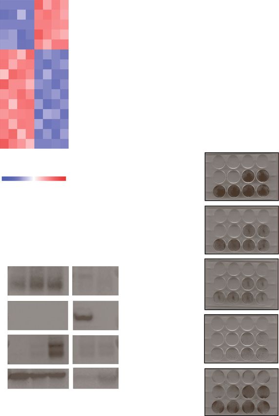
Fig. 2 Cells can be trained to metabolize fructose for proliferation. a Schematic for the positive selection strategy to generate fructolytic cell lines. b PC3
and PC3 passage 10 (P10) cells were seeded into 96-well plate (1500 cells/well) and cultured in media containing various amounts of sugar. Cell density
(% confluency) was monitored over time using live cell imaging (n = 2 per condition). c Schematic for the competition growth assay between PC3-red
(parental PC3 cells transduced with RFP reporter) and fructose-trained cell lines. d 40,000 of PC3, semi-trained PC3 passage 20 (P20), and trained-PC3
cells in 10 mM fructose or 11 mM glucose over time (n = 2 per condition). e Cells from d were grown in 10 mM fructose or 11 mM glucose for 96 h.
They were then fixed and stained with crystal violet solution (n = 2 per condition). f 20,000 PC3-red and 20,000 trained PC3 cells were seeded in the
same well and cultured for 96 h in 10 mM fructose or 10 mM glucose-containing media. Live fluorescent imaging was performed and the proportion of
PC3-red cells to total PC3 cells is shown over time (n = 2 per condition). Supplemental Video 1 and Supplemental Video 2 are of competition assays
monitored with live cell imaging
trained” (Figure S3A-B). We next removed glucose com- (Fig. 2f, Supplemental Video 1-2). We next asked if the
pletely from culture media of the “semi-trained” cell acquired ability to proliferate with fructose was lost
lines in hopes of selecting for cells that best proliferated when cells were grown in glucose for long periods of
in fructose (Fig. 2c). Recovered cells initially proliferated time. Even after 5 passages in media devoid of fructose,
slowly, but after 1–2 passages, “trained” cells proliferated the cells completely retained their fructolytic ability (Fig-
equally well in glucose and fructose (Fig. 2d-e, Supple- ure S3C-E).
mental video 1).
To control for plating and media conditions, we co- GLUT5 protein and mRNA abundance correlate with
plated the parental PC3 line with the trained cells in a fructolytic ability
competition assay [36] (Fig. 2c). Parental cells were la- We cultured the parental and semi-trained PC3 cells
beled with an RFP reporter and plated at a 1:1 ratio with for either 24 or 48 h in media containing either 11 mM
trained cells. In glucose-media, the final number of par- glucose (full RPMI) or 1 mM glucose plus 10 mM fruc-
ental and trained cells were equal, but in fructose-media, tose (Figure S4A). We then extracted RNA and per-
the parental cells only constituted 10–15% of total cells formed next-generation sequencing to analyzeLiang et al. Cancer & Metabolism (2021) 9:12 Page 8 of 13
expression across the transcriptome (RNA-seq) to cap- GLUT5 overexpression increases fructose flux into
ture intrinsic differences between the cells. Small differ- glycolysis
ences in gene expression between the parental and To measure differences in fructose metabolism between
semi-trained cells would presumably be enhanced in non-fructolytic and fructolytic cells, we cultured paren-
the trained cells. tal, semi-trained, and trained cells in media containing
The RNA-seq results were first summarized in a 3- 10 mM [U-13C]-fructose and traced its metabolic fate.
dimensional principal components analysis (PCA), which The trained cells demonstrated increased levels of
revealed unique clusters separating the parental from fructose-derived carbon into F1P, lactate, and TCA
semi-trained cells as well as the different media condi- cycle intermediates (Fig. 4a, b, Figure S8A-B). Measur-
tions (Figure S4B). Only fifteen genes were differentially able amounts of fructose were also detected in PC3
expressed between the parental and semi-trained cells, cells, suggesting that fructose can be imported into cells
even when using a generous statistical threshold (q=0.4 but does not meet the concentration necessary to sus-
and log2 fold change >1.1), confirming that the cells tain proliferation.
remained very similar despite being separated for > 20 In order to gain real-time insight into the ability of fructose
passages (Fig. 3a, Figure S4C). We validated the expres- to acidify the media (presumably via lactate production), we
sion of these 15 genes together with several fructolytic measured the extracellular acidification rate (ECAR) using
and glycolytic enzymes using cDNA from parental, parental and trained PC3 cells (Fig. 4c,d). While both cell
semi-trained, and trained cells (Fig. 3b, S4D, S4F). From types had similar ECAR in response to glucose, trained cells
these data, we observed that the expression of SLC2A5 had much higher ECAR in response to fructose. Semi-
had the highest fold change difference and correlated trained cells showed an intermediate phenotype, as expected.
with fructolytic ability. There was a >30× fold increase Interestingly, 2-deoxyglucose (2-DG), a competitive inhibitor
in semi-trained cells and >200× increase in trained PC3 for HK, immediately extinguished both glucose- and
cells (Fig. 3b). GLUT5 protein levels were also increased fructose-induced ECAR. This fact led us to hypothesize that
in trained PC3 cells compared to their parental PC3 cells fructose flux to lactate is mediated by HK rather than the ca-
(Fig. 3c). We further showed that the increased level of nonical fructose-metabolism protein, KHK.
GLUT5 expression was not due to an increase in
SLC2A5 copy number (Figure S4E). Cells proliferate with fructose through hexokinase
Using CRISPR-Cas9, we generated a KHK-/- line using
293T cells (293T KHK-/-) (Figure S9A-B). We then over-
GLUT5 overexpression rescues growth with fructose expressed either an empty vector (EV) or V5-tagged
across cell lines of different origin independent of KHK GLUT5 in the parental and KHK-/- cells (Figure S9C).
To test whether GLUT5 permits fructolytic growth in The resulting cells were cultured in 10 mM [U-13C]-
other cell lines, we overexpressed GLUT5 in the fructose prior to recovery of polar metabolites for meta-
brain, breast, prostate, colon, and liver cancer cell bolomics. GLUT5 overexpression greatly increased the
lines and repeated the proliferation assays. The over- abundance of F1P and its proportion of fructose-derived
expression of GLUT5 was sufficient to permit cellular carbons in the parental but not the KHK-/- cells (Figure
proliferation in fructose without affecting expression S9D-E). However, the abundance and isotopic labeling
of other fructolytic or glycolytic genes (Fig. 3d, Figure patterns of lactate and TCA cycle intermediates were
S5A-C). However, this proliferation required at least 5 similar between GLUT5-overexpressing parental and
mM fructose in the media (Figure S6A-D). We quan- KHK-/- cells (Figure S9D-E). Moreover, the absence of
tified the fructose-mediated proliferation at 96 h and F1P did not affect cellular proliferation with fructose, as
found that fructose contributed significantly to prolif- GLUT5 overexpression rescued fructose-mediated pro-
eration in the trained and GLUT5-overexpressing cells liferation in both the parental as well as the KHK-/- cells
only in the absence of glucose (Figure S6E-F). These (Fig. 4e). We therefore conclude that KHK is dispensable
data suggest that the proliferative contributions of for fructose-mediated cell proliferation.
glucose and fructose are through metabolism by a We capitalized on the kinetic properties of HK to dis-
common molecular enzyme that preferentially binds cern whether fructose-mediated cell proliferation was
glucose. mediated by KHK or HK. HK has a higher affinity for
KHK has been described as a rate-limiting enzyme for glucose than it does for fructose [38]. Therefore, we hy-
fructose metabolism in tumor and normal tissue [9, 15, pothesized that if cells used KHK for growth, then they
37]. To test whether KHK overexpression rescues would be more resistant to the HK inhibitor, 2-DG,
fructose-mediated cell growth, we overexpressed KHK-A when cultured in fructose as compared to glucose. Alter-
in non-fructolytic RKO cells and saw no rescue of cell natively, we hypothesized that if cells primarily used HK
proliferation (Figure S7A-B). for growth, then they would be more sensitive to 2-DGLiang et al. Cancer & Metabolism (2021) 9:12 Page 9 of 13
A B
SLC2A5 (Glut5)
SLC2A5
400
CNDP2 **** ****
Fold change/parental
VAMP8
*
MTLN 300 PC3-red
STAP2
semi-trained PC3
DDA1 200
NPTXR trained PC3
DNASE1L2
KLHL5
100
UBE2QL1
ZSWIM4 0
CCDC69
2
5
5-
t
lu
t
G
LHX1-DT
lu
G
RNF182
TNS1 EV Glut5
Parental
Parental
Parental
Parental
Trained
Trained
Trained
Trained
0 sugar
D RKO 10F
l l l (Colorectal)
-2 0 +2 10G
relative intensity
semi-trained PC3
0 sugar
PC3
trained PC3
10F
(Prostate)
C 10G
Msucle
Liver
PC3
0 sugar
Glut1 U118MG 10F
(Brain)
10G
Glut2
0 sugar
HUH7 10F
(Liver)
Glut5 10G
GAPDH 0 sugar
MCF7 10F
(Breast)
10G
Fig. 3 GLUT5 overexpression rescues cellular proliferation in fructose. a Normalized expression of genes that are differentially expressed (q = 0.4, >1.1
log2 fold change) between PC3 and semi-trained PC3 cells (passage 20) presented in heatmap form. b Relative expression of SLC2A5 transcript in semi-
trained PC3 and trained PC3 cells as compared to the parental PC3 line. Two primer sets were used (n = 2 per condition). Two-way ANOVA with
Fisher’s LDS test. *P < 0.05, ****P < 0.0001. c Immunoblot of the indicated proteins using lysates from PC3, semi-trained PC3, and trained PC3 cells. The
murine liver and muscle used as controls. d GLUT5 or an empty vector (EV) were overexpressed in the indicated cells lines. The cells were plated at
20,000-30,000 cells/well and then grown in the presence of no sugar, 10 mM fructose, or 10 mM glucose. After 3 days, the cells were fixed and stained
with crystal violet solution (n = 2 per condition)
when cultured in fructose as compared to glucose. We Discussion
treated cells with increasing concentrations of 2-DG in Cells preferentially metabolize the nutrients available in
media containing either 10 mM fructose or 10 mM glu- their microenvironment. Transformed cells acquire the
cose and found that cells in the fructose media were 5– ability to metabolize novel nutrients which allow them
33× more sensitive to 2-DG (Fig. 4f). At lower levels of to outgrow their neighbors and survive in sites of metas-
sugar (5 mM), the fructose-treated cells remain more tasis. Understanding how tumor cells acquire this ability
sensitive to 2-DG than glucose-treated cells; however, is valuable given the growing interest in metabolic and
this effect is lost when the sugars are given together (Fig. dietary interventions as anti-cancer therapy [39]. Here,
4g). Therefore, we conclude that cells can adapt to we show that human cancer cells from a wide range of
metabolize fructose through upregulation of GLUT5 and origins can acquire the ability to metabolize fructose
metabolism through HK instead of KHK. simply by stable overexpression of GLUT5. These dataLiang et al. Cancer & Metabolism (2021) 9:12 Page 10 of 13
Fructose F1P Lactate
150 150 **** 150 **** M+6
A **** ** M+5 C Glycolytic Stress Test
M+4 Glucose Oligo 2-DG
% labelling
100 100 100 25
M+3
M+2 20 PC3-red
50 50 50 M+1
semi-trained PC3
M+0 15
trained PC3
0 0 0 10
3
3
3
3
3
3
3
3
3
PC
PC
PC
PC
PC
PC
PC
PC
PC
d
d
ed
ed
d
ed
5
ne
ne
ne
in
in
in
i
i
i
ra
ra
tra
tra
ra
tra
i-t
i-t
i-t
ECAR (mpH/min)
m
m
m
0
se
se
se
0 20 40 60 80
Fructose F1P Lactate
5000 2000 ** 40000 *
D Fructolytic Stress Test
Fructose Oligo 2-DG
B 4000 * 1500
** 30000
15
Abundance
3000
1000 20000 10
2000
500 10000
1000
5
0 0 0
3
3
3
3
3
3
3
PC
PC
3
PC
PC
PC
PC
3
PC
PC
PC
d
ed
d
ed
d
ed
ne
ne
ne
in
in
in
0
i
i
i
ra
tra
ra
tra
ra
tra
i-t
i-t
0 20 40 60 80
i-t
m
m
m
se
se
se
minutes elapsed
7.3-EV 7.3-Glut5
E F 1.5 Trained PC3
1.5
H508
0 sugar
1.0 1.0
293T 10F IC50 = .00560 mM IC50 = .0931 mM IC50 = .198 mM IC50 = 6.48 mM
0.5 0.5
10G
0.0 0.0
-12 -10 -8 -6 -4 -2 0 -12 -10 -8 -6 -4 -2 0
293T-GLUT5 293T KO2-GLUT5
fold change viability/dmso
0 sugar 1.5
10 mM glucose
1.5
10 mM fructose
293T KO2 10F 1.0 1.0
IC50 = .111mM IC50 = .550 mM IC50 = .059 mM IC50 = 1.75 mM
10G 0.5 0.5
0.0 0.0
-12 -10 -8 -6 -4 -2 0 -12 -10 -8 -6 -4 -2 0
0 sugar
Trained PC3
293T KO3 10F
G 1.5 5 mM glucose + 0.04 mM fructose
5 mM glucose + 0.5 mM fructose
1.0 5 mM glucose + 5 mM fructose
10G 0 mM glucose + 5 mM fructose
0.5
0.0
-12 -10 -8 -6 -4 -2 0
log (M) 2-deoxyglucose
Fig. 4 Fructose fluxes through HK, not KHK, in order to sustain cellular proliferation. a Percent of heavy isotope (13C) incorporation into fructose,
fructose 1-phosphate (F1P), and lactate as detected by LC/MS from polar extracts of PC3, semi-trained PC3, and trained PC3 cells (n = 2-3). The
isotopic labelling is indicated by M+# designation indicated in the legend where the # represents the amount of [12C] replaced by [13C]. Two-
tailed unpaired t tests were used between parental and trained cells (M+3 for lactate, M+6 for fructose/F1P). *P < 0.05, **P< 0.01, and ****P <
0.0001. b Total abundance of fructose, F1P, and lactate as detected by LC/MS from polar extracts of PC3, semi-trained PC3, and trained PC3 cells
(n = 2–3). Two-tailed unpaired t tests were used between parental and trained cells. *P < 0.05, **P< 0.01, and ****P < 0.0001. c Extracellular
acidification rate (ECAR) over time of PC3, semi-trained PC3, and trained PC3 cells under basal conditions and following the addition of glucose,
oligomycin (Oligo), and 2-deoxyglucose (2-DG) at the indicated times. Data are the mean and SEM from 6 replicates. d ECAR over time of PC3,
semi-trained PC3, and trained PC3 cells under basal conditions and following the addition of fructose, Oligo, and 2-DG at the indicated times.
Data are the mean and SEM from 6 replicates. e GLUT5 or an empty vector (EV) were overexpressed in 293T or 293T KHK-/- cells. The cells were
plated at 20,000 cells/well and then grown in the presence of no sugar, 10 mM fructose, or 10 mM glucose. After 7 days, the cells were fixed,
stained with crystal violet solution (n = 2 per condition). f Fold change in cell viability as assessed by ATP concentration (Cell Titer Glo) of the
indicated fructolytic cell lines grown in either 10 mM glucose or 10 mM fructose containing the specified concentrations of 2-DG after 72 h (n =
3 per concentration). The half maximal inhibitory concentration (IC50) is displayed on the graph for each curve. g Fold change in cell viability as
assessed by ATP concentration (Cell Titer Glo) of the trained PC3 grown in the specified sugar conditions containing the specified concentrations
of 2-DG after 96 h. (n = 2 per concentration). The half maximal inhibitory concentration (IC50) is displayed on the graph for each curve
suggest that sugar uptake can be a limiting factor pre- the glucose transporters, GLUT1 and GLUT4, control
venting fructose-mediated cell proliferation. skeletal muscle glucose uptake at rest, and in response
Sugar uptake is also a key regulatory node for glucose to contraction or insulin [40, 41]. Additionally, the ex-
metabolism and growth. For example, the expression of pression of GLUT1 and GLUT3 in tumors is associatedLiang et al. Cancer & Metabolism (2021) 9:12 Page 11 of 13 with enhanced glucose uptake and oncogenic growth by KHK, but this may be unique to non-proliferative [42–44]. Tumor cells continue to regulate the flux of cells in the liver, intestine, and kidney. Proliferating cells glucose at the levels of phosphorylation by HK, fructose- typically switch to less fructolytic isoforms of KHK. For 1,6-bisphosphate production by phosphofructokinase, example, liver cancer cells convert from the high affinity and lactate export [45]. In this study, we show that fruc- KHK-c variant (Km = 0.7 mM), to the low affinity iso- tose phosphorylation by KHK is not required for fruc- form, KHK-a (Km = 7 mM), that may play a role in de tose metabolism and cell growth; however, we speculate novo nucleotide biosynthesis [49]. On average, the cell that other regulatory nodes exist. lines we profiled in this study expressed >160× more Our conclusions are supported by clinical evidence KHK-a than KHK-c (Fig. 1e, Table S1). Furthermore, the from subjects with cancer. GLUT5 is significantly upreg- expression of HK (Km for fructose 1–4 mM) is greater ulated in tumors from patients with the colon, lung, and than KHK-a in these cells, which may explain the prefer- breast adenocarcinoma, acute myeloid leukemia, ovarian ence for this route of metabolism [38, 50]. Our data sug- carcinoma, and glioma where it contributes to malig- gests that this route is most relevant in tissues such as nancy and poor survival [16, 21, 23, 25–27]. Many of the liver, kidney, seminal vesicles, and prostate, where these studies investigated fructose metabolism in the ab- fructose levels achieve concentrations higher than 5 mM sence of glucose, using a wide range of fructose concen- [17, 51, 52]. trations (ref 22: 6 mM, ref 24: 25 mM, ref 26: 1.5-6 mM, The exact role of KHK and F1P in these cell lines re- ref 27: 3 mM, ref 28: 6 mM). It is worth noting that main unclear. KHK-mediated fructose metabolism may these studies were able to discern physiologically rele- become more important when HK is saturated or inhib- vant findings despite modelling fructose-mediated ited by high concentrations of glucose and glucose 6- growth in the absence of glucose in vitro. phosphate. However, it is unclear if glucose ever reaches Our data confirms that GLUT5 overexpression is these high concentrations in poorly vascularized solid sufficient to promote cellular proliferation in fructose, tumors [53]. For example, pancreatic adenocarcinomas but the abundance of the GLUT5 transcript in our ini- in mice have significantly less glucose in the tumor tial profiling did not predict the fructolytic index interstitial fluid relative to plasma [54]. These poorly across cell lines. For example, H508 (fructolytic) and vascularized tumors also receive less oxygen from the RKO (non-fructolytic) cells are from the same colorec- blood [53], and the resulting hypoxia enhances the en- tal origin with similar levels of GLUT5 yet have vastly dogenous production of fructose and the expression of different abilities to proliferate in fructose. Other fructolytic genes [9, 55–58]. groups have shown that the stability of GLUT5 mRNA In conclusion, our study defines fructose uptake as a and the location of GLUT5 protein can be modulated limiting factor for fructose-mediated cell proliferation. by distinct signaling pathways [5, 46]. Therefore, we We describe a previously unappreciated role of HK to conclude that GLUT5 expression needs to be analyzed permit fructolytic cell growth. These findings advance in tandem with other, currently unknown, cellular fea- our basic understanding of fructose metabolism in can- tures in order to determine fructose-mediated prolif- cer cells and highlight a limitation of directly targeting eration a priori. KHK for anti-cancer therapy. Our data supports the conclusion that GLUT5 is a ro- bust determinant of fructose-mediated cell proliferation. However, we were unable to identify how the semi- Conclusions trained and trained cells upregulated this message. There The intent of this study was to find the determinants of was no difference in SLC2A5 copy number in the gen- fructose-mediated proliferation in cell lines. We have omic DNA and minimal change in the expression of found that fructose-dependent proliferation of cancer cells known SLC2A5-regulating fructose-response elements is not determined by tissue of origin nor expression of any like Chrebpβ (Fig. 3, Figure S3). Due to the specificity of individual fructolytic gene. Using a positive selection ap- the SLC2A5 overexpression, we hypothesize that the up- proach, we were able to train PC3 cells to proliferate with regulation stems from epigenetic or genetic modifica- fructose. We saw that GLUT5 was strongly upregulated in tions at the SLC2A5 locus. trained cells and that overexpressing GLUT5 allowed Our data suggest that KHK is dispensable for fructose- non-fructolytic cell lines of several different origins to pro- mediated proliferation. Instead, we show that cancer liferate in fructose. Lastly, we showed that cells metabolize cells metabolize fructose using HK, as is the case in fructose through hexokinase, not ketohexokinase, to sus- lower order organisms. For example, Hk is the only fruc- tain proliferation and glycolysis. This study sheds light on tokinase in yeast and the flux through HK sustains the cell-autonomous fructose metabolism and suggests that high activity of nectarivore flight muscles [47, 48]. In targeting fructose metabolism may require inhibition of humans, fructose is thought to be primarily metabolized both KHK as well as HK.
Liang et al. Cancer & Metabolism (2021) 9:12 Page 12 of 13
Supplementary Information Competing interests
The online version contains supplementary material available at https://doi. L.C.C. is a founder and member of the board of directors of Agios
org/10.1186/s40170-021-00246-9. Pharmaceuticals and is a founder and receives research support from Petra
Pharmaceuticals. M.D.G. reports personal fees from Novartis, Petra
Pharmaceuticals, and Bayer. He receives research support from Pfizer. L.C.C.
Additional file 1: Supplemental Figure 1. Cell growth in fructose is
and M.D.G. are inventors on patents (pending) for Combination Therapy for
heterogeneous. Supplemental Figure 2. Gene expression does not
PI3K-associated Disease or Disorder, The Identification of Therapeutic Inter-
determine the fructolytic index. Supplemental Figure 3. Cells can
ventions to Improve Response to PI3K Inhibitors for Cancer Treatment, and
stably utilize fructose for proliferation. Supplemental Figure 4. SLC2A5
Anti-Fructose Therapy for Colorectal and Small Intestine Cancers. L.C.C. and
copy number, validated RNA-seq transcripts (excluding SLC2A5), and
M.D.G. are co-founders and shareholders in Faeth Therapeutics. All other au-
selected metabolic enzyme transcripts do not correlate with fructolytic
thors report no competing interests.
ability. Supplemental Figure 5. Selected metabolism genes are not
changed with GLUT5 overexpression. Supplemental Figure 6. Serum
Author details
concentration of glucose overshadows fructose contributions to 1
Division of Endocrinology, Weill Department of Medicine, Weill Cornell
proliferation rate. Supplemental Figure 7. KHK overexpression does not
Medicine, New York, NY 10065, USA. 2Meyer Cancer Center, Department of
rescue the ability to proliferate in fructose. Supplemental Figure 8.
Medicine, Weill Cornell Medicine, New York, NY 10065, USA. 3Weill Cornell
Trained PC3 have increased fructose flux into the TCA cycle.
Graduate School of Medical Sciences, Weill Cornell Medicine, New York, NY
Supplemental Figure 9. Trained PC3 have increased fructose flux into
10065, USA. 4Weill Cornell/Rockefeller/Sloan Kettering Tri-I MD-PhD program,
the TCA cycle. Supplemental Table 1: Clinical and genomic data of
New York, NY 10065, USA. 5Division of Infectious Diseases, Weill Department
profiled cell lines in order of fructolytic index. Related to Figure 1.
of Medicine, Weill Cornell Medicine, New York, NY 10065, USA. 6Center for
Supplemental Table 2. qPCR data for each cell line using primers from
Molecular Oncology, Memorial Sloan Kettering Cancer Center, New York, NY
Supplemental File 1. (n = 2 per gene per sample, 2^dCt values shown).
10065, USA. 7Department of Pathology, Memorial Sloan Kettering Cancer
Related to Figure 1. Supplemental Table 3. qPCR primers for selected
Center, New York, NY 10065, USA. 8Weill Cornell Medical College, Weill
metabolic genes, CRISPR-cas9 primers, and qPCR primers for gDNA.
Cornell Medicine, New York, NY 10065, USA.
Related to Figures 1, 4 and Supplemental Figures 4, 8. Supplemental
Table 4. qPCR primers for RNA-seq hits, related to Figure 3 and
Received: 13 July 2020 Accepted: 8 February 2021
Supplemental Figure 4.
Additional file 2: Supplemental Video 1. Trained PC3 cells
outcompete parental PC3 cells in fructose media. 20,000 PC3-red and
20,000 trained PC3 cells were seeded in a 6-well dish containing 10 mM References
fructose. Cells were monitored with live cell imaging for 4 days. 1. United States Department of Agriculture, Economic Research Service. USDA
Sugar Supply: Table 50: US Consumption of Caloric Sweeteners. 2019;
Additional file 3: Supplemental Video 2. Trained PC3 cells grow at
Available from: https://www.ers.usda.gov/data-products/sugar-and-
the same rate as parental PC3 cells in glucose media. 20,000 PC3-red and
sweeteners-yearbook-tables/sugar-and-sweeteners-yearbook-tables/
20,000 trained PC3 cells were seeded in a 6-well dish containing 10 mM
#World%20Production,%20Supply,%20and%20Distribution
glucose. Cells were monitored with live cell imaging for 4 days.
2. Hannou SA, Haslam DE, McKeown NM, Herman MA. Fructose metabolism
and metabolic disease. J Clin Invest. 2018;128(2):545–55.
3. Khitan Z, Kim DH. Fructose: a key factor in the development of metabolic
Abbreviations syndrome and hypertension. J Nutr Metab [Internet]. 2013;2013 [cited 2020 May
HK: Hexokinase; KHK: Ketohexokinase; ALDOB: Aldolase B; F1P: Fructose-1- 31]. Available from: https://www.ncbi.nlm.nih.gov/pmc/articles/PMC3677638/.
phosphate; FBS: Fetal bovine serum; dFBS: Dialyzed FBS; PCA: Principle 4. Diggle CP, Shires M, Leitch D, Brooke D, Carr IM, Markham AF, et al.
components analysis; 2-DG: 2-Deoxyglucose; EV: Empty vector; SEM: Standard Ketohexokinase: expression and localization of the principal fructose-metabolizing
error of the mean enzyme. J Histochem Cytochem Off J Histochem Soc. 2009;57(8):763–74.
5. Douard V, Ferraris RP. Regulation of the fructose transporter GLUT5 in
health and disease. Am J Physiol Endocrinol Metab. 2008;295(2):E227–37.
Acknowledgements
6. Jang C, Hui S, Lu W, Cowan AJ, Morscher RJ, Lee G, et al. The small intestine
We would like to thank all members of the Goncalves Lab and the Cantley
converts dietary fructose into glucose and organic acids. Cell Metab. 2018;
Lab, especially Drs. Ted Kastenhuber and Andrés Quieroz, for the thoughtful
27(2):351–361.e3.
discussion and advice. The R25 AI140472 provided educational resources for
7. Funari VA, Herrera VLM, Freeman D, Tolan DR. Genes required for fructose
metabolomics. We would like to thank Dr. Feng Zhang (Broad Institute) for
metabolism are expressed in Purkinje cells in the cerebellum. Brain Res Mol
pSpCas9(BB)-2A-Puro (PX459) V2.0. The pDONR221_SLC2A5 plasmid was a
Brain Res. 2005;142(2):115–22.
gift from RESOLUTE Consortium & Giulio Superti-Furga. pLenti-U6-tdTomato-
8. Funari VA, Crandall JE, Tolan DR. Fructose metabolism in the cerebellum.
P2A-BlasR (LRT2B) was a gift from Lukas Dow. We would like to thank Weill
Cerebellum Lond Engl. 2007;6(2):130–40.
Cornell Medicine Genomics Core for conducting RNA-seq.
9. Mirtschink P, Krishnan J, Grimm F, Sarre A, Hörl M, Kayikci M, et al. HIF-
driven SF3B1 induces KHK-C to enforce fructolysis and heart disease. Nature.
Authors’ contributions 2015;522(7557):444–9.
Conceptualization: R.J.L and M.D.G.; Methodology: R.J.L., S.T., L.C.C., K.Y.R., and 10. Oppelt SA, Zhang W, Tolan DR. Specific regions of the brain are capable of
M.D.G.; Investigation: R.J.L., S.T., N.N., C.J.M., E.D., S.C., T.H., S.R., R.G., S.H., B.N.; fructose metabolism. Brain Res. 1657;2017(15):312–22.
Formal analysis: R.J.L. and E.D.; Data curation: R.J.L. and J.S.; Visualization: R.J.L. 11. Song (宋志林) Z, Roncal-Jimenez CA, Lanaspa-Garcia MA, Oppelt SA,
and M.D.G.; Investigation and validation: M.D.G.; Software: S.T., C.J.M, and J.S.; Kuwabara M, Jensen T, et al. Role of fructose and fructokinase in acute
Supervision: L.C.C., K.Y.R., and M.D.G.; Writing, review, and editing: R.J.L. and dehydration-induced vasopressin gene expression and secretion in mice. J
M.D.G.; Resources: L.C.C., K.Y.R., and M.D.G.; Funding acquisition: L.C.C., K.Y.R., Neurophysiol. 2017;117(2):646–54.
and M.D.G. The authors read and approved the final manuscript. 12. Charrez B, Qiao L, Hebbard L. The role of fructose in metabolism and
cancer. Horm Mol Biol Clin Invest. 2015;22(2):79–89.
13. Fan X, Liu H, Liu M, Wang Y, Qiu L, Cui Y. Increased utilization of fructose has a
Funding positive effect on the development of breast cancer. PeerJ. 2017;5:e3804.
This work was supported by the NIH R35 CA197588 (L.C.C.), NIH/NIAID R25 AI 14. Jiang Y, Pan Y, Rhea PR, Tan L, Gagea M, Cohen L, et al. A sucrose-enriched
140472 (K.Y.R), SU2C-AACR-DT22-17 (L.C.C.), NIH K08 CA230318 (M.D.G.), and diet promotes tumorigenesis in mammary gland in part through the 12-
NIH P50 CA211024 (M.D.G.). B.N. is supported by a National Science Founda- lipoxygenase pathway. Cancer Res. 2016;76(1):24–9.
tion (NSF) Graduate Research Fellowship and a National Cancer Institute 15. Gao W, Li N, Li Z, Xu J, Su C. Ketohexokinase is involved in fructose
(NCI) of the National Institutes of Health (NIH) under the F99/K00 Career utilization and promotes tumor progression in glioma. Biochem Biophys Res
Transition Fellowship (F99CA234950). Commun. 2018;503(3):1298–306.Liang et al. Cancer & Metabolism (2021) 9:12 Page 13 of 13
16. Su C, Li H, Gao W. GLUT5 increases fructose utilization and promotes tumor 41. Ren JM, Marshall BA, Gulve EA, Gao J, Johnson DW, Holloszy JO, et al.
progression in glioma. Biochem Biophys Res Commun. 2018;500(2):462–9. Evidence from transgenic mice that glucose transport is rate-limiting for
17. Carreño D, Corro N, Torres-Estay V, Véliz LP, Jaimovich R, Cisternas P, et al. glycogen deposition and glycolysis in skeletal muscle. J Biol Chem. 1993;
Fructose and prostate cancer: toward an integrated view of cancer cell 268(22):16113–5.
metabolism. Prostate Cancer Prostatic Dis. 2019;22(1):49–58. 42. Birsoy K, Possemato R, Lorbeer FK, Bayraktar EC, Thiru P, Yucel B, et al.
18. Jin C, Gong X, Shang Y. GLUT5 increases fructose utilization in ovarian Metabolic determinants of cancer cell sensitivity to glucose limitation and
cancer. OncoTargets Ther. 2019;12:5425–36. biguanides. Nature. 2014;508(7494):108–12.
19. Hsieh C-C, Shyr Y-M, Liao W-Y, Chen T-H, Wang S-E, Lu P-C, et al. Elevation 43. Onodera Y, Nam J-M, Bissell MJ. Increased sugar uptake promotes
of β-galactoside α2,6-sialyltransferase 1 in a fructose-responsive manner oncogenesis via EPAC/RAP1 and O-GlcNAc pathways. J Clin Invest. 2014;
promotes pancreatic cancer metastasis. Oncotarget. 2016;8(5):7691–709. 124(1):367–84.
20. Liu H, Huang D, McArthur DL, Boros LG, Nissen N, Heaney AP. Fructose 44. Yun J, Rago C, Cheong I, Pagliarini R, Angenendt P, Rajagopalan H, et al.
induces transketolase flux to promote pancreatic cancer growth. Cancer Glucose deprivation contributes to the development of KRAS pathway
Res. 2010;70(15):6368–76. mutations in tumor cells. Science. 2009;325(5947):1555–9.
21. Goncalves MD, Lu C, Tutnauer J, Hartman TE, Hwang S-K, Murphy CJ, et al. 45. Tanner LB, Goglia AG, Wei MH, Sehgal T, Parsons LR, Park JO, et al. Four key
High-fructose corn syrup enhances intestinal tumor growth in mice. steps control glycolytic flux in mammalian cells. Cell Syst. 2018;7(1):49–62.e8.
Science. 2019;363(6433):1345–9. 46. Gouyon F, Caillaud L, Carriere V, Klein C, Dalet V, Citadelle D, et al. Simple-sugar
22. Chen W-L, Jin X, Wang M, Liu D, Luo Q, Tian H, et al. GLUT5-mediated meals target GLUT2 at enterocyte apical membranes to improve sugar
fructose utilization drives lung cancer growth by stimulating fatty acid absorption: a study in GLUT2-null mice. J Physiol. 2003;552(Pt 3):823–32.
synthesis and AMPK/mTORC1 signaling. JCI Insight. 2020;5(3):e131596. 47. Emmerich W, Radler F. The anaerobic metabolism of glucose and fructose
23. Weng Y, Zhu J, Chen Z, Fu J, Zhang F. Fructose fuels lung adenocarcinoma by Saccharomyces bailii. Microbiology. 1983;129(11):3311–8.
through GLUT5. Cell Death Dis [Internet]. 2018;9(5) [cited 2020 May 24]. 48. Welch KC, Chen CCW. Sugar flux through the flight muscles of hovering
Available from: https://www.ncbi.nlm.nih.gov/pmc/articles/PMC5945656/. vertebrate nectarivores: a review. J Comp Physiol B. 2014;184(8):945–59.
24. Weng Y, Fan X, Bai Y, Wang S, Huang H, Yang H, et al. SLC2A5 promotes 49. Li X, Qian X, Peng L-X, Jiang Y, Hawke DH, Zheng Y, et al. A splicing switch
lung adenocarcinoma cell growth and metastasis by enhancing fructose from ketohexokinase-C to ketohexokinase-A drives hepatocellular carcinoma
utilization. Cell Death Dis. 2018;4:38. formation. Nat Cell Biol. 2016;18(5):561–71.
25. Bu P, Chen K-Y, Xiang K, Johnson C, Crown SB, Rakhilin N, et al. Aldolase B 50. Grossbard L, Schimke RT. Purification and comparison of soluble forms, vol.
mediated fructose metabolism drives metabolic reprogramming of colon 16; 1966.
cancer liver metastasis. Cell Metab. 2018;27(6):1249–1262.e4. 51. Owen DH, Katz DF. A review of the physical and chemical properties of
26. Jin X, Liang Y, Liu D, Luo Q, Cai L, Wu J, et al. An essential role for GLUT5- human semen and the formulation of a semen simulant. J Androl. 2005;
mediated fructose utilization in exacerbating the malignancy of clear cell 26(4):459–69.
renal cell carcinoma. Cell Biol Toxicol. 2019;35(5):471–83. 52. Helsley RN, Moreau F, Gupta MK, Radulescu A, DeBosch B, Softic S. Tissue-specific
27. Chen W-L, Wang Y-Y, Zhao A, Xia L, Xie G, Su M, et al. Enhanced fructose fructose metabolism in obesity and diabetes. Curr Diab Rep. 2020;20(11):64.
utilization mediated by SLC2A5 is a unique metabolic feature of acute 53. Vaupel P. Tumor microenvironmental physiology and its implications for
myeloid leukemia with therapeutic potential. Cancer Cell. 2016;30(5):779–91. radiation oncology. Semin Radiat Oncol. 2004 Jul 1;14(3):198–206.
28. Zhao P, Huang J, Zhang D, Zhang D, Wang F, Qu Y, et al. SLC2A5 54. Sullivan MR, Danai LV, Lewis CA, Chan SH, Gui DY, Kunchok T, et al.
overexpression in childhood philadelphia chromosome-positive acute Quantification of microenvironmental metabolites in murine cancers reveals
lymphoblastic leukaemia. Br J Haematol. 2018;183(2):242–50. determinants of tumor nutrient availability. DeBerardinis R, van Lohuizen M,
DeBerardinis R, Frezza C, editors. eLife. 2019;8:e44235.
29. Barretina J, Caponigro G, Stransky N, Venkatesan K, Margolin AA, Kim S, et al.
55. Andres-Hernando A, Johnson RJ, Lanaspa MA. Endogenous fructose
The Cancer Cell Line Encyclopedia enables predictive modelling of
production: what do we know and how relevant is it? Curr Opin Clin Nutr
anticancer drug sensitivity. Nature. 2012;483(7391):603–7.
Metab Care. 2019;22(4):289–94.
30. Tate JG, Bamford S, Jubb HC, Sondka Z, Beare DM, Bindal N, et al. COSMIC:
56. Armitage EG, Kotze HL, Allwood JW, Dunn WB, Goodacre R, Williams KJ.
the Catalogue Of Somatic Mutations In Cancer. Nucleic Acids Res. 2019;
Metabolic profiling reveals potential metabolic markers associated with
47(Database issue):D941–7.
Hypoxia Inducible Factor-mediated signalling in hypoxic cancer cells. Sci
31. Bairoch A. The cellosaurus, a cell-line knowledge resource. J Biomol Tech
Rep. 2015;5(1):15649.
JBT. 2018;29(2):25–38.
57. Hamann I, Krys D, Glubrecht D, Bouvet V, Marshall A, Vos L, et al. Expression
32. Zafra MP, Schatoff EM, Katti A, Foronda M, Breinig M, Schweitzer AY, et al.
and function of hexose transporters GLUT1, GLUT2, and GLUT5 in breast
Optimized base editors enable efficient editing in cells, organoids and mice.
cancer—effects of hypoxia. FASEB J. 2018;32(9):5104–18.
Nat Biotechnol. 2018;36(9):888–93.
58. Kucharzewska P, Christianson HC, Belting M. Global profiling of metabolic
33. Naito Y, Hino K, Bono H, Ui-Tei K. CRISPRdirect: software for designing CRIS
adaptation to hypoxic stress in human glioblastoma cells. PLoS ONE
PR/Cas guide RNA with reduced off-target sites. Bioinformatics. 2015;31(7):
[Internet]. 2015;10(1) [cited 2020 Jun 10]. Available from: https://www.ncbi.
1120–3.
nlm.nih.gov/pmc/articles/PMC4310608/.
34. Rosa R, Monteleone F, Zambrano N, Bianco R. In vitro and in vivo models
for analysis of resistance to anticancer molecular therapies. Curr Med Chem.
2014;21(14):1595–606. Publisher’s Note
35. Yudkin J. Origin of Acquired Drug Resistance in Bacteria. Nature. 1953; Springer Nature remains neutral with regard to jurisdictional claims in
171(4352):541–6. published maps and institutional affiliations.
36. Eekels JJM, Pasternak AO, Schut AM, Geerts D, Jeeninga RE, Berkhout B. A
competitive cell growth assay for the detection of subtle effects of gene
transduction on cell proliferation. Gene Ther. 2012;19(11):1058–64.
37. Ishimoto T, Lanaspa MA, Le MT, Garcia GE, Diggle CP, MacLean PS, et al.
Opposing effects of fructokinase C and A isoforms on fructose-induced
metabolic syndrome in mice. Proc Natl Acad Sci U S A. 2012;109(11):4320–5.
38. Cárdenas ML, Rabajille E, Niemeyer H. Fructose is a good substrate for rat
liver “glucokinase” (hexokinase D). Biochem J. 1984;222(2):363–70.
39. Lien EC, Vander Heiden MG. A framework for examining how diet impacts
tumour metabolism. Nat Rev Cancer. 2019;19(11):651–61.
40. Hansen PA, Gulve EA, Marshall BA, Gao J, Pessin JE, Holloszy JO, et al.
Skeletal muscle glucose transport and metabolism are enhanced in
transgenic mice overexpressing the Glut4 glucose transporter. J Biol Chem.
1995;270(4):1679–84.You can also read