Printable and flexible graphene pH sensors utilising thin film melanin for physiological applications - IOPscience
←
→
Page content transcription
If your browser does not render page correctly, please read the page content below

2D Materials

PAPER • OPEN ACCESS

Printable and flexible graphene pH sensors utilising thin film melanin for
physiological applications
To cite this article: Z Tehrani et al 2020 2D Mater. 7 024008

View the article online for updates and enhancements.

                                This content was downloaded from IP address 176.9.8.24 on 12/04/2020 at 18:51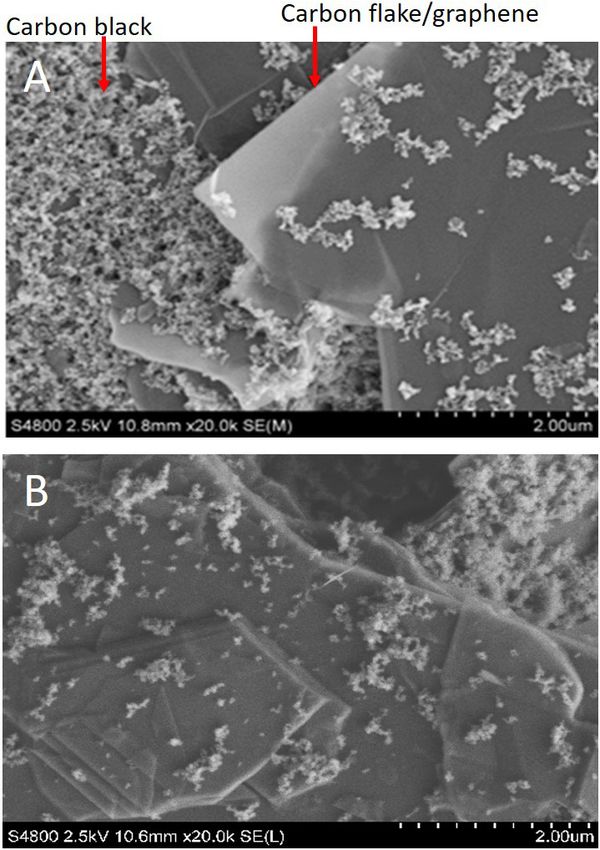
2D Mater. 7 (2020) 024008                                                                    https://doi.org/10.1088/2053-1583/ab72d5

OPEN ACCESS                  PAPER

                             Printable and flexible graphene pH sensors utilising thin film
RECEIVED
                             melanin for physiological applications
20 October 2019
RE VISED
                             Z Tehrani1,5 , S P Whelan1,5, A B Mostert2 , J V Paulin3, M M Ali1, E Daghigh Ahmadi1, C F O Graeff4 ,
15 January 2020
                             O J Guy2 and D T Gethin5
ACCEP TED FOR PUBLICATION
                             1
4 February 2020                  Centre for Nano Health, College of Engineering, Swansea University, Swansea SA2 8PP, United Kingdom
                             2
                                 Department of Chemistry, College of Science, Swansea University, Swansea SA2 8PP, United Kingdom
PUBLISHED
                             3
28 February 2020                 Post-Graduate Program in Materials Science and Technology, São Paulo State University (UNESP), Bauru, Brazil
                             4
                                 Department of Physics, São Paulo State University (UNESP), School of Sciences, Bauru, Brazil
                             5
                                 Welsh Centre for Printing and Coating, College of Engineering, Swansea University, Swansea SA1 8EN, United Kingdom
Original content from
this work may be used        E-mail: Z.Tehrani@swansea.ac.uk and D.T.Gethin@swansea.ac.uk
under the terms of the
Creative Commons             Keywords: graphene, pH Sensor, melanin, screen print, blood plasma, low cost manufacturing
Attribution 4.0 licence.
                             Supplementary material for this article is available online
Any further distribution
of this work must
maintain attribution
to the author(s) and the
title of the work, journal   Abstract
citation and DOI.
                             The application of highly sensitive pH sensors manufactured in volume at low cost has great
                             commercial interest due to an extensive array of potential applications. Such areas include industrial
                             processing, biotechnology and medical diagnostics particularly in the development of point of
                             care (POC) devices. A novel printable electrochemical pH sensor based on graphene and pigment
                             melanin (PGM), was designed and produced by using a screen printing process that enables up
                             scaling for potential commercial application. We demonstrate a highly sensitive pH sensor (62
                             mV pH−1  ±  7) over a pH range from 5 to 8, with high stability and superior performance when
                             compared with a number of existing devices and making it suitable for physiological applications.

                             1. Introduction                                                     is changing, or the active material of a pH sensor is eas-
                                                                                                 ily contaminated or the loss/degradation of the sensing
                             The measurement of the pH of a solution is of universal             substances occurs [13]. The ubiquitous glass electrode,
                             importance and is used extensively across a range of                as an example, is fragile and requires time to build up
                             disciplines (e.g. chemistry, biology, environmental                 a hydrate layer before usage. Furthermore, the ability
                             science), both in the laboratory as well as in the field [1–        to optimise it is challenging since remodelling options
                             5]. As such, multiple techniques have been developed                are limited [13].
                             to measure the pH [6]. The most common applications                      As a result, there is a continuing active interest in
                             for pH determination are based on potentiometry,                    finding new methods for measuring pH. Recent stud-
                             usually using a glass electrode [7, 8]. Film electrodes             ies show many material options (organic and inor-
                             and ion selective membranes are also used to measure                ganic) that can be used as ion sensitive layers (see
                             pH potentiometrically as are ion selective field effect             table 1 for examples). Ideally, a pH sensor needs to have
                             transistors [9, 10]. pH may also be measured using a                the following properties if it is to be used widely:
                             variety of ductometric [11] and optical [12] methods
                             including measuring colour changes in pH sensitive                       1.	Easy to produce in a commercial setting.
                             indicator dyes. This wide variety of ways to measure                     2.	Made of readily available materials.
                             the pH is indicative of its universal importance i.e.                    3.	Be sensitive, i.e. a high electrochemical voltage
                             its presence as a variable in many different kinds of                        versus pH gradient across a wide pH range.
                             systems.                                                                     Failing that across a specified, widely studied pH
                                 However, there are drawbacks to the various                              range.
                             approaches. Many of these methods are subject to sen-                    4.	That the sensor is stable over the pH range as
                             sors with a limited lifetime or their sensing time does                      well as over time (little degradation).
                             not comply with the speed at which the pH of a system                    5.	Ideally fast response time.

                             © 2020 The Author(s). Published by IOP Publishing Ltd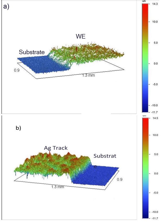
2D Mater. 7 (2020) 024008                                                                                         Z Tehrani et al

    The purpose of this paper is to demonstrate a com-       Table 1. Examples of different sensing materials used as active
                                                             components in pH sensors.
mercially viable, robust and sensitive pH sensor that
rivals that of 3D graphene with HfO2 & IrO2/RGO                                     Sensitivity       Range
(table 1) but utilising readily available materials.          Sensor material       (mV pH−1)         (pH)         Ref.

                                                              IrO2                  −51.1             1.5–12       [14]
1.1. Screen printing
                                                               (SM2O3)              56.2              2–12         [15]
One of the ways to achieve low cost manufacturing
                                                              C/PANI                50                4–10         [16]
to produce in a commercial setting is via a screen-
                                                              RuO2/SnO2             56.5              2–12         [17]
printing process. The added benefit of this process is
                                                              GO                    31.8              4–10         [18]
that it opens new avenues for other kinds of sensors.
                                                              IrO2/RGO              62                2–12         [19]
     Screen-printing is an attractive technique for the
                                                              3D—G with HfO2        71  ±  7          3–9          [20]
production of sensors, because the technology is well
                                                              RGO                   40  ±  5          4–10         [21]
established and allows for rapid production. Screen
printing is often used for the fabrication of electrodes      SnO2                  56–58             2–12         [22]

due to the low cost and simplicity of the technique           ZnO                   38                2–12         [23]

[28, 29].                                                     ITO                   55                1–11         [24]

     Furthermore, the base electrode material of inter-       PANI/PVSA             58                2–12         [25]
est for this application is based on carbon, since it has     CNT                   50.9              3–13         [26]
been studied extensively and is often used in electro-        Melanin/ITO/Au         48.9             2–12         [27]
chemistry due to its electrical conductive properties,        PGM                    62  ±  7         5–8          This work
low density and low thermal expansion [30]. Specifi-
                                                             G: graphene, CNTs: carbon nanotubes, GO: graphene oxide,
cally, we target graphene as the main electrode due to       RGO: graphene oxide, IrO2: iridium oxide, SM2O3: samarium
its reported use as an electronic material [31], sensor in   oxide, RuO2/SnO2: ruthenium oxide/tin oxide, PANI/PVSA:
bioelectronics [32, 33] and in applications based on its     polyaniline/poly vinyl sulfonic acid, ZnO: zinc oxide, ITO: indium
high carrier mobility and high carrier concentration.        tin oxide, Au: gold, HfO2: hafnium oxide.
Also it has, biocompatibility, atomic thickness, electro-
chemical stability and mechanical reliability [34].          conductivity [44–47], free radical properties [48–51],
     However, as will be shown below, graphene on its        metal ion chelation [52] and a broad band UV–Vis
own is not sensitive enough in this specific application.    absorbance [53, 54]. As a result melanin has been
Instead, this study explores the possibility of using        studied for applications in UV filters [55], solid-state
a melanin derivative as the pH sensitive medium in           organic electrochemical transistors [56], metal-
combination with a printed graphene (PG) working             insulator devices [57], flexible supercapacitors [58],
electrode. The graphene electrode has been chosen due        extended gate field effect transistors (EGFETs) for pH
to it good electrical conductivity and the ability to con-   sensing [27], engineered electrodes [59] and edible
trol its printed topography on which melanin is spin         batteries [60].
coated to form a very thin layer.                                What makes melanin particularly attractive as an
                                                             active electrochemical sensing material is its reported
1.2. D-melanin                                               sensitivity as a function of pH (~  −50 mV pH−1)
The melanins are a class of conjugated                       [27, 61]. The electrochemistry is due to the hydroxyl
biomacromolecule [35]. The most common form,                 groups of quinone moieties and the interconversion
eumelanin, is considered the archetypal melanin              between the various oxidative states [46, 47] (figure 2).
[35] and forms the basis of our discussion for the rest      Furthermore, melanin films have been used previously
of the manuscript. As such, we will follow standard          as an active layer in a pH sensing EGFET producing a
literature nomenclature and refer to it as ‘melanin’.        higher sensitivity to pH than indium tin oxide [27].
In nature, melanin has many biological functions             Additionally, the material is cheap to synthesise and
and properties including photoprotection [35–37],            is very robust. However, it has one flaw, it is partially
neuroprotection [38], free radical scavenging [39]           soluble at moderate to high pH.
and structural colouration [40]. They are commonly               As a consequence, we turn to a melanin derivative
present in biological systems and they can also be           referred to as DMSO melanin (figure 3). DMSO mela-
produced synthetically [35, 40]. Both naturally              nin or D-melanin, is melanin that is synthesised in
occurring and synthetic melanins have recently been          DMSO instead of water, the standard solvent [64, 65].
receiving attention as versatile biomolecules with the       This material has several advantages over standard
potential for various biomedical applications [41].          melanin. These include ease of spin coating of both
The structure of melanin is believed to be composed          hydrophobic and hydrophilic surfaces and stability
of macromolecules of 5,6-dihydroxyindol-quinone              in aqueous solutions of differing pH, including alka-
(DHI) and 5,6-dihydroxyindole-2-carboxylic acid              line solutions [66]. The former property means that
(DHICA) as can be seen in (figure 1). Indeed, melanin        DMSO melanin can be spin coated on substrates which
is a unique physio-chemical system with properties           require minimal preparation and the latter property
including hydrophilicity [42, 43], hydration dependent       makes it very attractive for pH sensing.

                                                2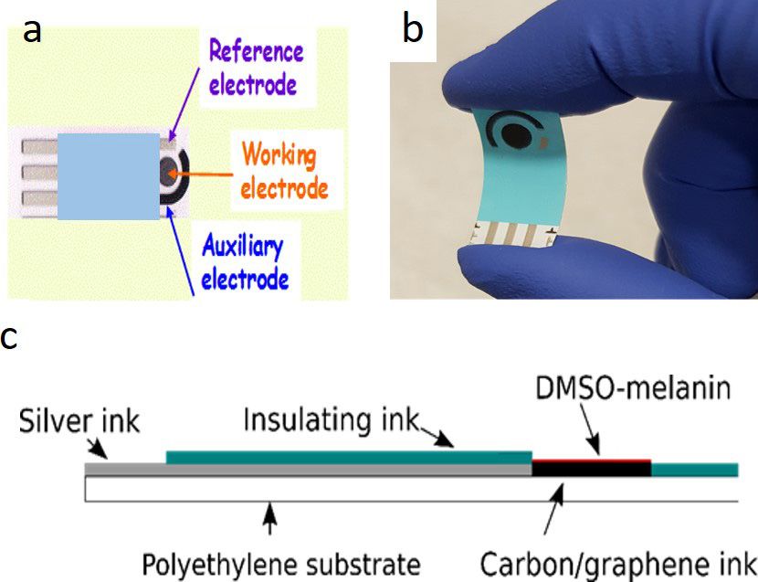
2D Mater. 7 (2020) 024008                                                                                                Z Tehrani et al

   Figure 1. Eumelanin, the brown-black pigment, is synthesised initially from the hydroquinone monomer, 5,6-dihydroxyindole
   (DHI, i.e. R  =  H) and 5,6-dihydroxyindole-2-carboxylic acid (DHICA, i.e. R  =  COOH) [35]. The various differing oxidative
   states for the monomer are also given, since as melanin is formed under oxidative conditions, these other monomers also become
   incorporated into the polymer.

   Figure 2. (a) The comproportionation reaction. Moieties of two differing oxidative states (hydroquinone and quinone) react
   together with water to form an intermediate oxidative state (the semiquinone) and hydronium [62] (b) the (de)protonation of the
   semiquinone. (c) Melanin is an unusual quinone system in that its quinones can undergo tautomerization to the quinone imine [63]
   (d) the final reaction is the (de)protonation of the quinone imine.

                                                       3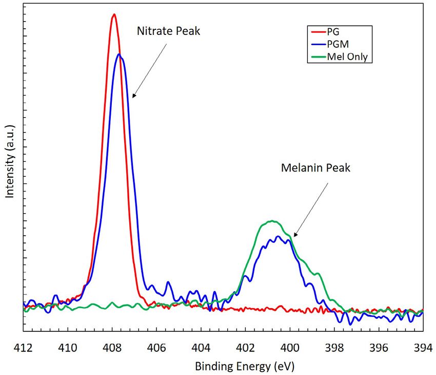
2D Mater. 7 (2020) 024008                                                                                       Z Tehrani et al

2. Materials and fabrication

2.1. Materials for screen printing
Polyethylene terephthalate (PET) with a thickness of
175µm (HIFI Melinex 339) was used as the substrate.
This film is heat stabilized to provide high temperature
resistance and hence dimensional stability during
processing [68]. This was used as is.
    Screen printed electrodes were prepared by first
printing conductive silver (SunTronic ref: AST6025)
or silver/silver chloride (Gwent ref: C2130809D5).
This highly conductive silver polymer ink was selected
for the printable refence electrode. It has been devel-
oped for a wide range of electronic applications [69]
and has the following characteristics necessary for
this application: compatibility with carbon inks, high
stability and long-term storage, excellent adhesion to       Figure 3. Basic monomeric moieties proposed for DMSO
                                                             melanin [67]. In addition to the standard moieties indicated
PET substrates after sintering. Carbon and graphene          in figure 1, many moieties are believed to form methylated
ink mixture (Gwent ref: C2171023D1) were used as a           sulfonate groups at the reactive sites of the initial starting
working electrode and finally a layer of insulating ink      monomer with various degrees of methylation.
(Gwent ref: D214011D5) to protect the electrode.
                                                           the electrode were covered with an insulator ink. The
2.2. Synthesis of melanin                                  design of the PGM sensor is shown in figure 4.
Melanin was synthesized from 3,4-dihydroxyphenyl-              There are many process settings that can influence
DL-alanine (DL-Dopa; Sigma-Aldrich, ⩾98%)                  the printed layer, deposition thickness, uniformity and
with dimethyl sulfoxide (DMSO; PA, Vetec, 99.9%)           topography, where final parameters settings are sum-
following a literature procedure [67]. 1.50 g of DL-       marised below.
Dopa and 0.93 g of benzoyl peroxide (Vetec, 75.0%–             The different parameters are the following:
80.0%) were dissolved in 200 ml of DMSO and kept           50 mm · s−1 forward speed, 10 mm · s−1 reverse speed,
under magnetic stirring for 58 d at room temperature       5.0 front squeegee pressure, 2.0 mm print gap and no
(≈27 °C.). Afterwards, the reaction solution was           snap-off distance (0.3 mm · s−1 snap speed) figure 1S
concentrated to 1/4 of the initial volume by increasing    in supplementary information (SI) (stacks.iop.org/
the temperature to 140 °C. Once it cooled down to          TDM/7/024008/mmedia) for details of printing.
room temperature, 150 ml of acetonitrile (Synth,           Through a comprehensive experimental programme,
99.5%) was added to the concentrated solution to           this was optimised to achieve print to print consist-
allow the melanin to be separated from synthetic by-       ency, thus assuring a path to full scale manufactur-
products. After two days, the solution was centrifuged     ing. When these were established, they were found to
at 2500 rpm for 15 min and the precipitate dried in an     deposit layers having consistent thickness and appro-
oven at 90 °C. The black/dark brown powder obtained        priate topography. Screens made of polyester having
was used to make a 30 mg ml−1 solution for film            appropriate thread count and thickness for each ink
fabrication.                                               layer were used and the associated oven temperature
                                                           for the drying regime is summarised in table 2.
2.3. pH-sensor fabrication
                                                               For the final step, 30 mg of DMSO melanin was dis-
2.3.1. Fabrication of electrodes by screen-printing
                                                           solved in 1 ml of anhydrous DMSO (Sigma-Aldrich,
The components of the sensor were printed using a
                                                           ⩾99.9%) which was stirred for 1 hour at 50 ºC and
R29 series screen printer from Reprint (UK) under
                                                           then filtered with a 0.45 µm Hydrophobic PTFE fil-
ambient air conditions. Basically, during the screen-
                                                           ter (Cole-Parmer). This solution was then spin coated
printing process, the ink is applied on the substrate
                                                           using two steps: first step, at 1000 rpm for 60s; and the
through a screen imaged according to the sensor
                                                           second step at 4000 rpm for 30s to form the final PGM.
layer requirements. The substrate was cleaned using
                                                               Thus, a very thin film of melanin was deposited on
isopropyl alcohol (IPA) before printing the bottom and
                                                           the screen-printed graphene electrode (PGM) by spin
top electrodes of the sensor in a stack configuration.
                                                           coating and the sensor was ready for testing.
The printing of electrode comprises four steps that
include silver/silver chloride printed as a reference
electrode, carbon printed as a current electrode and       2.4. Characterisation techniques
graphene as a working electrode, thin film melanin         The layers that formed the electrodes was characterized
covered the graphene layer and finally some parts of       for their thickness and topography using different

                                              4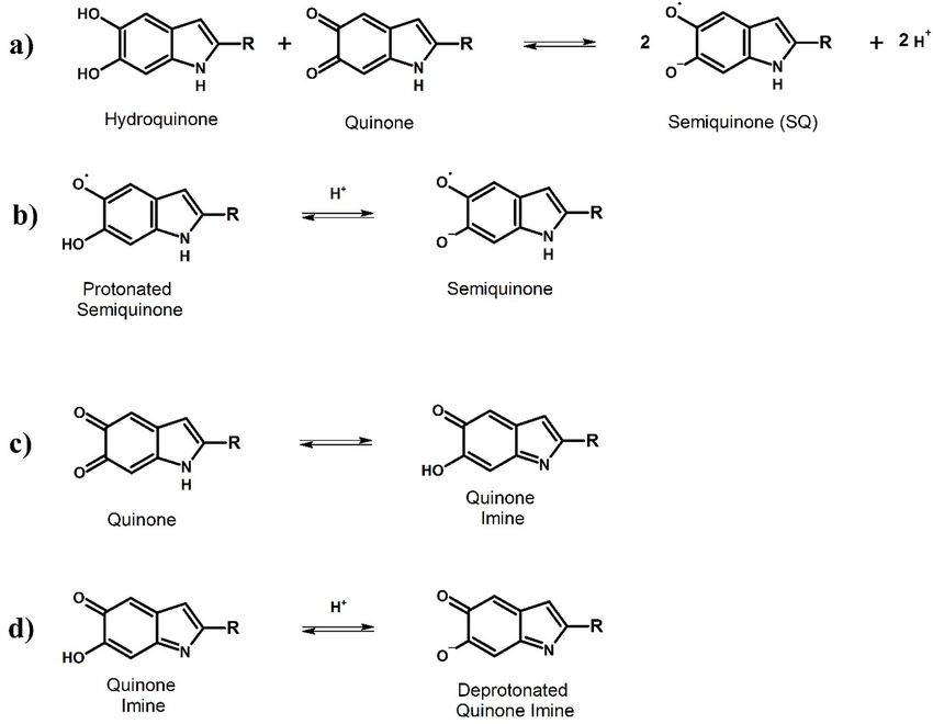
2D Mater. 7 (2020) 024008                                                                                                          Z Tehrani et al

                Figure 4. (a) Top view of electrode (b) optical image of the flexible sensor (c) cross section of PGM electrode.

          Table 2. Summary of printing and drying settings.                  The Fourier-transform infrared (FTIR) measure-
                        Screen used-                                     ments were obtained between 4000 and 400 cm−1, at
 Ink                    polyester       Drying regime in oven            room temperature, this technique has been employed
                                                                         to analyse D-Melanin in the sensor (SI figures 3S and
 Silver                 120–43          15 min @ 100 °C–110 °C           4S).
 Carbon                  61–64          15 min @ 100 °C–110 °C               Raman mapping measurements were taken with
 Graphene/carbon         61–64          10 min @ 90 °C–100 °C            an Renishaw system utilising a 532 nm excitation laser
 Insulator               61–64          25 min @ 100 °C                  with approximately 10mW of power on the sample,
                                                                         before and after melanin deposition (SI figure 5S).
techniques to check the quality of the printed layers.                       Electrochemical analysis was performed using the
These techniques include atomic force microscopy                         advanced potentiostat (PGSTAT-302 from Autolab,
(AFM), white light interferometry (WLI) and scanning                     Metrohm Autolab, Runcorn, UK) with the scanning
electron microscopy (SEM). These were applied at the                     voltage in the range of 0.0 V–0.8 V for evaluating the
different stages of fabrication.                                         electrochemical performance of PG and PGM elec-
    AFM was carried out using a JPK NanoWizards II.                      trodes. The standard 3-electrode system was used for
(Dimension-3100 Multimode, Bruker, Billerica, MA,                        the electrochemical evaluation, where printed gra-
USA), using a non-contact AFM tip with resonant                          phene/melanin was used as the working electrode,
frequency, spring constant and tip radius of 320 kHz,                    Ag/AgCl as the reference electrode and printed car-
40 N m−1 and 8 nm respectively operated on AC mode.                      bon as the counter electrode. 5 mM K3[Fe(CN)6]/
    WLI was performed using a Veeco Wyke NT9300                          K4[Fe(CN)6] in solution phosphate buffer saline
optical profiling system.                                                (PBS) solution (pH  =  7.4) was used as an electrolyte.
    Scanning electron microscopy (SEM; Ultra-High-                       To check the consistency of the sensor, four different
Resolution FE-SEM S-4800, Hitachi) was carried out                       samples were tested (SI figures 6S and 7S).
at 2.5 kV acceleration voltage and a 9.8 mA emission                         To test pH measurement, sensors were connected
current. The magnification was  ×20 k and working                        to the Ana-Pot from Zimmer and Peacock ltd set at a
distance was 10.8 mm.                                                    current range of 100 nA and measured for 600s in dif-
    X-ray photoelectron spectroscopy (XPS) measure-                      ferent buffer solutions (SI figure 9S).
ments were taken with a Kratos Axis Supra XPS system                         Electrical measurements were performed using
with a monochromatic Al Kα x-ray source, with an                         a 4-probe ‘EverBeing probe station’ with a 2636B
emission current of 15 mA.                                               Keithley Unit.

                                                          5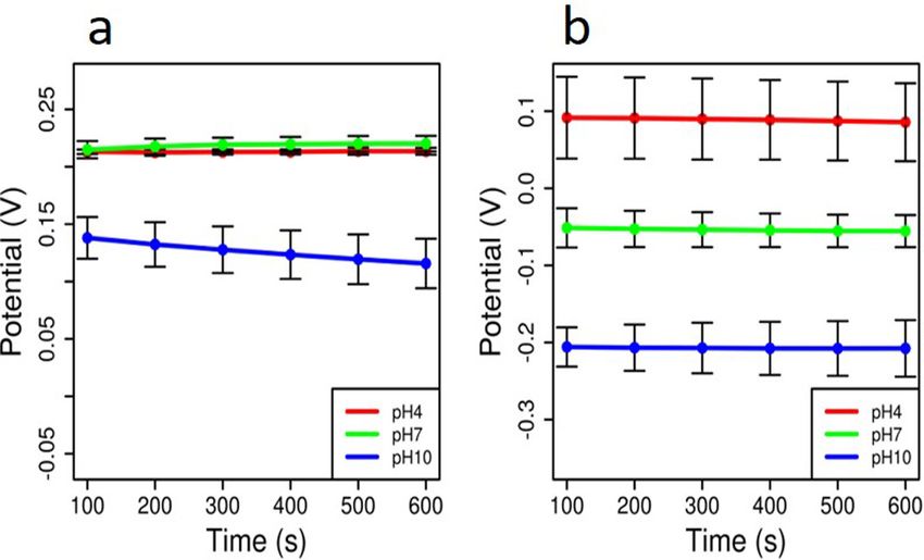
2D Mater. 7 (2020) 024008                                                                                                Z Tehrani et al

          Figure 5. An example of an axonometric profile of the surface using WLI. (a) Carbon–graphene electrode (b) Ag track.

         Table 3. Summary of thickness of different layers.          electrode (Metrohm Drop Sense DRP-110GPH) is
 Layer                    Thickness(µm)         Roughness(µm)
                                                                     shown in at table 3. Five measurements were taken at
                                                                     three rows and three columns in three different sheets
 Ag/AgCl                  10.63  ±  0.8         0.42  ±  0.19        of printing resulting in a total of 90 measurements col-
 Ag/AgCl110GPH            11.33                 —                    lected (for each layer: silver and graphene) the results
 PG                       8.61  ±  0.9          1.24  ±  0.12        are shown in at table 3. Where the standard deviation
 G-110GPH                 10.625                2.15                 is noted to be very small, thus confirming dimensional
 Insulator                14.5  ±  1.09         0.41  ±  0.05        consistency (SI figure 16S).
 PGM                      0.268  ±  0.03        282.66  ±  0.019         The roughness of the commercial sensor electrode
                                                                     (2.1 µm) is rougher than the PG (1.2 µm see table 3).
G: grapheme.
                                                                     However, the PG sensor has a more uniform roughness
3. Results and discussion                                            and the smoother surface is advantageous for the com-
                                                                     plete coverage of the electrode by a very thin melanin
3.1. Characterization of graphene-melanin                            layer (figures 12S–14S)
electrode: surface morphology                                            The SEM images show the uncoated printed gra-
The layers that formed the electrodes fabricated using               phene (PG) and the melanin coated graphene (PGM).
screen printing was characterized for their thickness                The small particles (carbon black) and the flake like
and topography.                                                      materials are present in both of the images taken
    WLI was used to measure the thickness and rough-                 before and after melanin coating took place. This is
ness of different parts of the sensors (figure 5).                   expected as the melanin molecules are extremely small
    As a benchmark, a comparison of thickness and                    and therefore not detectable by our SEM. These images
roughness between the in house printed graphene/car-                 show the surface structural characteristics of the work-
bon (PG) electrode with a commercial screen-printed                  ing electrode and how they appear to be unchanged

                                                        6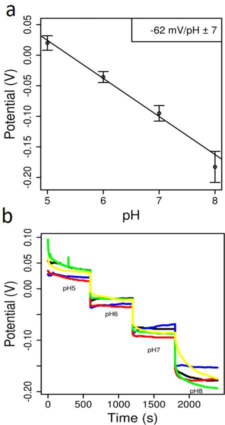
2D Mater. 7 (2020) 024008                                                                                             Z Tehrani et al

          Figure 6. Morphology top view of electrode with SEM (a) before coating- PG (b) after coating the melanin (PGM).

visually at the SEM level following melanin coating.               intensity ratio increase from 0.0744 to 0.0914. The
This shows that the amount of melanin that is required             D-peaks are most likely from the additional carbon
for the electrode to become functionalised and there-              compounds that are found in the carbon ink and from
fore sensitive to pH is extremely small (figure 6).                the application of melanin. The Raman system detects
    The effect of surface morphology of thin film of               these additional carbon compounds, especially after
melanin was also explored using AFM, figure 7.                     melanin application, resulting in an increased signal in
    The surface roughness of PG substrates before                  the D-band [70].
and after melanin deposition (figure 7) shows clear                    The intensity ratio between the 2D-peak and
changes in surface topography. The Root Mean Square                G-peak in monolayer graphene is approximately 2
roughness of 102.1 nm before deposition (figure 7(a))              [71]. However, this ratio decreases to approximately
increases to 292.5nm after deposition (figure 7(b)).               0.398, in the printed graphene electrode, denoting that
                                                                   there are multiple layers of graphene present on the
3.2. Characterization of graphene-melanin                          electrode [72].
electrode: surface chemistry                                           We obtained high-resolution XPS measurements
Raman mapping measurements were taken with a                       for N1s spectra as a reference for the chemical compo-
Renishaw inVia system using a 532nm laser, before                  sition of the blank surface (figure 9, red curve). We then
and after melanin deposition. The ratio between the                did a comparison test by obtaining data for d-melanin
G-peak, at 1580 cm−1, and the D-peak, at 1350 cm−1,                alone and then a melanin-coated electrode. In figure 9,
were mapped out using a custom MATLAB script (SI                   it can be seen that melanin has only one broad nitro-
figure 4S).                                                        gen-peak ranging from 397 to 403 eV related to amine
    As seen in (figure 8), the intensity of the D-peak is          groups in its aromatic structure (figure 1), in agree-
significantly higher, resulting in the D-peak/G-peak               ment with previous studies [67] while the graphene

                                                    7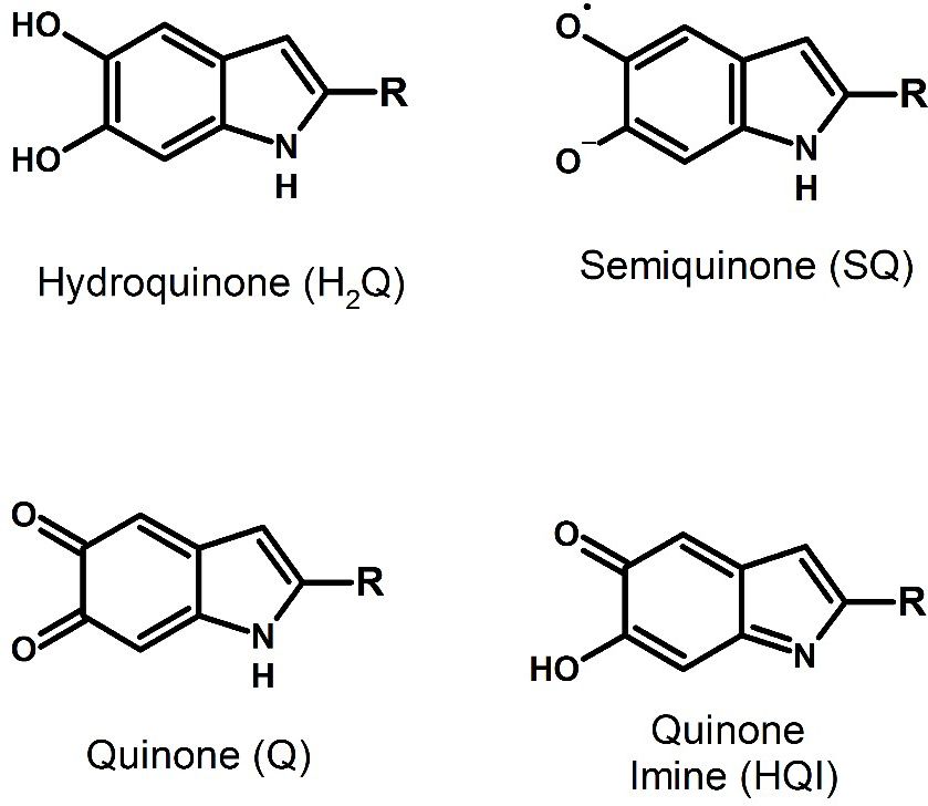
2D Mater. 7 (2020) 024008 Z Tehrani et al
Figure 7. Morphology of electrode with AFM (a) before coating- PG (b) after coating the melanin (PGM).
Figure 8. (a) Raman spectra of the electrode before (red) and after melanin coating (blue).
ink also has only one nitrogen peak at 408.2 eV from working electrode can be found with the maximum/
nitrate compounds. When the graphene electrode is peak of the curve. As can be seen, there is a clear dif-
coated with a melanin layer however, both nitrogen ference between the electrochemical behaviour of just
peaks can be observed. This indicates the presence of the bare electrode and afterwards when it is coated
the melanin on the graphene. The small shift towards with D-melanin. There are shifts in current and voltage
lower binding energies found for the peak at 408 eV, peaks, clearly showing that the D-melanin was depos-
when melanin is present, is probably related to struc- ited with good electrical contact, this test was carried
tural and/or electronic interaction between melanin out on multiple samples (table 4).
and graphene [73]. (SI for atomic concentration of
carbon and both nitrogen components in table 1S). 3.3. Four-point resistance measurements
We performed Square Wave Voltammetry ( figure IV curves using four probes revealed that there was
10) to characterise the electrochemistry of the a change in the resistance across the surface of the
electrodes. In this type of curve, the potential of the blank screen-printed electrode following melanin
8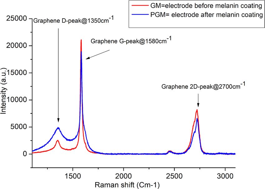
2D Mater. 7 (2020) 024008                                                                                                  Z Tehrani et al

   Figure 9. High-resolution N1s XPS spectra for melanin, graphene ink and melanin coated SPE. The presence of the nitrogen peak
   around 401 eV indicates that melanin is present at the surface of the sensor working electrode.

                                                                      Table 4. Peak current and voltage of electrode at different layers.

                                                                      Electrode         Peak current                Peak voltage

                                                                      Blank             0.05  ±  0.000 85 mA        0.34  ±  0.003 36 V
                                                                      PGM               0.55  ±  0.042 21 mA        0.15  ±  0.005 81 V

                                                                    pH4, pH7 and pH10, to see stability of the device over
                                                                    time. The difference between the voltage recorded with
                                                                    each reference buffer is presented in (figure 12).
                                                                         As can be seen, the PG electrode does not show
                                                                    any pH dependence at low pH. At high pH there is a
                                                                    dependence, but the electrode voltage decays over
                                                                    time. In contrast, the PGM (D-melanin coated) elec-
                                                                    trode shows a clear and significant change as a func-
                                                                    tion of pH. In addition, and importantly, the potential
                                                                    is stable over time, showing a robust and instantaneous
   Figure 10. An example Square Wave Voltammogram                   response to the pH.
   showing the change in peak current and peak voltage: for PG
   (red) and PGM (with melanin) (blue).                             3.5. Measured potential relationship to pH
                                                                    The potential of the PGM electrodes were recorded
deposition. Five blank PG and five PGM were tested.                 continously in solution as the pH was changed (figure
Each electrode was tested at five different positions on            13(a)) to demonstrate that the sensor can detect
the electrode to account for any heterogeneity in film              changes in the pH in real time. As can be seen, there
deposition. Five measurements were taken at each                    are clear changes in the potential at every pH change.
position resulting in a total of 250 data points collected          The sensitivity of the devices 35  ±  12 mV pH−1 over
(125 PG, 125 PGM) the results are shown in (figure 11).             the pH range from 2 to 12 (SI figure 9S).
The data clearly indicates that the melanin is present by                We did a further real time monitoring test by check-
the way it modifies the resistance measured.                        ing the hysterysis of the electrode behaviour. We did a
                                                                    test of the pH response going from pH 4 to 10 and then
3.4. Potentiometric measurements                                    from 10 to 4 (figure 13(b)). As can be see, the potential
Having shown the characterisation of the electrodes,                measured at the given pH and shows little hysterysis.
we now go on to demonstrate its sensing capabilities.                    The above test were done to demonstrate the sen-
The electrodes’ potential (PG and PGM) was                          sitivity and robustness of the devices across a wide
measured as a function of time in reference buffers                 range of pH. However, we are particualrly interested in

                                                      9
2D Mater. 7 (2020) 024008 Z Tehrani et al
Figure 11. Average resistance measurements on melanin (PGM) on non-melanin (PG) coated electrodes, error bars represent the
standard error multiplied by 2, with 125 data points for each condition.
Figure 12. The potential difference of electrode in pH specific reference buffers with averages (n = 3). (a) PG: before deposition of
melanin (b) PGM: after deposition of melanin to the electrode.
applying these devices for physiological measurements easuring the potential (figure 14(a)). The sensitiv-
m
since there is a high demand for measurement of pH in ity value we obtained is 62 ± 7 mV pH−1 (table 5),
medical settings. As such, we narrowed the range of pH an excellent result rivaling those of 3D graphene with
to be studied to between pH 5 and 8. HfO2 & IrO2/RGO [19, 20] (table 1).
The open circuit potential (OCP) is measured The major advantage of our sensor, in contrast to
using the potentiostat. No voltage is applied. The volt the above mentioned sensors is that it was made from
age potential that is measured is the difference between commonly avaiable materials coupled to a commeri-
the working electrode and the reference electrode, this cally viable low energy process for fabrication. We now
difference changes based upon the pH of the solution move onto testing of the stability and repeatability of
that the sensor is immersed in. The counter electrode the PGM electrode at physiological pH values. Five
balances the charge on the working electrode. PGM electrode were tested using a solution at pH
We obtained a sensitivity curve across this range 5 for 10 min followed by pH 5, pH 6, pH 7 and pH 8
by selecting the pH with a reference buffer and then solutions. As can be seen, a consistent change in the
102D Mater. 7 (2020) 024008 Z Tehrani et al
Figure 13. pH sensing performance of PGM (a) at extreme
pH ranges 2–12, (b) the PGM electrode tested for its ability to
recover following exposure to a high pH solution. Figure 14. Sensing results and sensor characterization,
(a) the slope was calculated using weighted least squares
regression to an uncertainty of two times the standard error
measured potential was seen across all the replicates with each solution indicated that the sensor remains highly
(figure 14(b)), indicating that there is consistency sensitive to pH changes following exposure to pH levels for
physiological environment (b) stability and repeatability of
across the fabrication process. the pH sensor. Data from the electrode was reanalysed using
the standard error ×2 in order to produce the error bars.
3.6. Mechanism of action
We now turn to the potential mechanism of operation
for the DMSO melanin pH sensor. The first thing to
note is that the pH sensitivity that has been recorded is
in line with previous pH/E data published on standard
melanin [27, 61]. This initial observation is surprising
at first, since one would expect a difference between
standard melanin and DMSO melanin which contains
additional sulfonated moieties (figure 3). However,
the pH/E behaviour is most likely due to the reduction
potentials of the one electron redox reactions for
the quinone/semiquinone and semiquinone/
hydroquinone pairs [47]. Both these reactions together
yield the comproportionation reaction (figure 2(a)).
This contrasts with our previous suppositions that
the pKas of the various constituents are responsible
for the effect [73]. Instead, both the the pKas and the
positions of the redox potentials is what drives the
redox chemistry and ultimately making our device a
sensitive pH sensor.
With the above, it is apparent that the DMSO mela-
nin electrochemically acts as a standard melanin, i.e.
the standard comproportionation reaction is active.
Considering that not all moieties in DMSO melanin
will be sulfonated, there will then still exist a melanin Figure 15. Potentials measured with pH7.2, pH7.3 and
fraction to undergo the electrochemistry. In contrast, pH7.4 reference buffers for 10 min intervals (a) Fresh
the sulfonated moieties may in turn only be specta- electrode (b) with the PGM sensors following testing with
plasma.
tors electrochemically. Overall, it appears that DMSO
112D Mater. 7 (2020) 024008 Z Tehrani et al
Table 5. Sensitivity of the electrode at different pH range. This suggests that the sensor will be well suited for
Stage Sensitivity (mV pH ) −1
pH range
applications that involve biological application and
may also have potential applications within a clinical
Blank 12.65 ± 20 2–12 setting. The sensor is sensitive enough to detect clini-
PGM -fresh 35 ± 12 2–12 cally relevant changes in blood plasma pH which the
PGM -reuse 33.8 ± 12 2–12 preliminary test started.
PGM -fresh 62 ± 7 5–8 In conclusion, a novel and highly sensitive pH sen-
PGM -reuse 60.24 ± 7 5–8 sor is demonstrated based on printed graphene coated
with a thin film of melanin derivative, DMSO melanin.
melanin has the same sensitivity as standard melanin In short, we have produced a pH sensor that has the
due to similar redox chemistry, but with the additional ‘best of both worlds’: low cost and reliable (i.e. com-
advantages of material processibility and stability. mercially viable) while being very sensitive.
The downside would be the presence of fewer stand-
ard melanin moieties. However, since the potential Acknowledgments
is what matters and not the total current, this is not a
great problem and further device engineering should This research was funded by Knowledge Economy
enhance the accuracy. Skills Scholarships (KESS). ZT acknowledges the
joint financial support from Welsh Government and
3.7. Preliminary tests with clinical samples. European Commission under European Regional
Since we are particulary interested in applying Development Funds (ERDF) through Sêr Cymru II
these sensors in physiological conditions, we have Fellowships (Project Number: 80761-su-100). ABM is
performed preliminary tests of the sensors on blood a Sêr Cymru II fellow and the results in this work have
plasma samples obtained from healthy volunteers received funding from the European Union’s Horizon
since plasma is commonly used as a clinical sample 2020 research and innovation program under the
type. To test the sensors, plasma was pipetted onto the Marie Sklodowska-Curie grant agreement no 663830.
surface of the sensor and the OCP was measured over JVP and CFOG would also like thank the financial
a 10 minute period. Following exposure to plasma, support of São Paulo Research Foundation (FAPESP;
the sensors were rinsed with distilled deionised grants 2015/23000-1, 2018/02411-1). The authors
water and measured with pH 7.2, pH 7.3 and pH 7.4 wish to thank to Ms Zelia Lagors and Mr Hugo Spieser
buffers (five PGM sensor were tested) to confirm their from Grenoble INP-Pagora France.
correct functioning (figure 15). The PGM sensor The authors would also like to acknowledge Dr
continued to exhibit sensitivity to the different pH Martin Peacock from Zimmer and Peacock Ltd.
buffers after being exposed to blood plasma, showing The authors declare no conflicts of interest.
that the sensor is robust enough to be considered as a
candidate in the development of applications where ORCID iDs
exposure to a clinical plasma sample is desired. This is
an example curve for one of the five sensors that were Z Tehrani https://orcid.org/0000-0002-5069-7921
tested (figure 14). A B Mostert https://orcid.org/0000-0002-9590-
2124
4. Conclusions C F O Graeff https://orcid.org/0000-0003-0162-
8273
We have produced printed graphene electrodes with
high reproducibility and good electrical conductivity
using a novel screen printing process. The fabricated References
printed electronic sensor exhibited improved
[1] Abdel-Rahman M A, Tashiro Y and Sonomoto K 2013 Recent
performance and properties when compared to a advances in lactic acid production by microbial fermentation
commercial sensor (DRP-110GPH) specifically with processes Biotechnol. Adv. 31 877–902
regards to reproducibility and homogeneity. [2] Guinovart T et al 2014 Bandage-based wearable
potentiometric sensor for monitoring wound pH
Furthermore, in using the active material DMSO
Electroanalysis 26 1345–53
melanin, a derivative of the pigment melanin (PGM), [3] Lemos S G et al 2007 Soil calcium and pH monitoring sensor
and which lends itself to the low energy fabrication, system J. Agric. Food Chem. 55 4658–63
we are able to create a sensitive pH sensor. The sensor [4] Phulara S C et al 2019 Modulation of culture medium confers
high-specificity production of isopentenol in Bacillus subtilis
itself demonstrates high levels of both accuracy and
J. Biosci. Bioeng. 127 458–64
precision within the physiologically relevant pH levels [5] Yang J et al 2016 Digital pH test strips for in-field pH
between pH 5 through to pH 8 (62 ± 7 mV pH−1). monitoring using iridium oxide-reduced graphene oxide
The above research provides the foundation for the hybrid thin films ACS Sensors 1 1235–43
[6] Yuqing M, Jianrong C and Keming F 2005 New technology for
later development and optimisation of the pH sensi-
the detection of pH J. Biochem. Biophys. Methods 63 1–9
tive properties of the electrode to further increase its [7] Baucke F 1985 The glass electrode—applied electrochemistry
accuracy and sensitivity. of glass surfaces J. Non-Cryst. Solids 73 215–31
122D Mater. 7 (2020) 024008 Z Tehrani et al
[8] Palleschi G et al 1994 Bioelectrochemical determination of [33] Tehrani Z et al 2014 Generic epitaxial graphene biosensors for
lactic and malic acids in wine Talanta 41 917–23 ultrasensitive detection of cancer risk biomarker 2D Mater.
[9] Janata J 1990 Potentiometric microsensors Chem. Rev. 1 025004
90 691–703 [34] Geim A K 2009 Graphene: status and prospects Science
[10] Komaba S et al 1997 Potentiometric biosensor for urea 324 1530–4
based on electropolymerized electroinactive polypyrrole [35] Meredith P and Sarna T 2006 The physical and chemical
Electrochim. Acta 42 383–8 properties of eumelanin Pigm. Cell Res. 19 572–94
[11] Contractor A et al 1994 Conducting polymer-based biosensors [36] Gray-Schopfer V, Wellbrock C and Marais R 2007 Melanoma
Electrochim. Acta 39 1321–4 biology and new targeted therapy Nature 445 851–7
[12] Ben-David O et al 1997 Simple absorption optical fiber pH [37] Lin J Y and Fisher D E 2007 Melanocyte biology and skin
sensor based on doped sol-gel cladding material Chem. Mater. pigmentation Nature 445 843–50
9 2255–7 [38] Zucca F A et al 2004 The neuromelanin of human substantia
[13] Li J-P, Peng T-Z and Fang C 2002 Screen-printable sol–gel nigra: physiological and pathogenic aspects Pigm. Cell Res.
ceramic carbon composite pH sensor with a receptor zeolite 17 610–7
Anal. Chim. Acta 455 53–60 [39] Panzella L et al 2013 Atypical structural and pi-electron
[14] Huang W-D et al 2011 A flexible pH sensor based on the features of a melanin polymer that lead to superior free-
iridium oxide sensing film Sensors Actuators A 169 1–11 radical-scavenging properties Angew., Commun. 52 12684–7
[15] Wu M-H et al 2009 Structural properties and sensing [40] d’Ischia M et al 2015 Melanins and melanogenesis: from
performance of high-k Sm2O3 membrane-based electrolyte– pigment cells to human health and technological applications
insulator–semiconductor for pH and urea detection Sensors Pigm. Cell Melanoma Res. 28 520–44
Actuators B 138 221–7 [41] Meredith P et al 2013 Electronic and optoelectronic materials
[16] Rahimi R et al 2016 A low-cost flexible pH sensor array for and devices inspired by nature Rep. Prog. Phys. 76 034501
wound assessment Sensors Actuators B 229 609–17 [42] Mostert A B et al 2010 Gaseous adsorption in melanins:
[17] Manjakkal L et al 2015 Sensing mechanism of RuO2–SnO2 hydrophilic biomacromolecules with high electrical
thick film pH sensors studied by potentiometric method and conductivities Langmuir 26 412–6
electrochemical impedance spectroscopy J. Electroanal. Chem. [43] Clulow A J et al 2017 The structural impact of water sorption
759 82–90 on device-quality melanin thin films Soft Matter 13 3954–65
[18] Melai B et al 2016 A graphene oxide pH sensor for wound [44] Mostert A B et al 2012 Role of semiconductivity and ion
monitoring 2016 38th Annual Int. Conf. of the IEEE Engineering transport in the electrical conduction of melanin Proc. Natl
in Medicine and Biology Society (IEEE) Acad. Sci. USA 109 8943–7
[19] Yang J et al 2016 Iridium oxide-reduced graphene oxide [45] Wünsche J et al 2015 Protonic and electronic transport in
nanohybrid thin film modified screen-printed electrodes as hydrated thin films of the pigment eumelanin Chem. Mater.
disposable electrochemical paper microfluidic pH sensors J. 27 436–42
Vis. Exp. 2016 e53339 [46] Sheliakina M, Mostert A B and Meredith P 2018 Decoupling
[20] Ameri S K, Singh P K and Sonkusale S R 2016 Three ionic and electronic currents in melanin Adv. Funct. Mater.
dimensional graphene transistor for ultra-sensitive pH sensing 28 1805514
directly in biological media Anal. Chim. Acta 934 212–17 [47] Motovilov K A et al 2019 Redox chemistry in the pigment
[21] Salvo P et al 2017 Temperature and pH sensors based on eumelanin as a function of temperature using broadband
graphenic materials Biosens. Bioelectron. 91 870–7 dielectric spectroscopy RSC Adv. 9 3857–67
[22] Chi L-L et al 2000 Study on extended gate field effect transistor [48] Mostert A B et al 2013 Hydration-controlled X-band EPR
with tin oxide sensing membrane Mater. Chem. Phys. 63 19–23 spectroscopy: a tool for unravelling the complexities of
[23] Batista P and Mulato M 2005 ZnO extended-gate field-effect the solid-state free radical in eumelanin J. Phys. Chem. B
transistors as p H sensors Appl. Phys. Lett. 87 143508 117 4965–72
[24] Chou J-C, Chiang J-L and Wu C-L 2005 pH and procaine [49] Batagin-Neto A, Bronze-Uhle E S and Graeff C F O 2015
sensing characteristics of extended-gate field-effect transistor Electronic structure calculations of ESR parameters of
based on indium tin oxide glass Japan. J. Appl. Phys. 44 4838 melanin units Phys. Chem. Chem. Phys. 17 7264–74
[25] Vieira N C et al 2011 Nanostructured polyaniline thin films as [50] Mostert A B et al 2018 The photoreactive free radical in
pH sensing membranes in FET-based devices Sensors Actuators eumelanin Sci. Adv. 4 eaaq1293
B 160 312–7 [51] Paulin J V, Batagin-Neto A and Graeff C F O 2019
[26] Chien Y-S et al 2012 A novel pH sensor of extended-gate Identification of common resonant lines in the EPR spectra of
field-effect transistors with laser-irradiated carbon-nanotube melanins J. Phys. Chem. B 123 1248–55
network IEEE Electron. Device Lett. 33 1622–4 [52] Hong L and Simon J D 2007 Current understanding of the
[27] da Silva M P et al 2014 Melanin as an active layer in biosensors binding sites, capacity, affinity, and biological significance of
AIP Adv. 4 037120 metals in melanin J. Phys. Chem. B 111 7938–47
[28] Hart J P and Wring S A 1997 Recent developments in the [53] Tran M L, Powell B J and Meredith P 2006 Chemical and
design and application of screen-printed electrochemical structural disorder in eumelanins: a possible explanation for
sensors for biomedical, environmental and industrial analyses broadband absorbance Biophys. J. 90 743–52
TRAC Trends Anal. Chem. 16 89–103 [54] Meredith P et al 2006 Towards structure–property–function
[29] Cui G et al 2000 A disposable amperometric sensor screen relationships for eumelanin Soft Matter 2 37–44
printed on a nitrocellulose strip: a glucose biosensor [55] Gallas J M 1987 Optical lens system incorporating melanin as
employing lead oxide as an interference-removing agent Anal. an absorbing pigment for protection against electromagnetic
Chem. 72 1925–9 radiation US Patent no. US5036115A
[30] Kahlert H 2008 Functionalized carbon electrodes for pH [56] Sheliakina M, Mostert A B and Meredith P 2018 An all-
determination J. Solid State Electrochem. 12 1255–66 solid-state biocompatible ion-to-electron transducer for
[31] Wu Y et al 2012 State-of-the-art graphene high-frequency bioelectronics Mater. Horiz. 5 256–63
electronics Nano Lett. 12 3062–7 [57] Ambrico M et al 2011 Melanin layer on silicon: an attractive
[32] Ameri S K, Singh P and Sonkusale S 2014 Utilization of structure for a possible exploitation in bio-polymer based
graphene electrode in transparent microwell arrays for metal-insulator-silicon devices Adv. Mater. 23 3332
high throughput cell trapping and lysis Biosens. Bioelectron. [58] Kumar P et al 2016 Melanin-based flexible supercapacitors
61 625–30 J. Mater. Chem. C 4 9516–25
132D Mater. 7 (2020) 024008 Z Tehrani et al
[59] Kim Y J et al 2013 Biologically derived melanin electrodes in [67] Paulin J V et al 2018 Structural and optical properties of
aqueous sodium-ion energy storage devices Proc. Natl Acad. soluble melanin analogues with enhanced photoluminescence
Sci. USA 110 20912–7 quantum efficiency Polym. Int. 67 550–6
[60] Bettinger C J and Whitacre J F 2015 A water-activated, [68] HIFI Melinex 339 PET film (www.hififilm.com/product/
ingestible battery US Patent no. US20150118526A1 melinex-339)
[61] Horak V and Weeks G 1993 Poly (5, 6-dihydroxyindole) [69] Tehrani Z et al 2017 Large-area printed supercapacitor technology
melanin film electrode Bioorganic Chem. 21 24–33 for low-cost domestic green energy storage Energy 118 1313–21
[62] Felix C et al 1978 Interactions of melanin with metal ions. [70] Taylor C E, Garvey S D and Pemberton J E 1996 Carbon
Electron spin resonance evidence for chelate complexes of contamination at silver surfaces: surface preparation
metal ions with free radicals J. Am. Chem. Soc. 100 3922–6 procedures evaluated by Raman spectroscopy and x-ray
[63] Szpoganicz B et al 2002 Metal binding by melanins: photoelectron spectroscopy Anal. Chem. 68 2401–8
studies of colloidal dihydroxyindole-melanin, and its [71] Ferrari A and Robertson J 2001 Resonant Raman spectroscopy
complexation by Cu(II) and Zn(II) ions J. Inorg. Biochem. of disordered, amorphous, and diamondlike carbon Phys. Rev.
89 45–53 B 64 075414
[64] Dezidério S N et al 2004 Thin films of synthetic melanin [72] Das A, Chakraborty B and Sood A 2008 Raman spectroscopy
J. Non-Cryst. Solids 338–40 634–8 of graphene on different substrates and influence of defects
[65] Bronze-Uhle E S et al 2013 Synthesis and characterization of Bull. Mater. Sci. 31 579–84
melanin in DMSO J. Mol. Struct. 1047 102–8 [73] Gargiulo V et al 2015 Supplementing π-systems: eumelanin
[66] Albano L G S et al 2016 Novel insights on the physicochemical and graphene-like integration towards highly conductive
properties of eumelanins and their DMSO derivatives Polym. materials for the mammalian cell culture bio-interface
Int. 65 1315–22 J. Mater. Chem. B 3 5070–9
14You can also read