Proteo-Genomic Analysis Identifies Two Major Sites of Vulnerability on Ebolavirus Glycoprotein for Neutralizing Antibodies in Convalescent Human ...
←
→
Page content transcription
If your browser does not render page correctly, please read the page content below
ORIGINAL RESEARCH
                                                                                                                                                    published: 16 July 2021
                                                                                                                                          doi: 10.3389/fimmu.2021.706757

                                             Proteo-Genomic Analysis Identifies
                                             Two Major Sites of Vulnerability on
                       Edited by:
              Abdul Qader Abbady,
                                             Ebolavirus Glycoprotein for
 Atomic Energy Commission of Syria,
                             Syria
                                             Neutralizing Antibodies in
                         Reviewed by:
                           Axel T Lehrer,
                                             Convalescent Human Plasma
         University of Hawaii at Manoa,
                           United States     Pavlo Gilchuk 1‡, Adrian Guthals 2†‡, Stefano R. Bonissone 3‡, Jared B. Shaw 4,
                            Victor Greiff,   Philipp A. Ilinykh 5,6, Kai Huang 5,6, Robin G. Bombardi 1, Jenny Liang 7, Ariadna Grinyo 7,
             University of Oslo, Norway      Edgar Davidson 7, Elaine C. Chen 8, Bronwyn M. Gunn 9†, Galit Alter 9, Erica Ollmann Saphire 10,
                  *Correspondence:           Benjamin J. Doranz 7, Alexander Bukreyev 5,6,11, Larry Zeitlin 2, Natalie Castellana 3
                  James E. Crowe Jr.         and James E. Crowe Jr.1,8,12*
               james.crowe@vumc.org
                                             1 The Vanderbilt Vaccine Center, Vanderbilt University Medical Center, Nashville, TN, United States, 2 Mapp
                    †
                   Present address:
                                             Biopharmaceutical, Inc. San Diego, CA, United States, 3 Abterra Biosciences (formerly Digital Proteomics LLC), San Diego,
                      Adrian Guthals,
                                             CA, United States, 4 Environmental Molecular Sciences Laboratory, Pacific Northwest National Laboratory, Richland, WA,
   Environmental Molecular Sciences
                                             United States, 5 Department of Pathology, University of Texas Medical Branch, Galveston, TX, United States, 6 Galveston
Laboratory, Pacific Northwest National
                                             National Laboratory, Galveston, TX, United States, 7 Integral Molecular, Inc., Philadelphia, PA, United States, 8 Department of
       Laboratory, WA, United States
                                             Pathology, Microbiology, and Immunology, Vanderbilt University Medical Center, Nashville, TN, United States, 9 Ragon
                   Bronwyn M. Gunn,
                                             Institute of MGH, MIT, and Harvard, Cambridge, MA, United States, 10 Center for Infectious Disease and Vaccine Research,
Paul G. Allen School of Global Animal
                                             La Jolla Institute for Immunology, La Jolla, CA, United States, 11 Department of Microbiology & Immunology, University of
 Health, Washington State University,
                                             Texas Medical Branch, Galveston, TX, United States, 12 Department of Pediatrics, Vanderbilt University Medical Center,
         Pullman, WA, United States
                                             Nashville, TN, United States
     ‡
      These authors have contributed
             equally to this work and
                share first authorship        Three clinically relevant ebolaviruses – Ebola (EBOV), Bundibugyo (BDBV), and Sudan
                                             (SUDV) viruses, are responsible for severe disease and occasional deadly outbreaks in
                  Specialty section:
                                             Africa. The largest Ebola virus disease (EVD) epidemic to date in 2013-2016 in West Africa
        This article was submitted to
Vaccines and Molecular Therapeutics,         highlighted the urgent need for countermeasures, leading to the development and FDA
              a section of the journal       approval of the Ebola virus vaccine rVSV-ZEBOV (Ervebo®) in 2020 and two monoclonal
             Frontiers in Immunology
                                             antibody (mAb)-based therapeutics (Inmazeb® [atoltivimab, maftivimab, and odesivimab-
              Received: 08 May 2021
             Accepted: 28 June 2021
                                             ebgn] and Ebanga ® (ansuvimab-zykl) in 2020. The humoral response plays an
             Published: 16 July 2021         indispensable role in ebolavirus immunity, based on studies of mAbs isolated from the
                             Citation:       antibody genes in peripheral blood circulating ebolavirus-specific human memory B cells.
 Gilchuk P, Guthals A, Bonissone SR,
                                             However, antibodies in the body are not secreted by circulating memory B cells in the
       Shaw JB, Ilinykh PA, Huang K,
     Bombardi RG, Liang J, Grinyo A,         blood but rather principally by plasma cells in the bone marrow. Little is known about the
     Davidson E, Chen EC, Gunn BM,           protective polyclonal antibody responses in convalescent plasma. Here we exploited both
      Alter G, Saphire EO, Doranz BJ,
    Bukreyev A, Zeitlin L, Castellana N
                                             single-cell antibody gene sequencing and proteomic sequencing approaches to assess
     and Crowe JE Jr. (2021) Proteo-         the composition of the ebolavirus glycoprotein (GP)-reactive antibody repertoire in the
      Genomic Analysis Identifies Two
                                             plasma of an EVD survivor. We first identified 1,512 GP-specific mAb variable gene
         Major Sites of Vulnerability on
            Ebolavirus Glycoprotein for      sequences from single cells in the memory B cell compartment. Using mass spectrometric
             Neutralizing Antibodies in      analysis of the corresponding GP-specific plasma IgG, we found that only a portion of the
        Convalescent Human Plasma.
          Front. Immunol. 12:706757.
                                             large B cell antibody repertoire was represented in the plasma. Molecular and functional
    doi: 10.3389/fimmu.2021.706757           analysis of proteomics-identified mAbs revealed recognition of epitopes in three major

Frontiers in Immunology | www.frontiersin.org                                        1                                             July 2021 | Volume 12 | Article 706757
Gilchuk et al.                                                                                            Ebolavirus Antibodies From Proteo-Genomics Studies

                                          antigenic sites - the GP head domain, the glycan cap, and the base region, with a high
                                          prevalence of neutralizing and protective mAb specificities that targeted the base and
                                          glycan cap regions on the GP. Polyclonal plasma antibodies from the survivor reacted
                                          broadly to EBOV, BDBV, and SUDV GP, while reactivity of the potently neutralizing mAbs
                                          we identified was limited mostly to the homologous EBOV GP. Together these results
                                          reveal a restricted diversity of neutralizing humoral response in which mAbs targeting two
                                          antigenic sites on GP – glycan cap and base – play a principal role in plasma-antibody-
                                          mediated protective immunity against EVD.
                                          Keywords: ebolavirus, ebolavirus infection, glycoprotein, proteo-genomics, convalescent plasma, viral antibodies,
                                          neutralizing antibodies, epitope mapping

INTRODUCTION                                                                      binds to domain C of its endosomal receptor, the protein
                                                                                  Niemann-Pick C1 (NPC1-C). The GP2 subunit contains the
Ebolaviruses are responsible for severe disease and occasional                    internal fusion loop (IFL) and stalk and is anchored into the
deadly outbreaks in Africa posing a significant health threat.                     viral membrane by a transmembrane domain (15–17).
The Ebolavirus genus consists of six species, including Zaire                         Recent improvement of instruments and technologies for high-
ebolavirus [represented by Ebola virus (EBOV)], Sudan                             throughput single B cell analysis enabled isolation of thousands of
ebolavirus [Sudan virus (SUDV)], Bundibugyo ebolavirus                            ebolavirus GP-reactive mAbs from the circulating memory B cells
[Bundibugyo virus (BDBV)], Taï Forest ebolavirus [Taï Forest                      of EVD survivors or vaccinees (18–20). Hundreds of mAbs were
virus (TAFV)], Reston ebolavirus [Reston virus (RESV)] (1), and                   characterized at the molecular level in studies that revealed a
Bombali ebolavirus [Bombali virus (BOMV)] (2). EBOV, BDBV,                        diverse landscape of epitope recognition in which distinct classes
and SUDV are the medically important causative agents of                          of mAbs recognized the MLD, glycan cap, GP1 head, GP1/GP2
symptomatic infections and ebolavirus disease (EVD) in                            trimer base, IFL, or stalk regions on the GP (21). This and other
humans. A total of 41 confirmed EVD outbreaks have been                            studies also demonstrated that only a fraction of ebolavirus GP-
documented, and the largest EVD epidemic to date occurred in                      reactive mAbs protects against infection or disease in vivo (18, 22,
2013-2016 in West Africa with a total of 28,610 disease cases and                 23). Nevertheless, protection is mediated principally by antibodies
11,308 deaths reported (3). The unpredictable nature of EVD                       circulating in serum that are secreted by long-lived plasma cells in
outbreaks and public health challenges stemming from the severity                 the bone marrow, not by circulating memory B cells (24). Bulk
of the disease underscores the need for development of medical                    serological analysis of plasma from four EVD survivors suggested
countermeasures and systematic studies to elucidate correlates of                 preferential binding of polyclonal plasma antibodies to
immune response-mediated protection against EVD.                                  proteolytically-cleaved form of GP (18). The landscape of
    The evidence to date suggests an indispensable role for                       epitope recognition by protective polyclonal antibody responses
antibody-mediated immunity in the protection against EVD.                         and their prevalence in convalescent plasma remains unknown.
Several investigational treatments based on human monoclonal                          We recently described a proteo-genomic approach for
antibodies (mAb) showed therapeutic efficacy in animal models of                   identifying sequences of antigen-specific polyclonal antibodies in
EVD (4–8) and clinical trials in the Democratic Republic of Congo                 animal serum (25) and reported methods for large-scale antiviral
outbreak demonstrated high efficacy of antibody-based                              human mAb discovery from the memory B cell repertoire (26). In
therapeutics for acute EVD treatment in patients (9). By 2020,                    this study, we characterize the circulating antibody response to
two monoclonal antibody-based therapeutics – ansuvimab-zykl                       ebolavirus GPs at the molecular level by identifying mAbs that are
(Ebanga®) and atoltivimab + maftivimab + odesivimab-ebgn                          present in convalescent plasma collected from an EVD survivor
(Inmazeb®) – were developed and approved by the Food and                          (Figure 1). For these mAbs we report the reactivity breadth,
Drug Administration (FDA) for clinical use (10, 11). A landmark                   epitope specificity, prevalence in plasma, functional activities,
achievement was the development and FDA approval of a                             and capacity to mediate in vivo protection.
recombinant viral vector-based vaccine (Ervebo®) for prevention
of EVD (12, 13), vaccination with which has been shown to induce
long-lasting antibody responses in clinical trials (14).                          MATERIALS AND METHODS
    The key target for protective antibodies is the ebolavirus
glycoprotein (GP), which is a single surface protein of the viral                 Human Samples
envelope. GP forms a trimer, in which each protomer consists of                   Human PBMCs and plasma were obtained at Vanderbilt
two subunits, designated GP1 and GP2. The GP1 subunit contains                    University Medical Center in Nashville, TN, USA, from a
a heavily glycosylated mucin-like domain (MLD) and a glycan cap,                  survivor of the 2014 EVD epidemic after written informed
which shields the host receptor binding site (RBS). The RBS is                    consent. The studies were approved by the Vanderbilt
exposed after proteolytical cleavage in the host endosome and                     University Medical Center Institutional Review Board. PBMCs

Frontiers in Immunology | www.frontiersin.org                                 2                                       July 2021 | Volume 12 | Article 706757
Gilchuk et al. Ebolavirus Antibodies From Proteo-Genomics Studies
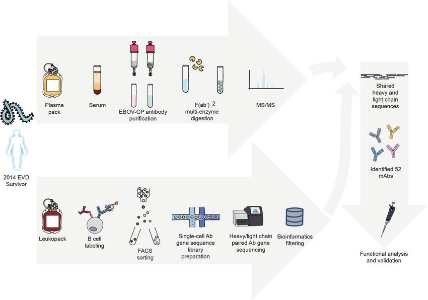
FIGURE 1 | Proteo-genomics workflow for the discovery of ebolavirus GP-specific monoclonal antibodies in convalescent human plasma. A cartoon representation
is shown of key workflow steps, which included collection of convalescent plasma from EVD survivor, affinity purification of polyclonal GP-specific antibodies from
plasma, mass-spectrometric analysis of plasma antibody protein sequences, isolation of GP-specific B cells from the peripheral blood mononuclear cells of an EVD
survivor, single-cell V(D)J gene sequence analysis, bioinformatics analysis to identify shared proteo-genomic antibody heavy and light chain variable region
sequences, recombinant expression of identified monoclonal antibodies, and functional analysis of recombinant antibodies.
and plasma were collected after the illness had resolved. A male EBOV GP and THP-1 cells were cultured in RPMI 1640 (Gibco)
human survivor of the 2014 EVD outbreak in Nigeria was age 31 medium supplemented with 10% FBS and 1% penicillin-
when infected and age 32 when PBMCs and plasma were streptomycin in 5% CO2, at 37°C.
collected 15 months later. At time of blood collection, plasma
samples were tested by qRT-PCR and found to be negative for Viruses
the presence of viral RNA. The mouse-adapted EBOV Mayinga variant (EBOV-MA,
GenBank: AF49101) (27), authentic EBOV Mayinga variant
Cell Lines expressing eGFP (28), and the infectious vesicular stomatitis
Vero-E6, Jurkat, Vero CCL-81, and THP-1 cells were obtained viruses rVSV/EBOV GP (Mayinga variant) (29) were used for
from the American Type Culture Collection (ATCC). Vero-E6 mouse challenge studies or neutralization assays. Viruses were
cells were cultured in Minimal Essential Medium (MEM) grown and titrated in Vero cell monolayer cultures.
(Thermo Fisher Scientific) supplemented with 10% fetal bovine
serum (FBS; HyClone) and 1% penicillin-streptomycin in 5% Monoclonal Antibodies
CO2, at 37°C. ExpiCHO (hamster, female origin) and FreeStyle MAbs EBOV-515, EBOV-520, and DENV 2D22 were described
293F cell lines were purchased from Thermo Fisher Scientific previously (19). Recombinant EBOV GP-specific mAbs 13C6
and cultured according to the manufacturer’s protocol. The and 4G7, and influenza HA-specific mAb C05 were produced as
Jurkat-EBOV GP (Makona variant) cell line stably transduced described below based on the variable gene sequences publicly
to display EBOV GP on the surface (Davis et al., 2019) was a kind available for these antibodies. Generation of new mAbs identified
gift from Carl Davis (Emory University, Atlanta, GA). Jurkat- by proteo-genomic approach is described below.
Frontiers in Immunology | www.frontiersin.org 3 July 2021 | Volume 12 | Article 706757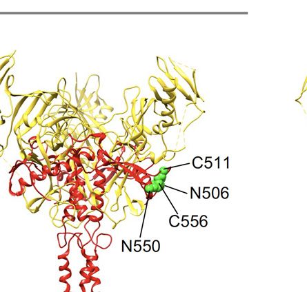
Gilchuk et al.                                                                               Ebolavirus Antibodies From Proteo-Genomics Studies

GP Expression and Purification                                          and then buffer-exchanged into PBS with a 0.5-mL Zeba spin
For B cell labeling, flow cytometric sorting, and purification of        column (Thermo Fisher Scientific) and coupled to Streptavidin
polyclonal antibodies from plasma, we used EBOV GP that was            Sepharose (GE Healthcare) at 1 mg/mL to prepare the affinity
produced in Drosophila Schneider 2 (S2) cells. Briefly,                 resin. Plasma was diluted 2 times in PBS, filtered through a 0.2-
recombinant ectodomain of EBOV GP DTM in a modified                     mm filter and applied to the 5 mL HiTrap protein G HP column
pMTpuro vector was transfected into S2 cells followed by               (Cytiva) and the GP-reactive IgG protein fraction was purified
stable selection of transfected cells with 6 mg/mL puromycin.          using vendors protocol. Purified antibodies were buffer-
GP ectodomain expression was induced with 0.5 mM CuSO4 for             exchanged into PBS. Approximately 1 mL of EBOV-GP-
4 days. Protein was purified using Strep-Tactin resin (Qiagen)          coupled resin was washed with PBS containing 0.5 M sodium
via an engineered strep II tag and purified further by Superdex         chloride, loaded with PBS, and used for affinity purification of
200 (S200) column chromatography. For ELISA studies, the               GP-reactive antibodies as follows. Unbound proteins were
ectodomains of EBOV GP DTM (residues 1-636; strain                     washed with 20 resin volumes of PBS followed by 10 resin
Makona; GenBank: KM233070), BDBV GP DTM (residues 1-                   volumes of 0.1 M glycine-HCI at pH 3.5. Bound antibodies
643; strain 200706291 Uganda; GenBank: NC_014373), SUDV                were eluted with 5 resin volumes of 0.1 M glycine-HCI at pH 1.8
GP DTM (residues 1-637; strain Gulu; GenBank: NC_006432),              and neutralized with 1M Tris buffer to adjust the pH to 7.4. The
and MARV GP DTM (residues 1-648; strain Angola2005;                    GP-reactive IgG protein fraction was buffer-exchanged into PBS
GenBank: DQ447653) were expressed using the FreeStyle 293F             and applied to a Streptavidin Sepharose column to remove
cell line and purified as described before (19).                        antibodies reactive to streptavidin. Antibody protein that was
                                                                       retained in the flow through fraction was collected, concentrated,
                                                                       quantified, and stored at 4°C until use.
Memory B Cell Isolation
PBMCs from a leukopak were isolated with Ficoll-Histopaque by
density gradient centrifugation. The cells were cryopreserved in       In-Solution Digestion and LC-MS/MS
the vapor phase of liquid nitrogen until use. Total B cells were       F(ab′)2 fragments were prepared from purified IgG protein using
enriched by negative selection from PBMCs using EasySep                IdeS cysteine protease containing polyhistidine tag (Promega)
Human Pan-B Cell Enrichment Kit (StemCell Technologies).               using the vendor’s protocol. F(ab′)2 fragments were separated
The EBOV GP-reactive memory B cells were labeled with the              from cleaved Fc fragments using protein A agarose (Pierce), and
recombinant EBOV GP protein that was produced in Drosophila            IdeS was removed using Talon Metal Affinity Resin (Takara).
S2 cells as described above and purified by flow cytometric cell         F(ab′)2 protein was buffer-exchanged into PBS and stored at
sorting using an SH800 cell sorter (Sony) as described                 -80°C until use. For mass spectrometric analysis, F(ab′)2 protein
previously (6).                                                        samples were denatured in 6 M guanidine HCl and 30 mM TCEP
                                                                       (tris(2-carboxyethyl)phosphine) at 60°C for 45 min. The
                                                                       denatured samples were alkylated with 30 mM iodoacetamide
Generation of Antibody Variable-Gene                                   for 30 minutes in the dark. After alkylation, the samples were
Libraries From Sorted B Cells                                          diluted or exchanged into appropriate buffers in accordance with
For paired antibody variable region gene sequencing, cells were        the manufacture’s protocols. The manufacturer’s protocols were
resuspended into DPBS containing 0.04% non-acetylated bovine           used for digestion conditions and enzyme ratio. Trypsin, LysC,
serum albumin (BSA), split into four replicates, and separately        GluC, AspN, and chymotrypsin were purchased from Promega.
added to 50 mL of RT Reagent Mix, 5.9 mL of Poly-dt RT Primer,         Elastase were purchased from Sigma Aldrich and pepsin was
2.4 mL of Additive A and 10 mL of RT Enzyme Mix B to complete          purchased from Worthington. All other chemicals were obtained
the Reaction Mix, as per the vendor’s protocol. The reactions          from Sigma Aldrich. Tandem mass spectra were acquired using a
then were loaded onto a Chromium chip (10x Genomics).                  Q Exactive HF Orbitrap mass spectrometer (Thermo Fisher
Chromium Single Cell V(D)J B-Cell-enriched libraries were              Scientific, Bremen, Germany) modified to enable 193 nm
generated, quantified, normalized and sequenced according to            ultraviolet photodissociation (UVPD) (31). Briefly, the
the User Guide for Chromium Single Cell V(D)J Reagents kits            instrument firmware was modified to enable triggering of an
(CG000086_REV C). Amplicons were sequenced on an Illumina              excimer laser (Excistar XS 500, Coherent, Inc.) that was aligned
Novaseq 6000, and data were processed using the CellRanger             on-axis with the HCD cell and irradiated precursor ions trapped
software v3.1.0 (10X Genomics). Bioinformatics filtering steps          in the HCD cell. Peptide separations were performed using a
were performed as described previously (26). The identities of         Waters nanoAcquity UPLC (Waters Corporation). Mobile phase
gene segments and CDRs from germlines were determined by               A and B were 0.1% formic acid in water and 0.1% formic acid in
alignment using the ImMunoGeneTics database (30).                      acetonitrile, respectively. Samples were prepared at 0.1 µg/µL and
                                                                       5 µL was injected and washed on a trap column (10 cm x 300 µm
                                                                       ID, C18, packed in-house) for 5 min at 5 µL/min with 99%
Purification of GP-Reactive Polyclonal                                  mobile phase A. Peptides were eluted from the trap column and
Antibodies From Plasma                                                 separated on an analytical column (70 cm x 75 µm ID, C18,
Purified EBOV GP was biotinylated at a 1:20 molar ratio in PBS          packed in-house) at 300 nL/min using a 100-minute gradient
using EZ-Link NHS-PEG4-Biotin (Thermo Fisher Scientific),               from 1% to 40% mobile phase A. Nanoflow UPLC was interfaced

Frontiers in Immunology | www.frontiersin.org                      4                                     July 2021 | Volume 12 | Article 706757
Gilchuk et al.                                                                                 Ebolavirus Antibodies From Proteo-Genomics Studies

with the mass spectrometer using an etched fused silica                  was clone-distinguishing peptide coverage over 50% of the CDR3
electrospray emitter (360 µm OD, 20 µm ID) and electrospray              and full peptide coverage of the CDR3.
voltage of 2.2 kV. Data-dependent top 5 acquisitions for HCD
and UVPD used 2E5 AGC target, 30K resolving power at m/z
200, and three microscans while 1E6 AGC target, 1 microscan,             Antibody Gene Synthesis
and 120K resolving power at m/z 200 was used for MS1. UVPD               For recombinant mAb production, cDNA encoding the genes of
was performed using a single laser pulse at 2 mJ, and HCD was            heavy and light chains were synthesized and cloned into DNA
performed with a normalized collision energy of 28. EThcD                plasmid expression vectors encoding IgG1 heavy chain and
tandem mass spectra were also acquired using an Orbitrap                 kappa or lambda light chain (32) and transformed
Fusion Lumos mass spectrometer (Thermo Fisher Scientific).                into E. coli cells to produce DNA for mammalian cell
Data dependent EThcD acquisition parameters included MS1                 expression. MAb proteins were produced following transient
resolution of 60k and AGC target of 5E5. EThcD MS2                       transfection of ExpiCHO cells following the manufacturer’s
parameters included resolution of 30k, 3 microscans, AGC                 protocol and were purified as described below.
target of 5E4, isolation width of 2 m/z, collision energy of 15,
and a cycle time of 3 seconds.
                                                                         MAb Production and Purification
                                                                         For parallel production of recombinant mAbs, we used
In-Gel Digestion and LC-MS/MS                                            approaches designated as ‘micro-scale’ or ‘midi-scale’ (26). For
F(ab′)2 protein was deglycosylated using PNGase F (New                   ‘micro-scale’ mAbs expression, we performed transfection
England Biolabs) per manufacturer’s protocols. 7 x 3 mg of               (~1 mL per antibody) of CHO cell cultures using a protocol for
sample was loaded into SDS-PAGE 4-12% Bis-Tris NuPage                    deep 96-well blocks (Thermo Fisher Scientific), as we previously
Mini-gel with the MOPS buffer system (Thermo Fisher                      described (26). For high-throughput micro-scale mAb
Scientific). Light chain and heavy chain protein bands were               purification, clarified culture supernatants were incubated with
excised for further processing using multi-enzyme digestion.             MabSelect SuRe resin (Cytiva), washed with PBS, eluted, buffer-
Excised gel bands were washed, reduced in 10 mM                          exchanged into PBS using Zeba Spin Desalting Plates (Thermo
dithiothreitol (DTT), alkylated in 10 mM iodoacetamide and               Fisher Scientific) and stored at 4°C until use. For ‘midi-scale’
digested. One mg of enzyme was used to digest each gel band,             mAbs expression, we performed transfection (~35 mL per
with the following incubation buffers: 25 mM NH4HCO3                     antibody) of CHO cell cultures as described by the vendor.
(trypsin, chymotrypsin, and Glu-C); 50 mM Tris-HCl, pH 8.0               MAbs were purified form culture supernatants using HiTrap
(elastase); 25 mM Tris-HCl, pH 8.0 (Asp-N); 25 mM Tris-HCl, 1            MabSelect SuRe columns (Cytiva). Purified mAbs were buffer-
mM EDTA, pH 8.5 (Lys-C); 0.1% formic acid (pepsin). Arg-C                exchanged into PBS, concentrated using Amicon Ultra-4 50 KDa
was incubated in 50 mM Tris-HCl, pH 7.9, 5 mM CaCl2, and                 Centrifugal Filter Units (Millipore Sigma) and stored at 4°C until
2 mM EDTA followed by activation in 5 mM Tris-HCl, pH 7.9,               use. To quantify purified mAbs, absorption at 280 nm (A280) was
5 mM DTT, and 0.2 mM EDTA. All enzymes were sourced from                 measured using a NanoDrop (Thermo Fisher Scientific).
Promega, except pepsin which was obtained from Worthington.
Each gel digest was analyzed by nano LC-MS/MS with a Waters
NanoAcquity HPLC system interfaced Orbitrap Velos Pro                    Cell-Surface-Displayed GP Antibody
(Thermo Fisher Scientific). Peptides were loaded on a trapping            Binding Assays
column and eluted over a 75 mm analytical column at 350 nL/              Alexa Fluor 647 NHS ester (Thermo Fisher Scientific) was used
min; both columns were packed with Luna C18 resin                        for antibody labeling. Binding of purified polyclonal or
(Phenomenex). The mass spectrometer was operated in data-                monoclonal antibodies to Jurkat-EBOV GP or Jurkat-EBOV
dependent mode, with MS performed in the Orbitrap at 60,000              GPCL cells was assessed by flow cytometry using an iQue
FWHM resolution. CID, ETD and HCD data were collected for                Screener Plus high throughput flow cytometer (Intellicyt
each precursor mass, CID and ETD were collected in the ion trap          Corp.) as described previously (6, 19). Briefly, ˜50,000 cells
and HCD data were collected in the Orbitrap at 7500 FWHM.                were added per each well of V-bottom 96-well plate (Corning)
The five most abundant ions were selected for MS/MS.                      in 5 mL of the DPBS containing 2% heat-inactivated ultra-low
                                                                         IgG FBS (Gibco) (designated as incubation buffer). Serial
                                                                         dilutions of antibody were added to the cells in replicates for a
Proteo-Genomic Data Analysis                                             total volume of 50 mL per well, followed by 1 h incubation at
We used the proprietary proteogenomic platform Alicanto for              ambient temperature, or 4°C in some experiments. Unbound
data analysis and visualization (25). 1,512 paired heavy and light       antibody was removed by washing with 200 mL of the incubation
chain antibody sequences derived from antigen-sorted memory              buffer. Staining of cells was measured by flow cytometric analysis
B cells were analyzed directly by Alicanto. The tandem mass              using an IntelliCyt iQue Screener Plus high throughput
spectra dataset was mapped by Alicanto to the repertoire of              cytometer (Intellicyt Corp.). Data for up to 20,000 events were
paired variable heavy and light region antibody variable region          acquired, and data were analyzed with ForeCyt (Intellicyt Corp.)
sequences. Antibodies in the cDNA gene sequence repertoire               software. Dead cells were excluded from the analysis on the basis
were defined as being present as proteins in the plasma if there          of forward and side scatter gate for viable cell population.

Frontiers in Immunology | www.frontiersin.org                        5                                     July 2021 | Volume 12 | Article 706757
Gilchuk et al.                                                                                 Ebolavirus Antibodies From Proteo-Genomics Studies

Binding to un-transduced Jurkat cells or binding of dengue                 For dose-response assays, serial dilutions of plasma of
antigen-specific mAb DENV 2D22 served as negative controls               purified polyclonal antibodies were applied to antigen-coated
for most experiments.                                                   wells in triplicate, and the assay was performed as described
    Cells that display cleaved GP were prepared as described            above. Non-linear regression analysis was used for curves fitting.
previously (6, 18, 19). Briefly, Jurkat-EBOV GP cells were washed
with DPBS containing calcium and magnesium (DPBS++),
resuspended at 106 cells/mL in DPBS containing 0.5 mg/mL of             Epitope Mapping Using an EBOV GP
thermolysin (Promega), and incubated for 20 min at 37°C.                Alanine-Scan Mutation Library
Cleavage reaction was inhibited by washing cells with the               Epitope mapping was carried out essentially as described
incubation buffer containing DPBS, 2% of heat-inactivated FBS           previously (33) using an alanine-scan mutation library of
and 2 mM EDTA (pH 8.0). The GP cleavage was confirmed by                 EBOV GP (Yambuku-Mayinga variant; Uniprot accession
loss of mAb 13C6 binding and high-level of binding that                 number Q05320) lacking the mucin-like domain (residues 311-
assessed with RBD-specific mAb MR78 relative to intact                   461). Our previous mapping of almost 200 anti-EBOV mAbs
Jurkat-EBOV GP antibody binding. Antibody binding to un-                identified 131 mutant GP clones validated as representing critical
transduced Jurkat (mock) cells served as a control for specificity       epitope residues. These GP cDNA clones were arrayed into 384-
of antibody staining.                                                   well plates, one mutant per well, transfected into HEK-293T cells
    For screening of the 52 micro-scale purified mAbs, cells were        and allowed to express for 22 hours. Cells were fixed in 4%
incubated with individual mAbs at a single 1:10 dilution, and the       paraformaldehyde in PBS containing calcium and magnesium
bound antibodies were detected using goat anti-human IgG                and incubated with antibody diluted in 10% normal goat serum
antibody conjugated with phycoerythrin (Southern Biotech).              (Sigma-Aldrich) for 1 h at ambient temperature, followed by a
    For the plasma antibody competition-binding assay, cells            0.5 h incubation with Alexa Fluor 488-conjugated secondary
were pre-incubated with 20 µg/mL of indicated mAb for which             antibodies (Jackson ImmunoResearch Laboratories) in 10%
the epitope specificity is known, followed by incubation with 20         normal goat serum. Cells were washed twice with PBS
µg/mL of Alexa Fluor 647-labeled polyclonal antibodies without          without calcium or magnesium and resuspended in
washing of unlabeled mAb and flow cytometric analysis.                   CellStripper (Cellgro) containing 0.1% BSA (Sigma-Aldrich).
Competition was quantified by comparing labeled polyclonal               Cellular fluorescence was detected using an Intellicyt high
antibody binding in the presence of indicated competing mAb to          throughput flow cytometer. Background fluorescence was
the level of maximal binding estimated from binding of labeled          determined by fluorescence measurement of vector-transfected
polyclonal antibodies in the presence of the dengue virus-specific       control cells. MAb reactivities against each mutant EBOV GP
mAb DENV 2D22.                                                          clone were calculated relative to wild-type EBOV GP reactivity
    For the epitope mapping competition-binding assay, cells            by subtracting the signal from mock-transfected controls and
were pre-incubated with 20 µg/mL of purified mAb followed                normalizing to the signal from wild-type GP-transfected
by incubation with 2 µg/mL of Alexa Fluor 647-labeled mAb for           controls. Mutated residues within critical clones were identified
which the epitope specificity is known and flow cytometric                as critical to the mAb epitope if they did not support reactivity of
analysis. Competition was quantified by comparing labeled                the test mAb but did support reactivity of other control
mAb binding in the presence of indicated competing mAb to               EBOV mAbs.
the level of maximal binding estimated from binding of labeled
mAb alone. Tested mAbs were considered competing if their
presence reduced the reference antibody binding to less than 30%        Neutralization Assays
of its maximal binding.                                                 A virus neutralization screening assay was performed under
                                                                        maximum biosafety level 4 (BSL-4) containment using
                                                                        recombinant EBOV-eGFP virus, as described previously (34).
ELISA Binding Assays                                                    Briefly, mAbs were mixed with virus and applied to Vero-E6 cell
Wells of 96-well microtiter plates were coated with 1 µg/mL of          monolayer cultures. In the absence of mAb neutralizing activity,
purified recombinant GP (produced in the FreeStyle 293F cell             the infection resulted in uniform eGFP fluorescence from the
line) in DPBS at 4°C overnight. Plates were blocked with 2% non-        monolayer of cells that was detected readily by fluorescence
fat dry milk (Bio-Rad Laboratories) and 2% normal goat serum            microscopy. For rapid screening analysis, micro-scale-purified
(Gibco) in DPBS containing 0.05% Tween-20 (DPBS-T) for 1 h.             mAbs were tested at a single 1:6 dilution; concentrations were
For rapid screening analysis, micro-scale purified mAbs were             not normalized. The results were expressed as percent virus
assessed at single 1:10 dilution in blocking buffer, added to the       neutralization relative to the infected cells control.
wells and incubated for 1 h at ambient temperature. The bound              Dose-response mAb neutralization studies were performed
antibodies were detected using goat anti-human IgG conjugated           using a plaque reduction neutralization test using infectious
with horseradish peroxidase (Southern Biotech) and TMB                  recombinant rVSV/EBOV-GP or rVSV/EBOV-GP CL , as
substrate (Thermo Fisher Scientific). Color development was              described previously (19). rVSV/EBOV-GPCL was generated by
monitored, 1N hydrochloric acid was added to stop the reaction          rVSV/EBOV-GP treatment with thermolysin (Promega), as
and the absorbance was measured at 450 nm using a                       described previously (19). MAbs were tested at four-fold
spectrophotometer (Biotek).                                             dilutions starting at 200 µg/mL in triplicate. Half maximal

Frontiers in Immunology | www.frontiersin.org                       6                                      July 2021 | Volume 12 | Article 706757
Gilchuk et al.                                                                                     Ebolavirus Antibodies From Proteo-Genomics Studies

inhibitory concentration (IC50) values were determined by                   RESULTS
nonlinear regression analysis using Prism software.
                                                                            Proteo-Genomic Identification of
Antibody-Mediated Cellular Phagocytosis                                     Ebolavirus Glycoprotein-Specific mAbs
by Human Monocytes                                                          From Human Plasma
The assay was performed as described before using EBOV GP-                  Peripheral blood mononuclear cells (PBMCs) and plasma were
coupled Alexa Fluor 488 Neutravidin beads and the THP-1 cell                collected from a human survivor of the 2014 EVD outbreak in
line (19). Micro-scale-purified mAbs were tested at a single 1:6             Nigeria 15 months after infection. Plasma from this individual
dilution; concentrations were not normalized. Results were                  exhibited a high level of GP-specific binding and broad reactivity
expressed as a phagocytic score that was determined using the               for diverse ebolavirus species as measured by ELISA using
percentage of Alexa Fluor 488+ cells and the median fluorescence             EBOV, BDBV, and SUDV recombinant soluble GP proteins
intensity (MFI) of the Alexa Fluor 488+ cells. A recombinant                (containing the full extracellular domain but lacking a
antibody based on the variable gene sequences of EBOV GP-                   transmembrane domain) (Figure 2A). In two separate studies,
specific mAb 13C6 was used as a positive control, and a                      we identified sequences of ebolavirus GP-specific antibodies
recombinant antibody based on the variable gene sequences of                from circulating plasma IgG protein and memory B cells.
the influenza virus A hemagglutinin-specific mAb C05 was used                     Total plasma IgG protein was purified using a protein G-
as a negative control.                                                      coupled affinity chromatography column followed by ebolavirus-
                                                                            reactive IgG purification using an EBOV GP-coupled affinity
Mouse Challenge                                                             column. EBOV GP-purified polyclonal IgG demonstrated high
Seven- to eight-week old female BALB/c mice were obtained from              specificity and broad reactivity to the three ebolavirus GPs by
the Jackson Laboratory. Mice were housed in microisolator cages             ELISA (Figure 2B) similarly to the GP reactivity and breadth
and provided food and water ad libitum. Challenge studies were              identified in the whole plasma IgG binding assay (Figure 2A).
conducted under maximum containment in an animal biosafety                  We next defined groups of GP-specific antibodies that bound to
level 4 (ABSL-4) facility of the Galveston National Laboratory,             common major antigenic sites in the purified EBOV GP-reactive
UTMB. Groups of mice (n = 5 per group) were inoculated with                 IgG fraction. We used a competition-binding assay with Jurkat
1,000 PFU of the EBOV-MA by the intraperitoneal (i.p.) route.               cells stably transduced to express EBOV GP on their surface
Mice were treated i.p. with 75 mg per mouse of individual mAbs              (Jurkat-EBOV GP) or the same cells that had been treated with
on the first day after virus inoculation (dpi). Antibody DENV                thermolysin to generate cell surface-displayed proteolytically
2D22 served as a control. Mice were monitored twice daily from 0            cleaved GP (Jurkat-EBOV GPCL). Cells were pre-incubated
to 14 dpi for illness, survival, and weight loss, followed by once          with EBOV-GP-reactive mAbs for which the epitope specificity
daily monitoring from 15 dpi to the end of the study at 28 dpi.             is known, including antibodies that recognize glycan cap (13C6)
The extent of disease was scored using the following parameters:            (33), base region (4G7, EBOV-515, and EBOV-520) (19, 33),
score 1 – healthy; score 2 – ruffled fur and hunched posture; score          receptor binding site (MR78 that is specific to Marburg virus
3 – a score of 2 plus one additional clinical sign such as orbital          [MARV] GP and recognize EBOV GP CL ) (35, 36), stalk
tightening and/or >15% weight loss; score 4 – a score of 3 plus one         (BDBV317), or mAb DENV 2D22 specific to dengue virus
additional clinical sign such as reluctance to move when                    (37). The level of competition binding was estimated using
stimulated, or any neurologic signs (seizures, tremors, head tilt,          fluorescently-labeled EBOV-GP-purified plasma IgG by
paralysis, etc.), or >20% weight loss. Animals reaching a score of 4        comparing to binding in presence of DENV 2D22. This study
were euthanized as per the IACUC-approved protocol. All mice                revealed that polyclonal antibody responses in this survivor
were euthanized on day 28 after EBOV challenge.                             targeted glycan cap and base region epitopes on intact GP and
                                                                            the GP base and RBS regions on GPCL, with a high prevalence of
Statistical Analysis                                                        base- and RBS-specific antibodies directed against GP CL
The descriptive statistics mean ± SEM or mean ± SD were                     (Figure 2C). Binding of EBOV-GP-purified polyclonal IgG to
determined for continuous variables as noted. Survival curves               Jurkat-EBOV GP or GPCL demonstrated that most reactivity in
were estimated using the Kaplan-Meier method and curves                     the plasma is mediated by antibodies recognizing epitopes on
compared using the two-sided log rank test (Mantel-Cox) with                GPCL (Figure 2D).
subjects right censored, if they survived until the end of the study.           F(ab′)2 fragments were prepared from the purified EBOV GP-
To correct for multiple comparisons Bonferroni-corrected                    reactive IgG protein fraction using IdeS cysteine protease that
threshold for significance level was determined. The comparisons             digests antibodies at a specific site below the hinge. The resulting
for plasma antibody competition-binding assay was performed                 fragments were subjected to high-resolution liquid chromatography
using ordinary one-way ANOVA with Dunnett’s multiple                        coupled to tandem mass-spectrometry. Multiple in-gel and in-
comparisons test. In neutralization assays, IC50 values were                solution protease digestion products were analyzed with
calculated after log transformation of antibody concentrations              higher energy collisional dissociation/ultraviolet photodissociation
using a four-parameter log-logistic (4PL) analysis. Technical and           (HCD/UVPD) on a customized Thermo Q Exactive HF
biological replicates are indicated in the figure legends. Statistical       and EThcD on a Orbitrap Fusion Lumos mass spectrometer
analyses were performed using Prism v8.4.3 (GraphPad).                      (Thermo Fisher Scientific). Bottom-up and middle-down MS/MS

Frontiers in Immunology | www.frontiersin.org                           7                                      July 2021 | Volume 12 | Article 706757
Gilchuk et al.                                                                                                                                                                                                                       Ebolavirus Antibodies From Proteo-Genomics Studies

                 A                                                                                                                         B
                                                                       Reactivity of plasma                                                                                        Reactivity of EBOV GP-purified
                                                                          antibodies                                                                                           4          IgG from plasma
                           Optical density at 450 nm

                                                                                                                                           Optical density at 450 nm
                                                          4

                                                          3                                                                                                                    3
                                                                                                                        EBOV GP
                                                                                                                        BDBV GP
                                                          2                                                                                                                    2                                                                                           EBOV GP
                                                                                                                        SUDV GP
                                                                                                                                                                                                                                                                           BDBV GP
                                                          1                                                             Normal serum                                           1                                                                                           SUDV GP
                                                                                                                        + EBOV GP
                                                                                                                                                                                                                                                                           Streptavidin
                                                          0                                                                                                                    0
                                                              1            2              3                 4       5         6                                                    -1             0                    1             2                  3
                                                              Log10 reciprocal plasma dilution                                                                                     Polyclonal antibody concentration
                                                                                                                                                                                              (log10ng/mL)

                 C                                                                                              Jurkat EBOV GP                                                                                                            Jurkat EBOV GPCL
                                                                                                                Jurkat                                                                                                                    Jurkat + thermolysin
                     antibody binding (MFI x 105)
                                  647 polyclonal

                                                 200000
                                                    2.0                                                                                                                        800000
                                                                                                                                                                                    8

                                                                                                                                                                                                                                                                 P=0.004
                                                                       P
Gilchuk et al.                                                                                    Ebolavirus Antibodies From Proteo-Genomics Studies

spectra yielded peptides between 6 and 40 amino acid of length.            of light chain variable region CDR3s identified. Thus, only a
From 60 mL of plasma, we obtained ˜0.16 mg of EBOV-GP-reactive             small portion of the large memory B cell antibody repertoire we
F(ab′)2 fragments that resulted in >205,260 mass spectra and 42 h of       obtained was detected as IgG protein in the plasma from this
LC-MS/MS time.                                                             survivor at the depth of proteomics and genomics analysis
    In parallel study, circulating B cells were enriched from              we achieved.
PBMCs by negative selection using magnetic beads
(STEMCELL Technologies). EBOV GP-reactive CD19+ B cells
were identified after labeling with biotinylated recombinant
soluble EBOV GP protein followed by detection with                         MAbs Identified in the Proteo-Genomic
allophycocyanin-conjugated streptavidin. EBOV-GP-labeled B                 Approach Recognize Ebolavirus GP,
cells were isolated by sorting in bulk using a Sony flow                    Demonstrate Diverse Reactivity to the GPs
cytometer. Isolated antigen-specific B cells were loaded on a               of Three Medically Important Ebolaviruses,
microfluidics device for single cell partitioning and barcoding             and Exhibit Varying Fv- and Fc Region-
(Chromium Controller; 10X Genomics) followed by reverse                    Mediated Functional Activities
transcription with PCR and sequence analysis to obtain paired              The mAbs identified in the approach described above were
heavy and light chain antibody variable gene sequences. A                  produced as recombinant IgG1 in transiently-transfected
detailed protocol for this antibody discovery workflow has                  Chinese hamster ovary (CHO) cells for functional antibody
been described previously (26). From ˜108 PBMCs we sorted                  analysis using previously described high-throughput
∼20,000 EBOV GP-reactive B cells and identified 1,512 paired                approaches for rapid antibody production and purification
antibody heavy and light chain variable region sequences                   from small sample volumes designated as microscale (26).
(Table S1).                                                                Fifty-one of 52 recombinant antibodies expressed sufficiently
    Using the Alicanto proteo-genomic analysis approach (25),              well to characterize their activity (Table S4). For initial screening
we next determined the sequences of protein antibodies in                  purposes, each mAb was tested at a single dilution from
plasma that were shared between plasma IgG and memory B                    microscale purification. The mAb concentration ranged from 1
cell antibody variable gene repertoires. Antibodies in the                 to 60 mg/mL for GP binding assays, and from 2 to 100 mg/mL for
repertoire were identified as being present in the plasma if                in vitro functional assays. The reactivity of individual mAbs was
there was clone-distinguishing peptide coverage over 50% of                assessed by ELISA using recombinant EBOV, BDBV, or SUDV
the complementarity determining region 3 (CDR3) and general                GP proteins. All identified mAbs exhibited ebolavirus GP-
peptide coverage over 100% of the CDR3 (Figure S1). We                     specific binding and revealed diverse reactivity profiles, in
compared the amino acid sequences from the plasma                          which the majority of mAbs reacted to EBOV or EBOV and
proteomics experiments with the inferred amino acid                        BDBV GP, and a smaller fraction of mAbs cross-reacted to the
sequences based on paired cDNA antibody sequences from                     GP of all three ebolaviruses (Figure 3A). Binding analysis using
memory B cells in the database obtained with the 10X                       cell-surface-displayed EBOV GP of EBOV GPCL suggested
Genomics single-cell experiments. This approach identified 5                recognition of diverse epitopes on the GP (Figure 3B). We
individual heavy and 48 individual light chain variable region             next assessed the functional activities of mAbs by performing
sequences in plasma (for a total of 53 heavy or light chain                Fc-mediated effector function and virus neutralization in vitro
sequences), that were ranked based on the distinct peptide count           assays, because the activities measured by these assays may
for peptides covering the corresponding CDR3 (Table S2).                   contribute to in vivo antibody function. We performed an
Heavy chain variable region CDR3s had between 1 to 15                      antibody-dependent cellular phagocytosis (ADCP) assay that
CDR3-covering peptides, and light chain CDR3s had between                  used beads coupled with recombinant EBOV GP to determine
1 to 10 peptides (Table S2). Of the 53 heavy or light chain                the capacity of bound mAb to activate human effector
variable region protein sequences identified in plasma, we                  cells in vitro. ADCP profiling revealed diverse activation
identified one cognate pair for which both the heavy and light              patterns with low, intermediate, or high activities observed for
chain proteins were both detected in plasma and also found as              individual mAbs (Figure 3C). A relatively small fraction (9 of 52)
paired cDNA sequences from a single circulating memory B cell.             of mAbs from the panel possessed detectable neutralizing activity
For the remaining 4 heavy or 47 light chain variable region                against live EBOV (Figure 3D) based on a >30% virus
protein sequences that were identified in plasma, we identified              neutralization cutoff for a single tested mAb dilution
either a matching heavy or light chain cDNA sequence in the                (Table S4). The reactivity of most (8 of 9) neutralizing mAbs
single-cell paired sequence database. Therefore, we identified a            we identified was limited to the homologous EBOV GP or EBOV
panel of 52 individual mAbs that we identified in both plasma               and BDBV GP, and one neutralizing mAb cross-reacted to GPs
and memory B cells with at least one chain match (Table S3).               of all three tested ebolaviruses. Several non-neutralizing mAbs
While the overall complexity of plasma and memory B cell                   bound efficiently to cell-surface-displayed GP and exhibited high
repertoires was difficult to assess, the overlap between these              ADCP activity, suggesting these mAbs may also contribute to
two repertoires was small as evidenced by the small number of              protective immunity. Together, these studies revealed diverse
proteomically-identified sequences and the small number of                  patterns of recognition of ebolavirus GP by mAbs that were
heavy chain variable region CDR3s compared to the number                   present in the plasma of the EVD survivor and identified

Frontiers in Immunology | www.frontiersin.org                          9                                      July 2021 | Volume 12 | Article 706757
Gilchuk et al.                                                                                                                                                    Ebolavirus Antibodies From Proteo-Genomics Studies

                                                                             A                                           B             C        D
                                                                             GP                                      Cell surface Fc-mediated Virus
                                                                          reactivity                                displayed GP    function neutrali-
                                                                           (ELISA)                                     binding      (ADCP)    zation

                                                                                                                                          CL
                                                                                                      MARV GP

                                                                                                                                                 EBOV GP
                                                            EBOV GP

                                                                                        SUDV GP

                                                                                                                      EBOV GP

                                                                                                                                EBOV GP
                                                                          BDBV GP

                                                                                                                                                           EBOV
                                            Antibody
                                              EBOV-1130
                                              EBOV-1131
                                              EBOV-1132
                                              EBOV-1133
                                              EBOV-1134
                                              EBOV-1135
                                              EBOV-1136
                                              EBOV-1137
                                              EBOV-1138
                                              EBOV-1139
                                              EBOV-1140
                                              EBOV-1141
                                              EBOV-1142
                                              EBOV-1143
                                              EBOV-1144
                                              EBOV-1145
                                              EBOV-1146
                                              EBOV-1147
                                              EBOV-1148
                                              EBOV-1149
                                              EBOV-1150
                                              EBOV-1151
                                              EBOV-1152
                                              EBOV-1153
                                              EBOV-1154
                                              EBOV-1155
                                              EBOV-1156
                                              EBOV-1157
                                              EBOV-1158
                                              EBOV-1159
                                              EBOV-1160
                                              EBOV-1161
                                              EBOV-1162
                                              EBOV-1163
                                              EBOV-1164
                                              EBOV-1165
                                              EBOV-1166
                                              EBOV-1167
                                              EBOV-1168
                                              EBOV-1169
                                              EBOV-1170
                                              EBOV-1171
                                              EBOV-1172
                                              EBOV-1173
                                              EBOV-1174
                                              EBOV-1175
                                              EBOV-1176
                                              EBOV-1177
                                              EBOV-1178
                                              EBOV-1179
                                              EBOV-1180
                                              EBOV-1181
                                                EBOV-520
                                            (EBOV) 13C6
                                          (Influenza) C05

                                                                                                                                                              Neutralizing
                                                            Low                                        High          Low          High         Low High       Non-neutralizing
                                                                      1             2             3             4    4 5 6                     100
                                                                                                                                                150

 FIGURE 3 | Reactivity and functional activity of 52 monoclonal antibodies identified in plasma by proteomic studies. Antibody sequences that were present in both
 genomic and proteomic repertoires were determined, generated as synthetic antibody-encoding cDNA in mammalian immunoglobulin IgG expression vectors, and
 produced as recombinant IgG1 using transiently-transfected CHO cells and a micro-scale expression protocol. Micro-scale purified antibodies were assessed for
 binding and functional activities at a single dilution (see Methods and Table S4). The broadly reactive neutralizing mAb EBOV-520, and recombinant forms of EBOV
 GP monospecific mAb 13C6 and influenza A hemagglutinin-specific mAb C05 were used as controls. (A) MAb binding to each of four GP proteins, including EBOV,
 BDBV, SUDV, and MARV GP. The figure shows a heatmap for binding of 52 mAbs expressed recombinantly, representing optical density (O.D.) values measured at
 450 nm for each antigen (range, 0.1 – 3.7). White indicates a lack of detectable binding, while blue indicates moderate binding and darker blue indicates higher
 binding. (B) MAb binding to Jurkat-EBOV or Jurkat-EBOV GPCL cells determined by flow cytometric analysis. The figure shows a heatmap for binding of 52 tested
 mAbs, representing log10 median fluorescence values for each tested condition (range, 3.6 – 6.4). White indicates background binding, and the gradient of blue
 indicates moderate binding and darker blue indicates the extent of specific binding. Binding of human antibody was detected using secondary goat anti-human IgG-
 specific antibodies conjugated to phycoerythrin. (C) Antibody Fc region-mediated cellular phagocytosis activity determined using EBOV-GP-coupled fluorescent
 beads and THP-1 monocyte human cell line. The figure shows a heatmap for binding of 52 tested mAbs, representing phagocytic score values (range, 62 to 195).
 (D) Screening for neutralizing antibody activity using biosafety level 4 recombinant EBOV encoding enhanced green fluorescent protein (eGFP). Green indicates
 neutralizing activity (>30% virus neutralization at the single mAb dilution tested), and white designates lack of neutralizing activity (
Gilchuk et al. Ebolavirus Antibodies From Proteo-Genomics Studies
functional mAbs that possessed neutralizing and/or the Fc- glycan cap (Figure 4). Several previous studies postulated an
mediated effector function activities. indispensable role in EBOV protective mAbs for Fc-mediated
effector functions of glycan-cap-specific mAbs that may be
sufficient alone or complement neutralizing activity (21, 38,
Major Antigenic Binding Sites Targeted by 39). Of 14 characterized glycan cap-specific mAbs, most
Class-Representative Plasma mAbs demonstrated strong reactivity to both EBOV and BDBV GP
Revealed Common Features by ELISA, many mAbs exhibited moderate or high Fc-mediated
To determine the molecular features of GP recognition by effector function activities, and four mAbs neutralized live EBOV
representative plasma antibodies, we produced 25 mAbs from (Figures 3A, C, D), suggesting these mAbs would have in vivo
the panel in a larger scale. For this study, we selected all of the functions. For individual mAbs, alanine scanning mutagenesis
neutralizing mAbs and also mAbs that demonstrated strong studies identified key binding site residues that mostly were
binding to cell-surface-displayed EBOV GP or EBOV GPCL located on the top part of the glycan cap (Figures 4, 5B). Many of
(Figure 3). Previous findings by us and others showed that these residues were defined previously by us and others as key
efficient binding to cell-surface-displayed GP is often associated contact residues for potent neutralizing glycan cap-specific
with antibody protective functions (18, 19). To define key contact human mAbs BDBV-289 (W275), EBOV-237 (N278), EBOV-
residues in the binding site, we used alanine scanning 337 (W275), EBOV-442 (W275, P273), and EBOV-548 (T240,
mutagenesis of the GP and tested the binding of individual T270, I274, W275), weakly neutralizing protective murine mAb
mAbs to each member of a shotgun mutagenesis alanine 1H3 (K276), or the non-neutralizing protective mAb 13C6
mutation library of the EBOV GP displayed in cells. In (T270, K272) (6, 33, 40, 41). We also assessed mAbs from this
addition, we performed competition-binding analysis using study in a competition-binding assay and demonstrated
reference EBOV-GP-reactive mAbs for which the epitope competition with mAb 13C6 for GP binding, which confirmed
specificity is known and cell-surface-displayed EBOV GP to the glycan cap specificity of these mAbs (Figure 4).
identify major binding sites recognized by the tested mAbs Ebolavirus entry involves cathepsin-mediated cleavage of GP
(Figure 4). This analysis revealed that the representative into GPCL in the endosome (42). Cleavage removes the glycan
plasma mAbs recognized three major binding sites, which cap and mucin-like domain of GP ectodomain, thereby exposing
included the base region (8 mAbs), the head domain/RBS the RBS for engagement of the NPC1 in the endosome (43, 44).
region (3 mAbs), and the glycan cap (14 mAbs). The other highly-represented class of EBOV GP-reactive IgGs in
Of the 9 strongly neutralizing mAbs in this panel, 5 mapped plasma included mAbs that recognize head domain/RBS. These
to the base region of the GP (Figure 4), along with 3 non- Abs exhibited broader reactivity to recombinant diverse GPs
neutralizing mAbs. Most of the base-specific mAbs (7 of 8) when compared to those identified for the base-region specific
exhibited stronger binding to EBOV GPCL than to uncleaved class of mAbs (Figures 3A, 4). Of three characterized mAbs, two
EBOV GP (Figure 3B), which was reminiscent of the binding were non-neutralizing and one mAb (EBOV-1133) possessed
pattern of polyclonal antibodies to cleaved GP (Figures 2C, D), weak neutralizing activity (Figure 4). Lack of potent virus
although these mAbs also bound strongly to intact cell-surface- neutralization by this class of mAbs likely is explained by
displayed GP. The majority (4 of 5) of neutralizing base-region- recognition of cryptic epitopes on the intact cell-surface-
specific mAbs bound strongly to EBOV but not to BDBV or displayed GP. Thus, all three mAbs bound preferentially to
SUDV GP. One neutralizing mAb (EBOV-1177) cross-reacted cleaved GP (Jurkat-EBOV GPCL) and showed weak or no
strongly to the GP of all three ebolaviruses (Figure 3A), detectable binding to uncleaved cell-surface-displayed GP
suggesting that this mAb could mediate broad neutralizing (Figure 3B), in a binding pattern similar to that of polyclonal
activity. Another common feature of these class-representative antibodies to cleaved GP (Figures 2C, D). Alanine scanning
mAbs was the similar location of key binding site residues. mutagenesis studies identified that, in addition to the binding site
These included residues positioned near the IFL-heptad repeat residues for these mAbs located on the GP head, three residues
1 (HR1) interface, and GP2 residues N550, D552, G553, C511 (I113, K114, and G118) mapped to a place in the RBS that is
and C556 (which form a disulfide bridge) that were identified exposed only after GP cleavage (Figures 4, 5C, D). In agreement
previously as key binding site residues for the potently with these results, one identified mAb (EBOV-1173) competed
neutralizing base-region murine mAbs 2G4 and 4G7 for binding to GPCL with the RBS-specific Marburg virus
(Figures 4, 5A) (33). MAbs 2G4 and 4G7, with the glycan (MARV) GP mAb MR78 that recognizes not only MARV GP
cap-specific mAb 13C6, formed the basis of therapeutic three- but also EBOV GPCL (36). Two other mAbs likely have distinct
antibody cocktail ZMapp™ used for treatment of human EVD epitopes within the RBS, such as they did not compete with
(8, 9). In agreement with the epitope residues mapped using MR78 (Figure 4).
alanine scan mutagenesis studies, the base-specific mAbs Together, these studies revealed that antibodies recognizing
competed with mAb 4G7 and/or the base-specific mAbs three major antigenic sites - base region, glycan cap, and head
EBOV-515 or EBOV-520 for the GP binding, as was shown domain/RBS - dominate the convalescent plasma IgG protein
by the cell-surface-display GP assay (Figure 4). repertoire of the EVD survivor and suggested preferential “hot
Another class of EBOV GP-reactive IgGs that was highly- spot” binding site residues on the GP for recognition by class-
represented in plasma comprised of mAbs that recognize the representative plasma mAbs.
Frontiers in Immunology | www.frontiersin.org 11 July 2021 | Volume 12 | Article 706757Gilchuk et al. Ebolavirus Antibodies From Proteo-Genomics Studies FIGURE 4 | Epitope mapping identified common features of plasma antibodies recognizing three major antigenic sites on EBOV GP. aMajor antigenic sites that were recognized by plasma-represented mAbs include base region, head domain/receptor binding site (RBS), and glycan cap. bGP mutations that reduce binding for indicated antibodies to the GP identified by alanine-scanning mutagenesis of cell surface-displayed EBOV GP library. Amino acids and their positions for key binding site residues are indicated. Residues that form a disulfide bridge in the GP subunit near the internal fusion loop (IFL)-heptad repeat 1 (HR1) interface (green), the Niemann-Pick C1 (NPC1) receptor binding site residues (magenta), and residues of the glycan cap that are commonly recognized by glycan cap-specific mAbs are (blue) are highlighted in respective colors. cCompetition-binding was assessed by measuring binding of fluorescently-labeled antibodies to Jurkat-EBOV GP cells that were pre-incubated with indicated unlabeled competing antibody. Competition was defined as
Gilchuk et al.                                                                                                 Ebolavirus Antibodies From Proteo-Genomics Studies

                  A                                                                   B

                  C                                                                                               D

 FIGURE 5 | Epitope residues of class representative plasma antibodies recognizing three major antigenic sites on EBOV GP. Amino acids and their positions for key
 binding site residues (green) are indicated for representative base-specific (A), glycan cap-specific (B), or head domain/RBS-specific (C) antibodies and shown on a
 ribbon diagram of the EBOV GP trimer (PDB ID: 5JQ3) for one protomer. GP1 is in yellow, and GP2 is in red. (D) The NPC1 receptor binding site residues of the
 RBS (PDB ID: 5F1B) are shown in orange and NPC1 contact region is indicated within a dashed orange shape on GPCL (top view). RBS residues I113, K114, and
 G118 identified as critical for binding of head domain/RBS-specific antibodies are shown in green.

Class-Representative Plasma mAbs                                                     Antibodies EBOV-1171 and EBOV-1176 potently neutralized
Exhibited Varying Levels of Neutralizing                                             rVSV/EBOV-GP with half maximal inhibitory concentration
Activity Against Chimeric VSV Displaying                                             (IC50) values below 100 ng/mL, indicating the presence of high-
GP or GPCL                                                                           potency neutralizing mAbs in the plasma of this EVD survivor.
Given many mAbs differentially recognized GP and GPCL in our                             In contrast, two of three head domain/RBS that recognized
Jurkat cell surface-displayed GP binding screening assay, we next                    cell surface-displayed GPCL only and did not neutralize EBOV,
examined in more detail the neutralizing capacity of class-                          exhibited a large (˜600-fold or higher) increase in neutralizing
representative mAbs (Figure 6). We used a replication-competent                      potency against VSV displaying cleaved GP, and partial (40 to
recombinant vesicular stomatitis virus (rVSV) displaying a full-                     90%) neutralization of rVSV/EBOV-GP at the highest
length EBOV GP in a place of VSV glycoprotein (rVSV/EBOV-GP)                         concentration tested (200 mg/mL) (Figure 6, middle panel).
or thermolysin-treated rVSV/EBOV-GP that generates a cleaved                         We concluded that the antigenic sites for these two mAbs are
intermediate form of GP (GPCL) displayed on the virion surface                       occluded on intact GP and become accessible only after
(rVSV/EBOV-GPCL) to estimate dose-response neutralization by                         proteolytic priming in the endosome of infected cells.
class-representative mAbs. We assessed four base region-specific                          Two EBOV neutralizing mAbs that recognize the glycan cap,
mAbs and two glycan cap-specific mAbs that were selected based on                     EBOV-1130 or EBOV-1164, neutralized rVSV/EBOV-GP with
their capacity to neutralize live EBOV in the initial screening study                high (IC50 = 47 ng/mL) or moderate (IC50 = 3,801 ng/mL)
and all three head domain/RBS-specific mAbs that did not show                         potency, respectively (Figure 6, bottom panel). EBOV-1164
detectable EBOV neutralization (Figure 3).                                           neutralized rVSV/EBOV-GPCL ∼60-fold less efficiently when
    Base region-specific mAbs, in agreement with their ability to                     comparing the IC50 value to the corresponding IC50 value from
efficiently recognize both Jurkat cell surface-displayed GP and                       neutralization of rVSV/EBOV-GP by this mAb, indicating that
GPCL, showed similar neutralization activity for both rVSV/                          the binding site for EBOV-1164 is largely altered after GP
EBOV-GP and rVSV/EBOV-GP CL (Figure 6, top panel),                                   cleavage. Neutralization of rVSV/EBOV-GP and rVSV/EBOV-
indicating that antigenic sites for this class of mAbs are                           GPCL by EBOV-1130 was similar, indicating that thermolysin
accessible on intact GP for antibody binding and neutralization.                     treatment did not alter the binding site for this mAb.

Frontiers in Immunology | www.frontiersin.org                                   13                                          July 2021 | Volume 12 | Article 706757
You can also read