Response of pathogenic yeast Cryptococcus neoformans to hypoxia - Palacký University, Olomouc Faculty of Medicine and Dentistry Department of ...
←
→
Page content transcription
If your browser does not render page correctly, please read the page content below
Palacký University, Olomouc
                  Faculty of Medicine and Dentistry
                    Department of Microbiology

Response of pathogenic yeast Cryptococcus neoformans to hypoxia

                             PhD thesis

                   Author: Mgr. Zuzana Moráňová
            Supervisor: MUDr. Vladislav Raclavský, Ph.D.

                           Olomouc 2013
I declare, that I have prepared this PhD thesis independently under supervision of MUDr.
Vladislav Raclavský Ph.D. and that I have acknowledged all literature used and also all
literature sources that are part of my thesis.

In Olomouc, June 20th 2013                                …………………….

                                             2
ACKNOWLEDGEMENTS

This work was partly completed at the Department of Microbiology, Faculty of
Medicine and Dentistry, Palacký University in Olomouc, Czech Republic and
partly at the Medical Mycology Research Centre, Chiba University in Chiba,
Japan.

My thanks belong to everybody who participated and contributed to accomplish
this work. First, I would like to thank to Vladislav Raclavský M.D., PhD, my
supervisor and to my colleagues at the Department of Microbiology for their help
and advice.

My special thanks belong to Prof. Susumu Kawamoto and members of his
laboratory in Chiba, where I spent 10 months and achieved most of the results
presented in this dissertation work. In particular, I would like to thank Misako
Ohkusu, Eric Virtudazo and Kiminori Shimizu for their great help not only in the
laboratory.

Czech Science Foundation (310/06/0645), Ministry of Education, Youth and
Sports, Czech Republic (MSM6198959223) and the Internal Grant Agency of
Palacky University Olomouc (LF_2013_012) supported this work. Infrastructural
part of this project (Institute of Molecular and Translational Medicine) was
supported from the Operational Programme Research and Development for
Innovations (project CZ.1.05/2.1.00/01.0030). This work was also partially
supported by Grants-in-Aid for Scientific Research from the Ministry of
Education, Science, Sports, and Culture of Japan (to Susumu Kawamoto) and
Cooperative Research Program of Medical Mycology Research Center, Chiba
University (to Susumu Kawamoto). The support of the Japan Society for the
Promotion of Science (JSPS) to Zuzana Moranova is highly acknowledged (JSPS
Postdoctoral Fellowship for Foreign Researchers [Short-term]). My thanks belong
to Dr. J. Kwon-Chung for providing the A. tumefaciens strain EHA 105
harbouring kanamycin resistance plasmid pYCC716, and to Dr. J. Heitman for
providing the pJAF1 plasmid harbouring the neomycin resistance marker cassette.

                                       3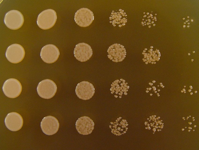
TABLE OF CONTENTS

1. INTRODUCTION .................................................................................................................. 6

2. REVIEW OF CURRENT LITERATURE ............................................................................. 8

   2.1. Genus Cryptococcus and Cryptococcus neoformans ...................................................... 8

     2.1.1. Classification and clinical importance ..................................................................... 8

     2.1.2. Cryptococcus neoformans species complex, ecology and epidemiology ................ 9

     2.1.3. Description, morphological characterisation and identification of C. neoformans 10

     2.1.4. Cryptococcosis ....................................................................................................... 11

     2.1.5. Clinical presentation and treatment of cryptococcosis ........................................... 14

   2.2. Virulence factors ........................................................................................................... 16

     2.2.1. Capsule ................................................................................................................... 17

     2.2.2. Melanin................................................................................................................... 20

     2.2.3. Ability to grow at human body temperature .......................................................... 22

   2.3. Cell cycle ....................................................................................................................... 24

     2.3.1. Regulation of cell cycle .......................................................................................... 24

     2.3.1.1. Cell cycle activation ............................................................................................ 25

     2.3.1.2. Cell cycle inhibition ............................................................................................ 26

     2.3.1.3. Cell cycle checkpoints ......................................................................................... 26

     2.3.1.3.1. G1 Restriction Checkpoint ............................................................................... 27

     2.3.1.3.2. G2 Checkpoint.................................................................................................. 27

     2.3.1.3.3. Morphogenesis checkpoint ............................................................................... 27

     2.3.1.3.4. Metaphase Checkpoint-Spindle Assembly Checkpoint ................................... 28

     2.3.2. Cell cycle control in C. neoformans ....................................................................... 28

   2.4. Response to hypoxia in C. neoformans ......................................................................... 29

   2.5. Strategy of unravelling the molecular mechanism of hypoxia-induced slowdown of
   proliferation and G2-arrest in C. neoformans ...................................................................... 39

   Appendix 1 ........................................................................................................................... 42

   Peculiar Clusters of Daughter Cells Observed in Cryptococcus neoformans Grown in
   Sealed Microtiter Plates ....................................................................................................... 42

   Appendix 2 ........................................................................................................................... 46

   Growth strategy of the pathogenic yeast Cryptococcus neoformans submerged culture
   under different cultivation formats ....................................................................................... 46

3. AIMS OF THESIS ............................................................................................................... 52

4. MATERIAL AND METHODS ........................................................................................... 53

     4.1. Strains and cultivation ............................................................................................... 53

     4.2. Random insertional mutagenesis - Agrobacterium mediated transformation ........... 53

     4.3. Screening system for hypoxia response in C. neoformans........................................ 53

     4.4. Identification of the insertional locus ........................................................................ 53

     4.5. Targeted deletion strategy ......................................................................................... 54

     4.5.1. Construction of CNAG_00156.2 deletion cassette for biolistic transformation .... 54

     4.5.2. Biolistic transformation .......................................................................................... 56

     4.5.3. Colony PCR............................................................................................................ 57

     4.5.4. Southern Blot Analysis........................................................................................... 57

     4.5.5. Reconstitution of CNAG_00156.2 deletion strain ................................................. 58

     4.6 Hypoxia phenotype .................................................................................................... 59

     4.7. Cell death and cell cycle assays ................................................................................ 59

     4.8. Biofilm formation ...................................................................................................... 60

5. RESULTS............................................................................................................................. 61


                                                                       4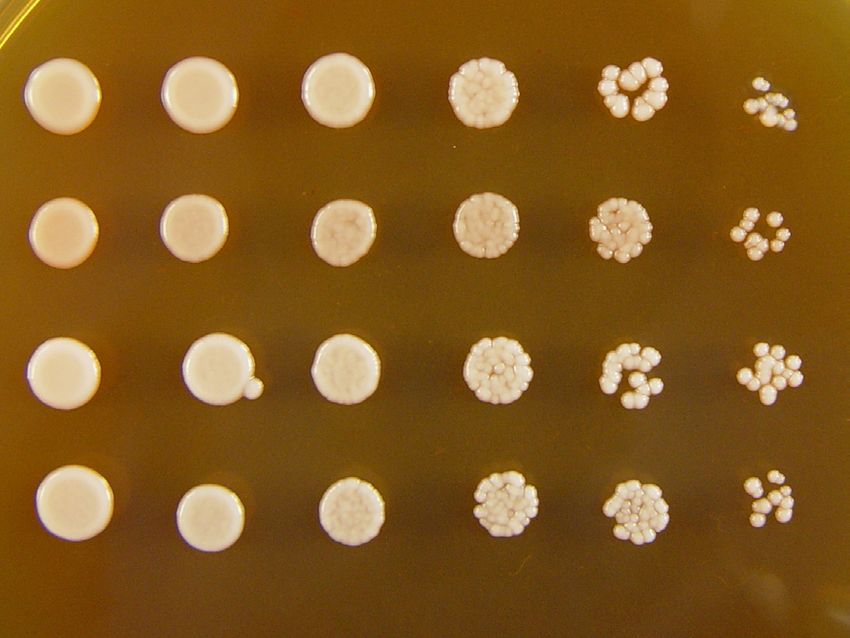
5. 1. Results obtained by screening of mutants prepared by Agrobacterium mediated
   transformation ...................................................................................................................... 61

      5.1.1. Random insertional mutagenesis - Agrobacterium mediated transformation ........ 61

      5.1.2. Screening system for hypoxia response in C. neoformans and gene identification61

      5.1.2.1. Screening for mutants showing hypoxia-specific phenotypes ............................ 61

      5.1.2.2. Identification of the insertional locus .................................................................. 61

      5.1.2.3. CNAG_00156.2 sequence characteristics and alignment ................................... 63

      5.1.3. Growth comparison of the wild type strain IFM49144, CNAG_00156.2 targeted
             deletion strain and reconstituted strains on different media at different
             temperatures. .......................................................................................................... 65

      5.1.3.1. Growth on solid YPD with different pH ............................................................. 65

      5.1.3.2. Growth at 37°C in liquid medium ....................................................................... 65

      5.1.4. Hypoxia phenotype ................................................................................................ 67

      5.1.5. Sensitivity to cell wall perturbing agents: Sodium Dodecyl Sulphate (SDS),
             CaclofluorWhite (CFW), Congo red (CR) ............................................................ 68

      5.1.6. Sensitivity to salts: NaCl, CaCl2, LiCl. .................................................................. 70

      5.1.7. Cell death and cell cycle assays ............................................................................. 71

      5.1.8. Biofilm formation ................................................................................................... 73

      5.1.9. Antifungal susceptibility testing............................................................................. 73

      5.1.10. Melanin formation and capsule induction assay .................................................. 73

6. DISCUSSION ...................................................................................................................... 75

7. CONCLUSIONS .................................................................................................................. 83

8. REFERENCES ..................................................................................................................... 84


                                                                      5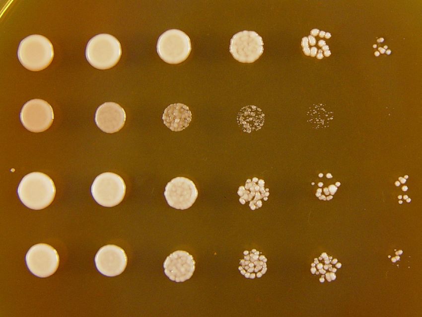
INTRODUCTION

                                    1. INTRODUCTION

       Cryptococcus neoformans is an obligate aerobic basidiomycetous fungus that
predominantly infects immunocompromised patients. The incidence of infections caused by
this encapsulated yeast has risen markedly over the past years as a result of the HIV epidemic
and increasing use of immunosuppressive therapies. Mortality from HIV associated
cryptococcal meningitis remains high (10–30%), even in developed countries, because of the
inadequacy of current antifungal drugs and combinations.
       C. neoformans is an environmental saprophyte. The rarity of its isolation as a human
commensal and of human-to-human transmission suggests that human infection is an
accidental dead-end event in its life cycle. However, uniquely amongst the thousands of
basidiomycetous fungi, C. neoformans, under selective pressure in the environment, has
evolved a number of characteristics that, perhaps by chance, also enable it to survive within
human and other mammalian and avian hosts. During pathogenesis, it relies on well-
established virulence factors: a capsule, melanin and the ability to grow at human body
temperature and others.
       Well-ventilated lungs are the primary sites of infection in this yeast, which is typically
followed by spread to the central nervous system that represents a tissue with considerably
lower oxygen level. For C. neoformans as obligate aerobic yeast oxygen is a growth-limiting
nutrient. Therefore, to sense oxygen concentrations and respond to its different levels is
essential and must rely on a delicate oxygen sensing system that controls cell proliferation.
       In past years a unique hypoxia-response has been described in C. neoformans,
represented by cell cycle arrest partly in unbudded G2 as well as G1. When oxygen
concentration drops in culture under conditions of limited aeration, cryptococcal cells slow
growth and delay the onset of budding to G2 that is gradually prolonged, resulting in
unbudded G2-arrest in some strains. It has been also demonstrated that during spread to CNS,
cryptococcal cells stay in very close proximity to blood vessels when crossing the blood-brain
barrier. Later the cells are able to invade brain parenchyma, however, still remain mainly
close to capillaries. Large cystic lesions are formed only 10 days later; most of them again
develop in close proximity to blood vessels. Moreover, deletion of genes identified to be
important for hypoxia signalling was shown to result in decreased virulence in a murine
model of cryptococcosis.

                                               6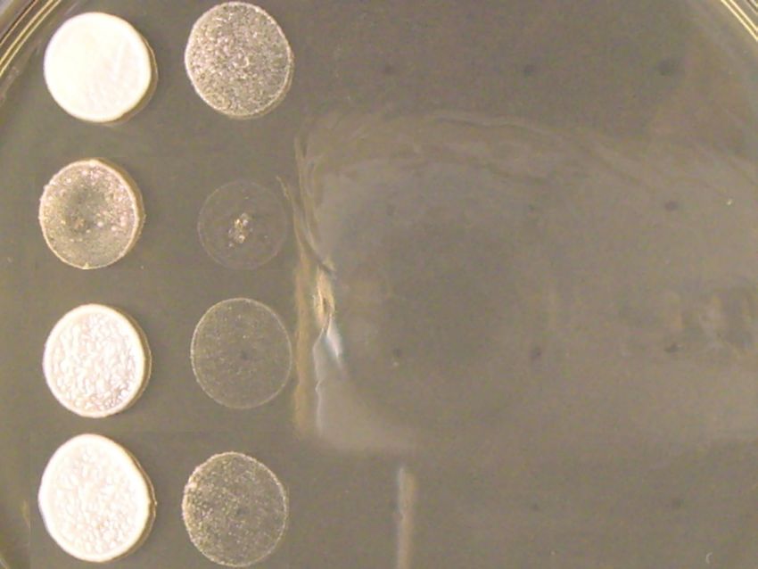
INTRODUCTION

       Therefore this work focuses on the potential new virulence factor – ability to adapt to
low oxygen levels. In our work we focused on identification of genes that are required for
slowdown of proliferation in response to limited aeration in submerged culture.
       Random insertional mutagenesis of C. neoformans was performed by transformation
with Agrobacterium tumefaciens harbouring kanamycin resistance plasmid. Transformants
were screened according to hypoxia response with regard to cell cycle progress to identify
components of hypoxia-induced G2 arrest on molecular level.
       We report that the cryptococcal homologue of yeast CRZ1 appears to play an
important role when C. neoformans is required to adapt its proliferation to limited aeration.
The participation of cryptococcal CRZ1 homologue in the slowdown of proliferation under
limited aeration represents both a novel biological role for this conserved protein and also
opens a new field in the study of hypoxia sensing and signalling in C. neoformans.

                                              7
REVIEW OF CURENT LITERATURE

                        2. REVIEW OF CURRENT LITERATURE

                   2.1. Genus Cryptococcus and Cryptococcus neoformans

2.1.1. Classification and clinical importance

Kingdom:        Fungi
Phylum:         Basidiomycotina
Order:          Filobasidiales
Family:         Filobasidiaceae
Genus:          Filobasidiella (Cryptococcus)

         The genus Cryptococcus includes at least 39 species of variously encapsulated yeast-
like fungi that reproduce by budding rather than by producing spores. Among these, only
Cryptococcus neoformans species complex is considered as pathogenic (Casadevall, 1998).
Many non-pathogenic species of Cryptococcus are commonly found in the soil and on the
skin and mucous membranes of healthy people.
         C. neoformans is dimorphic basidiomycetous pathogenic fungus. The perfect (sexual)
form or teleomorph is called Filobasidiella, but the imperfect (asexual) form or anamorph is
called Cryptococcus. Mostly, the asexual form has been described in association with clinical
specimens (Casadevall, 1998). The sexual form is characterized by the presence of
basidiospores and is observed during mating.
         Cryptococcus was identified as a human pathogen in 1894 by a pathologist named
Busse who isolated the yeast from the tibia of a 31-year-old woman and published the case
report that same year. The following year, a surgeon named Buschke reported the same isolate
from the same patient, thus establishing the early eponym of Busse-Buschke disease. This
single case served to identify new yeast and to prove its pathogenic potential (King, 2007).
         Since the first discovery, researchers have identified the diverse spectrum of host
responses to cryptococcal infection. The responses range from a harmless colonization of the
airways and asymptomatic infection in laboratory workers (resulting in a positive skin test
result) to meningitis or disseminated disease.
         Although virulence varies among cryptococcal strains, virulence probably plays only
partial role in the outcome of an infection. The other crucial factor is the immune status of the

                                                 8
REVIEW OF CURENT LITERATURE

host. The most serious infections usually develop in patients with defective cell-mediated
immunity. For example, patients with AIDS, patients undergoing organ transplantation,
patients with reticuloendothelial malignancy, patients undergoing corticosteroid treatment
(but not those with neutropenia or immunoglobulin deficiency), and patients with sarcoidosis
develop the most serious infections (King, 2007).
      With the global emergence of AIDS, the incidence of cryptococcosis is increasing and
now represents a major life-threatening fungal infection in these patients.

2.1.2. Cryptococcus neoformans species complex, ecology and epidemiology

       Conventional     nomenclature    classified   C.   neoformans     into   three   varieties
(C. neoformans, C. gattii, C. grubii) and five serotypes (A, B, C, D, including the hybrid
serotype AD) based on antigenic specificity of the capsular polysaccharide, which differ in
ecological, morphological-physiological and molecular features, geographical distribution,
epidemiology and virulence properties. C. neoformans and C. gattii diverged from a common
ancestor an estimated 37 million years ago (Fig.1; Xu, 2000).
       Some authors have suggested establishing C. neoformans var. gattii (B, C serotype) as
a new species C. gattii. Also DNA genetic typing techniques recognised C. neoformans var.
gattii as species, based on genetic variability and lack of evidence for genetic recombination
between neoformans and gattii (Kwon-Chung, 2002). According to this C. neoformans var.
neoformans and C. neoformans var. grubii should form the species C. neoformans sensu
stricto (Kwon-Chung, 2006). But Bovers et al. (2008) recently proposed that these two
varieties should also be described as different species. Further studies are required to analyse
the ongoing speciation events within this clade.
       Most clinical and environmental isolates belong to serotype A that is responsible for
over 95% of cryptococcosis cases worldwide (Lin, 2009) and most of them are mating type
alpha. C. neoformans var. grubii (serotype A) and C. neoformans var. neoformans (serotype
D) cause disease mainly in immunocompromised patients, whereas C. neoformans var. gattii
(serotypes B, C) rarely infects people with HIV infection and other immunosuppressed
patients. Patients infected with C. neoformans var. gattii are usually immunocompetent,
respond slowly to treatment, and are at risk for developing intracerebral mass lesions (e.g.,
cryptococcomas) (King, 2007). Soil contaminated with bird (mainly pigeon) excreta was
traditionally known as the natural habitat of C. neoformans var. grubii and var. neoformans
(Sorrell, 1997). High concentration of the organism was also found in woody debris in

                                               9
REVIEW OF CURENT LITERATURE

hollows of aged trees (Lazera, 1996; Lazera, 2000; Randhawa, 2001). C. neoformans can
utilize this woody material which is rich in lignin, polyphenole compounds and cellulose as a
substrate for growth due to its laccase, phenol oxidase and cellulase activities (Botes, 2009).

Fig. 1. The C. neoformans species complex. The Cryptococcus species complex contains at
least two subspecies: C. neoformans and C. gattii, which diverged from a common ancestor
an estimated 37 million years ago. The serotypes A and D diverged from each other 18.5
million years ago while serotypes B and C diverged from each other 9.5 million years ago (Xu
et al., 2000).

       Whereas C. neoformans var. grubii and var. neoformans are ubiquitous,
C. neoformans var. gattii can be primary found in tropical and subtropical regions where it is
associated mainly with eucalyptus trees (Ellis, 1990). C. gattii isolates can also cause
infection in regions with a temperate climate (Colom, 2005; Montagna, 1997) and is
responsible for the ongoing outbreak of cryptococcosis on Vancouver Island, Canada (Kidd,
2004) and has recently been detected in other areas in the Pacific Northwest, USA
(MacDougall, 2007).
       Based on these techniques C. neoformans complex is further divided into 9 major
molecular types or genotypes.

2.1.3. Description, morphological characterisation and identification of C. neoformans

       C. neoformans cells form round yeast like cells that are 3-6 µm in diameter and
reproduce by multilateral budding. The capsule is distinctive feature that sets C. neoformans

                                               10
REVIEW OF CURENT LITERATURE
apart from the other medically important yeasts and it is easily visualized in a dilute
suspension of India ink. The capsule size varies according to the strain and culture conditions
with most isolates having a medium sized capsule resulting in a total diameter 4-10 µm. Due
to the capsule, cultures on solid media have a mucous appearance.
C. neoformans forms smooth, convex, and yellow or tan colonies at 20-37°C on most
bacteriological and mycological (Sabouraud dextrose agar) solid media. In liquid media
bottom growth and ring formation can be observed, occasionally a pellicle, often the entire
content of the flasks changes into a slimy mass. The growth rate is somewhat slower than
Candida and usually takes 48 to 72 h.
This fungus is identified based on its microscopic appearance, biochemical test results,
and ability to grow at 37°C. Most non-pathogenic Cryptococcus strains do not grow at this
temperature. Although C. neoformans grows at 37°C, a temperature of 30–35°C is optimal.
Identification based on biochemical tests uses ability of urease production. Ability to
ferment sugars is completely lacking and in addition, C. neoformans does not assimilate
lactose and nitrates or produce pseudomycelia on cornmeal or rice-Tween agar.
Another useful biochemical characteristic of C. neoformans, which distinguishes it
from nonpathogenic strains, is its ability to produce melanin. The fungal enzyme phenol
oxidase acts on certain substrates (e.g., dihydroxyphenylalanine DOPA, caffeic acid) to
produce melanin. On special media, such as birdseed agar or caffeic acid ferric citrate
(CAFC) agar, which contain diphenolic compounds, cryptococcal laccase leads to formation
of melanin and brown colonies. Concanavine–glycine thymol blue agar can be used to
discriminate C. gattii from C. neoformans isolates.
Serotyping is possible with commercial kits using monoclonal antibodies. Antibodies
to C. neoformans are not useful in diagnostics. In disseminated cryptococcosis, measurable
levels of cryptococcal products (antigens) are present in the body fluids of the patients and are
diagnostic for cryptococcosis (Gordon, 1966).
In tissue sections, cryptococcal cells are positive with Gomori methenamine silver and
periodic acid–Schiff staining. Mucicarmine, which stains the capsule red, and Fontana–
Masson, which stains fungal melanin reddish-brown, are more specific for the organism.
2.1.4. Cryptococcosis
Cryptococcosis occurs in both animals and humans and in both immunocompetent and
in immunosuppressed individuals. The organism is primarily transmitted via the respiratory
11REVIEW OF CURENT LITERATURE
route and not directly from human to human. Small airborne propagules (basiodiospores or
poorly encapsulated or desiccated yeast cells) are believed to be the infectious propagule,
however, its true nature remains unsolved. The ciliary action of the lung epithelium removes
particles larger than 5 µm, such as yeast cells. Spores, which are less than 2 µm, can penetrate
and lodge in the alveoli of the lung; therefore spores might represent the infectious propagules
for C. neoformans.
The primary pulmonary infection is usually asymptomatic and in normal host may be
eradicated or contained within granulomata. Depending on host factors, inoculum, and
possibly isolate virulence, the organism may disseminate either acutely or after a period of
latency to extrapulmonary sites, with a preference for the brain. The nature of neurotropism of
C. neoformans is an active research topic and remains an open question (Lin, 2006). The
meningoencephalitis caused by Cryptococcus is the most severe form of the disease and it is
fatal if untreated.
Cryptococcal cells can colonize host respiratory tract for months or years without
causing any clinical symptoms (Garcia-Hermoso, 1999; Salyer, 1974). In
immunocompromised individuals these dormant cryptococcal cells can be reactivated and
disseminate hematogenously to cause systemic infections that could involve any organs such
as the skin, eyes, bones, lungs, prostate gland, or urinary tract (Perfect, 1989; Garcia-
Hermoso, 1999; Goldman, 2001). Also acute cryptococcosis can develop after exposure to a
large number of fungal cells (Nosanchuk, 2000).
The host response to cryptococcal infection includes both cellular (macrophage and
Th1 activation) and humoral (activation of the complement system and the induction of
antibodies, Fig. 2) components. In the initial stage, C. neoformans interacts with and is
internalized by lung phagocytes (mainly macrophages). Alveolar macrophages (AM)
represent the first line of defence to C. neoformans, followed by the inflammatory response
with an influx of neutrophils and monocytes, which builds the second line of defence.
A cryptococcal polysaccharide capsule plays important role in cryptococcosis since it
has antiphagocytic properties and may be immunosuppressive. The antiphagocytic properties
of the capsule block recognition of the yeast by phagocytes and inhibit leukocyte migration
into the area of fungal proliferation. Glucuronoxylomannan (GXM) is able to persist in
monocytes/macrophages and can downregulate T cell responses by interfering with the
antigen-presentation process (Feldmesser, 2001; Feldmesser, 2001). Galactoxylomannan
(GalXM) has recently been found to suppress T cell proliferation and function and to induce
T cell apoptosis (Pericolini, 2006). The ability to withstand phagocytosis by alveolar
12REVIEW OF CURENT LITERATURE
macrophages is an important component in pulmonary infection and dormancy. Subsequently,
when the individual becomes immunocompromised, C. neoformans can start proliferating
inside the macrophage, followed by macrophage lyses and release of yeasts. The released
organism can enter other macrophages, causing dissemination and increased proliferation.
But normally, in an immunocompetent individual, a T-cell mediated immune response
(driven by CD4+ cells) develops. This leads to activation of macrophages via cytokine release
and granuloma formation, resulting in either destruction of the intracellular fungus or
containment in a latent state.
Secretion of proinflammatory cytokines and chemokines by monocytes/macrophages
and dendritic cells is important for initiating the adaptive immune response, the third line of
defence. T and B cell infiltration to the infection site in response to chemokines results in
production of lymphokines and specific antibodies, which help activate phagocytes to gain
enhanced efficiency in killing of C. neoformans. T lymphocytes can directly kill
C. neoformans and C. neoformans – infected phagocytes (Levitz 1994, 1995, 1995) or
indirectly activate phagocytes to display enhanced killing efficiency (Kawakami, 2001).
Fig. 2. Network of immune cell interaction and activation in host immune response to
cryptococcosis. Macrophage/monocyte and microglial cells can engulf C. neoformans serve
as APCs to present antigens to T lymphocytes. Activated macrophages/monocytes and
microglial cells also communicate with lymphocytes through chemokine such as CCL2 and
13REVIEW OF CURENT LITERATURE
MCP-1, and cytokines such as IFN-g and TNF-a. Activated effector cells gain enhanced
fungicidal activity that results in inhibition and killing of C. neoformans both intracellulary
and extracellularly by oxidative molecules such as NO or through killing pathways mediated
by perforin and granulysin. (Zhou, 2006).
2.1.5. Clinical presentation and treatment of cryptococcosis
C. neoformans can infect any organ with a predilection for the lung and the CNS. The
lung is the usual port of entry and symptoms range from asymptomatic colonization to severe
pneumonia. Meningitis is the most common clinical form, accounting for up to 85% of the
total number of cases; however the clinical signs are rarely dramatic. Symptoms usually
develop slowly over several months, and initially include headache, followed by drowsiness,
dizziness, irritability, confusion, nausea, vomiting, neck stiffness and focal neurological
defects, such as ataxia. Diminishing visual acuity and coma may also occur in later stages of
the infection. Complications are common; raised intracranial pressure in the absence of
ventricular dilatation may cause profound visual or hearing loss.
Untreated cryptococcal meningitis is uniformly fatal, although survival can range from
years in those without apparent immunocompromise to only a few weeks in HIV-associated
infection (Mwaba, 2001). Optimal current therapy is amphotericin B 0.7–1 mg/kg/day plus
flucytosine 100 mg/kg/day for 2 weeks, followed by fluconazole 400 mg/day for 8 weeks and
200 mg/day thereafter (Bicanic, 2004).
Amphotericin B (Fig. 3) was originally extracted from Streptomyces nodosus, a
filamentous bacterium, in 1955. It is a polyene often used intravenously for systemic fungal
infections. Mechanism of action is based on interaction of its hydrophobic double bonds with
ergosterol in the fungal plasma membrane, which increases permeability to protons and
monovalent cations such as potassium and magnesium (Ghannoum, 1999). Antifungal activity
may also be due to stimulation of the generation of reactive forms of oxygen in fungal cells as
well. The actual mechanism of action may be more complex and multi-faceted. Amphotericin
B is well-known for its severe and potentially lethal side effects. It can cause unpleasant side
effects including chills, fever and a lowering of blood pressure and nephrotoxicity. Saline
loading reduces amphotericin B nephrotoxicity.
14REVIEW OF CURENT LITERATURE
Fig. 3. Amphotericin B. Chemical structure. (www.drugs.com)
Flucytosine (5-fluorocytosine, Fig.4), a fluorinated pyrimidine analogue, is a synthetic
antimycotic drug. Because of its analogue status, the drug is actively taken up by the fungal
cells where it is metabolised to fluorouracil. It inhibits fungal protein synthesis by replacing
uracil by 5-flurouracil in fungal RNA. Flucytosine also inhibits thymidylate synthetase via
5-fluorodeoxy-uridine monophosphate and thus interferes with fungal DNA synthesis.
Amphotericin B and flucytosine combination has proven to be favourable in treatment of
cryptococcal meningitis (Dismukes, 1987; Larsen, 1990). Flucytosine is excreted via the
kidneys and efficient drug levels are achieved in body fluids such as CSF.
Fig. 4. Flucytosine, chemical structure.
(http://www.bmb.leeds.ac.uk/mbiology/ug/ugteach/icu8/antibiotics/antifungals.html)
15REVIEW OF CURENT LITERATURE
Fluconazole is a triazole antifungal drug used in the treatment and prevention of
superficial and systemic fungal infections. Like other imidazole- and triazole-class
antifungals, fluconazole inhibits the fungal cytochrome P450 enzyme 14α-demethylase (Saag,
1988). Mammalian demethylase activity is much less sensitive to fluconazole than fungal
demethylase. This inhibition prevents the conversion of lanosterol to ergosterol, an essential
component of the fungal cytoplasmic membrane, and subsequent accumulation of 14α-methyl
sterols.
Fig. 5. Fluconazol, chemical structure. (www.drugbank.ca)
2.2. Virulence factors
Virulence factors increase the degree of pathogenicity of a microbe. The virulence of
an isolate cannot be attributed to any single factor, but rather it is combination of many factors
that work in unison to cause progressive disease.
For strains of C. neoformans to cause disease, cells must be able to produce small
particles that can reach the alveolar spaces, must be able to grow at 37°C at a pH of 7.3 to 7.4
in an atmosphere of approximately 5% CO2, must have an intact calcineurin pathway, and
possibly be an α-mating type. All together plus the ability to produce a large capsule and shed
great amounts of capsular material into the body fluids makes this organism highly virulent.
Other factors, such as melanin, mannitol, superoxide dismutase, protease, and phospholipase
production, may enhance the pathogenicity of C. neoformans. But the effectiveness of
cryptococcal virulence factors depends on the status of the host’s defensive mechanisms.
C. neoformans has three major well established virulence factors that we will discuss
here further: a capsule, melanin production and the ability to grow at human body
temperature.
16REVIEW OF CURENT LITERATURE
2.2.1. Capsule
The capsule is a key virulence factor for C. neoformans. Well-known aspects of the
capsule include its structure, antigenic properties and its function as a virulence factor
(Zaragoza, 2009). The capsule is composed primarily of two polysaccharides, a high-
molecular- weight polysaccharide called glucuronoxylomannan (GXM), which represents
88% of the total capsule weight, and galactoxylomannan (GalXM), which represents the other
12% in addition to a smaller proportion of mannoproteins (MPs) (Cherniak, 1980; Cherniak,
1994). The capsular polysaccharides are attached to the cell wall of the yeast by glucan
bridges (Reese, 2003). The capsule also serves as the major diagnostic feature of
cryptococcosis, because its components can be detected in the bloodstream and it can be
visualized with light microscopy by using India ink staining. The presence of capsule
excludes the ink particles and forms characteristic halos (Fig.6).
Fig. 6. India ink staining of C. neoformans isolated from mouse brain (Ohkusu, unpublished,
2009).
The capsule present in environmental isolates varies greatly depending on the
environmental conditions and might protect cells from dehydration. In nature, cryptococcal
cells rarely display the large capsule seen in clinical isolates. Capsule synthesis is tightly
regulated and can be induced by serum, iron limitation and physiological CO2 levels
(Zaragoza, 2004). The size of capsule also depends on carbon source, glucose and NaCl
concentration, presence of certain amino acids, on pH, and on the growth phase. Capsule is
continually shed from the fungus and has myriad detrimental effects on host immune cellular
functions such as complement activation and depletion, antibody unresponsiveness, inhibition
of leukocyte migration and inhibition of phagocytosis (Kozel, 1976). During mammalian
17REVIEW OF CURENT LITERATURE
infection, the size of the capsule increases dramatically (Fig. 6), and it is thought that this
phenomenon is necessary for cryptococcal pathogenesis. Encapsulated isolates have varying
degrees of virulence and are more resistant to phagocytosis, whereas acapsular mutants are
typically avirulent (Chang, 1996; Chang, 1998), because they are readily phagocytosed and
destroyed. Capsular components are considered key virulence determinants in C. neoformans,
which has motivated their use in vaccines and made them targets for monoclonal antibody
treatments.
Glucuronoxylomannan (GXM)
GXM has a backbone of -1,3-D-mannopyranan units with single residues of ß-D-
xylopyranosyl and ß-D-glucuronopyranosyl attached and 6-O-acetyl branching (Cherniak,
1994; Ma, 2009). Cherniak and colleagues (1998) defined six elementary structures named
‘‘structure reporter groups’’ (SRG) ranging from M1 to M6, which define GXM’s constitutive
repeat elements (Cherniak, 1998) (Fig.7). Each strain’s GXM structure is composed of a
major SRG (specific to each serotype) and one or several minor SRGs, thus defining eight
distinct chemotypes (Cherniak, 1998). The degree of xylose substitution is strain dependent
(Cherniak, 1998). Also acetylation of mannose in the GXM backbone varies between strains
Bhattacharjee, 1984; Janbon, 2001).
GXM determines the serotypes. The capsule structure differs from serotype to
serotype, but it is also not completely identical for two strains of the same phenotype. Thus,
the relative proportions of the various SRGs change from one strain to another within a
serotype, although the major SRG is serotype-specific (Cherniak, 1998).
GXM display wide range of properties. It is shed from the cell as a large molecule
(1-7 MDa) (Cherniak, 1994). Mutant strains lacking surface GXM are avirulent (Fromtling,
1982; Chang, 1994), making this polymer a major virulence factor of C. neoformans. It is
antiphagocytic (Blumer, 1968; Kozel, 1982) and poorly immunogenic (Kozel, 1977; Murphy,
1989). In vitro, GXM inhibits leukocyte migration (Dong, 1995), enhances human
immunodeficiency virus (HIV) infection in human lymphocytes (Orendi, 1994; Pettoello-
Mantovani, 1992), induces the release of tumor necrosis factor alpha by peripheral-blood
mononuclear cells (Chaka, 1997; Levitz, 1994) and promotes L-selectin shedding from
neutrophils (Dong, 1995).
18REVIEW OF CURENT LITERATURE
Fig. 7. Schematic representation of the structure of capsular polysaccharides. The positions of
the O-acetyl residues on GXM are still unknown in all serotypes except the serotype D, for
which a genetic approach has been used (Janbon, 2001).
Galactoxylomannan (GalXM)
GalXM is the minor polysaccharide of cryptococcal capsule, with a molecular weight
of 105 Da (Cherniak, 1994; McFadden, 2006). Based on the study with a mutant cxt1 (Klutts,
2008) that produces a form of GalXM without xylose, Doering, (2009) proposed new name
and new structure of GalXM. In the revised structure the galactose backbone is modified with
side branches composed of galactose, mannose, xylose, and glucuronic acid (the original
structure had xylose in place of the glucuronic acid residue, but was otherwise identical).
They proposed this polymer to be renamed glucuronoxylomannogalactan (GXMGal) instead
of galactoxylomannan (GalXM) (Heiss, 2009). Fig. 8.
19REVIEW OF CURENT LITERATURE
Fig. 8. The structure of glucuronoxylomannogalactan (GXMGal), formerly named GalXM.
Trisaccharide side branches occur at every other galactose and may be substituted on 0–3
moieties; the two extreme cases are shown. Linkages are indicated on the diagram. In both
panels all sugars are in pyranose form, and single polymer repeat units are shown. (Doering,
2009).
In addition to their structural roles in the polysaccharide capsule, these molecules have been
associated with many deleterious effects on the immune response. Multiple reports suggest
that this polymer also interferes significantly with the host immune response (Delfino, 1996;
1997).
2.2.2. Melanin
The ability to produce pigments from a variety of available substrates is considered a
survival advantage. Melanin pigments are used by many microorganisms for myriad purposes,
including cell wall stability, protection against UV radiation, protection against heat and cold,
resistance against heavy metals, and reduced susceptibility to amoeboid predators
(Nosanchuk, 2003; Steenbergen, 2001). Production of melanin has been also associated with
virulence in diverse microorganisms. Melanin appears to contribute to the virulence by
reducing the susceptibility of melanised microbes to host defence mechanisms. Generally,
melanin is a brown-black pigment of uncharacterized structure, negatively charged,
hydrophobic with high molecular weight formed by oxidative polymerization of phenolic
compounds. Eumelanin found in fungi is derived from tyrosine or the L-enantiomer of 3,4-di-
hydroxyphenylalanine (DOPA). Oxidation of this precursor by phenoloxidase enzymes
initiates the melanization pathway (Butler, 1998).
20REVIEW OF CURENT LITERATURE
Fig. 9. Proposed pathway for melanin synthesis in C. neoformans (Polacheck, 1988).
C. neoformans has two phenoloxidase enzymes responsible for the pigment formation.
First, laccase Lac1 (CNLAC1) was found to be membrane bound (Polacheck, 1982) and
initiates oxidation from DOPA. The succeeding steps are very rapid and considered to occur
spontaneously through autooxidation. The enzyme is regulated in response to various
environmental signals including the metals calcium, iron and copper, and repressed by
nutrients such as glucose and nitrogen as well as by elevated temperature and is mediated
through multiple signal transduction pathways. The Gα-cAMP-PKA signalling pathway
modulates laccase expression in C. neoformans, sensing extracellular stimuli such as
hormones and nutrients to regulate cell development and adaptation. The cAMP pathway also
controls mating and capsule production.
Second laccase gene, LAC2, which is adjacent to LAC1 also contributes to the
polymerization of diphenolic molecules to form melanin, but to a lesser extent than Lac1.
Melanin formation is an essential virulence factor in C. neoformans that is expressed by this
fungus in the brain (Kwon-Chung, 1982). During disease development, melanin can be
formed by the oxidation of host catecholamines that are mainly found in the brain, and it
protects the fungus from toxic free radicals that are produced by the host defence. Recently,
growing number of substrates for melanization in C. neoformans have been reported. These
include several catecholamines, such as methyl-DOPA and norepinephrine (Garcia-Rivera,
2005). In addition, C. neoformans can use substrates produced by bacteria for melanization
(Frases, 2006, 2007). C. neoformans deficient in melanin production is less invasive and
survives poorly in the spleen, liver or brain of infected animals (Williamson, 1994; Salas,
21REVIEW OF CURENT LITERATURE
1996). Laccase activity is also required for extrapulmonary dissemination but not for
pulmonary persistence of the organism (Noverr, 2004).
2.2.3. Ability to grow at human body temperature
The ability of fungi to grow at human body temperature is a fundamental characteristic
of invasive human fungal pathogens. The human body temperature represents a primary
protective factor for even the severely immunosuppressed host (Perfect, 2006). Recently,
many strategies have been used to identify genes and many genes have already been identified
to be necessary for high-temperature growth. The understanding of high-temperature growth
at a molecular level is dramatically accelerating.
The ability to grow at human body temperature is also essential for the virulence of
C. neoformans. Although some cryptococcal species posses capsule and/or produce melanin,
only rarely are they capable of in vitro growth at 37°C, and thus none of them causes
consistent infection in mammals (Perfect, 2005). Even within the various strains and varieties
of C. neoformans the tolerance for high temperature can vary. According to study of Martinez
et al. (2001), C. neoformans var. grubii (serotype A) strains generally have a better tolerance
for high temperature than C. neoformans var. neoformans strains. This might also explain the
geographic differences between serotype A and D strains and for the dermatotropism of
serotype D strains. The SAGE used by Steen et al. (2002) also revealed transcriptome
differences between the two strains (serotype A and D). Although C. neoformans is found in
bird guano, the birds don't become infected, probably because C. neoformans doesn't grow
well at the avian body temperature of 40-42°C (Mitchell, 1995).
High-temperature growth has become a very productive focus for the study of
molecular pathogenesis in C. neoformans and at least 20 genes have been identified and
validated to be necessary for high-temperature growth and pathogenesis in C. neoformans.
These genes have all been already confirmed by creation of site-directed mutants and their
pathobiological importance has been studied in animal models.
Kraus et al. (2004) used a shotgun genomic-DNA microarray to analyse regulation of
genes at the human body temperature of 37°C compared to lower temperatures in
C. neoformans. Genes with orthologs involved in stress responses were induced during
growth at 37°C, suggesting that a conserved transcriptional program is used by C. neoformans
to alter gene expression during stressful conditions. Among the induced genes are orthologs
of trehalose synthase, catalase, superoxide dismutase, and a presumptive sensor of cell wall
22REVIEW OF CURENT LITERATURE
stress. The critical regulation also depends on the yeast growth stage at the time of
temperature exposure and also on the time of exposure to the environmental temperature
(Kraus et al., 2004).
Also many cryptococcal genes are known to be regulated by temperature, although
they are not necessarily required for high-temperature growth and vice versa. In fact, the
temperature sensitive mutant (cna1) does not have temperature regulation of its gene (CNA1)
but mutant in this gene is unable to survive in vitro conditions that mimic the host
environment (elevated temperature, 5% CO2 or alkaline pH) and are no longer pathogenic in
an animal model of cryptococcal meningitis (Odom, 1997). On the other hand, virulence
genes such as AOX1 (Akhter, 2003), SOD1 (Cox, 2003), SKN7 (Wormley, 2004) and MGA2
(Kraus, 2004) have been identified as transcriptionally regulated by temperature with
requirement for high-temperature growth. However, temperature regulated genes may not
have a virulence phenotype (CBP1; Gorlach, 2000).
There are several observations regarding temperature-sensitive (ts) genes of C. neo-
formans on pathobiology. All null ts-mutants for growth at 37°C appear to be attenuated in
virulence in studies within mammalian models. But there is possibility that all ts-mutants will
not be killed within the host at the same rate. Furthermore, some of these genes will have an
effect on the virulence composite through mechanisms unrelated to their ts-phenotype
(Perfect, 2006).
Temperature also appears to be one of the most important factors regulating
morphology. Thermal dimorphisms in which fungi grow in yeast state at high temperatures
(e.g. 37°C) and in filamentous state at ambient temperatures (e.g. 25°C) appears to be
common among all environmental dimorphic fungal pathogens, including C. neoformans (Sia,
2000). Thermally regulated dimorphism occurs in C. neoformans during mating and haploid
fruiting and in self-fertile diploid strains but is not a requirement for pathogenesis (Alspaugh,
2000; Sia, 2000). Although a temperature-dependent morphogenetic switch is not essential for
the virulence of C. neoformans, the ability to grow at 37°C is considered an established
virulence attribute (Casadevall, 1998).
Temperature also affects the susceptibility of C. neoformans biofilms to antifungal
agents (Pettit, 2009). Since an intact calcineurin pathway is necessary for high-temperature
growth of C. neoformans, signalling cascade involving calcineurin has been proposed to
regulate the response to elevated temperature (Odom, 1997).
23REVIEW OF CURENT LITERATURE
2.3. Cell cycle
The cell cycle is a series of events that take place in a cell leading to its duplication
(replication) and division. The cell cycle is best studied in Saccharomyces cerevisiae and
Schizosaccharomyces pombe. Therefore, the following description of cell cycle regulation
will be based on the data available mainly in S. cerevisiae. Evolutionary C. neoformans is
closer to S. pombe (Casadevall a Perfect, 1998) but in contrast to Cryptococcus, S. pombe
divides by fission. Thus, morphologically, the division of Cryptococcus resembles more that
of S. cerevisiae.
Mitotic cell cycle progression is accomplished through a reproducible sequence of
events, DNA replication (S phase) and mitosis (M phase) separated temporally by gaps (G1
and G2 phases). G1, S, and G2 phases are collectively known as interphase. Cells in
exponential phase of growth have very short G1 and G2 phase and the cell cycle progression
is very fast. Under stress conditions the cell cycle extends and the phases are prolonged and,
eventually, cell cycle may arrest.
Yeast cells sense the amount and quality of external signals through multiple
interconnected signalling networks, which allow them to adjust their metabolism,
transcriptional profile and developmental program readily and appropriately to changing
environmental conditions. Regulation of the cell cycle involves processes crucial to the
survival of a cell, including the detection and repair of genetic damage as well as the
prevention of uncontrolled cell division.
2.3.1. Regulation of cell cycle
Two key classes of regulatory molecules, cyclins and cyclin-dependent kinases
(CDKs), determine a cell's progress through the cell cycle. S. cerevisiae possesses 5 CDKs
(Cdc28, Pho85, Kin28, Ssn3, Ctk1) but Cdc28 is the central coordinator of the major events
of the yeast cell division cycle. Cdc28 is the catalytic subunit of the cyclin-dependent kinase
(Lew, 1997; Mendenhall, 1998). The gene coding for Cdc28p is essential and the level of
Cdc28p is constant through cell cycle. Homologs include CDK1 in animals and cdc2 in
S. pombe.
Waves of CDK activity drive events of the cell cycle through phosphorylation of key
substrates (Lew, 1997; Loog, 2005). To accomplish these waves of activity, Cdc28p
associates with different regulators throughout the cell cycle. The expression of several of
24REVIEW OF CURENT LITERATURE
these regulators is periodic which serves to limit their window of action to the proper time in
the cell cycle (Wittenberg, 2005).
2.3.1.1. Cell cycle activation
Full activation of CDKs generally requires two events - cyclin binding and stimulatory
phosphorylation of Cdc28 at the position T169 that proceeds cyclin binding. Phosphorylation
at this position requires CDK-activating kinase Cak1p (Espinoza, 1996). Cak1 activity
appears to be constitutive throughout the cell cycle (Sutton, 1997).
Cdc28 activation at G1 phase requires association with G1-cyclins Cln1 to Cln3, while
B-type cyclins Clb1 to Clb6 regulate Cdc28 during S, G2, and M phases. Cln3p/Cdc28p
activity is required for setting the size threshold at which cells pass through START
(commitment to duplication and division) (Cross, 1988). CLN3 transcription is not periodic
during cell cycle but there is a small rise over the basal levels at the G1/M border (Cross,
1993). Once the decision has been made, Cln3p/Cdc28p inactivates a repressor of G1
transcription, Whi5p, which in turn leads to activation of SBF (Swi4p-Swi6p) and MBF
(Mbp1p-Swi6p), transcription factors that promote transcription of CLN1, CLN2, and genes
required for S-phase progression (deBruin, 2004).
Cln1 and Cln2 interaction with Cdc28 promotes activation of B-type cyclin associated
CDK, which drives DNA replication and entry into mitosis. Cln1p and Cln2p are important
for initiating polarized growth at the site of bud emergence, promoting spindle pole body
(SPB) duplication and inhibiting Sic1p and Cdh1p, two CDK inhibitors (Lew 1993, 1995).
During G1, Sic1p binds and inhibits the growing pool of Cdc28p/B-type cyclin complexes.
Towards the end of G1, Cdc28p/Cln1p and Cdc28p/Cln2p complexes phosphorylate Sic1p
and target it for degradation (Lew, 1995; Schneider, 1996). The absence of Sic1p allows a
wave of CDK/B-cyclin activity that drives DNA replication and entry into mitosis (Verma,
1997).
Once DNA is segregated, exit from mitosis (spindle disassembly, cytokinesis and
transition to the next G1) requires that mitotic CDK activity be turned off. This is
accomplished by degradation of mitotic cyclins and inhibition of remaining mitotic activity by
Sic1p. In the absence of mitotic CDK activity, G1 cyclins can once again accumulate (Irniger,
2002; Harper, 2002).
25REVIEW OF CURENT LITERATURE
2.3.1.2. Cell cycle inhibition
CDC28 activation is opposed by the binding of inhibitory proteins, the cyclin kinase
inhibitors (CKIs), and by inhibitory phosphorylation events. Phosphorylation on the CDK
catalytic subunit at positions corresponding to T18 and Y19 of Cdc28 inhibits the activity of
CDKs. Tyrosine 19 (Y19) is phosphorylated by Swe1p (Wee1 in S. pombe) in CDC28 with
maximal levels in S and G2 and being undetectable in M and G1. The action of Swe1 is cyclin
specific. It phosphorylates Cdc28-Clb2 but not Cdc28 Cln2. Tyrosine phosporylation delays
entry into mitosis. In fungal systems, tyrosine phosphorylation alone seems sufficient to meet
known regulatory needs. This phosphorylation is removed by the phosphatase Mih1p
(homolog of Cdc25 in S. pombe).
CKIs play key roles in coordinating cell proliferation and development. They also
function to control critical cell cycle transitions and as effectors of checkpoint pathways. The
activity of CKIs is tightly controlled through the cell cycle and in response to various signals
and the levels or availability of the CKIs generally affects regulation. Mechanisms controlling
CKI function include the regulation of transcription, translation and proteolysis. There are two
main proteins with inhibitory function in S. cerevisiae, Sic1p a Far1p (Chang, 1990, Verma,
1997). Far1p inhibits the Cln/Cdc28p complex and Sic1p inhibits the Clb/Cdc28p complex.
2.3.1.3. Cell cycle checkpoints
Cell cycle checkpoints are regulatory pathways that control the order and timing of
cell cycle transitions and ensure that critical events such as DNA replication and chromosome
segregation are completed with high fidelity. Multiple checkpoints have been identified, but
some of them are less understood than others. Checkpoints exist at every single point in the
cell cycle to verify whether the processes at each phase of the cell cycle have been accurately
completed before progression to the next phase. Checkpoints respond to damage by arresting
the cell cycle to provide time for repair and by inducing transcription of genes that facilitate
repair.
26REVIEW OF CURENT LITERATURE
2.3.1.3.1. G1 Restriction Checkpoint
The first checkpoint is located at the end of the cell cycle's G1 phase, just before entry
into S phase, making the key decision of whether the cell should divide, delay division, or
enter a resting stage. The G1 checkpoint is where eukaryotes typically arrest the cell cycle if
environmental conditions make cell division impossible or if the cell passes into G0 for an
extended period. In yeast cells it is called the START point. There is a phase late in G1 phase
called the restriction point (RP, or the restriction checkpoint).
2.3.1.3.2. G2 Checkpoint
The second checkpoint is located at the end of G2 phase, triggering the start of the M
phase (mitosis). In order for this checkpoint to be passed, the cell has to check a number of
factors to ensure the cell is ready for mitosis. If this checkpoint is passed, the cell initiates the
many molecular processes that signal the beginning of mitosis. The CDKs associated with this
checkpoint are activated by phosphorylation of the CDK by the action of a "Maturation
promoting factor" (or Mitosis Promoting Factor, MPF).
The molecular nature of this checkpoint involves an activating phosphatase, known as
Mih1, homolog of Cdc25 in S. pombe, which under favourable conditions removes the
inhibitory phosphates present within the MPF complex. However, DNA is frequently
damaged prior to mitosis, and to prevent transmission of this damage to daughter cells, the
cell cycle is arrested via inactivation of the Mih1 phosphatase (via phosphorylation with other
protein kinases).
2.3.1.3.3. Morphogenesis checkpoint
The morphogenesis checkpoint maintains coordination between the process of bud
formation and the nuclear events of the cell cycle in yeast. Swe1p, the S. cerevisiae homolog
of Wee1, inhibits the mitosis-promoting CDK by phosphorylating it on a conserved
tyrosine 19 (Booher, 1993). This checkpoint can monitor actin organization, septin
organization, the presence of a bud and even the size of a bud.
Swe1p accumulates during late G1 and S phase and is localized to the nucleus. After
bud emergence, a subpopulation of Swe1p is recruited to the bud side of the neck by Hsl1p
and Hsl7p (Longtine, 2000). Hsl1p together with the protein methyltransferase Hsl7p are
27You can also read