Synthesis of high-quality monolayer tungsten disulfide with chlorophylls and its application for enhancing bone regeneration - Nature
←
→
Page content transcription
If your browser does not render page correctly, please read the page content below
www.nature.com/npj2dmaterials

                  ARTICLE                  OPEN

                  Synthesis of high-quality monolayer tungsten disulfide with
                  chlorophylls and its application for enhancing bone
                  regeneration
                                                                                                                                                                    5✉
                  Yi-Wen Chen1,2, Ming-You Shie3,4, Chien-Hsuan Hsiao5, Yu-Chun Liang5, Ben Wang6,7,8 and I-Wen Peter Chen

                      Due to the population explosion of the 21st century, nearly one billion people are over 64 years of age and bone fracture is one of
                      the most frequent problems facing both sexes because of osteoporosis. However, difficulty in enhancing bone regeneration to
                      repair bone fracture poses challenges and thus, a two-dimensional monolayer material (i.e. tungsten disulfide (WS2)) could be one
                      of the candidates offering a possible solution to the problem. Here, we prepare high-quality monolayer WS2 thin sheets in a large
                      quantity with the assistance of extracted chlorophyll molecules, the natural pigment used in photosynthesis, via a liquid-phase
                      exfoliation method. Then, the exfoliated WS2 sheets were mixed with polycaprolactone (PCL)/calcium silicate (CS) to form a
                      biocompatible WS2-based composite. The in vivo experiments show that the bone regeneration of the WS2-based composite was
                      120% superior to commercially available mineral trioxide aggregate (MTA) bone cement. Moreover, the mechanical properties of
                      the WS2-based composite exhibited ~300% enhancement over PCL/CS, which is one of the most commonly used bone
1234567890():,;

                      regeneration materials. Our findings highlight the prospects for the composite of WS2 towards the improvement of bone
                      regeneration applications.
                      npj 2D Materials and Applications (2020)4:34 ; https://doi.org/10.1038/s41699-020-00168-y

                  INTRODUCTION                                                                              functional WS2 nanotubes that can be dispersed in polar liquids16.
                  In 2020, the world’s population is close to eight billion1.                               In 2018, they also found that the functionalised WS2 is biocompatible
                  Humankind is facing two critical challenges: population ageing                            and may come into contact with blood, indicating a high degree of
                  and global climate change2. Regarding the first, nearly one billion                        biocompatibility17. However, there have only been a limited number
                  people are over 64 years of age, and bone fracture is one of the                          of studies focussing on the preparation of WS2 thin sheets via liquid-
                  most frequent problems for all people in this demographic                                 phase exfoliation and the application of these sheets18–21. There has
                  because of osteoporosis3. In response, many synthetic biocompa-                           yet to be found a simple, eco-friendly, facile method for producing
                  tible materials have been developed and used in bone tissue                               WS2 thin sheets in large quantities owing to its insolubility in
                  engineering. However, most biocompatible materials are insuffi-                            common solvents, and a low degree of compatibility with common
                  cient in their mechanical properties4, lack bioactivity, have                             polymers16. In this study, the authors report on an environmentally
                  uncontrolled degradation rates5, and thus, cannot help with                               friendly and effective strategy for scalable production of high-quality
                  native bone regeneration for certain physiological conditions6.                           WS2 thin sheet suspensions with the assistance of a natural pigment
                  Therefore, developing biocompatible materials with superior                               that exists in abundance—chlorophyll molecules. With the strategy
                  mechanical properties and osteogenic bioactivity has attracted                            used in this study, exfoliated WS2 thin sheets can be produced in
                  significant attention in the field of bone tissue regeneration7,8. To                       high concentrations. They can be thoroughly mixed with poly-
                  do so, discovering functional materials is fundamental to resolving                       caprolactone (PCL) and calcium silicate (CS), which are Food and
                  the issues mentioned above.                                                               Drug Administration (FDA) approved materials, to form a WS2/PCL/
                     Transition metal dichalcogenides (TMDs) have attracted much                            CS biocompatible composite possessing an extraordinary bone
                  attention over the past ten years due to their intrinsic properties9–11.                  regeneration efficiency of 120%, which is superior to commercially
                  As the most representative TMD, molybdenum disulfide (MoS2) has                            available mineral trioxide aggregate (MTA) bone cement, and it is
                  been intensively studied. Although tungsten disulfide (WS2) has a                          non-toxic for stem cells. To the best of the authors’ knowledge, the
                  crystal structure analogous to that of MoS2, it exhibits superior                         current study is the first on a WS2/PCL/CS composite to demonstrate
                  biocompatibility and physical and photoelectrochemical proper-                            an excellent degree of bone regeneration efficiency.
                  ties12–14. Lalwani et al.15 demonstrated that WS2 nanotubes can
                  uniformly disperse in biodegradable polymers and are excellent
                  reinforcing agents compared with carbon-based materials (e.g.,                            RESULTS AND DISCUSSION
                  carbon nanotubes). However, in vitro experimental results for WS2                         Stability of WS2 thin sheet suspension
                  nanotubes are lacking. Raichman et al. used a highly electrophilic                        Images of the chlorophyll-assisted exfoliated WS2 thin sheet
                  acidic Vilsmeier-Haack reagent to produce polycarboxylated                                suspensions are shown in Fig. 1a. Figure 1b shows that no decay

                  1
                   χ-Dimension Centre for Medical Research and Translation, China Medical University Hospital, 2 Tuh-Der Road, Taichung City 40447, Taiwan. 2Graduate Institute of Biomedical
                  Sciences, China Medical University, Taichung City, Taiwan. 3School of Dentistry, China Medical University, Taichung City, Taiwan. 4Department of Bioinformatics and Medical
                  Engineering, Asia University, Taichung City, Taiwan. 5Department of Applied Science, National Taitung University, 369, Sec. 2, University Road, Taitung City 95092, Taiwan.
                  6
                   Georgia Tech Manufacturing Institute, Georgia Institute of Technology, Atlanta, GA 30332, USA. 7School of Industrial and Systems Engineering, Georgia Institute of Technology,
                  Atlanta, GA 30332, USA. 8School of Materials Science and Engineering, Georgia Institute of Technology, Atlanta, GA 30332, USA. ✉email: iwchen@nttu.edu.tw

                  Published in partnership with FCT NOVA with the support of E-MRS
Y.-W. Chen et al.
                  2

                      Fig. 1 Synthesis of high-quality WS2 thin sheet suspension via chlorophyll-assisted liquid-phase exfoliation. a Images of 1000 mL
                      suspensions of bulk WS2 in acetone with extracted chlorophyll molecules (sample A) and without extracted chlorophyll molecules (sample B);
                      b Comparative stability of sample A (□) and sample B (Δ). ‘A0’ represents the initial absorbance of the suspension, and ‘A’ depicts the
                      measured absorbance after standing for certain periods of time; and c Characteristic absorbance of exfoliated WS2 thin sheets and extracted
                      chlorophyll molecules.
1234567890():,;

                                                                                              Fig. 3 XPS and zeta potential measurement of the exfoliated WS2
                                                                                              thin sheets. a XPS spectra showing peak regions of W 4f and W5p;
                      Fig. 2 Characterisation of chlorophyll-assisted exfoliated WS2          b S 2p core level for exfoliated WS2 thin sheets; and c Zeta potential
                      thin sheets. a LRTEM image. Scale bar: 400 nm; b HRTEM image of         of exfoliated WS2 thin sheets.
                      exfoliated WS2 monolayer (Scale bar: 2 nm) showing basal planes
                      (100) and (110) with a repeat distance of 0.27 and 0.15 nm,             Supplementary Fig. 3 exhibit reflection spots, while the pattern of
                      respectively. The inset scale bar used is 0.5 nm. The point group of    the thin sheets indicates that the crystallinity and hexagonal
                      the single layer is D3h65. c FFT image; and d Raman spectrum of
                      exfoliated monolayer WS2 thin sheet.                                    structure were not significantly affected by probe sonication.
                                                                                              Figure 2d shows the unpolarised Raman spectrum measured at
                      in the absorbance of the suspension was observed, indicating the        532 nm of excitation. The dominant peaks at ~350 and ~420 cm−1
                      superior stability of this study’s chlorophyll-assisted liquid-phase    were attributed to the E12g and A1g modes, respectively. Most
                      exfoliation method in the scalable production of WS2 thin sheets.       importantly, the B2g1 phonon (~310 cm−1) peak has been reported
                      Figure 1c shows the optical absorbance spectra of the exfoliated        to correspond to layer-layer interaction, which is active for
                      WS2 thin sheet suspensions and extracted chlorophyll solution,          multilayers but inactive for monolayers23. Figure 2d shows that
                      which exhibited characteristic excitonic transitions. According to      the B2g1 phonon peak did not appear in the exfoliated high-
                      the related empirical equation22, the layer number (N) of the           quality WS2 thin sheets, indicating that they are monolayer in
                      sheets in the dispersions was close to unity. The low-resolution        structure23–27. These results validate the successful large-scale
                      TEM (Fig. 2a and Supplementary Fig. 1) images show that the bulk        production of monolayer WS2 thin sheets having a relatively high
                      WS2 was exfoliated into thin sheets with the help of extracted          crystallinity.
                      chlorophyll molecules. Supplementary Fig. 2 shows a high-angle             To further examine the chemical composition of the sheets,
                      annular dark-field scanning transmission electron microscopy             X-ray photoelectron spectroscopy (XPS) was utilised to measure
                      (HAADF-STEM) image, and the elemental mapping results,                  the binding energy of W and S. Figure 3a shows the core-level
                      indicating that the W and S atoms were homogeneously                    spectrum of W 4 f and W 5p on the sheets. Three prominent peaks
                      distributed. The atomic percentages of W and S were 28.9% and           appear at 32.9, 35.1, and 37.8 eV, which correspond to the W 4f7/2,
                      54.7%, respectively. In the HRTEM image with atomic resolution          W 4f5/2, and W 5p3/2 components, respectively. The peak positions
                      (Fig. 2b), the sharp edge corresponds to a monolayer single-crystal     correspond to a trigonal prismatic configuration of W atoms and
                      WS2 structure. The fast Fourier transformation (FFT) in Fig. 2c and     are identical to those of the semiconducting-phase WS2 grown

                      npj 2D Materials and Applications (2020) 34                                  Published in partnership with FCT NOVA with the support of E-MRS
Y.-W. Chen et al.
                                                                                                                                                                  3
                  through other methods28–30. Meanwhile, the peak at 35.8 eV is           semiconducting form is not perceived as a good hydrogen
                  ascribed to the characteristics of W6+. The appearance of W6+           evolution reaction (HER) electrocatalyst18,30,38–40, but here a
                  peaks might be on account of limited surface oxidisation of the         superior electrocatalytic performance was achieved with the
                  sheets during exfoliation30–32. Similarly, in the core-level spectrum   chlorophyll-assisted exfoliated WS2 thin sheets. Either a too short
                  of S 2p, Figure 3b shows two prominent peaks at 162.2 and               or a too long an assembly time led to a decrease in HER activity on
                  163.2 eV, corresponding to the S 2p3/2 and S 2p1/2 components of        the WS2 film. A possible reason for this is that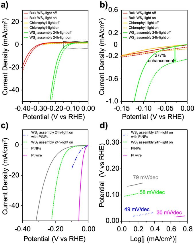
Y.-W. Chen et al.
4
                                                                               In vitro cell behaviour effects of WS2/PCL/CS scaffold
                                                                               To evaluate the effect of cells on WS2/PCL/CS scaffold, we used the
                                                                               human mesenchymal stem cells (hMSCs) to analyse Col I secretion,
                                                                               cell viability, alkaline phosphatase (ALP), and cell morphology.
                                                                               Figure 6a shows the amount of Col I adsorbed onto the scaffolds
                                                                               of a cell culture well (the control, abbreviated as Ctl), the PCL/CS,
                                                                               and the WS2/PCL/CS. The concentration of Col I on the WS2/PCL/
                                                                               CS scaffold was over two times higher than the result of Ctl,
                                                                               indicating the effective binding of Col I on the scaffold with the
                                                                               added WS2. Col I contains several cell-binding sub-units, which in
                                                                               turn bind to components of cell membranes to positively
                                                                               influence the cell behaviour, such as adhesion and proliferation60.
                                                                               An understanding of the biocompatibility of the WS2/PCL/CS
                                                                               scaffold is required, as the efficiency of cell viability is one of the
                                                                               critical issues affecting the results of tissue regeneration. Figure 6b
                                                                               shows significant enhancement of the viability of the cultured
                                                                               hMSCs on the WS2/PCL/CS scaffolds, as compared to the Ctl at
                                                                               days 1, 3, and 7 (p < 0.05). This was especially true for the WS2/
                                                                               PCL/CS scaffolds as a nearly 30% enhancement of the cell viability,
                                                                               as compared with the PCL/CS scaffolds, was observed at Day 7.
                                                                               This is attributed to the WS2/PCL/CS scaffold offering better micro-
                                                                               environmental conditions for hMSCs viability56. To evaluate the
                                                                               osteoblastic differentiation efficiency in detail, the useful marker of
                                                                               ALP activity was performed. Figure 6c shows that the ALP activity
                                                                               for all scaffolds increased with time, and the ALP activity from
                                                                               hMSCs in WS2/PCL/CS scaffolds was significantly higher than that
                                                                               of the Ctl and the PCL/CS scaffold at days 3, 7, and 14 (p < 0.05),
                                                                               indicating that the WS2/PCL/CS scaffold ably supports the early
                                                                               steps of osteoblastic differentiation in hMSCs. Figure 6d shows
    Fig. 4 Electrochemical performance of the exfoliated WS2 thin
    sheets. a Polarisation curves for the bulk WS2 (red), extracted            that hMSCs had a better adhesion to the WS2/PCL/CS scaffold than
    chlorophyll molecules (orange), and WS2 assembly 24 h (green)              to the PCL/CS scaffold after 12 h. The authors believe that the
    sample with (dashed line), and without (solid line) simulated              WS2/PCL/CS scaffold can adsorb more Col I on the surface so
    sunlight irradiation (100 mW cm−2); b) Magnification of dotted              enhancing the initial cell attachment ability. Various studies have
    rectangle in (a); c Polarisation curves; and d Tafel slope for Pt wire,    demonstrated that Col I can regulate the attachment, proliferation,
    PtNPs, and assembled composites including the sample of WS2                and osteogenic differentiation of stem cells56. Overall, the cell
    assembly 24-h-light on-with PtNPs and WS2 assembly 24-h-light on.          adhesion and viability data demonstrate that the chlorophyll-
                                                                               assisted exfoliated WS2 thin sheets did not exhibit any cytotoxic
    printed scaffolds had a well-designed morphology (Fig. 5a). The            responses to the hMSCs. Thus, this study opens possibilities in the
    interconnected macro-pore structure promotes cell in-growth and            development of chlorophyll-assisted exfoliated WS2 thin sheets for
    nutrient transportation, and their bioactive components can lead           tissue regeneration applications.
    to osteogenesis and angiogenesis56. To understand the homo-
    geneity of the chlorophyll-assisted exfoliated WS2 thin sheets in          In vivo bone regeneration efficacy of WS2/PCL/CS scaffold
    the WS2/PCL/CS composite, non-destructive Raman measure-
                                                                               In vivo experiments were carried out (Supplementary Fig. 8) by
    ments were used. Figure 5b shows that the mapping spectrum
                                                                               evaluating bone regeneration in femoral defects in rabbits after
    of the scaffold had a uniform WS2 signal intensity, indicating that
    the exfoliated WS2 thin sheets were homogeneously distributed in           the implantation of PCL/CS, WS2/PCL/CS, and MTA scaffolds.
    the WS2/PCL/CS composite. To further investigate the reinforce-            Figure 7a shows micro-computed tomography (µCT) images of the
    ment effect of the sheets in the scaffold, compression testing was         three types of scaffolds after implantation at four and eight weeks.
    carried out. Figure 5c shows that the compression strength was             The implanted WS2/PCL/CS scaffold specimen generated much
    enhanced from 2.0 MPa for a PCL/CS scaffold to 6.0 MPa for the             more bone around the scaffold compared to the PCL/CS and MTA
    WS2/PCL/CS scaffold, a 300% improvement. The Young’s modulus               scaffolds. The BV/TV (Fig. 7b) and Tb.Th (Fig. 7c) values for the
    of the PCL/CS scaffold was 32 MPa. The Young’s modulus of the              WS2/PCL/CS scaffold were much higher than those of the PCL/CS
    composite was significantly improved to 145 MPa, which is nearly            and MTA scaffolds (p < 0.05). In comparison to MTA, the best
    a 450% improvement. The results of the compressive modulus                 therapeutic efficiency (scored as the extent of in vivo bone
    and strength of the scaffold reached the range of human                    regeneration) was exhibited by the WS2/PCL/CS scaffold. The
    cancellous bone tissue57. Such reinforcement-enhancement is                results of the undecalcified specimens of PCL/CS, WS2/PCL/CS, and
    related to the original properties of the exfoliated sheets58,             MTA scaffolds stained by the haematoxylin and eosin (HE) stain,
    indicating the superior degree of dispersion of the sheets in the          Von Kossa (VK) stain, and Masson’s trichrome (MT) stain are shown
    biocompatible matrix. In addition to understanding the biocom-             in Fig. 7d. The HE staining showed the tissue regeneration was
    patibility and bioactivity of WS2/PCL/CS scaffold, precipitated            well formed within the defect area in the WS2/PCL/CS scaffold,
    spherical mineral aggregates were observed on the surfaces of the          whereas tissue was minimal in the PCL/CS scaffold. The MT
    PCL/CS and WS2/PCL/CS scaffolds using FE-SEM. Full mineral                 staining in the WS2/PCL/CS group at eight weeks shows that the
    aggregates coating both types of scaffold showed that apatite              collagen area and the lumen of the blood vessels supported active
    formation was not inhibited by the added WS2 (Fig. 5d). Apatite            osteogenesis around the scaffolds. VK staining is known to be
    layers are responsible for the biocompatibility and bioactivity of         highly visible in formed bone tissue61. In particular, the images of
    CS-based materials that are part of the interface of the scaffold          the WS2/PCL/CS scaffold in Fig. 7d exhibit the most intense VK
    and natural bone tissue59.                                                 staining, which indicates much more extensive bone regeneration

    npj 2D Materials and Applications (2020) 34                                     Published in partnership with FCT NOVA with the support of E-MRS
Y.-W. Chen et al.
                                                                                                                                                       5

Fig. 5 Characterisation of WS2/PCL/CS composite properties. a Photograph of WS2/PCL/CS scaffold. The scale bar used is 2 mm; b Raman
mapping image of WS2/PCL/CS scaffold by extracting the frequency of the characteristic peak of WS2 thin sheets; c Mechanical properties of
PCL/CS and WS2/PCL/CS scaffolds. Data presented as mean ± s.d. (s.d. standard deviation), n = 6 for each group; and d FE-SEM images of the
surface microstructures of PCL/CS and WS2/PCL/CS scaffolds, before and after being immersed in SBF.

in the WS2/PCL/CS sample, demonstrating that the formed bone             calcium oxide (CaO, Sigma-Aldrich), silica (SiO2, High Pure Chemicals,
was generated around the bone defect.                                    Saitama, Japan), MTA (MTA, Sigma-Aldrich), aluminium oxide (Al2O3,
   In summary, this study demonstrates an eco-friendly and easy          Sigma-Aldrich), phosphate-buffered saline (PBS, Caisson, North Logan, UT),
route to the scalable production of high-quality WS2 thin sheets         paraformaldehyde (Sigma-Aldrich), Alexa Fluor 488 dye (InvitrogenTM,
for bone regeneration and photoelectrochemical hydrogen                  ThermoFisher), 4’,6-diamidino-2-phenylindole (InvitrogenTM, Thermo-
generation applications. The extracted chlorophyll is a useful           Fisher), and p-nitrophenyl phosphate (Sigma-Aldrich) were all used as
pigment from nature for preparing a high-concentration WS2 thin          received. Acetone (HPLC grade) and sulfuric acid (H2SO4, 99.5%, HPLC
                                                                         grade) were purchased from ECHO chemical Co., Ltd. All solvents were
sheet dispersion with an excellent degree of stability in a volatile
                                                                         used without further purification.
solvent. The self-assembled exfoliated WS2 thin sheets on an Au
surface exhibited unusual HER photoelectrocatalytic activity with
an onset potential of −14 mV and a Tafel slope of 49 mV dec−1,
                                                                         Preparation of chlorophyll extracts
the lowest onset potential achieved with such a low Tafel slope
                                                                         Green leaves (20 g) were ground in a mortar and pestle and 500 mL
value that has been reported to date. According to the in vivo
                                                                         acetone was poured into the mortar. After standing for 8 h, the chlorophyll
experiments, the bone regeneration of the WS2/PCL/CS scaffold
                                                                         extracts were filtered through a 0.22 μm pore size polyvinylidene fluoride
was 120% superior to that of commercially available MTA material.        membrane to remove impurities. The filtered solution was centrifuged at
The mechanical properties of the WS2/PCL/CS scaffold exhibited           3000 rpm for 30 min and the supernatants of the chlorophyll extract
~300% enhancement over PCL/CS, which is one of the most                  solution were collected. The concentration of the chlorophyll extracts
commonly used materials. The WS2 added to the PCL/CS scaffold            was ~5 mg L−1.
enabled the scaffold to interact with cells, resulting in excellent
osteoblast adhesion and apatite deposition. The authors thus
believe that these versatile WS2 thin sheets may provide an              Chlorophyll-assisted liquid-phase exfoliation of WS2
opportunity to help resolve issues related to population ageing          WS2 powder (5 g), 10 mL of extracted chlorophyll solution, and 240 mL
and global climate change.                                               acetone were added to a 500 mL glass serum bottle at a controlled
                                                                         temperature, and sonicated with 150 W horn sonication (Q700,
                                                                         QSONICA) in pulse mode for 5 h. To measure the concentration of the
METHODS                                                                  WS2 suspension, 100 mL of solution was filtered through a 0.2 μm
Chemicals                                                                polyvinylidene fluoride membrane, and the weight of the WS2 measured.
WS2 powder (99% metals basis; ~325 mesh powder) was purchased from       The suspension concentration of the exfoliated WS2 thin sheets
Alfa Aesar. PCL (PCL; MW 43,000–50,000, Polysciences, Warrington, PA),   was ~1 mg mL−1.

Published in partnership with FCT NOVA with the support of E-MRS                                  npj 2D Materials and Applications (2020) 34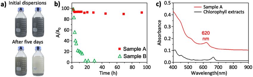
Y.-W. Chen et al.
6

    Fig. 6 In vitro cell behaviour effects of WS2/PCL/CS scaffold. a Col I secretion; b Viability; and c ALP activity of hMSCs cultured on PCL/CS
    and WS2/PCL/CS scaffolds. ‘*’ indicates a significant difference (p < 0.05) compared to Ctl. ‘#’ indicates a significant difference (p < 0.05)
    compared to the PCL/CS scaffold; d F-actin filaments (green) and nuclei (blue) staining of hMSCs cultured on scaffold at different time-points.
    Scale bar is 200 µm.

    Synthesis of PtNPs                                                               better mechanical properties and cell behaviours. First, 47.5 mg PCL was
    The platinum nanoparticles (PtNPs) were synthesised in glassware with a          placed in a 150 °C oven for 2 h. Then, 47.5 mg CS powder and 5 mg of the
    magnetic stir bar. 1 mL of 16 mM hexachloroplatinate (H2PtCl6), 1 mL of          exfoliated WS2 thin sheets were suspended in 5 mL alcohol and dropped
    40 mM tri-sodium citrate, and 38 mL of deionised water were separately           into the PCL. The sample was labelled is 5% WS2/PCL/CS. Supplementary
    added to a 200 mL two-necked bottle and stirred for a half-hour to obtain        Fig. 7 shows the cell viability of different ratios of the WS2 scaffold. The 5%
    a colourless mixture. Then, 200 μL of 50 mM sodium borohydride (NaBH4)           WS2/PCL/CS composite showed a suitable condition for cell viability and
    was added slowly into the two-necked bottle. The colourless mixture              hence, it was used in the study. The WS2/PCL/CS composites were loaded
    instantaneously turned to brownish-yellow. Finally, the brownish-yellow          into a steel syringe and heated to 100 °C in a three-dimensional printer
    solution was continuously stirred at room temperature for 1 h. The solution      (3DP) system (BioScaffolder 3.1, GeSiM, Grosserkmannsdorf, Germany) to
    was then stored in the dark until use. All glassware used in the PtNPs           produce the printable WS2/PCL/CS composites, which were injected by
    synthesis experiments was thoroughly cleaned with aqua regia, rinsed with        applying a pressure of 500 kPa to produce a 400 µm diameter straight line.
    deionised water (Elga Ltd., High Wycombe, UK), and then dried in an oven         Seven lines were printed in parallel with a gap of 400 µm between the
    before use.                                                                      lines, forming one layer. The 3DP scaffold was plotted layer-by-layer, up to
                                                                                     16 layers, for bone engineering applications. The weight of the 16 layers
                                                                                     scaffold was 0.4 g. Mechanical testing was done in accordance with ASTM
    Electrochemical measurements                                                     D695‐02a. The EZ Test machine (Shimadzu, Kyoto, Japan) was used to
    HER experiments were measured using a CHI7279E electrochemistry                  determine the mechanical properties of each scaffold at a loading rate of
    workstation. Electrochemical measurements were carried out with a three-         1 mm min−1. The scaffolds were printed into a 6 mm×6 mm × 10 mm
    electrode configuration using 0.5 M H2SO4 as an electrolyte, and an Ag/           rectangle. The average values and standard deviations (s.d.) of Young’s
    AgCl electrode and a graphite electrode as the reference and counter             modulus, and the maximum compressive strength were evaluated from
    electrodes, respectively. Linear sweep voltammetry (LSV) was performed at        the recorded stress-strain curves. Data are presented as mean ± s.d., n = 6
    a rate of 1 mV s−1 for the polarisation curves. The I-t curve test was carried   for each group.
    out with a controlled bias voltage.
                                                                                     Cell adhesion and viability
    Preparation of the calcium silicate (CS) powder                                  All 3DP scaffolds were soaked in 75% ethanol followed by irradiation for
    The method used for the preparation of CS powder has been described              1 h under UV light before the cell culture experiment. The human
    elsewhere62. In brief, CaO, SiO2, and Al2O3 powders were used as matrix          mesenchymal stem cells (hMSCs) were purchased from Sciencell Research
    materials (composition: 70% CaO, 25% SiO2, and 5% Al2O3). The oxide              Laboratories (Sciencell, Carlsbad, CA) and developed in medium (Sciencell).
    mixtures were sintered at 1400 °C for 2 h using a high-temperature furnace,      The cultured medium consisted of 500 mL of basal medium, 25 mL of
    and then the CS powder was ball-milled in ethyl alcohol using a centrifugal      foetal bovine serum, 5 mL of mesenchymal stem cell growth supplement
    ball mill (S100, Retsch, Hann, Germany) for 6 h.                                 and 5 mL of penicillin/streptomycin solution. The cells were seeded onto
                                                                                     the specimens at a concentration of 5 × 10⁴ cells per scaffold. After
                                                                                     culturing for 1 and 3 h, the cultured medium was collected, and Col I
    Fabrication of the WS2/PCL/CS composite scaffolds                                secreted from hMSCs was analysed by an enzyme-linked immunosorbent
    The CS matrices were produced using the thermal pressing method63. In a          assay kit (ELISA, Invitrogen), following the instructions in the manufac-
    previous study64, we demonstrated that the ratio of PCL/CS = 50/50 had           turer’s manual. In addition, cell viability was determined using PrestoBlue

    npj 2D Materials and Applications (2020) 34                                            Published in partnership with FCT NOVA with the support of E-MRS
Y.-W. Chen et al.
7
Fig. 7 In vivo bone regeneration efficacy of WS2/PCL/CS scaffold. a Cross-sectional images of µCT; b Bone mass volume (BV/TV);
c Trabecular thickness (Tb.Th) of rabbit femoral defects after four and eight weeks with PCL/CS, WS2/PCL/CS, and MTA scaffolds. Scale bar is
3 mm. ‘*’ indicates a significant difference (p < 0.05) compared to the PCL/CS scaffold. ‘#’ indicates a significant difference (p < 0.05) compared
to the MTA scaffold; and d HE staining images: haematoxylin and eosin (HE) stain (left); Masson’s trichrome (MT) stain (centre); and Von Kossa
(VK) stain (right) of regenerated bone mass after four and eight weeks for in vivo experiments. Scale bar is 400 µm.
assay (Invitrogen) after being directly cultured for 1, 3, and 7 days. The In vivo bone regeneration
PrestoBlue solution (30 μL) and 300 μL DMEM were added separately to All in vivo studies and protocols were done in accordance with relevant
each of the 96-well plates and incubated for 30 min, after which, 100 μL laws and regulations. The animal use protocol listed below has been
was extracted from each well and placed into a fresh 96-well plate and the reviewed and approved by the Institutional Animal Care and Use
absorbance was measured at 570 nm with a reference wavelength of Committee of China Medical University, Taichung, Taiwan. The white
600 nm. Cells cultured without scaffold were used as the control. New Zealand rabbits (2.0–2.5 kg) underwent surgery to create a standard
critical-sized femoral defect. The PCL/CS, WS2/PCL/CS, and commercially
Osteogenic differentiation available CS-based product (MTA) scaffolds (d = 7 mm and h = 8 mm) were
After cell seeding, the osteogenic differentiation was induced using a implanted into the defects to assess the bone regeneration capacity.
specific osteogenic medium from StemPro™ (osteogenesis differentiation Afterwards, the periosteum and related tissues were sutured, and the
kit, Invitrogen) and ALP secretion was measured on days 3 and 7. The ALP rabbits were kept under close examination until they recovered from the
activity of each sample was characterised using p-nitrophenyl phosphate effects of the anaesthesia. They initially limped but were able to walk with
as the substrate with 1 mol L−1 diethanolamine buffer. The reaction was a proper gait after four weeks. They had free access to food and water
terminated by adding 5 N NaOH and the absorbance recorded at 405 nm. during the period of observation. After the four weeks, the rabbits were
Analysis of blank disks was used as the control. euthanised through CO2 asphyxiation. The scaffolds and bone specimens
of the rabbits were then harvested and subsequently fixed using 10%
Fluorescent staining formalin. Images of all in vivo specimens were taken with a high 360°
The cultured cells were washed with PBS, fixed in 4% paraformaldehyde spatial resolution micro-CT camera (µ-CT, SkyScan 1076, Skyscan Inc.,
for 15 min, and then permeabilised with a 0.1% Triton X-100 PBS solution Kontich, Belgium). The SkyScan software was used to recognise bone tissue
at 25 °C. The F-actin filaments were labelled with phalloidin conjugated to and analyse the bone volume per tissue volume (BV/TV) and trabecular
Alexa Fluor 488 dye (Invitrogen) for 1 h. The cell nuclei were stained with thickness (Tb.Th) in the 3D model. The specimens were then embedded in
300 nM 4′,6-diamidino-2-phenylindole (DAPI) for 30 min. After washing OCT (KMA-0100-00A, CellPath Ltd., Newtown, Wales, UK), without being
thoroughly, the morphology was imaged using a white-light laser confocal decalcified, and sectioned (6 µm). The sections were stained with a
microscope (Leica TCS SP8 X, Leica Microsystems, Heidelberg, Germany). haematoxylin and eosin stain kit, a modified Masson’s Trichrome stain kit
Published in partnership with FCT NOVA with the support of E-MRS npj 2D Materials and Applications (2020) 34Y.-W. Chen et al.
8
(ScyTek Lab., West Logan, UT, USA), and a Von Kossa kit (ScyTek) in 11. Rao, C. N. R., Matte, H. S. S. R. & Maitra, U. Graphene analogues of inorganic
accordance with the manufacturers’ instructions. layered materials. Angew. Chem. Int. Ed. 52, 13162–13185 (2013).
12. Jamison, W. E. & Cosgrove, S. L. Friction characteristics of transition-metal dis-
ulfides and diselenides. ASLE Trans. 14, 62–72 (1971).
Characterisation 13. Sobczynski, A. et al. Tungsten disulfide: a novel hydrogen evolution catalyst for
The exfoliated 2D material thin sheets were deposited onto a silicon water decomposition. J. Phys. Chem. 92, 2311–2315 (1988).
substrate for Raman and attenuated total reflectance Fourier-transform 14. Teo, W. Z., Chng, E. L. K., Sofer, Z. & Pumera, M. Cytotoxicity of exfoliated
infrared spectroscopy (ATR-FTIR) studies. Raman spectra were recorded transition-metal dichalcogenides (MoS2, WS2, and WSe2) is lower than that of
using an iHR550 spectrometer (Horiba Jobin Yvon) with a 532 nm laser graphene and its analogues. Chem. Eur. J. 20, 9627–9632 (2014).
source in ambient conditions, a laser power ofY.-W. Chen et al.
9
40. Shifa, T. A. et al. A vertical-oriented WS2 nanosheet sensitized by graphene: an 64. Lin, Y.-H. et al. The synergistic effects of graphene-contained 3D-printed calcium
advanced electrocatalyst for hydrogen evolution reaction. Nanoscale 5, silicate/poly-ε-caprolactone scaffolds promote FGFR-induced osteogenic/angio-
14760–14765 (2015). genic differentiation of mesenchymal stem cells. Mater. Sci. Eng. C: Mater. Biol.
41. Laursen, A. B., Kegnæs, S., Dahl, S. & Chorkendorff, I. Molybdenum sulfides- Appl. 104, 109887 (2019).
efficient and viable materials for electro- and photoelectrocatalytic hydrogen 65. Wirtz, A. M.-S. L. Phonons in single and few-layer MoS2 and WS2. Phys. Rev. B 84,
evolution. Energy Environ. Sci. 5, 5577–5591 (2012). 155413 (2011).
42. Wang, T. et al. Enhanced electrocatalytic activity for hydrogen evolution reaction
from self-assembled monodispersed molybdenum sulfides nanoparticles on an
Au electrode. Energy Environ. Sci. 6, 625–633 (2013). ACKNOWLEDGEMENTS
43. Kristensen, J., Zhang, J., Chorkendorff, I., Ulstrup, J. & Ooi, B. L. Assembled monolayers This research is supported by the Ministry of Science and Technology, Taiwan (MOST
of Mo3S4+ clusters on well-defined surfaces. Dalton Trans 33, 3985–3990 (2006). 107-2628-M-143-001-MY2; 109-2811-M-143-500; 109-2628-M-143-001-MY3; 109-
44. Bourg, M.-C., Badia, A. & Lennox, R. B. Gold sulfur bonding in 2D and 3D self- 2218-E-039-001) and China Medical University, Taiwan (CMU109-MF-01). We grate-
assembled monolayers: XPS characterization. J. Phys. Chem. B 104, 6562–6567 (2000). fully thank Ms. Chia-Ying Chien, Su-Jen Ji, and Yin-Mei Chang of the Ministry of
45. Cao, S.-W. et al. Preparation of Au-BiVO4 heterogeneous nanostructures as highly Science and Technology (National Taiwan University and National Tsing-Hua
efficient visible-light photocatalysts. ACS Appl. Mater. Interfaces 4, 418–423 (2011). University) for their assistance in the electron microscopy experiments.
46. Yang, Y. Z., Chang, C.-H. & Idriss, H. Photo-catalytic production of hydrogen form
ethanol over M/TiO2 catalysts (M = Pd, Pt or Rh). Appl. Catal. B: Environ. 67,
217–222 (2006).
AUTHOR CONTRIBUTIONS
47. Chen, I.-W. P. et al. Scalable synthesis of two-dimensional nano-sheet materials
with chlorophyll extracts: enhancing the hydrogen evolution reaction. Green. I.-W.P.C. designed this work. I.-W.P.C., C.-H. H., Y.-C. L. and M.-Y.S. prepared the
Chem. 20, 525–533 (2018). materials and characterised them. I.-W.P.C. analysed the material characterisation and
48. Ko, C.-H., Huang, M.-J., Fu, M.-D. & Chen, C.-H. Superior contact for single- Raman spectroscopy data. M.-Y.S., Y.-W.C. and B. W. analysed the mechanical testing
molecule conductance: electronic coupling of thiolate and isothiocyanate on Pt, and bone regeneration data and prepared the relevant part of the manuscript. All
Pd, and Au. J. Am. Chem. Soc. 132, 756–764 (2010). authors discussed the experimental results and commented on the paper. I.-W.P.C.
49. Lu, J. et al. Multilayered graphene hydrogel membranes for guided bone wrote the manuscript.
regeneration. Adv. Mater. 28, 4025–4031 (2016).
50. Nieto, A. et al. Three dimensional graphene foam/polymer hybrid as a high
strength biocompatible scaffold. Adv. Funct. Mater. 25, 3916–3924 (2015). COMPETING INTERESTS
51. Qiu, K. et al. Electrophoretic deposition of dexamethasone-loaded mesoporous The authors declare no competing interests. All in vivo studies and protocols were
silica nanoparticles onto poly(L-lactic acid)/poly(ε-caprolactone) composite scaf- done in accordance with relevant rules and regulations. The animal use protocol
fold for bone tissue engineering. ACS Appl. Mater. Interfaces 8, 4137–4148 (2016). listed below has been reviewed and approved by the Institutional Animal Care and
52. Shie, M.-Y., Chiang, W.-H., Chen, I.-W. P., Liu, W.-Y. & Chen, Y.-W. Synergistic Use Committee of China Medical University, Taichung, Taiwan.
acceleration in the osteogenic and angiogenic differentiation of human
mesenchymal stem cells by calcium silicate-graphene composites. Mater. Sci. Eng.
C: Mater. Biol. Appl. 73, 726–735 (2017). ADDITIONAL INFORMATION
53. Wei, J. et al. Preparation and characterization of bioactive calcium silicate and Supplementary information is available for this paper at https://doi.org/10.1038/
poly(ε−caprolactone) nanocomposite for bone tissue regeneration. J. Biomed. s41699-020-00168-y.
Mater. Res. A 90, 702–712 (2009).
54. Wang, W. et al. Enhancing the hydrophilicity and cell attachment of 3D Printed Correspondence and requests for materials should be addressed to I.-W.P.C.
PCL/graphene scaffolds for bone tissue engineering. Materials 9, 992 (2016).
55. Middleton, J. C. & Tipton, A. J. Synthetic biodegradable polymers as orthopedic Reprints and permission information is available at http://www.nature.com/
devices. Biomaterials 21, 2335–2346 (2000). reprints
56. Kajave, N. S., Schmitt, T., Nguyen, T.-U. & Kishore, V. Dual crosslinking strategy to
generate mechanically viable cell-laden printable constructs using methacrylated Publisher’s note Springer Nature remains neutral with regard to jurisdictional claims
collagen bioinks. Mater. Sci. Eng. C: Mater. Biol. Appl. 107, 110290 (2020). in published maps and institutional affiliations.
57. Henkel, J. et al. Bone regeneration based on tissue engineering conceptions. Bone
Res. 1, 216–248 (2013).
58. Leffler, P. E. & Kazantzis, G. Handbook on the Toxicology of Metals 4E. Vol. 2
1297–1306 (2015).
59. Liu, X., Ding, C. & Chu, P. K. Mechanism of apatite formation on Wollastonite Open Access This article is licensed under a Creative Commons
coatings in simulated body fluids. Biomaterials 25, 1755–1761 (2004). Attribution 4.0 International License, which permits use, sharing,
60. Shie, M.-Y. & Din, S.-J. Integrin binding and MAPK signal pathways in primary cell adaptation, distribution and reproduction in any medium or format, as long as you give
responses to surface chemistry of calcium silicate cements. Biomaterials 34, appropriate credit to the original author(s) and the source, provide a link to the Creative
6589–6606 (2013). Commons license, and indicate if changes were made. The images or other third party
61. Wu, Y.-H. A. et al. 3D-printed bioactive calcium silicate/poly-ε-caprolactone material in this article are included in the article’s Creative Commons license, unless
bioscaffolds modified with biomimetic extracellular matrices for bone regen- indicated otherwise in a credit line to the material. If material is not included in the
eration. Int. J. Mol. Sci. 20, 942 (2019). article’s Creative Commons license and your intended use is not permitted by statutory
62. Chen, Y.-W., Yeh, C.-H. & Shie, M.-Y. Stimulatory effects of the fast setting and suitable regulation or exceeds the permitted use, you will need to obtain permission directly
degrading Ca-Si-Mg cement on both cementogenesis and angiogenesis differ- from the copyright holder. To view a copy of this license, visit http://creativecommons.
entiation of human periodontal ligament cells. J. Mater. Chem. B 3, 7099–7108 (2015). org/licenses/by/4.0/.
63. Chiu, Y.-C., Fang, H.-Y., Hsu, T.-T., Lin, C.-Y. & Shie, M.-Y. The characteristics of
mineral trioxide aggregate/polycaprolactone 3-dimensional scaffold with osteo-
genesis properties for tissue regeneration. J. Endod. 43, 923–929 (2017). © The Author(s) 2020
Published in partnership with FCT NOVA with the support of E-MRS npj 2D Materials and Applications (2020) 34You can also read