CD123 POSITIVE PLASMACYTOID DENDRITIC CELLS & CUTANEOUS LUPUS ERYTHEMATOSUS (LE)
←
→
Page content transcription
If your browser does not render page correctly, please read the page content below
CD123 POSITIVE PLASMACYTOID
DENDRITIC CELLS & CUTANEOUS
LUPUS ERYTHEMATOSUS (LE)

Ms Nandini Roy (3rd Year Medical Student)1
Dr. Garima Gupta (Specialist Registrar Histopathologist)2
Dr. Rathi Ramakrishnan (Consultant Histopathologist)2
Imperial College School of Medicine1 &
Imperial College Healthcare NHS Trust2
INTRODUCTION
Cutaneous LE – an autoimmune inflammatory dermatosis
affecting skin & subcutaneous tissue
Histologically- B & T cells, macrophages in perivascular &
adnexal area with interface dermatitis
Plasmacytoid dendritic cells (PDCs) are often associated
with LE producing interferons and CD123 antigen and are
linked to symptoms
Identification of PDCs – ? diagnosis value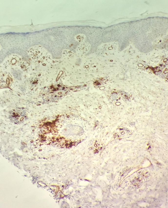
AIMS AND OBJECTIVES
To review cutaneous LE at our centre
To assess diagnostic value of CD123 staining
PDCs in CLE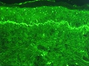
MATERIALS AND METHODS
CLE diagnosed at Charing Cross Hospital
between 2013 & 2017 were reviewed with respect
to histology & immunofluorescence (IMF)
Immunohistochemistry (IHC) to CD123 was
performed & semi-quantitative criteria were
developed for assessing staining intensity &
patterns of distributionSEMI-QUANTITATIVE CRITERIA FOR
ASSESSMENT
Intensity of staining
Weak Strong
• 1+ • 2+SEMI-QUANTITATIVE CRITERIA FOR
ASSESSMENT
Number of clusters
1+ 2+ 3+
•1-2 cluster •3-5 cluster •> 5 clusters
involvement involvementSEMI-QUANTITATIVE CRITERIA FOR
ASSESSMENT
Overall score generated
≤3 ≥4
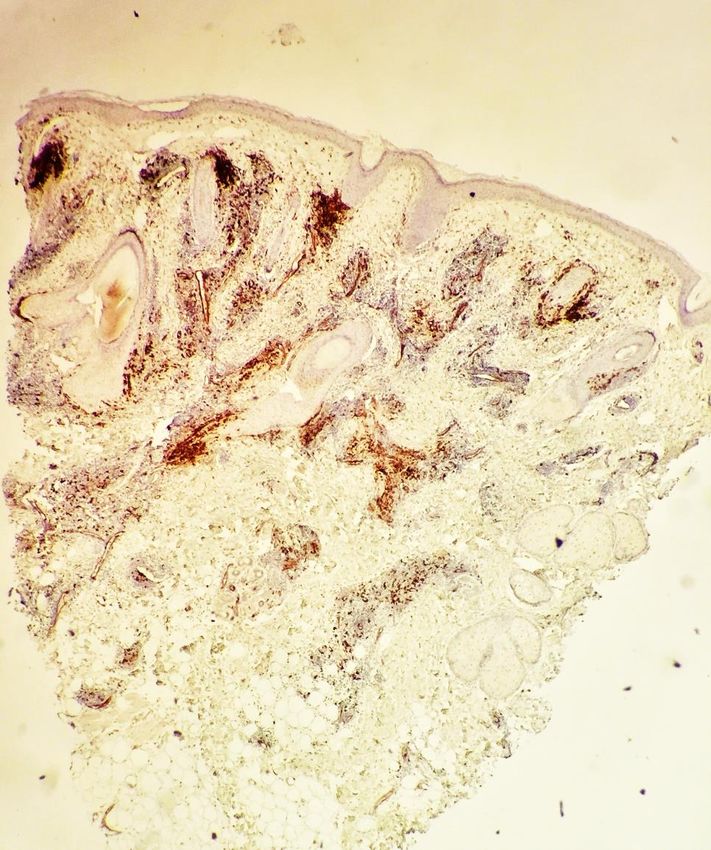
• Low expression • High expressionH&E STAINED SECTIONS OF CLE
Section showing moderate
Section showing moderate
perifollicular and perivascular
perifollicular lymphoid infiltrate
lymphoid infiltrateRESULTS: POSITIVITY BASED ON STAINING
INTENSITY
A total of 81 cases of
CLE were identified
72/81(88%) cases stained
for CD123
45/72 (62%) – 2+ Staining
27/72 (38%) – 1+
Strong(2+)
Weak(1+)RESULTS: PROPORTION OF CELLS
3+
26/72(36%) – 3+ 2+
1+
20/72 (28%) – 2+
26/72(36%) – 1+
1+ 2+ 3+
•1-2 cluster • 3-5 cluster •> 5 clusters
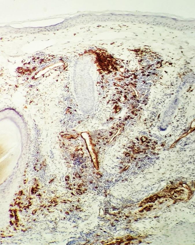
involvement involvementCD123 STAINING 1+ distribution with weak staining 2+ distribution with strong staining
3+ distribution with strong staining
RESULTS: OVERALL SCORE/EXPRESSION
28/72 (39%): Low Expression
expression
44/72 (61%): High
expression Low
HighRESULTS
CD123 positive PDCs were distributed in
perivascular, perifollicular, superficial dermal
and deep subcutis
Predominantly noted in perivascular 56/72(78%)RESULTS: CASES WITH IMF DATA
IMF available in 34/81
cases (42%). 47/81
(58%) no IMF
12/34 (35%) cases -
positive for IgG and C3,
22/34 (65%) – IMF
negative
Positive
19/22 (86%) with Negative
negative IMF were
positive for CD123KEY POINTS
High concordance between histologically
diagnosed CLE and CD123 staining;
Predominantly high overall score (61%)
Good diagnostic utility: CD123 staining present
even in cases lacking IMF to support histological
diagnosis (81% (38/47))
Helps in the distinction of lupus from other
neoplastic lymphoid proliferationsREFERENCES
1. Wenzel J, Proelss J, Wiechert A, Zahn S, Bieber T, Tuting T.
Cxcr3‐mediated recruitment of cytotoxic lymphocytes in lupus
erythematosus profundus. J. Am. Acad. Dermatol. 2007; 56; 648–
650.
2. Farkas L, Beiske K, Lund‐Johansen F, Brandtzaeg P, Jahnsen FL.
Plasmacytoid dendritic cells (natural interferon‐alpha/beta‐
producing cells) accumulate in cutaneous lupus erythematosus
lesions. Am. J. Pathol. 2001; 159; 237–243.
3. Tomasini D, Mentzel T, Hantschke M et al. Plasmacytoid dendritic
cells: an overview of their presence and distribution in different
inflammatory skin diseases, with special emphasis on Jessner's
lymphocytic infiltrate of the skin and cutaneous lupus
erythematosus. J. Cutan. Pathol. 2010; 37; 1132–1139.
4. Marshak‐Rothstein A. Toll‐like receptors in systemic autoimmune
disease. Nat. Rev. Immunol. 2006; 6; 823–835.
5. Barrat FJ, Meeker T, Gregorio J et al. Nucleic acids of mammalian
origin can act as endogenous ligands for toll‐like receptors and may
promote systemic lupus erythematosus. J. Exp. Med. 2005; 202;
1131–1139.THANK YOU
You can also read