Importance of Angomonas deanei KAP4 for kDNA arrangement, cell division and maintenance of the host bacterium relationship - Nature
←
→
Page content transcription
If your browser does not render page correctly, please read the page content below
www.nature.com/scientificreports
OPEN Importance of Angomonas deanei
KAP4 for kDNA arrangement,
cell division and maintenance
of the host‑bacterium relationship
Camila Silva Gonçalves1,2,5, Carolina Moura Costa Catta‑Preta3,5, Bruno Repolês4,
Jeremy C. Mottram3, Wanderley De Souza1,2, Carlos Renato Machado4* &
Maria Cristina M. Motta1,2*
Angomonas deanei coevolves in a mutualistic relationship with a symbiotic bacterium that divides
in synchronicity with other host cell structures. Trypanosomatid mitochondrial DNA is contained in
the kinetoplast and is composed of thousands of interlocked DNA circles (kDNA). The arrangement
of kDNA is related to the presence of histone-like proteins, known as KAPs (kinetoplast-associated
proteins), that neutralize the negatively charged kDNA, thereby affecting the activity of mitochondrial
enzymes involved in replication, transcription and repair. In this study, CRISPR-Cas9 was used to
delete both alleles of the A. deanei KAP4 gene. Gene-deficient mutants exhibited high compaction
of the kDNA network and displayed atypical phenotypes, such as the appearance of a filamentous
symbionts, cells containing two nuclei and one kinetoplast, and division blocks. Treatment with
cisplatin and UV showed that Δkap4 null mutants were not more sensitive to DNA damage and repair
than wild-type cells. Notably, lesions caused by these genotoxic agents in the mitochondrial DNA
could be repaired, suggesting that the kDNA in the kinetoplast of trypanosomatids has unique repair
mechanisms. Taken together, our data indicate that although KAP4 is not an essential protein, it plays
important roles in kDNA arrangement and replication, as well as in the maintenance of symbiosis.
The kinetoplast contains the mitochondrial DNA (kDNA) of trypanosomatids, which is arranged in a network
of several thousand minicircles categorized into different classes and several dozen maxicircles that are virtually
identical. Minicircles (0.5–10 kb) are physically connected to each other and also to maxicircles (20–40 kb) that
are usually interwoven into the network p eriphery1,2. Maxicircle sequences encode components of the respira-
tory chain and ribosomal proteins, but first, posttranscriptional editing of the generated mRNA is required. This
process is mediated in part by small noncoding guide RNAs (gRNAs) that are transcribed from m inicircles3,4.
The kDNA network is linked to the basal body through proteins that compose the tripartite attachment com-
plex (TAC)5. Usually, loss of kDNA is associated with mitochondrial dysfunction, which makes this structure a
potential chemotherapy target and diagnostic marker for t rypanosomiasis6–8.
In contrast to most eukaryotes, mitochondrial DNA replication in trypanosomatids is regulated during the
cell cycle, initiating immediately before nuclear DNA replication in S phase followed by network scission and
kinetoplast division during the G2 phase. The duplication cycle of the kinetoplast occurs in four steps: kDNA
synthesis; scission, when kDNA is cleaved into two networks; separation; and partitioning of kinetoplast between
the daughter cells during cytokinesis9. The kDNA network replication is a complex and unusual mechanism that
involves various enzymes, such as the mitochondrial topoisomerase II (mtTopo II), which detaches covalently
closed minicircles from the network. Minicircle replication initiates at the kinetoflagellar zone (KFZ), which com-
prises the region between the kDNA facing the basal body and the inner mitochondrial membrane. At the KFZ,
1
Laboratório de Ultraestrutura Celular Hertha Meyer, Instituto de Biofísica Carlos Chagas Filho, Universidade
Federal do Rio de Janeiro, IBCCF, CCS, UFRJ, Cidade Universitária, Rio de Janeiro, RJ CEP 21941‑590, Brazil. 2Centro
Nacional de Biologia Estrutural e Bioimagem, Rio de Janeiro, RJ, Brazil. 3Department of Biology, York Biomedical
Research Institute, University of York, Wentworth Way, Heslington, York YO10 5DD, UK. 4Laboratório de Genética
Bioquímica, Departamento de Bioquímica e Imunologia, Instituto de Ciências Biológicas, Universidade Federal
de Minas Gerais, Belo Horizonte, Brazil. 5These authors contributed equally: Camila Silva Gonçalves and Carolina
Moura Costa Catta-Preta. *email: crmachad@icb.ufmg.br; motta@biof.ufrj.br
Scientific Reports | (2021) 11:9210 | https://doi.org/10.1038/s41598-021-88685-8 1
Vol.:(0123456789)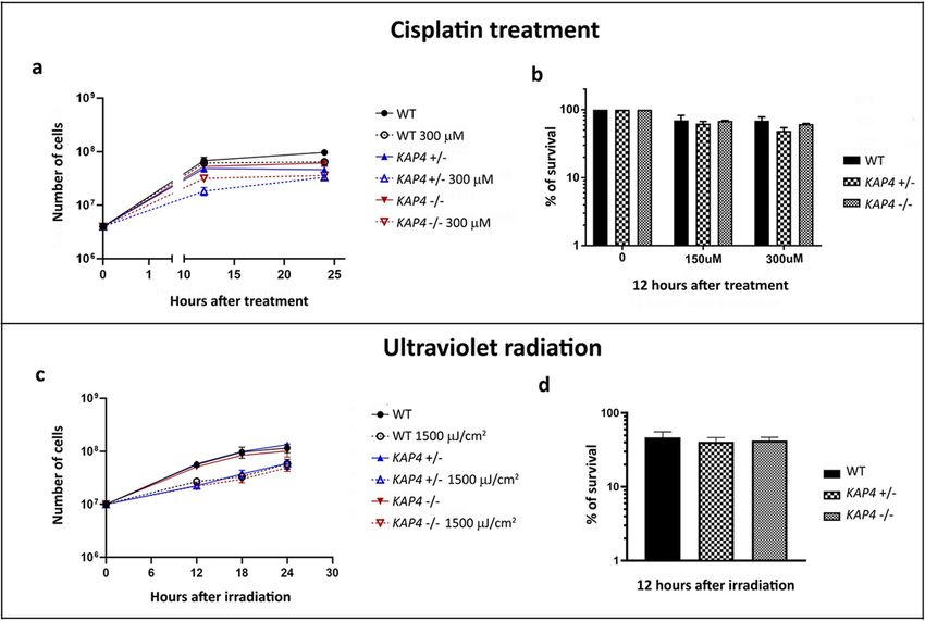
www.nature.com/scientificreports/
the minicircles duplicate as theta structures, by UMSBP, Pol 1B, and other proteins and subsequently migrate to
the antipodal sites. At this kinetoplast region, a primase enables the synthesis initiation of new DNA fragments
following kDNA replication that involves more than 100 enzymes, such as universal minicircle sequence-binding
protein (UMSBP) and polymerases. Next, each newly replicated minicircle is reattached to the network by the
mtTopoII, maintaining at least one nick/gap that is filled by proteins, such as Pol β-PAK and DNA ligase kα,
prior to the network scission. Later, the duplicated network is separated by the basal body distance, since the
kDNA is connected to it via the TAC structure. This minicircle replication model was primarily based on find-
ings obtained with Trypanosoma brucei and Crithidia fasciculata4.
The kDNA arrangement varies according to species and developmental stages, ranging from densely packed
fibers to a looser distribution in the kinetoplast m atrix10–12. The proteins involved in this intriguing phenom-
enon have not been fully characterized. Kinetoplast-associated proteins (KAPs) are homologous to small basic
histone H1-like proteins and nonhistone high-mobility group (HMG) box-containing proteins. KAPs have low
molecular weights, are highly basic, are rich in alanine and lysine residues and contain a cleavable nine amino
acid presequence involved in protein import to the kinetoplast in their amino-terminal r egion13. KAPs are
involved in kDNA duplication, transcription, packing and topological remodeling14–16. KAPs can also bind to
other proteins, such as UMSBP; in this case, they promote kDNA unpacking and facilitate the access of mtTopoII,
which liberates minicircles from the network for replication17.
The first model used to study the roles played by KAPs was the monoxenic Crithidia fasciculata, where the
disruption of the KAP1 gene generated viable cells with a phenotype of highly condensed kDNA fibers, which
was similar to that observed when trypanosomatids were treated with nalidixic acid, an inhibitor of prokaryote
topoisomerase II15,18. When both C. fasciculata alleles for KAP2 and KAP3 were disrupted separately, no detect-
able phenotypes were generated, and the same lack of phenotypes was observed to heterozygous cells (kap2⁄3+⁄−),
indicating a redundant function for these two encoded proteins. However, the double-knockout cells had notably
slow proliferation, atypical cell morphology, an increased copy number of mRNAs encoding for ATPase and a
significantly reduced r espiration15. These first findings obtained with knockout cells indicated that KAPs were
involved in distinct functions, such as kDNA arrangement and metabolism. Deletion of the KAP3 gene was also
performed in Trypanosoma cruzi by homologous recombination. Such null mutants did not exhibit changes in
cell proliferation, differentiation, kDNA arrangement and infectivity, suggesting that this KAP is not essential for
this parasite19. Later, the RNAi system was used to knockdown proteins associated with kDNA in Trypanosoma
brucei. Downregulation of KAP6 promoted cell growth arrest and inhibition of covalently closed minicircle
release, resulting in loss, shrinkage and disorganization of k DNA20.
Symbiont-harboring trypanosomatids (SHTs), such as Angomonas deanei (previously classified as Crithidia
deanei21), coevolve in a mutualist relationship with a single bacterium that divides in synchronicity with other
host cell structures and is usually observed close to the nucleus. During the protozoan cell cycle, the bacterium
is the first DNA-containing structure to divide, followed by the kinetoplast and the nucleus22–24. The symbiont
is a Gram-negative of the Alcaligenaceae family that contains a reduced genome, is enclosed by two membranes
and has a very reduced peptidoglycan layer25–27. Such species has been used to study the kinetoplast which, in
these cells, presents atypical shapes and a looser kDNA arrangement, which is more susceptible to topoisomer-
ase inhibitors and DNA-binding drugs11,18,28,29. Recently, phylogenetic analysis showed that SHTs present an
expanded repertoire of nuclear encoded KAPs and that genes for KAP4 and KAP7 are present in all trypanoso-
matid species analyzed to d ate11.
While mitochondrial DNA is subjected to the same damage sources as nuclear DNA, the reactive oxygen
species (ROS) generated by the oxidative phosphorylation metabolism usually results in higher mutation rates
in the mtDNA than does damage caused to nuclear DNA. In mammalian cells, base excision repair has been
described as a restoration mechanism in the mitochondrion with the identification of several glycosylases, such
as MYH, NEIL1, NEIL2 and UNG1, that are involved in the response of mtDNA to oxidative damage30–33. Other
proteins, such as APE1, APE2, FEN1, and DNA2, were also detected, suggesting that all steps of this repair
mechanism are present in the mitochondria of m ammalians34–38. Mismatch removal activity was also identified
39
in this organelle , although it has not been determined which proteins are involved in this process and whether
the same pathway is active in the nucleus. However, the most striking and unexpected feature in mammalian
cells is the lack of DNA repair mechanisms to address UV- and cisplatin-induced lesions on the m tDNA40–42.
In trypanosomatids, some proteins involved in DNA repair have been described in both nuclear DNA and
in kDNA metabolism. It was demonstrated that T. cruzi is able to remove oxidative lesions from both genomes,
although damage to the kDNA remains higher than that in the nucleus43–45. This parasite contains DNA glycosy-
lases that participate in the kDNA damage response43,44, as well as polymerases involved in the response to oxida-
tive stress, such as Polβ, Polβ-PAK46,47 and Polκ, which are able to interact with intermediates of the homologous
recombination48. Studies in T. brucei showed that the bloodstream form is able to deal with damage caused by
cisplatin, hydrogen peroxide and methylmethanesulfonate (MMS), suggesting that DNA repair pathways are
present in the parasite mitochondrion and that TbRad51 might be crucial to the response to alkylation lesions49.
In the present work, for the first time, we used the CRISPR-Cas9 system to analyze the role played by KAP in
a trypanosomatid protozoan. The results demonstrated that A. deanei Δkap4 mutants have reduced proliferation
and exhibit morphological and ultrastructural alterations. In KAP4 mutants, the kDNA network becomes highly
packed and cells have atypical phenotypes including filamentous bacterium and atypical numbers of nuclei and
kinetoplasts. Considering alterations in kDNA arrangement, gene deletion mutants were not more sensitive to
cisplatin and UV treatment than wild-type protozoa, but these genotoxic agents interfered with cytokinesis in
both cell types. Notably, cisplatin and UV lesions can be repaired in mitochondrial DNA, which suggests that
there are unique DNA repair mechanisms in the trypanosomatid kinetoplast.
Scientific Reports | (2021) 11:9210 | https://doi.org/10.1038/s41598-021-88685-8 2
Vol:.(1234567890)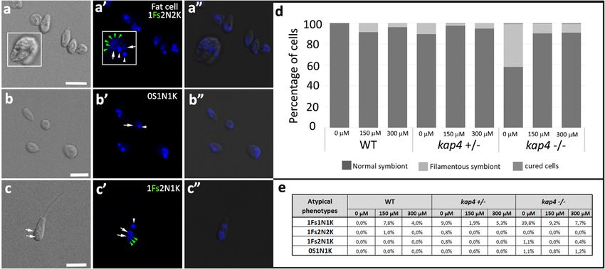
www.nature.com/scientificreports/
Materials and methods
Cell culture. The Angomonas deanei wild type (WT—ATCC 30255) strain was cultured in Warren´s
edium50 supplemented with 10% fetal bovine serum. Protists were maintained by weekly passages by inoculat-
m
ing 10% of an established cell culture in fresh medium. WT and T7RNAPol-SpCas9 cell lines were grown at 28 °C
for 24 h and cells with single or double deletions to kap4 genes were grown for 48 h, both cases corresponded
to the protozoan exponential growth phase. After this growth period, cells were used in assays or stored at 4 °C.
Analysis of cell growth and viability. For the growth curve, the initial cell concentration was 1 × 10⁶
cells/mL, and counts were made every 24 h up to 72 h. Cell density was determined by counting live protozoa
in a flow cytometer, where cell size was evaluated by detection of forward scatter on an SSA detector in a BD
Accuri C6 flow cytometer (Becton Dickinson Bioscience BDB, San Jose, CA, USA). The relative growth rate (μ,
expressed as h − 1) of the exponential phase was estimated by an exponential function y = AeBx , considering
the parameters of culture cell density (cells/mL) vs culture time (h) of each strain, when B = μ. Such graphics only
considered the cell density from 0 to 48 h of growth, which corresponds to the exponential phase, when all assays
in this study were performed. Cell duplication time (DT) was calculated according to the formula DT = ln2 µ.
To test cell viability, 5 × 10⁶ cells were washed once with filtered-sterilized PBS (phosphate-buffered saline)
pH 7.2 and incubated for 10 min with 20 μg/mL propidium iodide (PI). After this step, 10,000 events per sam-
ple were collected, and the fluorescence was detected on an FL-2 filter (488/630). The percentages of viable and
nonviable cells were determined using control assays of life and death, respectively. To check cell death, cells were
fixed in 4% paraformaldehyde for 10 min, washed with PBS, pH 7.2 and subsequently incubated with propidium
iodide (PI 1:100). To control for living cells, protozoa were washed in PBS, pH 7.2, but were not incubated with
PI. Cell fluorescence was detected as previously described. In such viability assays, as well as in growth curves,
cells were collected on a BD Accuri C6 flow cytometer (Becton Dickinson Bioscience BDB, San Jose, CA, USA)
using the manufacturer software.
Genotoxic treatment. WT and AdKAP4 mutants were compared by plating 1 × 10⁷ cells/mL in the pres-
ence or absence of genotoxic agents. For cisplatin treatment, cells were incubated with 150 and 300 μM of the
inhibitor for 1 h, washed three times with PBS at pH7.2 and resuspended in fresh medium. UVC irradiation
(254 nm) was performed with a germicidal lamp at a fluence rate of 1,500 µJ/cm2 (GS GeneLinker UV Chamber,
Bio-Rad). For growth curves, in all conditions, the number of surviving cells was determined at 0 h (immediately
before the treatment) and after 12 and 24 h of treatment, which corresponds to the A. deanei exponential p hase19.
Experiments were performed in triplicate. The cell number was determined in a hemocytometer chamber using
the erythrosine vital stain (0.4% diluted in 1 × PBS) to differentiate living and dead cells. Only dead cells were
stained, presenting a red color. The survival rate was calculated by comparing treated and control cells, which
were employed as references (considered as 100%).
Cell cycle analysis by flow cytometry. Protozoa were treated with cisplatin 150 and 300 μM for 1 h.
Next, the cells were washed twice with PBS, pH 7.2, and the culture medium was replaced as described above.
Protozoa were analyzed before treatment, as well as 1 h and 24 h after the incubation with the inhibitor. Approxi-
mately 5 × 10⁶ cells were pelleted, washed once with PBS and fixed in 0.25% paraformaldehyde at room tempera-
ture for 5 min. Next, the cells were permeabilized in 70% ethanol, in an ice bath, for 30 min and incubated with
100 μg/mL RNase and 25 μg/mL propidium iodide at 37 °C for 30 min. After this step, 10,000 events per sample
were collected, and the fluorescence was detected on an FL-2 filter (488/630) on a BD Accuri C6 flow cytometer
(Becton Dickinson Bioscience BDB, San Jose, CA, USA) using the manufacturer’s software. DNA histograms
were analyzed with the same software.
CRISPR‑Cas9 gene editing. Protozoa transformation. Angomonas deanei transfections were performed
by electroporation using the Amaxa 2B system program U-033 (Human T Cell Nucleofector™ Kit—Lonza),
as previously d escribed24. Cultures were immediately split into 2 populations, and recovered for 4 h at 26 °C
before the addition of suitable antibiotics. Motile cells in both populations were counted and diluted for distri-
bution in 96-well plates (200 µL of 1 or 0.5 cells/well). Clones were recovered after 5–8 days. Angomonas deanei
T7RNAPol-SpCas9 was engineered using the pTB007 plasmid previously employed for Leishmania species, and
SpCas9 expression was confirmed by Western blotting as in Beneke et al., 2 01751. Transgenic lines were main-
tained in the following antibiotics and respective concentrations: G418 (250 µg/mL) and hygromycin (300 µg/
mL).
CRISPR‑Cas9 DNA fragment preparation. CRISPR-facilitated mutants were obtained by transfection of PCR
fragments. The sgRNA sequence was obtained from E uPaGDT52, selected based on correct on-target sequence
(ADEAN_000063100)53 and fewer A. deanei genome off-target hits, as well as sgRNA predicted activity. The
sgRNA forward oligonucleotide is designed by flanking it with the T7RNAPol promoter (upstream) and the
first 20 nucleotides of the SpCas9 scaffold (downstream). This oligo is combined with a universal primer con-
taining the remaining sequence of SpCas9 backbone (OL00—Table 1). Amplification was performed in 20 µL
using 0.2 mM dNTPs, 2 µM of each primer in Q5 reaction buffer and high-fidelity polymerase (NEB). The PCR
program was set as 30 s at 98 °C followed by 35 cycles of 10 s at 98 °C, 30 s at 60 °C, and 15 s at 72 °C. The repair
template fragments were produced using primers containing annealing sequences compatible with pPLOT and
pT plasmids51 and 30 nucleotide homology arms at the 5′end of the oligonucleotide, both forward and reverse,
for recombination upstream and downstream of the DNA double strand break (DSB), respectively, at the UTR
Scientific Reports | (2021) 11:9210 | https://doi.org/10.1038/s41598-021-88685-8 3
Vol.:(0123456789)
www.nature.com/scientificreports/
Oligo name Description Sequence
aaaagcaccgactcggtgccactttttcaagttgataacggactagccttattttaactt-
OL00 Universal reverse primer for sgRNA amplification
gctatttctagctctaaaac
gaaattaatacgactcactataggCGGCGCTTACAGCATGTT
OL01 KAP4—5′ sgRNA primer
TAgttttagagctagaaatagc
gaaattaatacgactcactataggTTTCTGCTGTTTCCACAG
OL02 KAP4—3′ sgRNA primer
TTgttttagagctagaaatagc
TACTCTTATTATAATTAGTTTTTTTATAAAgtataatgca-
OL03 KAP4—Upstream Forward primer
gacctgctgc
TTTTTATTATTATTTGAATAGGTTTACCGCccaatttga-
OL04 KAP4—Downstream Reverse primer
gagacctgtgc
KAP4—Checking CDS presence/deletion/Neo integration
OL05 GTCTCATAGGAAAAGTACAC
(Forward)
OL06 KAP4—Checking CDS presence/deletion (Reverse) CGGCTTTTCTGCTGTTTC
OL07 Reverse at 5′ (ATG) of Neo resistance gene ACTAGTATGGGATCGGCCATTGAACAAG
Forward qPCR primer for large mitochondrial fragment
qPCRMitF TTTTATTTGGGGGAGAACGGAGCG
(approximately 10kB)
Reverse qPCR primer for large mitochondrial fragment
qPCRMitR TTGAAA CTGCTTTCCCCAAACGCC
(approximately 10kB)
Forward qPCR primer for small mitochondrial fragment
qPCRMitSmF CGCTCTGCCCCC ATAAAAAACCTT
(250 bp)
qPCRNucF Forward qPCR primer for large nuclear fragment GAGGCACTGCATACCATTCAAG
qPCRNucR Reverse qPCR primer for large and small nuclear fragment GTGGTCCTTCTTTGTCAATTTCAC
qPCRNucSmF Forward qPCR primer for small nuclear fragment ATATACACGGGATAAAGGCCAGC
Table 1. List of oligonucleotides for CRISPR-Cas9 in A. deanei, including sgRNA, repair template and
diagnostic PCR. Sequences are written in the 5′ to 3′ orientation.
of the gene. Fragments were amplified from 20 ng of p TNeo_v151 using the same reaction buffer described above
for sgRNA fragments in a final volume of 40 µL. PCR program was 10 min at 98 °C followed by 40 cycles of 30 s
at 98 °C, 30 s at 60 °C, 2 min 15 s at 72 °C, and a final elongation step of 10 min at 72 °C. Products were run
on 2% (sgRNAs) or 1% (repair templates) agarose gels in 0.5% Tris–Borate-EDTA (TBE) to confirm fragment
amplification and expected sizes. Primer sequences are detailed in Table 1. DNA for transfection was prepared
by combining sgRNA and repair templates followed by precipitation in a one-tenth volume of 3 M NaOAc, pH
5.5 and 2.5 volumes of ice-cold absolute ethanol, and washing in 70% ethanol thereafter. DNA was resuspended
in 10 µL of molecular biology grade water and immediately transfected.
Diagnostic PCRs. Genomic DNA (gDNA) was purified after clone cell culture amplification and kept under
antibiotic selection, using the DNeasy Blood & Tissue Kit (Quiagen) following the manufacturer’s instructions.
PCRs were set using 50 ng of gDNA using PCRBIO HS Taq Mix Red (PCR Biosystems) and 0.4 µM of prim-
ers to amplify the CDS locus or the integrated repair template containing the resistance marker gene (Neo).
The oligonucleotides OL05 + OL6 were used to detect KAP4 presence or absence, respectively. Oligonucleotides
OL05 + OL07 were used to confirm integration of the repair template containing the neomycin (Neo) resistance
marker at KAP4 loci. The PCR program used was 5 min at 95 °C followed by 25 cycles of 30 s at 95 °C, 30 s at
55 °C, 20 s at 72 °C and a final elongation step of 5 min at 72 °C. Reactions were directly run in a 0.8% agarose
gel in TBE to confirm genetic manipulation by comparing the presence or absence of WT and mutants PCR
products. Primer sequences are detailed in Table 1.
Fluorescence microscopy. DAPI staining. Protozoa were collected by centrifugation at 2000×g, washed
once with PBS (phosphate buffered saline) pH 7.4, fixed in 4% paraformaldehyde in the same solution, and
mounted on poly-l-lysine-coated circular microscope coverslips (14 mm diameter), next, the slides were washed
with PBS and incubated with 10 μg/ml 4′,6-diamidino-2-phenylindole (DAPI, from Molecular Probes, Oregon,
USA) for 10 min. After washing with PBS, slides were mounted using ProLong Gold (Molecular Probes), and
visualized using a TCS SP5 confocal laser scanning microscope (Leica, Germany). Confocal images were ob-
tained using an HCX PL APO 60 × objective for light microscope oil immersion with a numerical aperture of
1.4. Optical sections obtained from the whole cell were transformed into 2D images by maximum projection
in the manufacturer’s software (LAS-X). The cellular patterns were determined by counting DNA-containing
structures as nuclei, kinetoplasts and symbionts. Symbiont division was evaluated based on its form as described
previously22,24. Analyses were based on counts of 1000 cells of WT and KAP4 mutants.
Immunofluorescence with anti‑porin antibody. Protozoa were washed in PBS and fixed with freshly prepared
2% formaldehyde diluted in PBS, for 1 h. After fixation, cells were adhered to poly-l-lysine-coated microscope
coverslips and permeabilized with 4% Nonidet P-40 (NP-40) diluted in PBS for 45 min. Slides were incubated in
blocking solution containing 1.5% bovine serum albumin (BSA), 0.5% teleostean gelatin (Sigma Aldrich), and
0.02% Tween 20 diluted in PBS. Next, slides were incubated for 1 h with antibody produced against the symbi-
ont porin54 diluted 1:10 in blocking solution. After that step, the cells were washed with PBS and incubated for
Scientific Reports | (2021) 11:9210 | https://doi.org/10.1038/s41598-021-88685-8 4
Vol:.(1234567890)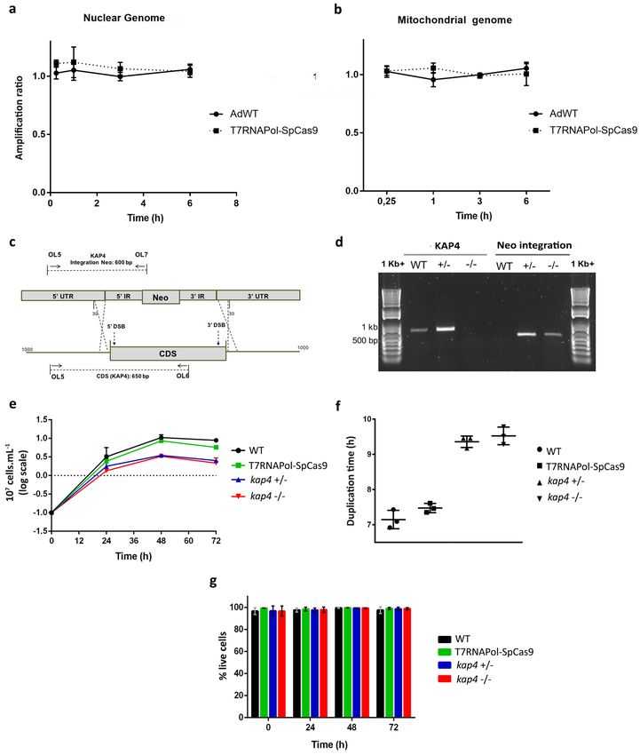
www.nature.com/scientificreports/
45 min with Alexa488-conjugated anti-mouse IgG (Molecular Probes, USA) diluted 1:200 in blocking solution.
Slides were mounted using the anti-fading reagent ProLong Gold containing 5 μg/mL of DAPI (4′,6-diamidino-
2-phenylindole, MolecularProbes). Serial image stacks (0.36-μm Z-increment) were collected at 64× (oil immer-
sion 1.4 NA) on an Elyra PS.1 microscope (Carl Zeiss) and three-dimensional projections were obtained on the
Zen Black program (Carl Zeiss).
In situ labeling of kDNA networks. Cells were centrifuged, washed, and fixed in 2% paraformaldehyde diluted
in PBS for 5 min. Next, cells were adhered to poly-l-lysine-coated slides for 10 min and washed twice in PBS
containing 0.1 M glycine for 5 min. After permeabilization in methanol for 1 h at 20 °C, cells were rehydrated
with three washes in PBS for 5 min and incubated for 60 min at room temperature in 25 μL of reaction solution
containing: TdT reaction buffer (Roche Applied Science), 2.0 mM CoCl2, 10 μM dATP, 2.5 μM Alexa Fluor 488-
dUTP (Molecular Probes) and 10 units of TdT (Roche Applied Science). The reaction was stopped with three
washes in 2xSSC for 5 min. Slides were mounted using the anti-fading reagent ProLong Gold containing 5 μg/
mL DAPI (4′,6-diamidino-2-phenylindole, MolecularProbes). Slides were examined on an Axiobserver micro-
scope (Carl Zeiss), and images were collected at 100× (oil immersion 1.4 NA). Analyses were based on counts
of 1000 cells of WT and KAP4 mutants considering the kDNA replication as described by Liu and E nglund55.
Electron microscopy. Scanning electron microscopy (SEM). Sample processing was performed using glass
coverslips precoated with 1 mg/mL poly-l-lysine. Protozoa were fixed for 1 h in 2.5% glutaraldehyde diluted in
0.1 M cacodylate buffer pH 7.2. Cells were subsequently adhered to coverslips, postfixed for 1 h with 1% os-
mium tetroxide diluted in cacodylate buffer, and dehydrated in a graded alcohol series (50%, 70%, 90%, and two
exchanges of 100% ethanol for 10 min each step). Samples were critical-point dried in a Leica EM CPD030 ap-
paratus (Leica, Wetzlar, Germany). Specimens were sputtered with gold in a Balzers FL9496 unit (Postfach 1000
FL-9496 Balzers Liechtenstein) and observed in an EVO 40 VP SEM (Zeiss, Germany). In all assays performed,
approximately 500 cells were observed.
Transmission electron microscopy (TEM). Protozoa were fixed for 1 h in 2.5% type II glutaraldehyde (Sigma,
Missouri, USA) diluted in 0.1 M cacodylate buffer, pH 7.2. The protozoa were washed twice in cacodylate buffer
and postfixed (1% osmium tetroxide, 0.8% potassium ferrocyanide, 5 mM calcium chloride diluted in 0.1 M
cacodylate buffer) for 1 h. Samples were then washed in cacodylate buffer, dehydrated in a graded series of
acetone solutions (50%, 70%, 90%, and two exchanges of 100% acetone) for 10 min at each step, and embedded
in Polybed resin. Ultrathin sections were stained with 5% uranyl acetate for 45 min and lead citrate for 5 min
before observation in a Jeol 1200 EX TEM operating at 80 kV. In all assays performed, approximately 500 cells
were analyzed.
Damage quantification by long‑range qPCR analysis. Parasite cultures were treated with the respec-
tive drug as reported above. After treatment, 1 × 10⁸ cells were harvested by centrifugation at 3000×g for 5 min at
the time points after treatment indicated on the graph. The first time point (0 h) was collected immediately after
the end of UV radiation exposure, and after the washes to remove cisplatin from the media in cisplatin treat-
ment. DNA extraction was performed by using the QIamp® DNA Mini and Blood Mini Kit (Qiagen, cat: 51104)
protocol for tissue extraction.
Amplification was performed using a Kappa LongRange HotStart PCR Kit (Sigma, cat: KK3501). Specific
primers for the mitochondrial coding region were used and are listed in Table 1. Amplification of the large
mitochondrial fragment (approximately 10 kB) was performed by using primers qPCRMitF and qPCRMitR.
Amplification of the small mitochondrial fragment (250 bp) was performed by using the primers qPCRMitSmF
and qPCRMitR. For the nuclear fragment analyses, the amplification of the larger fragment was performed using
the primers qPCRNucF and qPCRNucR. The smaller fragment was amplified using the primers qPCRNucSmF
and qPCRNucR.
The assay consists of the comparison of the amount of amplified material of treated cells with the amount
of amplified material within nontreated cells. The smaller fragment was used to normalize the amplification of
the large fragments and to avoid any bias from uneven loading of template DNA among the various PCRs. The
normalized value of treated and nontreated cells was compared, and the relative amplification was subsequently
calculated. These values were used to estimate the average number of lesions/10 kb of the mitochondrial genome
using a Poisson distribution. All the results presented are the mean of two technical replicates of amplification
and two different biological experiments. Details of the data analysis can be found in the literature56.
Results
To allow genetic manipulation in A. deanei facilitated by CRISPR-Cas9, we first generated an A. deanei mutant
expressing SpCas9 and T7RNAPol by transfecting log-phase cells with the pTB007, generously provided by Dr.
Eva Gluenz and previously used to generate a similar mutant in Leishmania sp52. Western blotting confirmed
SpCas9 expression in the mutants, using L. mexicana T7RNAPol-SpCas9 as a control (Supplementary Informa-
tion 1).
To verify whether the expression of SpCas9 in the AdT7RNAPol-SpCas9 strain could constitutively cut non-
specific sites, long-range qPCR quantification was performed to determine the amount of possible accumulation
of DNA damage in those cells. WT protozoa were used as a controls, since they do not contain the cassette con-
struction for the SpCas9 expression. If SpCas9 generated nonspecific DNA damage, it was expected to produce
a difference between the amplification ratio of the genetically modified strain in comparison with WT cells. The
amplification for both strains was approxemetly 1, indicating that the expression of SpCas9 on A. deanei did not
Scientific Reports | (2021) 11:9210 | https://doi.org/10.1038/s41598-021-88685-8 5
Vol.:(0123456789)www.nature.com/scientificreports/
generate DNA strand breaks in a nonspecific manner in either nuclear or mitochondrial genomes (Fig. 1a,b).
The confirmed mutant had a regular morphology, and SpCas9 expression was well tolerated. To delete KAP4, A.
deanei was cotransfected with a repair template containing the neomycin resistance gene and 30 nt homologous
to flanking KAP4 UTRs′, and 2 sgRNA templates were expressed in vivo by T7 RNA polymerase to insert DSBs
at the 5′ and 3′ ends of the gene. Cells were kept under G418 pressure and mutants were confirmed by diagnostic
PCR to detect the resistance cassette integration and KAP4 deletion (Fig. 1c). We were able to disrupt one or
both alleles of KAP4 by integrating a resistance marker (NEO), and enabling selection with neomycin (Fig. 1d),
thereby successfully validating our system.
Analyses of cell proliferation showed that WT, T7RNAPol-SpCas9 and KAP4 mutants cultivated for 48 h,
which corresponds to the peak of exponential phase, presented different proliferation profiles: when compared
to WT protozoa, T7RNAPol-SpCas9 strain had a reduction of 19% in proliferation, whereas these values were
equivalent to 67% and 69% to gene-deficient cells for one or both alleles of KAP4, respectively (Fig. 1e). The
duplication times of WT and T7RNAPol-SpCas9 were similar and equivalent to 7.1 and 7.4 h, respectively,
whereas values obtained for Δkap4 with single or double deletions were 9.3 h and 9.5 h, respectively (Fig. 1f).
Although WT cells, as well as the T7RNAPol-SpCas9 background and KAP4 mutants, exhibited distinct decreases
in proliferation after 48 h (Fig. 1e), the viability rate after 72 h of cultivation was similar to that of all cell types,
that is, approximately around 98.5% (Fig. 1g).
The morphological and ultrastructural analyses in this study used cells cultivated for 24 h, which is equivalent
to the exponential growth phase of A. deanei, whose generation time is equivalent to 6 h. Transmission electron
microscopy images showed that as in other trypanosomatids, the nucleus usually occupies a central position in
the cell body and contains a nucleolus surrounded by heterochromatin, which is also observed at the nuclear
periphery. The symbiont was usually observed close to the host cell nucleus and delimited by two membranes
(Fig. 2a). A. deanei WT displays a trapezoidal kinetoplast containing a looser arrangement of the kDNA fibers
in the central area and a more densely packed array in the region that faces the TAC and connects the mito-
chondrial DNA to the basal body (Fig. 2b). This same phenotype was observed in the CRISPR-Cas9 background
cell line that did not have alterations in kinetoplast shape or kDNA arrangement (Fig. 2d,e). Scanning electron
microscopy demonstrated that the WT and CRISPR-Cas9 background strains presented the typical choanomas-
tigotes of the Angomonas genus. The smooth cell surface often exhibited gentle undulations that corresponded
to mitochondrial branches (Fig. 2c–f).
In cells with a single deletion (kap4+⁄−) the kinetoplast shape was maintained; however, kDNA fibers of the
central area were broken in most cells, and the kinetoplast network was determined to be more condensed as
a whole than those observed in control cells. In some instances, the nucleus presented matrix loss and a more
condensed chromatin (Fig. 2g,i). Such protozoa showed unusually elongated symbionts, indicating that bacte-
rial division was impaired (Fig. 2h). In KAP4 null mutants, cells with division impairment phenotype usually
presented two flagella in the same flagellar pocket (Fig. 2m). The symbiotic bacterium was also affected in
these cells, which presented filamentous forms surrounded by small vacuoles (Fig. 2n). The kDNA packing was
severely compromised in the whole network, especially in the central area (Fig. 2o). Alterations in the nuclear
ultrastructure were rarely observed.
As a next step, analysis by scanning electron microscopy was performed by comparing KAP4 mutants and
WT protozoa. Cells with a single gene deletion (kap4+⁄−) had alterations in morphology, with many protozoa
showing a round shape with a shortened flagellum (Fig. 2j, white arrow). Part of the culture presented body shape
asymmetry during division (Fig. 2j, gray arrow), which resulted in the generation of daughter cells with different
dimensions (Fig. 2k). Protozoa with multiple cell bodies and flagella were also observed, indicating cytokinesis
impairment (Fig. 2l). Null mutants also presented morphological alterations, such as cell body shortening and
flagellar length reduction (Fig. 2p). A high number of cells with impaired cytokinesis was observed, thereby
generating a popcorn-like phenotype (Fig. 2q–r).
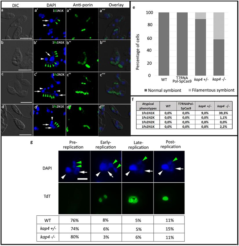
Analyses of cellular patterns were performed in A. deanei labeled with DAPI and with an anti-porin antibody
that recognizes the endosymbiont, considering the number of nuclei, kinetoplasts and symbionts, as well as the
shape of the bacterium (Fig. 3). As expected, in asynchronous cultures of WT cells, approximately 30%, presented
one rod-shaped symbiont, one kinetoplast and one nucleus (1S1K1N). Most cells, that is, approximately 50%, also
presented 1S1K1N; however, the symbiont presented a constricted or dividing format. The other part of the cul-
ture, approximately 20%, was composed of cells containing two rod-shaped symbionts. Such protozoa presented
one or two kinetoplasts and nuclei; however, kinetoplast division was always observed before the karyokinesis.
In KAP4 mutants cultivated for 24 h, protozoa presented atypical phenotypes as two nuclei, one kinetoplast and
one filamentous symbiont (1Sf2N1K) or two nuclei, two kinetoplasts and one filamentous symbiont (1Sf2N2K),
an indication of kDNA division and cytokinesis blockage, respectively (Fig. 3a–d). In KAP4 mutants cultivated
for 24 h, filamentous symbionts were observed in 3% kap4+⁄− cells and in 54% of kap4−⁄− protozoa, exhibiting
bacterium division impairment (Fig. 3e).
The counting of cell patterns in KAP4 mutants showed that the percentage of filamentous symbionts was
higher in cells containing one bacterium, one nucleus and one kinetoplast (1Sf1N1K) than in cells containing
two nuclei or two kinetoplasts, indicating that as the cell cycle progresses, the symbiont filamentation increases,
eventually leading to bacterial lysis. The percentage of cells containing one filamentous symbiont, two nuclei
and one kinetoplast (1Sf2N1K) was almost three times higher than in kap4 −⁄− protozoa when compared to
kap4+⁄− cells, indicating that in the double mutant, kinetoplast division was more affected (Fig. 3f). To check
whether the KAP4 mutant phenotype has an impact on kDNA replication, assays of dUTP incorporation by the
deoxynucleotidyl transferase terminal (TdT) were performed. The results showed that the percentage of cells
with the kDNA in the early replication stage was 62.5% lower in cells containing deletions of both KAP4 genes
than in WT protozoa. During this stage, the kinetoplast exhibits strong labeling in the antipodal sites but little
labeling in the kDNA network (Fig. 3g).
Scientific Reports | (2021) 11:9210 | https://doi.org/10.1038/s41598-021-88685-8 6
Vol:.(1234567890)www.nature.com/scientificreports/
Figure 1. Generation of KAP4 mutants, cell proliferation and viability. qPCR amplification showing that there was no damage to
the nuclear (a) and mitochondrial (b) DNA of the T7RNAPol-SpCas9 cells compared to WT cells. (c) Diagram representing the
sgRNA PCR transfection that allows for double strain breaks (DSBs) at the 5′ and 3′ ends of the genes and repair-templates mediated
recombination at the UTRs 30 nt upstream and downstream of the CDS. Diagnostic PCR oligonucleotides were designed to amplify
the integrated NEO repair template, binding upstream of the open reading frame (OL5) and internally to the NEO gene (OL6), and
the presence (WT and +/−) or absence (−/−) of KAP4 (650 bp, OL5 + OL7). (d) Diagnostic PCR showing KAP4 gene deletion and
Neo selectable marker integration in the A. deanei genome. (e) Growth curve for 72 h showed that KAP4 mutants present a reduced
proliferation in relation to WT and T7RNAPol-SpCas9 strains. Cell number was plotted on a logarithmic scale, and the presented data
are the mean ± s.d. of three independent cell cultures. After 48 h, when cells reached the peak of the exponential phase, a paired T test
(p < 0.05) was performed to compare control and mutant cells. (f) Duplication time of WT, T7RNAPol-SpCas9 and cells deleted for
KAP4. (g) The cell viability was similar among the strains analyzed and maintained even after 72 h of cultivation. The presented data is
a mean ± s.d. of three independent cell cultures. WT, wild-type cells, kap4+⁄−, cells with deletion for one allele, kap4−⁄−, null mutant.
Scientific Reports | (2021) 11:9210 | https://doi.org/10.1038/s41598-021-88685-8 7
Vol.:(0123456789)www.nature.com/scientificreports/
Figure 2. Ultrastructure and morphology of A. deanei. WT (a–c), T7RNAPol-SpCas9 (d–f) and KAP4 mutant ▸
cells with single (g–l) or double deletions (m–r). (a,b) Transmission electron microscopy of WT cells showed
typical characteristics of symbiont-harboring trypanosomatids, which were also observed in T7RNAPol-SpCas9
cells (d,e). kap4+⁄− and kap4−⁄− cells presented ultrastructural alterations as a high condensation of nuclear
DNA (g), a densely packed kDNA (i–o), a filamentous symbiont (h,n), dividing cells with two flagella in the
same flagellar pocket (m). Scanning electron microscopy showed the typical choanomastigote form in WT
and T7RNAPol-SpCas9 cells of mutant cells (c,f). kap4+⁄− mutants presented ultrastructure alterations such as
asymmetric division (j, yellow arrow), which generated cells with different dimensions (k) and protozoa with
multiple cell bodies and flagella (l). kap4−⁄− cells presented cytokinesis impairment that generated a popcorn-like
phenotype (q–r). In both mutant strains, cell bodies and flagellum shortening were observed (j,p, white arrows).
ht heterochromatin, k kinetoplast, lb lipid body, n nucleus, nu nucleolus, s symbiont, f flagellum, fs filamentous
symbiont, v vacuole. Brackets show the more densely packed kDNA.
Considering the structural results obtained in this work, we assumed that A. deanei KAP4 could participate in
kDNA metabolism. To confirm this hypothesis, WT and mutant cells were exposed to cisplatin or UV radiation to
verify the cell response to DNA damage. These agents cause distortions in the DNA that can impair transcription
and replication, with cisplatin lesions being more effective than UV light. Protozoa that had one or both KAP4
genes deleted were able to grow after treatment with cisplatin or exposure to UV, although in cisplatin treatment,
the mutant cells presented a slight decrease in cell proliferation compared to the WT strain after 12 h of treat-
ment, especially the single gene-deficient mutant treated with the highest inhibitor concentration (Fig. 4a–d).
Considering the cellular morphology and cellular organization, microscopy analyses were performed to test
whether KAP4 mutants presented atypical phenotypes in relation to WT cells after cisplatin treatment. This
compound interacts with DNA and proteins and forms intrastrand or interstrand DNA crosslinks that cause
distortions in the double helix, thereby blocking duplication and transcription. Transmission electron microscopy
images showed that WT cells did not present nuclear or kinetoplast changes after incubation with cisplatin, even
when a higher drug concentration (300 µM) was used (Fig. 5a–c). The same phenomenon was observed in the
background T7RNAPol-SpCas9 cell line (data not shown). Similarly, KAP4 mutants did not display topological
rearrangement on the kDNA network compared to WT cells treated with cisplatin (Fig. 5h,m). However, other
cellular structures suffered alterations in mutant protozoa. In kap4+⁄− cells, nuclear DNA unpacking was observed,
as well as myelin figures in the cytoplasm (Fig. 5f, black arrow) and mitochondrial swelling. The abundance of the
endoplasmic reticulum was noted and also its frequent association with the symbiont, which sometimes was seen
surrounded by this organelle, suggested an autophagic process (Fig. 5f, arrowheads). The symbiont also displayed
matrix loss and alterations in its DNA condensation (Fig. 5g, white arrows). In null mutants treated with 300 µM
cisplatin, the primary ultrastructural alteration was observed in the symbiont that presented membrane convolu-
tions (Fig. 5k, arrow), matrix loss and densely packed DNA fibers (Fig. 5l, white arrows). It is also worthwhile
to mention the presence of vacuoles around the symbiont, indicating that the bacterium had lysed (Fig. 5k–l).
Analyses by SEM showed that cultures of A. deanei WT cells presented a higher incidence of rounded proto-
zoa with a shortening flagellum after treatment with 150 and 300 µM cisplatin for 24 h (Fig. 5d,e). This phenotype
was also observed in mutant cells after treatment with both concentrations (Fig. 5i,j,n–o, white arrowheads).
Protozoa presenting a fat cell-like phenotype and lacking the flagellum (Fig. 5i, white arrow) were observed
after treatment with 150 µM cisplatin for 24 h. After using 300 µM of this drug, protozoa with the cytokinesis
phenotype were observed more frequently (Fig. 5j), indicating division impairment, as well as plasma membrane
blebs at the posterior end of the cell body (Fig. 5o). Protozoa that had one allele deleted seemed to have their
morphology more affected than null mutant when treated with this genotoxic agent.
Cells subjected to cisplatin treatment presented atypical phenotypes, as demontrated by fluorescence micros-
copy analysis. When treated with the lower inhibitor concentration (150 µM), WT trypanosomatids presented
rounded shapes with a reduced flagellum length and the fat cell phenotype. Symbionts were seen in the filamen-
tous format, presenting several nucleoids (Fig. 6a–a″). Mutants for KAP4 treated with cisplatin also presented
filamentous bacterium, but in this case, protozoa lacking the bacterium were also observed, as well as cells
presenting two nuclei and one kinetoplast (Fig. 6b–b″ and c–c″). Next, we counted the number of protozoa
containing a filamentous bacterium after cisplatin treatment for 24 h. In WT A. deanei, filamentous bacteria
were not identified in non-treated cells, as previously demonstrated. However, after incubation with 150 µM and
300 µM of cisplatin, 14% and 3% of the cells showed filamentous symbionts, respectively. In kap4+/− protozoa,
when both concentrations of cisplatin were used, the percentage of filamentous bacteria was similar, that is,
approximately 2%. In the null mutant (kap4−/−), values were equivalent to 14% and 8%, respectively (Fig. 6d).
These results indicate that treatment with cisplatin induced symbiont filamentation and that a higher con-
centration of the inhibitor (300 µM) augmented symbiont lysis, as also suggested by transmission electron
microscopy data. Counting of cellular patterns demonstrated that after treatment with cisplatin, the highest
percentage of bacterial filamentation was present in the double mutant cells containing one nucleus and one
kinetoplast (1Fs1N1K). In these cells, the percentage of protozoa with a filamentous symbiont decreased in a
concentration-dependent manner. The percentage of protozoa with bacterial filamentation also decreased with
the progression of the cell cycle, as in cells containing two nuclei, reinforcing the notion of the bacterial lysis.
Taken together, these data indicate that somehow genotoxic agents alter the cell division pattern in A. deanei and
that this effect is exacerbated in mutant cells (Fig. 6e). Cisplatin can block replication and trigger checkpoints
at the end of S phase and the beginning of G2 to repair lesions, thereby causing cell cycle arrest. However, when
cells treated with cisplatin were submitted to flow cytometry analysis, they did not show cell cycle alterations in
relation to control cells, even after treatment with 300 µM for 24 h (Supplementary Fig. 2).
Scientific Reports | (2021) 11:9210 | https://doi.org/10.1038/s41598-021-88685-8 8
Vol:.(1234567890)www.nature.com/scientificreports/
Scientific Reports | (2021) 11:9210 | https://doi.org/10.1038/s41598-021-88685-8 9
Vol.:(0123456789)www.nature.com/scientificreports/
Figure 3. Atypical phenotypes were observed in KAP4 mutant cells cultivated for 24 h after labeling with DAPI
and anti-porin antibodies. WT (a–a″); kap4+⁄− mutants containing one filamentous symbiont with multiple
nucleoids (Fs—green arrowhead), one nucleus (N-white arrows) and one kinetoplast (K-white arrowhead)
(b–b″) or two nuclei and two kinetoplasts and (c–c″); kap4−⁄− cells were seen with one filamentous symbiont,
two nuclei and one kinetoplast (d–d″). Bars 5 μm. (e) Counting of cellular patterns showing that filamentous
symbionts (Fs) are more frequent in kap4−⁄−. (f) Percentage of cells presenting atypical phenotypes. (g) In situ
labeling showing the different stages of kDNA network replication in WT and mutant cells (according to Liu
and Englund 2007)15. Green arrowheads indicate the symbiont, white arrows the nucleus and white arrowheads
the kinetoplast. Bars 1 μm. t test p-value < 0.005. A total of 1000 WT and KAP4 mutant cells were counted in 3
independent experiments.
The susceptibility of WT and mutant cells to UV radiation was also verified. Thus, protozoa were subjected to
UV-C irradiation, which affects the DNA replication and transcription and can be repaired by nucleotide exci-
sion. Ultrastructural and morphological analyses were performed after 24 h of protozoa irradiation at 1500 μJ/
m2. The results obtained by transmission electron microcopy were similar to those observed for WT and KAP4
mutant cells treated with cisplatin: nuclear DNA and kDNA did not suffer additional topological alterations in
relation to nonirradiated cells (Fig. 7a,b,f,g,k), and a close association of the ER with the symbiont occurred
Scientific Reports | (2021) 11:9210 | https://doi.org/10.1038/s41598-021-88685-8 10
Vol:.(1234567890)www.nature.com/scientificreports/
Figure 4. Cell growth and survival after cisplatin treatment or UV radiation. After 12 h, no remarkable
differences were observed in cell proliferation and survival when comparing WT and mutant protozoa after
treatment with 150 μM and 300 μM cisplatin (a,b) or exposure to UV radiation (c,d).
frequently, strongly indicating autophagy (Fig. 7a,f,k, white arrowheads). Notably, after irradiation, mutant cells
presented bacteria with a higher DNA condensation (Fig. 7a,g,k, white arrows). Furthermore, polynucleated
cells were observed (Fig. 7l).
Scanning electron microscopy analyses showed that WT protozoa suffered morphological alterations
after irradiation, exhibiting wrinkled cell surfaces and irregular forms that indicated cytokinesis impairment
(Fig. 7c–e). In single- and double-KAP4-deleted mutants, the morphological modifications were exacerbated:
many protozoa presented multiple interconnected cell bodies, reinforcing the notion that cytokinesis was blocked
(Fig. 7h–i). Such cells also presented wrinkled surfaces, and the flagellum was absent in some instances (Fig. 7h,j,
white arrows), as also observed in null mutants (Fig. 7m,o, white arrows). In this last case, a high number of
round cells were also observed (Fig. 7m–o).
Irradiated protozoa were also labeled with DAPI, exhibiting atypical phenotypes that were compatible with
asymmetric division and cytokinesis impairment, such as the presence of one kinetoplast and two nuclei in cells
containing two symbionts (Fig. 8a) and dyskinetoplastic cells. Such morphotypes were observed in WT cells,
as well as in mutant cells (Fig. 8b). Protozoa with filamentous bacterium were observed more frequently in WT
cells than in KAP4 mutants, on which symbiont division was probably more strongly affected. The absence of
the symbiont was observed in null mutants (Fig. 8c), which may have been related to the possible occurrence of
autophagy, in this case, a symbiophagy, that generated aposymbiotic cells, as suggested by transmission electron
microscopy. The very reduced number of WT cells presenting filamentous symbionts (1.4%) and the absence of
this phenotype in mutant cells reinforced this notion (Fig. 8d). The percentage of irradiated protozoa presenting
atypical phenotypes was low in all cell types (Fig. 8e).
To verify whether KAP4 was involved in kDNA repair, mutant and wild-type cells of A. deanei cells were
treated with cisplatin and UV radiation, as described before, and DNA repair kinetics were measured by long-
range qPCR assay. After treatment with 300 µM cisplatin, WT and both mutant strains presented the same levels
of DNA damage on the kDNA, which was approximately 1.5 lesions/10 kB. The repair kinetics were very similar
for all cell types: after 3 h of treatment, levels of kDNA damage were almost undetectable, reaching the slowest
point after 6 h (Fig. 9a,b, Supplementary Information 3). A similar phenotype was observed for the UV radia-
tion. The levels and DNA repair kinetics of mutant cells were very similar to those observed in WT cells. After
1 h of treatment, most damage had already been repaired, although it required 3 h after UV radiation to reach
the same level of repair that was observed in cisplatin-treated cells, with the lowest point being observed at 6 h
(Fig. 9c,d). Taken together, these results demonstrate that KAP4 was not directly involved in the removal of DNA
Scientific Reports | (2021) 11:9210 | https://doi.org/10.1038/s41598-021-88685-8 11
Vol.:(0123456789)www.nature.com/scientificreports/
Figure 5. Effects of cisplatin on the ultrastructure of mutant cells as revealed by TEM (a–c,f–h,k–m) and SEM
(d,e,i,j,n,o). A–E: WT cells treated with cisplatin did not present ultrastructural alterations by TEM. However,
SEM showed rounded cells with a shortening flagellum (d,e). (f–o) mutant cells treated with cisplatin. (f) Note
DNA unpacking in the nucleus (n), the proximity between the ER (black arrowhead) and the endosymbiont,
and mitochondrial branch swelling (m). (g,l) The symbiotic bacterium presents alterations in the nuclear matrix
and DNA condensation (white arrows). (k,l): The symbiont presented membrane convolutions (black arrow)
and was surrounded by vacuoles, an indication of autophagy. (h,m) In mutant cells, the kDNA arrangement
was not affected in relation to protozoa not submitted to cisplatin treatment. n nucleus, k kinetoplast, m
mitochondrial branch, s symbiont, v vacuole. (d,e,i,j,n,o) WT and mutant cells of both types treated with
cisplatin presented a rounded format containing a shortening flagellum. Other atypical phenotypes, such as fat-
cell shape (d), lack of flagellum (i,n, arrowheads) and plasma membrane blebs (o, arrows), were also observed.
damage generated by cisplatin and UV radiation but, notably, show that lesions generated by both genotoxic
agents could be repaired in mitochondrial DNA (Fig. 9a–d).
Discussion
In recent decades, A. deanei has been used as a model to investigate endosymbiosis and the origin of organelles.
Genome sequencing is a vailable26,27,57, and molecular tools for gene functional studies were developed, although
with limited use on the evaluation of gene essentiality and symbiosis m aintenance24,58,59. The recent application
of highly efficient CRISPR-Cas9 protocols to other trypanosomatids, such as Leishmania and T. cruzi , acceler-
ated functional studies with gene deletion51,59,60. In this study, for the first time, we describe gene depletion in
an endosymbiont-harboring trypanosomatid. Phylogenetic proximity with Leishmania enabled the successful
application of the CRISPR-Cas9 system developed by Beneke et al.51 to A. deanei, resulting in efficient deletion
of KAP4, a kinetoplast associated protein present in all trypanosomatids so far a nalyzed9.
KAPs can neutralize the negative DNA charge, thus facilitating the interaction of mitochondrial proteins with
kDNA, as those involved in replication and transcription. In this work, deletion of A. deanei KAP4 generated
trypanosomatids with reduced cell proliferation and generated cells with atypical phenotypes, as those presenting
two nuclei and one kinetoplast, as well as cytokinesis impairment. Cells containing aberrant numbers of nucleus
and kinetoplast were also observed in null mutants of C. fasciculata for KAP2 and KAP3 that presented cell divi-
sion block16. In T. brucei, the RNAi knockdown of a kDNA associated protein, resulted in reduced growth and
in the appearance of dyskinetoplastic c ells20. These results reinforce the importance of KAPs to cell proliferation
and kDNA network replication in order to guarantee that each new protozoa will receive one kinetoplast during
trypanosomatid division.
The coordinated division of the symbiont with the host cell nucleus was previously demonstrated in A.
deanei and in Strigomonas culicis, another symbiont-harboring t rypanosomatid22–24. In the present work, it was
interesting to observe in KAP4 mutant cells that the kDNA condensation, which is associated with kinetoplast
replication impediment, resulted in symbiont filamentation. This filamentation occurred most frequently in
Scientific Reports | (2021) 11:9210 | https://doi.org/10.1038/s41598-021-88685-8 12
Vol:.(1234567890)www.nature.com/scientificreports/
Figure 6. DAPI-stained mutant protozoa presented different atypical phenotypes when compared to WT
cells after treatment with cisplatin for over 24 h (a–c). (a–a″) WT cells treated with 150 µM cisplatin presented
rounded shapes, and the fat cell phenotype contained a symbiont with multiple nucleoids (white square, green
arrowheads). (b–b″) Ad kap4(+⁄−) cells treated with 300 µM cisplatin lacking the symbiont. (c–c″) Ad kap4(−⁄−)
cells treated with 300 µM cisplatin containing one filamentous symbiont, two nuclei and one kinetoplast. (d)
Counting of cellular patterns considering the presence of normal or filamentous symbionts. (e) Percentage of
cells with atypical phenotypes. Bars 5 μm. Fs Filamentous symbiont., N nucleus—white arrows; K-kinetoplast—
white arrowheads. A total of 1000 cells of WT and KAP4 mutant cells were counted in 3 independent
experiments.
mutant with two nuclei and one kinetoplast. Consistent with this notion, TdT labeling showed a lower percentage
of kap4−⁄− cells in the early replication phase when compared to the WT protozoa, indicating that in such cells
the mitochondrial DNA replication was delayed or even impaired. Since kDNA loss resulting in dyskinetoplastic
protozoa was not observed, it can be assumed that the impediment of mitochondrion DNA replication promoted
cytokinesis blockage. Taken together, the results indicate that bacterial division is also coordinated with kineto-
plast replication, but further studies are essential to confirm this hypothesis. Cell cycle checkpoints are not well
established for most trypanosomatids species, nor are the factors that coordinate the equal partitioning of single
copy organelles to daughter cells. Such questions are best studied in T. brucei, especially by investigating the role
of protein kinases in cell cycle progression, organelle positioning and protozoan morphology60–64. Recently, it
was shown that T. brucei UMSBP2, which is involved in kDNA replication and segregation, is also localized at
telomeres. The RNAi system showed that this protein not only participates in nuclear division but also plays a
role in the coordinated replication of DNA-containing organelles65.
In A. deanei KAP4 mutants, the high level of kDNA packing was associated with delay in cell proliferation and
in kDNA duplication at the early stage, when the covalently closed minicircles are released from the network to
initiate replication into the KFZ and then migrate to antipodal sites, where this process c ontinues4. Previously,
it was shown that the downmodulation of T. brucei P93, a kDNA-associated protein localized in antipodal sites,
resulted in loss of gapped minicircles and consequently in the network reduction66. Similarly, in TbKAP6 RNAi
cells, the levels of total minicircles and maxicircles decreased the total amount of nicked/gapped minicircles.
In such cells, the kinetoplast presented network shrinkage or elongation, but in both cases, two basal bodies
could be identified, indicating failures in kDNA replication and scission. Conversely, in protozoa overexpressing
TbKAP6, the minicircle decatenation was enhanced, indicating that a controlled expression of this protein is
required for proper kDNA replication20.
The kDNA arrangement and metabolism are the result of the coordinated activity of a set of mitochondrial
proteins that serve different functions. In addition to KAPs, other proteins are involved in the kDNA replica-
tion, such as the minicircle replication factor (MiRF172), which is supposedly involved in the reattachment of
replicated minicircles to the kDNA disc. Once depleted, T. brucei cells presented reduced kDNA content or even
a dyskinetoplastic p henotype67. Downregulation of mitochondrial heat shock proteins 70 and 40 also showed
impairment of minicircle replication and loss of kDNA, demonstrating the importance of chaperones to the
maintenance of the kinetoplast as a cellular structure68. In the present work, the generation of dyskinetoplastic
cells was not observed among KAP4 mutants. Although the gene deletion promoted increased kDNA compac-
tion, the data obtained by qPCR did not indicate loss of mitochondrial DNA. In UV-irradiated protozoa, a very
low percentage of cells without a kinetoplasts was observed.
The DNA repair kinetics showed no differences between KAP4 mutant cells and the WT strain. In both cases,
protozoa were able to efficiently repair the damage generated by cisplatin and UV radiation. In addition, differ-
ences in the long-term survival of these cells were not observed. For both genotoxic agent treatments, the kDNA
Scientific Reports | (2021) 11:9210 | https://doi.org/10.1038/s41598-021-88685-8 13
Vol.:(0123456789)You can also read


