Lima1 mediates the pluripotency control of membrane dynamics and cellular metabolism - Caltech Authors
←
→
Page content transcription
If your browser does not render page correctly, please read the page content below

ARTICLE
                  https://doi.org/10.1038/s41467-022-28139-5                 OPEN

                  Lima1 mediates the pluripotency control of
                  membrane dynamics and cellular metabolism
                  Binyamin Duethorn1, Fabian Groll2, Bettina Rieger3, Hannes C. A. Drexler 4, Heike Brinkmann1,
                  Ludmila Kremer5, Martin Stehling6, Marie-Theres Borowski 3, Karina Mildner7, Dagmar Zeuschner 7,
                  Magdalena Zernicka-Goetz 8,9, Marc P. Stemmler 10, Karin B. Busch3, Juan M. Vaquerizas 2,11,12 &
                  Ivan Bedzhov 1 ✉
1234567890():,;

                  Lima1 is an extensively studied prognostic marker of malignancy and is also considered to be
                  a tumour suppressor, but its role in a developmental context of non-transformed cells is
                  poorly understood. Here, we characterise the expression pattern and examined the function
                  of Lima1 in mouse embryos and pluripotent stem cell lines. We identify that Lima1 expression
                  is controlled by the naïve pluripotency circuit and is required for the suppression of mem-
                  brane blebbing, as well as for proper mitochondrial energetics in embryonic stem cells.
                  Moreover, forcing Lima1 expression enables primed mouse and human pluripotent stem cells
                  to be incorporated into murine pre-implantation embryos. Thus, Lima1 is a key effector
                  molecule that mediates the pluripotency control of membrane dynamics and cellular
                  metabolism.

                  1 Embryonic Self-Organization research group, Max Planck Institute for Molecular Biomedicine, Röntgenstraße 20, 48149 Münster, Germany. 2 Regulatory

                  Genomics group, Max Planck Institute for Molecular Biomedicine, Röntgenstraße 20, 48149 Münster, Germany. 3 Institut für Integrative Zellbiologie und
                  Physiologie, University of Münster, Schlossplatz 5, 48149 Münster, Germany. 4 Mass Spectrometry Unit, Max Planck Institute for Molecular Biomedicine,
                  Röntgenstraße 20, 48149 Münster, Germany. 5 Transgenic Facility, Max Planck Institute for Molecular Biomedicine, Röntgenstraße 20, 48149
                  Münster, Germany. 6 Flow Cytometry Unit, Max Planck Institute for Molecular Biomedicine, Röntgenstraße 20, 48149 Münster, Germany. 7 Electron
                  Microscopy Facility, Max Planck Institute for Molecular Biomedicine, Röntgenstraße 20, 48149 Münster, Germany. 8 Mammalian Embryo and Stem Cell
                  Group, Department of Physiology, Development, and Neuroscience, University of Cambridge, Downing Street, Cambridge CB2 3EG, UK. 9 Plasticity and Self-
                  Organization Group, Division of Biology and Biological Engineering, California Institute of Technology (Caltech), Pasadena, CA 91125, USA. 10 Department of
                  Experimental Medicine 1, Nikolaus-Fiebiger-Center for Molecular Medicine, FAU University Erlangen-Nürnberg, Erlangen, Germany. 11 MRC London Institute
                  of Medical Sciences, Du Cane Road, W12 0NN London, UK. 12 Institute of Clinical Sciences, Faculty of Medicine, Imperial College London, Hammersmith
                  Hospital Campus, Du Cane Road, London W12 0NN, UK. ✉email: ivan.bedzhov@mpi-muenster.mpg.de

                  NATURE COMMUNICATIONS | (2022)13:610 | https://doi.org/10.1038/s41467-022-28139-5 | www.nature.com/naturecommunications                                  1
ARTICLE                                                                 NATURE COMMUNICATIONS | https://doi.org/10.1038/s41467-022-28139-5

T
        he most frequent types of cancer originate from epithelial           mitochondrial function in a cell-autonomous manner and is
        cells, which aberrantly activate signalling cascades that            essential for the growth of solid tumours. Strikingly, ectopic
        promote cell proliferation and allow cells to evade                  expression of Lima1 in human pluripotent stem cells enabled
apoptosis1. The cancer cells often reprogramme their energy                  these cells to be incorporated into murine pre-implantation
metabolism, lose epithelial morphology and activate invasion and             embryos. Thus, Lima1 is a key effector molecule mediating
metastasis, enabling a benign adenoma to transform into an                   pluripotency control of membrane dynamics and cellular
invasive carcinoma, which correlates with poor patient prognosis.            metabolism.
The malignant transformation is associated with reduced inter-
cellular adhesion as well as cytoskeletal and signalling reorgani-
sation, in a process known as epithelial-to-mesenchymal                      Results
transition (EMT)2.                                                           Lima1 expression pattern during mouse embryonic develop-
   The process of EMT as well as the activation of several sig-              ment. The process of embryogenesis starts from a single cell
nalling pathways, such as Wnt, Notch and Hedgehog, are com-                  (zygote), which gives rise to both the embryonic and extra-
mon features in both embryonic development and cancer3. The                  embryonic lineages of the early embryo. After implantation, a
EMT, which mediates E-cadherin (E-cad) downregulation, is the                complex cascade of cell fate decisions and morphogenetic events
driving force enabling malignancy and is also a key step in the              form the different tissues and organs of the developing embryo.
process of gastrulation1. Similarly, the canonical Wnt/β-catenin             To analyse Lima1 expression during murine development, we
(β-cat) signalling is essential for the patterning of the early mouse        isolated E14.5 embryos, which were sectioned and stained for
embryo4,5. However, aberrant activation of this pathway can                  Lima1. Alongside this, we examined the localisation of the major
initiate tumorigenesis by promoting the expression of down-                  components of the CCC, namely E-cad, α-E-catenin (α-E-cat),
stream oncogenes6.                                                           N-cadherin (N-cad), α-N-catenin (α-N-cat) and β-cat. In E14.5
   E-cad and β-cat, together with several actin-binding proteins,            embryos, E-cad and α-E-cat were expressed mainly in epithelial
such as α-catenin (α-cat) and Lima1 (also known as EPLIN;                    compartments, such as the inner lining of the stomach (Fig. 1a,
epithelial protein lost in neoplasm), form the adherens junction             blue arrowhead). N-cad and α-N-cat were detected in the muscle
(AJ) complex1,7. The role of the main components of this com-                fibres of the stomach wall (Fig. 1a, magenta arrowhead) as well as
plex has been extensively studied in the context of both cancer              in the nervous system, whereas various levels of β-cat were
and embryogenesis, which substantially widens our under-                     broadly detectable throughout the embryo (Fig. 1a and S1).
standing of their expression pattern, functions and regulation.              Although Lima1 is considered to be an epithelial protein10,
Yet, it is so far unknown how Lima1, which is considered to be a             unexpectedly its expression was not confined only to epithelial
prognostic marker and tumour suppressor8, functions during                   tissues. Similar to β-cat, Lima1 was broadly expressed, occupying
embryonic development.                                                       both E-cad and N-cad expression domains (Fig. 1a and S1),
   From previous studies, the Lima1 locus is known to harbour                suggesting that the potential functions of Lima1 in the developing
two promoter regions, which drive the expression of the shorter              embryo may span beyond epithelial homoeostasis.
Lima1-α (600 amino acids) and the longer Lima1-β (759 amino                     Next, we analysed the expression of Lima1 during the pre-
acids) isoforms. Both isoforms contain two actin-binding                     implantation embryogenesis. Using available transcriptomics
domains and a central LIM domain9,10. Recombinant Lima1-α                    data20, we found that Lima1 mRNA is present in GV and MII
and -β proteins suppress the depolymerization of actin filaments              oocytes as maternal transcripts, and later the zygotic expression
and cross-link the filaments in bundles11. In epithelial cells, the           exhibits a sharp increase after the 16-cell stage (Fig. 1b). Staining
actin fibres are linked to the cadherin-catenin complex (CCC) via             GV and MII oocytes for Lima1 revealed that the protein is
α-cat on the AJ12. Lima1 can bind α-cat and F-actin, thereby                 localised in the cytoplasm and the cortex, with strong enrichment
mediating the contact of the CCC with the cytoskeleton7 and                  in the cortical cup overlying the spindle of the MII oocytes
acting as a mechanosensitive regulator13.                                    (Fig. 1c). Interestingly, in the zygote, Lima1 accumulated in the
   In the context of cancer, Lima1 downregulation is implicated in           extruded polar body, where it remained detectable during the
the progression of oral, prostate and breast cancer10,14–17. In              next cleavage stages. Later, Lima1 expression appeared stronger in
prostate cancer cells, Lima1 depletion promotes AJ disassembly               the inside cells of the 16-cell morula and the ICM of the early
and cell invasion18; conversely, in oesophageal and breast cancer            blastocyst (Fig. 1c). To validate these observations, we examined
cells, Lima1-α overexpression decreases tumour growth and                    the Lima1 expression pattern in combination with lineage
invasiveness, acting as a tumour suppressor16,17. Moreover,                  markers, discriminating the inner/outer and the ICM/trophecto-
Lima1 function is also involved in the cellular metabolism, as               derm (TE) cells of morula and early blastocyst stage embryos,
shown in the gut, where Lima1 binds to intracellular cholesterol             respectively. In comparison to E-cad, β-cat and α-E-cat, which
transporters and facilitates cholesterol absorption19.                       were overall ubiquitously expressed in both compartments, Lima1
   As the majority of studies examining Lima1 have been per-                 was enriched in the Sox2 +/Cdx2− inner cells of the pre-
formed in pathological situations, the role of Lima1 in a devel-             implantation embryo (Fig. 1d, e).
opmental context of non-transformed cells is still poorly                       At mid and late blastocyst stage, we found that the Sox17+
understood. Therefore, we used the mouse embryo and embryo-                  primitive endoderm (PE) cells toned-down Lima1 expression
derived stem cell lines to analyse the expression pattern and                (Fig. 1f, arrowhead), whereas the pre-implantation epiblast
function of Lima1.                                                           (Sox17−/Cdx2−) maintained a relatively high level of Lima1.
   Here, we show that Lima1 expression was not confined only to               In addition, Lima1 also became detectable in the TE (Fig. 1f).
the epithelial tissues of the foetus. During the pre-implantation            Conversely, after implantation at early egg cylinder stage (E5.5),
development, Lima1 was initially enriched in the polar body and              Lima1 expression was decreased in the extraembryonic ectoderm
later in the pluripotent inner cell mass (ICM) of the early embryo.          (ExE) and the post-implantation epiblast (Fig. 1f). Thus, Lima1
We determined that Lima1 is under the transcriptional control of             exhibited a dynamic expression pattern in the embryonic and
the naïve pluripotency circuit and is required for suppressing the           extraembryonic lineages during early mouse development. In the
formation of membrane blebs in mouse and human pluripotent                   next experiments we focused on examining Lima1 function in the
stem cells. In addition, we found that Lima1 is required for proper          pluripotent lineage.

2                NATURE COMMUNICATIONS | (2022)13:610 | https://doi.org/10.1038/s41467-022-28139-5 | www.nature.com/naturecommunications
NATURE COMMUNICATIONS | https://doi.org/10.1038/s41467-022-28139-5 ARTICLE
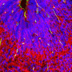
a E-cadherin α-E-catenin N-cadherin α-N-catenin β-catenin Lima1
E14.5
pancreas stomach liver
stomach
b E-cadherin
c d
Lima1
E-cad Sox2 DAPI
4000
Normalized expression
Lima1 DAPI
3000
2000
1000 Lima1 Sox2 DAPI
Lima1 DAPI
0
La bla tocy ll
8- ll
M bla 6-c ll
e
oo e
Ea Zy te
4- ll
2 - ll
La 2- ll
st
to t
id s e
ce
ce
te st st
te ce
ce
rly got
id e
II yt
as ys
cy
M 2-c
cy
M ooc
bl oc
1
V
G
rly
Ea
e E-cad Gata3 DAPI β-cat Gata3 DAPI
morula early blastocyst morula early blastocyst
f Lima1 Cdx2 Sox17 DAPI
early blastocyst mid blastocyst late blastocyst egg cylinder
α-E-cat Gata3 DAPI Lima1 Cdx2 DAPI
morula early blastocyst morula early blastocyst
Fig. 1 Lima1 expression pattern during mouse embryonic development. a E14.5 embryos sectioned and stained for E-cad, α-E-cat, N-cad, α-N-cat, β-cat or
Lima1. Blue and magenta arrows indicate epithelial and muscle layers, respectively. b Normalized expression levels of Lima1 and E-cad during mouse pre-
implantation development, based on Wang et al., 2004. c Oocytes and pre-implantation embryos stained for Lima1. Arrowheads indicate Lima1 localisation
on the cortical cap in MII oocyte and in the polar body at the zygote stage. d Blastocyst stage embryos stained for Sox2 and E-cad or Sox2 and Lima1.
e Morula and blastocyst stage embryos stained for AJ components, Lima1 and TE lineage markers (Gata3 or Cdx2). f Blastocyst and egg cylinder stage
embryo stained for Lima1, Cdx2, Sox17 and DAPI. The PE is indicated by an arrowhead. Scale bars, (a), 1000 μm, upper panel and 100 μm, lower panels;
(c), (d) and (e), 20 μm; (f), 10 μm. Experiments were repeated independently at least three times with similar results (a, c, d, e, f). Related to Figure S1.
NATURE COMMUNICATIONS | (2022)13:610 | https://doi.org/10.1038/s41467-022-28139-5 | www.nature.com/naturecommunications 3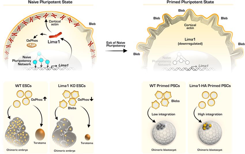
ARTICLE                                                                  NATURE COMMUNICATIONS | https://doi.org/10.1038/s41467-022-28139-5

Lima1 expression is under the control of the naïve plur-                      comparing WT and Lima1 KO ESC (Fig. 3d and S3A,
ipotency circuit. As Lima1 was enriched in the pre-implantation               Supplementary. Dataset 1). We found no substantial changes in
epiblast, we asked whether its expression is maintained in vitro, in          the expression of core, naïve or primed pluripotency markers
embryonic stem cells (ESC). Murine ESC are typically co-cultured              (Fig. 3e). In addition, Lima1 loss of function did not result in an
with mitotically inactivated mouse embryonic fibroblasts (MEFs)                upregulation of ecto-, endo- or mesodermal markers (Fig. S3B),
in serum-containing medium, supplemented with leukaemia                       showing that Lima1 depletion does not promote ESC
inhibitory factor (Lif)21; alternatively, ESC can be grown without            differentiation.
MEFs in serum-free N2B27 medium, supplemented with Lif and                       Interestingly, gene ontology (GO) analysis revealed the
2i (Gsk3 and Mek inhibitors)22. In the 2i/Lif culture conditions,             enrichment of gene expression associated with epithelial and
we found that the longer Lima1-β isoform was predominantly                    amoeboid-like cell migration as well as the downregulation of
expressed, whereas, in the presence of MEFs, both Lima1-α and -β              genes involved in cellular metabolism in Lima1 KO ESC. In
isoforms were detectable (Fig. 2a). However, MEFs themselves                  addition, GO terms such as DNA replication, nuclear division
showed only Lima1-α expression, indicating that Lima1-β is the                and chromosome segregation were reduced, suggesting that
main isoform present in pluripotent cells (Fig. 2a).                          Lima1 KO ESC may exhibit reduced rates of mitosis and
   The mouse blastocyst and egg cylinder stage embryos are also a             potentially a decrease in cell proliferation (Fig. 3f and S3C-S3D).
source of trophoblast stem cells (TSC), which are derived from the            To further explore Lima1 functions, we characterised the Lima1
trophoblast lineage23. As Lima1 expression was lower in the TE (early         interactome using APEX2 (peroxidase proximity labelling with
blastocyst) and ExE (egg cylinder), compared to the pre-implantation          ascorbate peroxidase) assay36. We fused the Lima1-β isoform (full
epiblast, we asked whether TSC and ESC recapitulate this expression           length) at the C-terminus to an HA-tag, followed by the APEX2
pattern in vitro. Similar to the pre-implantation embryo, Lima1 was           coding sequence (Fig. 3g, h). In the presence of biotin-phenol
enriched in ESC but not TSC, whereas E-cad was ubiquitously                   (BP), a pulse of hydrogen peroxide results in the generation of
expressed in both TSC and ESC (Fig. 2b and S2A). In addition, using           biotin-phenoxyl radicals by APEX2, which covalently labels
a forced expression of Cdx224, we reprogrammed the ESC to TSC-                proximal endogenous proteins (Fig. 3g, i). The subsequent mass
like cells (Fig. 2c) and found that, in contrast to the E-cad expression      spectrometry analysis revealed four main groups of potential
that remained unchanged, Lima1 was downregulated in the TSC-like              interaction partners: cytoskeletal proteins (e.g., actin and tubulin),
cells (Fig. 2d–f).                                                            actin-binding factors (e.g., cofilin-1 and spectrin), AJ components
   As ESC capture features of the naïve pluripotent lineage of the            (e.g., β-cat and p120) and factors involved in metabolism (e.g.,
blastocyst stage embryo25, we asked whether Lima1 is under the                pyruvate kinase and dihydropyrimidinase) (Fig. 3j, Supplemen-
transcriptional control of the naïve pluripotency network. To test this,      tary Dataset 2). Among these factors, we found proteins that have
we compared Lima1 expression in naïve ESC to more developmen-                 been previously shown to directly interact with Lima1, such as
tally advanced pluripotent stem cells. Briefly, we treated ESC with            actin37, or participate together in a larger molecular complex,
Fgf2/Activin, which causes cells to exit naïve pluripotency and               such as β-cat and p120 catenin7. Thus, altogether, the RNA-seq
transition into so-called epiblast-like cells (EpiLC)26 (Fig. 2g). These      and proteomics analysis suggest that Lima1 might play a role in
cells shut down the expression of naïve pluripotency transcription            cytoskeletal dynamics as well as in cellular metabolism.
factors, such as Nanog and Esrrb (Fig. 2k and S2B), establishing                 To examine the potential role of Lima1 in cell migration, we
an early post-implantation epiblast-like state26. Similar to Nanog and        dissociated Lima1 KO and WT ESC and analysed their behaviour on
Esrrb, Lima1 expression was diminished in EpiLC, whereas E-cad                a single-cell level. Strikingly, Lima1 KO ESC exhibited spherical
and β-cat protein levels remained overall unchanged (Fig. 2h, k               membrane protrusions (blebs), that were rarely detectable in
and S2B). In addition, Lima1 was also enriched in naïve human                 WT cells (Fig. 3k, Movies S1 and S2). The blebbing was exhibited
induced pluripotent stem cells (naïve hiPSC) in comparison to                 only upon individualisation of the cells and it faded away completely,
primed (conventional) hiPSC (Fig. S2C).                                       as the cells came together to form small colonies. Membrane blebbing
   During the transition from the naïve to the primed state of                has been subdivided into three phases, namely initiation (nucleation),
pluripotency, Lima1 was substantially downregulated on the                    expansion and retraction38, whereby bleb nucleation is established by
mRNA level, exhibiting similar transcriptional dynamics as the                cell membrane dissociation from the actin cortex39 or via local
naïve pluripotency factor Nanog (Fig. 2l). Moreover, analysis of              rupture of the cortex40,41. The expansion phase is driven by the
available ChIP-seq data on pluripotency transcription factor                  hydrostatic pressure of the cytoplasm, followed by the formation of a
binding in ESC27–35 (Table S1) revealed peaks for Nanog, Nr0b1,               new actin cortex under the blebbing membrane. Finally, the
Klf4, Sox2, Oct4, Sall4, β-cat and Tcf3 in the vicinity of the                recruiting of myosin to the newly assembled cortex allows the bleb
Lima1-β promoter (Fig. 2m). This suggests that Lima1 is a                     to retract38.
potential target gene of the naïve pluripotency circuit and may act              To examine the blebbing in WT and Lima1 KO ESC, we stained
as a downstream effector, mediating naïve pluripotency control of             the cells for the bleb marker phospho-ERM (ezrin, radixin and
cytoskeletal dynamics.                                                        moesin)38 and found that Lima1 KO ESC exhibit extensive pERM-
                                                                              positive membrane protrusions (Fig. 3L, white arrowheads). In non-
                                                                              blebbing WT ESC, Lima1 was uniformly localised on the cortex,
Lima1 controls the membrane dynamics in ESC. To analyse                       which appeared as a continuous ring (Fig. 3m, blue arrowhead).
Lima1 functions, we established Lima1 knockout (ko) ESC using                 However, in WT cells that formed blebs, Lima1 was absent from the
the CRISPR/Cas9 approach (Fig. 3a). We deleted the genomic                    blebbing membrane (Fig. 3m, yellow arrowheads). Accordingly,
region containing exons 4 and 5 to generate a premature stop                  genetic deletion of Lima1 converted the overall non-blebbing ESC
codon, thus abolishing Lima1 translation (Fig. 3b). Morphologi-               morphology to a highly blebbing phenotype (Fig. 3k, m and o). This
cally, Lima1 KO ESC formed typical dome-shaped colonies,                      indicates that Lima1 plays an integral role in stabilising the cortex,
undistinguishable from the wild-type (WT) ESC. The main                       suppressing the formation of membrane blebs.
components of the CCC, namely E-cad, β-cat and α-E-cat, were                     In contrast to the naïve ESC, primed pluripotent cells, such as
properly localised on the membrane, and Lima1 KO ESC did not                  conventional human ESC (hESC) and mouse epiblast stem cells
exhibit intercellular adhesion defects (Fig. 3c).                             (EpiSC), typically exhibit blebbing upon dissociation into single
   To understand whether the loss of Lima1 can affect the status              cells42. As bleb formation depends on actomyosin activation via
of undifferentiated stem cells, we performed RNA-seq analysis                 the Rho/ROCK pathway38, treatment with a ROCK inhibitor

4                 NATURE COMMUNICATIONS | (2022)13:610 | https://doi.org/10.1038/s41467-022-28139-5 | www.nature.com/naturecommunications
NATURE COMMUNICATIONS | https://doi.org/10.1038/s41467-022-28139-5                                                                           ARTICLE

Fig. 2 Lima1 expression in embryonic and extraembryonic stem cell lines. a Western blot analysis of Lima1 expression in ESC and MEFs. b ESC and TSC
stained for Lima1 and Oct4 (left panel) or E-cad and Eomes (right panel). c Schematic representation of ESC conversion to TSC-like cells. d Western blot
analysis of Lima1 expression in ESC and TSC-like cells. e Western blot analysis of E-cad expression in ESC and TSC-like cells. f ESC and TSC-like cells
stained for Oct4, Troma1 and Lima1. g Schematic representation of ESC conversion to EpiLC. h Western blot analysis of Lima1 expression in ESC and EpiLC.
i Western blot analysis of E-cad expression in ESC and EpiLC. j Western blot analysis of β-cat expression in ESC and EpiLC. k ESC and EpiLC stained for
Nanog (left panel) or E-cad and Lima1 (right panel). l Relative expression levels of Oct4, Nanog and Lima1 normalized to GAPDH in ESC and EpiLC. Mean
values from three independent repetitions, data represent mean ± SEM, unpaired Student’s t-tests, 2-sided. m ChIP-seq peaks of pluripotency factors
binding on the Lima1 locus in ESC. Polr2a peaks indicate the promoter region, ATAC-seq peaks indicate the accessible chromatin regions. Scale bars,
(b), (f), (k), 50 μm. Experiments were repeated independently two times (a, b, e, f, i, j, k) or three times (d, h) with similar results. Related to Figure S2.

NATURE COMMUNICATIONS | (2022)13:610 | https://doi.org/10.1038/s41467-022-28139-5 | www.nature.com/naturecommunications                                      5
ARTICLE                                                                NATURE COMMUNICATIONS | https://doi.org/10.1038/s41467-022-28139-5

(Y-27632) has been shown to supress the blebbing of primed                  Lima1 depletion in ESC results in reduced mitochondrial ATP
pluripotent cells42,43. Accordingly, Y-27632 treatment also                 production, decreased rates of teratoma growth and a reduced
diminished blebbing in Lima1 KO ESC (Fig. 3n, o). Altogether,               contribution to chimeras. Pluripotent ESC can form non-
this shows that Lima1 acts as an effector molecule downstream of            invasive tumours (teratomas) that consist of derivatives of the
the naïve pluripotency circuit, mediating the control of                    three germ layers44. Loss of Lima1 has been implemented in
membrane dynamics in ESC.                                                   malignancy10,14–17, but how Lima1 depletion affects the growth

6               NATURE COMMUNICATIONS | (2022)13:610 | https://doi.org/10.1038/s41467-022-28139-5 | www.nature.com/naturecommunications
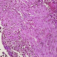
NATURE COMMUNICATIONS | https://doi.org/10.1038/s41467-022-28139-5                                                                      ARTICLE

Fig. 3 Lima1 depletion in ESC. a CRISPR/Cas9 targeting strategy. b Validation of Lima1 deletion in ESC by western blot. c Lima1 KO and WT ESC stained for
Lima1, E-cad, β-cat, α-E-cat or Nanog. d MA plot of gene expression in Lima1 KO vs. WT ESC, cultured in N2B27 2i/Lif. Statistical testing for differential
gene expression between conditions was performed using the default DESeq2 settings calculating the p-value using a two-tailed Wald-test with
Benjamini–Hochberg correction for multiple testing and independent filtering of results. e Expression of core, naïve and primed pluripotency genes in WT
and Lima1 KO ESC. f Gene ontology (GO) enrichment analysis “Biological processes”. Gene Ratio indicates the fraction of genes comprised by the GO term,
showing significant changes in their expression. g Scheme representation of APEX2 interactome analysis. h ESC expressing Lima1-HA-APEX2 construct,
stained for HA. i Western blot analysis of Lima1-HA-APEX2 ESC cultured in the presence of or without biotin-phenol (BP). The biotinylated proteins are
detected by streptavidin-HRP. j Example of putative Lima1 interaction partners identified by the APEX2 assay. k Membrane blebbing (arrows) in WT and
Lima1 KO ESC. l WT and Lima1 KO ESC stained for actin, pERM and DAPI. Arrowheads indicate membrane blebs. m WT and Lima1 KO ESC stained for
actin, Lima1 and DAPI. Blue arrowhead indicates Lima1 localisation on the cortex in control ESC; yellow arrowheads indicate loss of Lima1 on the membrane
blebs in control ESC; white arrowheads indicate membrane blebs in Lima1 KO ESC. n WT and Lima1 KO ESC treated with Y-27632. o Mean distribution of
cell membrane fluctuation counts per cell over 90 s of time-lapse imaging with 5 s intervals; n = 224 (WT), 133 (WT treated), 260 (Lima1 KO), 159 (Lima1
KO treated) cells, Mann–Whitney tests, 2-sided. Scale bar, (c) 20 μm; (h) 50 μm; (k), (l), (n), (o), 10 μm. Experiments were repeated independently two
times (c, h, l, m) or at least three times (b, i, k, n, o) with similar results. Related to Fig. S3.

of solid tumours is still obscure. To understand whether Lima1                  reactive oxygen species (Fig. 5f), suggesting that Lima1 depletion
depletion affects the growth rate and/or the composition of solid               is not associated with mitochondrial oxidative damage.
tumours, we subcutaneously injected WT ESC or Lima1 KO ESC                         Next, we conducted a mitochondrial stress test assay using an
into SCID mice to generate teratomas. Although Lima1 KO cells                   automatic flux analyser (Seahorse) to examine the oxygen
could give rise to teratomas, their size was substantially smaller              consumption rate (OCR) in WT vs. Lima1 KO cells. This assay
compared to the tumours derived from WT ESC (Fig. 4a, b).                       measures the OCR before and after applying inhibitors of
Nevertheless, Lima1 KO teratomas consisted of tissue derivatives                different electron transport chain (ETC) components, thereby
of all three germ layers, indicating that Lima1 depletion does not              providing an assessment of multiple respiration parameters. We
affect the ESC differentiation capacity (Fig. 4c).                              found that the basal and maximal respiration as well as the ATP-
   To further explore the behaviour of tissues derived from Lima1               linked respiration and the non-mitochondrial oxygen consump-
KO ESC, we generated chimeric embryos via aggregation of                        tion were substantially decreased in Lima1 KO cells (Fig. 5g, h).
morulae with nuclear tdTomato-labelled Lima1 KO ESC or                          This shows that Lima1 depletion results in a general reduction of
membrane tdTomato-labelled WT ESC. At E4.5, we found that                       the mitochondrial ATP production rate and energy efficiency in a
similar numbers of Lima1 KO and WT cells were incorporated in                   cell-autonomous manner. As it was previously reported that
the ICM of the chimeric blastocysts (Fig. 4d, e). However, after                primed pluripotent cells exhibit low mitochondrial respiration49,
implantation, at E6.5 and later at E13.5, we found a gradual but                we also compared the OCR of Lima1 KO ESC to WT EpiSC and
substantial decrease of Lima1 KO cells (Fig. 4f–i). Despite the                 found similar levels. Thus, although Lima1 KO ESC maintain the
reduced number, the Lima1 KO cells were present in all examined                 expression of naïve pluripotency transcription factors, they
foetal organs (Fig. 4j, k).                                                     exhibit low mitochondrial respiration akin to the primed
   Next, we aimed to determine why the tumour growth and                        pluripotent state.
chimerism were reduced in Lima1 KO cells. To understand                            As the reduction in cell proliferation of Lima1 KO ESC was not
whether the initial pool of Lima1-deficient pluripotent cells may                compensated by the presence of Y-27632 (Fig. 5b), we examined
have survival and/or proliferative disadvantage, we compared the                whether the reduction of the OCR is also Y-27632-independent.
cell death and proliferation of Lima1 KO vs. WT ESC. We found                   Accordingly, we found similar OCR levels in Lima1 KO ESC
that Lima1 depletion was not associated with an increase in                     cultured in the presence of or without Y-27632 (Fig. S4A). In
apoptosis or necrosis (Fig. 5a). However, we found a decrease in                addition, we differentiated WT and Lima KO ESC in vitro, using
the proliferation of Lima1 KO ESC cultured either in the presence               the embryoid body (EB) assay, and found no substantial
of or without Y-27632 (Fig. 5b).                                                difference in the OCR in WT and Lima1 KO EBs (Fig. S4B and
   As the transcriptional profiling of Lima1 KO ESC and the                      S4C). This suggests that the Lima1 loss of function effects on the
APEX2 interactome analysis indicated an interplay of Lima1 and                  mitochondrial energetics are context-dependent and are asso-
cellular metabolism, we asked whether the reduced cell prolifera-               ciated with the undifferentiated ESC state.
tion of Lima1 KO ESC is caused by a metabolic defect. The
APEX2 assay identified pyruvate kinase, which catalyses the last                 Lima1-mediated suppression of membrane blebbing enables
step of glycolysis, as a potential interaction partner of Lima1.                the incorporation of primed pluripotent cells into pre-
Nevertheless, we found that pyruvate kinase activity was not                    implantation embryos. Finally, we examined the gain of func-
altered in Lima1 KO ESC (Fig. 5c).                                              tion effects of Lima1 expression in mouse and human primed
   Previous studies have shown that Lima1 accumulates in                        pluripotent stem cells. To this end, we generated murine EpiSC
RasV12-transformed cells, which results in a decreased mito-                    and conventional hiPSC expressing the Lima1-HA construct (β
chondrial transmembrane potential (ΔΨm) in a non-cell-                          isoform). These cells did not exhibit changes in the level of E-cad
autonomous manner45–47. Thus, we asked whether depletion of                     expression and adhesion, and they formed colonies that were
Lima1 affects the transmembrane potential and energy produc-                    morphologically undistinguishable from the control lines
tion of the mitochondria. We analysed the mitochondrial                         (Fig. 6a–d, S5A and S5B).
transmembrane potential using the tetramethyl-rhodamine ethyl                      As the endogenous Lima1 is downregulated in primed cells, we
ester (TMRE) assay and found that the TMRE signal was reduced                   asked whether the forced expression of Lima1 in this context
in Lima1 KO ESC (Fig. 5d, e). As the energy required for ATP                    results in the activation of naïve pluripotency makers. Therefore,
production is derived from the ΔΨm48, this indicates that Lima1                 we analysed the transcriptomes of control and Lima1-HA
KO ESC exhibit a decreased energy availability for ATP synthesis                expressing EpiSC using RNA-seq (Fig. 6e–g, S5D–S5F, Supple-
in a cell-autonomous fashion. The reduction in the ΔΨm in                       mentary Dataset 3). Principal component analysis revealed that
Lima1 KO ESC did not result in an increase in the intracellular                 the Lima1-HA EpiSC clustered together with the control EpiSC,

NATURE COMMUNICATIONS | (2022)13:610 | https://doi.org/10.1038/s41467-022-28139-5 | www.nature.com/naturecommunications                                 7
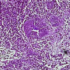
ARTICLE                                                                   NATURE COMMUNICATIONS | https://doi.org/10.1038/s41467-022-28139-5

Fig. 4 Developmental potential of Lima1 KO ESC. a Teratomas derived from Lima1 KO or WT ESC. b Growth of WT and Lima1 KO teratomas.
c Haematoxylin and eosin staining of sectioned teratomas. d Chimeric blastocysts containing control (WT) or Lima1 KO donor cells. e Quantification of
integrated donor cells at blastocyst stage, data represent mean ± SEM, WT n = 6 embryos, Lima1 KO n = 6 embryos, unpaired Student’s t-test, 2-sided.
f Chimeric E6.5 embryos containing control (WT) or Lima1 KO donor cells. g Quantification of the ectopic cells relative to the total epiblast cell number at
E6.5. WT (n = 40 embryos), Lima1 KO (n = 32 embryos), three independent experiments. Data represent mean ± SEM, Mann–Whitney test, 2-sided.
h Chimeric E13.5 embryos containing control (WT) or Lima1 KO donor cells. i Quantification of the ectopic cells relative to the total cell number of the
foetus, determined by flow cytometry. WT (n = 7 embryos), Lima1 KO (n = 7 embryos), three independent repetitions. Data represent mean ± SEM,
unpaired Student’s t-test, 2-sided. j Chimeric E13.5 embryos containing WT donor cells, stained for tdTomato, Podocalyxin and DAPI. k Chimeric E13.5
embryos containing Lima1 KO donor cells, stained for tdTomato, Podocalyxin and DAPI. Scale bars, (c) 50 μm; (d), 20 μm; (f) 50 μm; (h) 1000 μm;
(j), (k),100 μm; Experiments were repeated independently three times (c, j, k) with similar results.

apart from the naïve ESC (Fig. 6e). Accordingly, gene expression                  Next, we asked whether the ectopic expression of Lima1 in
analysis confirmed the ectopic upregulation of Lima1 (Fig. 6f),                 primed cells affects membrane blebbing. Control EpiSC and
which was not associated with an upregulation of naïve                         hiPSC formed blebs (Fig. 6k, o), which were efficiently suppressed
pluripotency transcription factors or downregulation of primed                 by Y-27632 treatment (Fig. 6l, p), in agreement with previous
pluripotency markers (Fig. 6g).                                                reports42,43. In contrast, blebbing was rarely observed in naïve
   In addition, we characterised the transcriptional profiles of                hiPSC (Fig. 6n). We found that formation of blebs was
control (conventional) hiPSC and Lima1-HA expressing hiPSC                     substantially reduced as a result of the ectopic Lima1 expression
(Fig. 6h–j, S5G–S5I, Supplementary Dataset 4). We also included                in EpiSC and conventional hiPSC (Fig. 6k–m, o–q). In addition,
in the analysis available transcriptional data of naïve hiPSC50. The           studies have reported that the membrane blebbing in conven-
principal component analysis showed that Lima1-HA and control                  tional human ESC (hESC) triggers an apoptotic response,
hiPSC clustered closely together, apart from the naïve hiPSC                   whereas treatment with a ROCK inhibitor protects the dissociated
(Fig. 6h). Similar to the Lima1-HA EpiSC, the ectopic expression               hESC from cell death42,43. Accordingly, we found that suppres-
of Lima1 in conventional hiPSC was not associated with                         sing membrane blebbing by Lima1 enhanced the survival of
activation of naïve pluripotency genes or downregulation of                    conventional hiPSC (Fig. S5C).
primed pluripotency factors (Fig. 6i, j). Altogether, this shows                  To further examine the effects of Lima1 expression on the
that the expression of Lima1-HA in both mouse and human                        membrane dynamics, we performed ultrastructural analysis to
primed pluripotent stem cells does not promote conversion to                   determine the subcellular localisation of Lima1 using immuno-
naïve pluripotency.                                                            gold labelling. We stained control and Lima1-HA EpiSC for the

8                  NATURE COMMUNICATIONS | (2022)13:610 | https://doi.org/10.1038/s41467-022-28139-5 | www.nature.com/naturecommunications
NATURE COMMUNICATIONS | https://doi.org/10.1038/s41467-022-28139-5                                                                      ARTICLE

Fig. 5 Lima1 is required for proper mitochondrial energetics. a Proportion of apoptotic and necrotic Lima1 KO and WT ESC. The cells were individualized
by trypsinization and cultured on cell-repellent plates for 2 h at 37 °C. After that, the cell death was determined by annexin V assay in combination with
DAPI, three independent experiments, data represent mean ± SEM, unpaired Student’s t-test, 2-sided. b Quantification of WT and Lima1 KO cell
proliferation after 96 h of culture. The cells were treated with or without Y-27632 upon dissociation. Untreated Lima1 KO (n = 6 samples) and WT
(n = 6 samples), three independent experiments. Treated with Y-27632 Lima1 KO (n = 6 samples), WT (n = 6 samples), four independent experiments.
Data represent mean ± SEM, unpaired Student’s t-tests, 2-sided. c Pyruvate kinase activity measured in WT and Lima1 KO ESC, n = 4 independent
experiments. Data represent mean ± SEM, unpaired Student’s t-test, 2-sided. d FACS analysis of WT and Lima1 KO ESC TMRE assay. Plot shows the TMRE
signal normalized to the MitoTracker signal. CCCP is a mitochondrial oxidative phosphorylation uncoupler used as a negative control. e Quantification of
TMRE signal intensity, normalized to MitoTracker signal intensity, n = 3 independent experiments. Data represent mean ± SEM, unpaired Student’s t-test,
2-sided. f Quantification of relative intracellular ROS levels in Lima1 KO ESC, relative to WT ESC. Three independent experiments, data represent
mean ± SEM, unpaired Student’s t-test, 2-sided. g OCR measurement using the Seahorse mitochondrial stress test assay. FCCP−Carbonyl cyanide-4
(trifluoromethoxy) phenylhydrazone; Rot Rotenone, AA−Antimycin. Three independent experiments, mean values ± SEM. h Quantification of the OCR
analysis, n = 3 independent experiments. Data represent mean ± SEM, 2-way ANOVA with Tukey’s multiple comparisons. Related to Fig. S4.

NATURE COMMUNICATIONS | (2022)13:610 | https://doi.org/10.1038/s41467-022-28139-5 | www.nature.com/naturecommunications                                 9
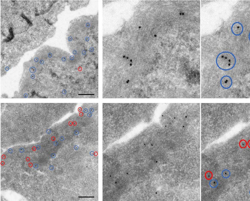
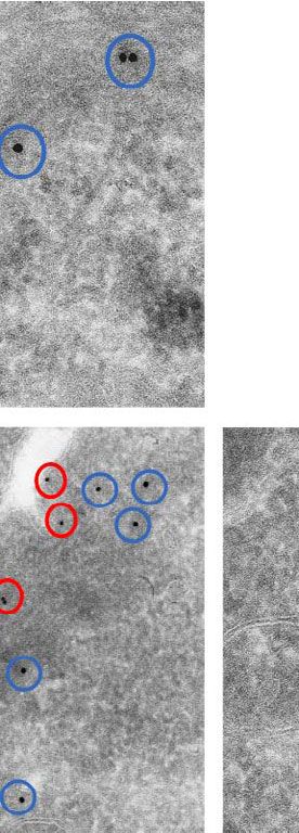
ARTICLE                                                                 NATURE COMMUNICATIONS | https://doi.org/10.1038/s41467-022-28139-5

HA-tag and found Lima1-HA signal on the cell cortex (Fig. 7a).               the control and Lima1-HA expressing cells (Fig. S6A–S6D),
We did not detect enrichment of Lima1 in the mitochondria                    indicating that the effects of Lima1 on the mitochondrial
(Fig. 7a, b), suggesting that Lima1 is not directly involved in the          energetics are associated with the naive pluripotent state.
mitochondrial composition. In addition, we examined the OCR                     To understand whether Lima1 is localised within the cortical
in WT EpiSC or WT hiPSC vs. Lima-HA EpiSC or Lima-HA                         actin, we carried out double immunogold staining using two
hiPSC, respectively and found no substantial difference between              different sizes of gold particles— 10 nm of diameter for detecting

10               NATURE COMMUNICATIONS | (2022)13:610 | https://doi.org/10.1038/s41467-022-28139-5 | www.nature.com/naturecommunications
NATURE COMMUNICATIONS | https://doi.org/10.1038/s41467-022-28139-5                                                                          ARTICLE

Fig. 6 Lima1 ectopic expression in primed pluripotent stem cells. a Control and Lima1-HA-expressing EpiLC stained for HA and DAPI. b Western blot
analysis of Lima1 expression in control and Lima1-HA EpiSC. c Control and Lima1-HA-expressing conventional hiPSC stained for HA and DAPI. d Western
blot analysis of Lima1 expression in control and Lima1 HA conventional hiPSC. e Principal component analysis of control EpiSC, Lima1-HA EpiSC, WT ESC
and Lima1 KO ESC transcriptomes. f MA plot of gene expression in Lima1-HA vs. control EpiSC, two-tailed Wald-test with Benjamini–Hochberg correction
for multiple testing and independent filtering of results. g Gene expression of naïve and primed pluripotency markers in WT ESC, Lima1 KO ESC, control
EpiSC, Lima1-HA EpiSC. h Principal component analysis of control conventional hiPSC, Lima1-HA hiPSC and naïve hiPSC (Giulitti et al., 2019)50
transcriptomes. i MA plot of gene expression in Lima1-HA vs. control conventional hiPSC, two-tailed Wald-test with Benjamini–Hochberg correction for
multiple testing and independent filtering of results. j Gene expression of naïve and primed pluripotency markers control conventional hiPSC, Lima1-HA
hiPSC and naïve hiPSC. k Dissociated control and Lima1-HA-expressing EpiSC. l Dissociated control and Lima1-HA-expressing EpiSC treated with Y-27632.
m Mean distribution of cell membrane fluctuation counts per cell over 90 s of time-lapse imaging with 5 s intervals from k and l; n > 177 cells for each
condition, Mann–Whitney tests, 2-sided. n Dissociated naïve hiPSC cultured without or treated with Y-27632. o Dissociated control and Lima1-HA-
expressing conventional hiPSC. p Dissociated control and Lima1-HA-expressing conventional hiPSC treated with Y-27632. q Mean distribution of cell
membrane fluctuation counts per cell over 90 s of time-lapse imaging with 5 s intervals from n, o and p; n = 92 (Control hiPSC), 89 (Control hiPSC treated)
88 (naïve hiPSC), 100 (naïve hiPSC treated), 294 (Lima1-HA hiPSC), 245 (Lima1-HA hiPSC treated) cells; 2-way ANOVA with Tukey’s multiple
comparisons. Scale bars, (a), (c), 50 μm; (k), (l), (n), (o), (p), 10 μm. Experiments were repeated independently two times (a, b, c, d) or three times (m, q)
with similar results. Related to Fig. S5.

Lima1 and 15 nm of diameter for detecting actin. The transmis-                     A typical feature of the AJ complex proteins E-cad and N-cad
sion electron microscopy confirmed that Lima1 was enriched on                    is their almost mutually exclusive expression pattern. Similarly, α-
the cortical actin filaments (Fig. 7b), in accord with the proposed              E-cat and α-N-cat are preferentially found in complex with E-cad
role of Lima1 in stabilisation of the cortex, supressing the                    and N-cad, respectively, whereas β-cat exhibits very broad
formation of membrane blebs.                                                    expression, as it binds directly to both cadherins and also plays a
   Lima1 protein harbours two actin-binding domains localised in                central role in the Wnt signalling pathway. During EMT, E-cad is
the N-terminal and C-terminal regions, as well as a central LIM                 downregulated and N-cad upregulated in a process known as
domain, which was suggested to mediate Lima1 dimerization8–10.                  cadherin switch1. Unexpectedly, we found Lima1 localisation in
To examine the role of these domains on the membrane                            both E-cad and N-cad expression domains, overall resembling the
dynamics, we generated deletion constructs, lacking the                         β-cat expression pattern. However, our immunohistochemistry
N-terminal or the C-terminal regions, or the LIM domain and                     analysis does not differentiate between the long or short isoforms
expressed these constructs in EpiSC (Fig. 7c, d). In comparison to              of Lima1. Therefore, Lima1-α and Lima1-β may still exhibit an
the full-length Lima1-HA, N- and C-terminal deletions, as well as               unaccounted differential expression pattern in the foetus. More-
monomeric Lima1 (ΔLIM), failed to suppress membrane blebbing                    over, we found β-cat and Tcf3 binding on the Lima1-β promoter
(Fig. 7e). Likewise, tethering Lima1 to the cell membrane by                    region, which indicates that in certain contexts, Lima1-β could be
fusing the full-length Lima1 to E-cad (lacking the intracellular                a direct target of the Wnt pathway. As Wnt signalling is aber-
domain) was also inefficient to abolish blebbing (Fig. 7c–e). This               rantly activated in cancer, this may help explain why upregulation
indicates that the cytoplasmic pool of dimeric Lima1 proteins,                  of Lima1-β is observed in some breast cancer cells lines, where the
bound to the cortical actin filaments, suppresses the formation of               shorter isoform is depleted10.
membrane blebs in pluripotent stem cells.                                          We found that the Lima1 transcripts and protein are already
   The establishment of a primed pluripotent state also results in a            present in the oocyte before fertilisation. During the transition
reduced incorporation efficiency of EpiSC and hiPSC into pre-                    from the GV to MII stage, Lima1 accumulates on the membrane
implantation embryos. To understand whether suppressing the                     above the spindle and later in the polar body. This specialised
membrane blebs via forced expression of Lima1 allowed for                       membrane domain, known as cortical actin cup, plays a critical
EpiSC integration into early embryos, we aggregated control or                  role in the asymmetric division during meiosis, which results in
Lima1-HA EpiSC with 8-cell stage morulae. We found a                            the extrusion of the polar body51. Several actin nucleators, such as
substantial increase in the number of chimeric blastocysts when                 ARP2/3 complex and formin-2, regulate the cytoskeletal remo-
we used donor EpiSC expressing Lima1-HA for aggregation                         delling during oocyte maturation52,53, and it would be interesting
(Fig. 8a–c). Moreover, ectopic upregulation of Lima1 in                         to address the potential role of Lima1 in this process, which
conventional hiPSC enabled the generation of chimeric blasto-                   would require depleting the maternal transcripts. Interestingly,
cysts (Fig. 8d–f). Alternatively, supplementing the embryo culture              Lima1 has already been shown to localise in the cleavage furrow
medium used for morula aggregation with a ROCK inhibitor                        in HeLa cells, where it is involved in the maintenance of the
allowed the control EpiSC or hiPSC to be incorporated into pre-                 contractile ring54. Therefore, Lima1 might also play a role during
implantation embryos (Fig. S7A–S7D). Altogether, this shows                     meiosis, particularly in the process of the polar body extrusion.
that suppression of membrane blebbing by Lima1 is sufficient to                     At the 16-cell stage, we found that Lima1 expression is enriched in
enable primed pluripotent cells engraftment into murine pre-                    the inside cells of the morula. These cells give rise to the ICM of the
implantation embryos.                                                           blastocyst, whereas the outside cells form the TE55. In contrast, E-cad,
                                                                                β-cat and α-E-cat exhibited overall uniform expression in the early
                                                                                lineages. Accordingly, Lima1 was also highly expressed in ESC,
Discussion                                                                      whereas E-cad and β-cat showed similar levels of expression in both
Lima1 is an epithelial actin-binding protein that has been                      ESC and TSC. Moreover, E-cad and β-cat expressions were not
implicated and extensively studied in the progression of various                affected by the exit of naïve pluripotency and the establishment of a
types of cancers8. It also binds α-cat and associates with the AJ               primed state, whereas Lima1 was downregulated in response of this
complex on the cell membrane7,13. We found that in the context                  transition. Essentially, we found that the Lima1 promoter region is
of the developing mouse embryo, Lima1 exhibits a pattern of                     bound by several pluripotency transcription factors, such as Nanog,
expression that is not confined only to epithelial compartments.                 Nr0b1, Klf4, Sox2, Oct4, Sall4, as well as β-cat and Tcf3, indicating

NATURE COMMUNICATIONS | (2022)13:610 | https://doi.org/10.1038/s41467-022-28139-5 | www.nature.com/naturecommunications                                    11
ARTICLE NATURE COMMUNICATIONS | https://doi.org/10.1038/s41467-022-28139-5 that Lima1 expression is under the control of the naïve pluripotency appeared to decrease Lima1 expression. A recent study of network (Fig. 9). This suggests that the establishment of the plur- Yanagida et al. showed that the PE cells exhibit larger surface ipotent fate activates Lima1 expression in the inner cells of the early fluctuations (membrane blebs), compared to the epiblast cells. embryo. Based on these results, a model was proposed suggesting that the At mid and late blastocysts stage, we found that the epiblast difference in the membrane dynamics is involved in the segre- cells retain a relatively high level of Lima1, whereas the PE cells gation of PE and epiblast cells during blastocyst maturation56. 12 NATURE COMMUNICATIONS | (2022)13:610 | https://doi.org/10.1038/s41467-022-28139-5 | www.nature.com/naturecommunications
NATURE COMMUNICATIONS | https://doi.org/10.1038/s41467-022-28139-5                                                                            ARTICLE

Fig. 7 Ultrastructural analysis and expression of Lima1 deletion constructs in EpiSC. a Transmission electron microscopy of immunogold staining for the
HA-tag in control and Lima-HA EpiSC. Arrowheads indicate the HA signal; mitochondria are marked in magenta. At least three technical replicates with
similar results. b Transmission electron microscopy of double immunogold staining for Lima1 and Actin in control and Lima-HA EpiSC. Blue and red circles
indicate the 15 nm and 10 nm gold particles, respectively; mitochondria are marked in magenta. At least three technical replicates with similar results.
c Maps of the Lima1 constructs. d Western blot analysis of the expression of the Lima1 constructs detected by anti-HA antibody. e Mean distribution of cell
membrane fluctuation counts per cell over 90 s of time-lapse imaging with 5 s intervals; n = 225 (Control), 245 (full-length Lima1-β), 68 (del LIM domain),
78 (del C-terminus), 51 (del N-terminus), 18 (E-cad-Lima1-β fusion) cells, one-way ANOVA. Scale bars, (a), (b) 1 μm. Related to Fig. S6.

Fig. 8 Engraftment of Lima1-HA-expressing primed pluripotent cell in mouse pre-implantation embryos. a E3.5 and E4.5 chimeric blastocysts generated
via morula aggregation using control or Lima1-HA-expressing EpiSC. b Quantification of the chimeric blastocysts containing control (22 embryos) or Lima1-
HA-expressing (26 embryos) EpiSC at E4.5. Data represent mean ± SEM, n = 3 independent experiments, unpaired Student’s t-test, 2-sided. c Chimeric
blastocysts generated via morula aggregation using control (upper panel) or Lima1-HA-expressing EpiSC (lower panel) and stained for Venus, Oct4 and
DAPI. Arrowhead indicates integrated EpiSC. d E3.5 and E4.5 chimeric blastocysts generated via morula aggregation using control or Lima1-HA-expressing
conventional hiPSC. e Quantification of the chimeric blastocysts containing control (30 embryos) or Lima1-HA-expressing (33 embryos) conventional
hiPSC at E4.5. Data represent mean ± SEM, n = 4 independent experiments, Mann–Whitney test, 2-sided. f Chimeric blastocysts generated via morula
aggregation using control (upper panel) or Lima1-HA-expressing conventional hiPSC (lower panel) and stained for Venus, Oct4 and DAPI. Arrowhead
indicates integrated hiPSC. Scale bars, (c), (f), 20 μm. Experiments were repeated independently three times (a, c, d) with similar results. Related to Fig. S7.

The PE cells displayed also elevated levels of pERM56, similarly to              growth of the Lima1 KO teratomas, as well as the proportion of
our observations of pERM enrichment in the ameboid membrane                      donor Lima1 KO cells in post-implantation embryos, were
protrusions in Lima1 KO ESC. Thus, a reduction of Lima1                          severely reduced. ESC exhibit bivalent metabolism, using both
expression may serve as an indicator of cells that exhibit dynamic               OxPhos and glycolysis, whereas somatic cells rely mainly on
membrane fluctuations. Moreover, the progressive changes in the                   OxPhos for ATP production to support their growth57. In addi-
expression pattern of Lima1 in the PE and epiblast cells of the                  tion, the transition from a naïve to a primed state of pluripotency
ICM may contribute to a surface fluctuations-driven segregation                   results in the reduction of OxPhos and a switch mainly to
of these lineages, which as proposed by Yanagida et al., resembles               glycolysis49,58,59. Loss of Lima1 did not cause cells to enter into
phase separation between active and passive particles in colloidal               primed pluripotency, but the mitochondrial ATP production rate
mixtures56.                                                                      was substantially reduced. Accordingly, Lima1 KO ESC exhibited
   Loss of Lima1 did not affect the expression of pluripotency                   decreased proliferation rate, which may contribute to the overall
markers nor the differentiation capacity of the ESC. However, the                smaller size of the teratomas. The exact underlying molecular

NATURE COMMUNICATIONS | (2022)13:610 | https://doi.org/10.1038/s41467-022-28139-5 | www.nature.com/naturecommunications                                      13
ARTICLE                                                                    NATURE COMMUNICATIONS | https://doi.org/10.1038/s41467-022-28139-5

Fig. 9 Lima1 mediates the pluripotency control of membrane dynamics and cellular metabolism. Lima1 expression is promoted by the naïve pluripotency
transcription factors, which occupy the Lima1 promoter region. In turn, the cytoplasmic pool of dimeric Lima1 proteins bound to the actin filaments
stabilises the cortex, thereby suppressing the formation of membrane blebs in naïve pluripotent cells. Lima1 is also involved in the mitochondrial energetics
and is crucial for growth of teratomas and embryonic chimerism of ESC. Upon exit of naïve pluripotency, the naïve transcriptional circuit is dismantled and
Lima1 is downregulated, resulting in the formation of membrane blebs. Accordingly, ectopic expression of Lima1 in mouse and human primed pluripotent
cells suppresses membrane blebbing and enables the incorporation of the primed cells into murine pre-implantation embryos. OxPhos, oxidative
phosphorylation; naïve pluripotency transcription factors are marked in blue; donor cells are marked in yellow.

mechanism remains open. As Lima1 exhibited a broad expression                   application of force to the E-cad complex promotes the activity of
in the foetus, loss of Lima1 may affect actin dynamics, membrane                AMP-activated protein kinase (AMPK), which plays a critical role
properties and adherens junctions in various tissues, which may                 in glucose and fatty acid uptake and oxidation63. As Lima1 can
also account for the reduced tumour growth and chimerism. In                    act as a mechanosensor13, potential changes in the mechan-
addition, Lima1 KO cells might face cell competition and                        otransduction in Lima1 KO cells may also, in turn, influence the
potentially be outcompeted by the host embryo’s cells, as a recent              cellular metabolism.
study showed that cells with mitochondrial dysfunction are                         Lima1 KO ESC exhibited membrane blebbing similarly to
eliminated in the post-implantation conceptus60.                                primed pluripotent cells, where the endogenous Lima1 expression
   Previously, Zhang et al. reported a conditional deletion of exon             was downregulated upon exiting the naïve state (Fig. 9). Ectopic
5 of the Lima1 locus via adenoviral EIIa promoter-directed Cre                  expression of Lima1 in EpiSC and conventional hiPSC was suf-
recombinase61 for the generation of heterozygous mice, which                    ficient to stabilise the actin cortex and prevent blebbing nuclea-
exhibited lower cholesterol absorption and decreased plasma total               tion. A stabilising role of Lima1 on the cortex has also been
cholesterol levels19. The homozygous exon 5 del/del mice were                   reported in human umbilical vein endothelial cells (HUVEC),
viable and also displayed decrease cholesterol absorption19. Here,              where downregulation of Lima1 results in gaps on the cortical
in addition to exon 5, we deleted also exon 4, which contains the               actin64. Interestingly, HUVEC form classical lamellipodia and
second transcription start site of the Lima1 locus. Whether this                junction-associated intermittent lamellipodia in which Lima1-α
targeting strategy may result in an embryonic phenotype remains                 controls the protrusion dynamics, whereas Lima1-β binds and
to be determined. Alternatively, Lima1 KO cells may exhibit a                   stabilizes stress fibres37. Moreover, it was reported that Erk-
developmental disadvantage in vivo only in a competitive envir-                 mediated phosphorylation of Lima1 reduces the affinity for actin
onment, such as in the context of chimeric embryos.                             filaments, which contributes to cell motility65. In 2i/Lif culture
   Understanding how Lima1 regulates mitochondrial function                     conditions, Erk activity is suppressed by the chemical inhibition
on a molecular level will require further investigation. Although               of the upstream Mek, whereas in primed pluripotency culture
we found several putative interaction partners involved in cellular             conditions, Erk activity is promoted by the stimulation of the Fgf
metabolisms, such as pyruvate kinase and dihydropyrimidinase,                   signalling22,26. Thus, although, Lima1 is transcriptionally down-
none of these candidates directly control any key steps of energy               regulated upon exiting naïve pluripotency, we cannot exclude that
homeostasis. Thus, it is possible that a yet unidentified Lima1                  additional fine-tunning of Lima1-actin interactions occur on a
interaction partner(s) is involved in this process. Alternatively,              post-translational level by the Fgf/Erk pathway.
the primary function of Lima1 as an actin-binding protein may                      We found that the Lima1-mediated suppression of membrane
indirectly affect the cellular metabolism. For instance, it has been            blebbing enabled primed pluripotent cells to be integrated into
shown that actin cytoskeleton dynamics regulate the activity of                 murine pre-implantation embryos. Interestingly, E-cad over-
aldolase A, which is a key glycolytic enzyme62. In addition,                    expression has also been shown to promote EpiSC

14                 NATURE COMMUNICATIONS | (2022)13:610 | https://doi.org/10.1038/s41467-022-28139-5 | www.nature.com/naturecommunications
NATURE COMMUNICATIONS | https://doi.org/10.1038/s41467-022-28139-5                                                                                   ARTICLE

incorporation66, but we found no substantial changes in the                            Conventional hiPSC75 were cultured on matrigel coated plates in MEF-
endogenous E-cad level upon exit of naïve pluripotency or upon                      conditioned medium, supplemented with 10 ng/ml Fgf2. The cells were split every
                                                                                    3 days using TrypLE Express (Gibco, 12604-013).
ectopic expression of Lima1 in EpiSC. Nevertheless, the stabili-                       Naïve hiPSC76 were cultured in PGXL medium consisting of 48% DMEM F-12
sation of the actin cortex by Lima1 gain of function may promote                    medium, Neurobasal (48%) medim, 1% B27 Supplement (Invitrogen, 17504044),
more stable F-actin engagement with the E-cad complex, thus                         10 U/ml penicillin/50 μg/ml streptomycin solution, 2 mM L-glutamine and
resembling the net effect of E-cad overexpression. In addition, it                  0.15 mM β-mercaptoethanol, supplemented with 1 μM PD0325901, 2 μM Gö6983
has been shown that human pluripotent stem cells can be inte-                       (Tebu-bio, 800088), 2 μM XAV939 (Sigma, X3004) and 250 U/ml Lif. The cells
                                                                                    were split every 3–4 days using Accutase and cultured on geltrex coated plates
grated into pre-implantation mouse embryos via suppression of                       (Thermo Fischer Scientific, A1413302).
cell death67–69. Accordingly, Lima1 expression, as well as treat-                      Xfect (Takara, 631320) or Lipofectamine 2000 (Invitrogen, 11668027) were
ment with a ROCK inhibitor, promoted the survival of dis-                           used for transfection, following the manufacturer’s instructions.
sociated hiPSC. This indicates that downregulation of the
endogenous Lima1 in primed hiPSC, which results in membrane                         Generation of Lima1 KO ESC via CRIPSR/Cas9-mediated deletion. Guide
blebbing and apoptotic response, is a key part of the interspecies                  RNAs (guide RNA 1 5'-3': CTGCTCACTTTGTCCTTATA, guide RNA 2 5'-3':
                                                                                    GTCGCATGTTAGCTGCAAGA) were designed using the MIT tool
engraftment barrier.                                                                (www.crispr.mit.edu). Each of the guide RNA coding sequences was synthesised as
   In tumour cells, membrane blebbing can mediate cell motility,                    a pair of complementary oligonucleotides, which were annealed and separately
which is used by the malignant cells as an alternative mechanism                    cloned into pSpCas9(BB)-2A-Puro (PX459) vectors77. The vectors were addition-
of migration41,70. This alteration in the morphology and beha-                      ally modified to express fluorescent reporters (GFP or Cerulean) in order to
                                                                                    facilitate the sorting and collection of double-positive cells. After re-seeding,
viour of the cancer cells is known as a mesenchymal–amoeboid                        individual ESC clones were manually picked and expanded. Homozygous deletion
transition (MAT)70. As blebbing motility does not require                           of Lima1 was confirmed by PCR genotyping (primer pair 1 5'-3':
degradation of the extracellular matrix, MAT allows tumour cells                    TCTTGTTGTTTGTGGCATAC and CACTCACTTTCCTAACATTGA—653 bp
to evade anti-cancer treatments, which rely on pharmacological                      product of Lima1 KO allele; primer pair 2 5'-3': GTTTTATCACTTCCTGCTTCA
protease inhibitors70,71. Our results in pluripotent cells show that                and AAAAGACATACTGTCCACACA —796 bp product of WT Lima1 locus), as
                                                                                    well as by sequencing and western blot analysis.
Lima1 depletion enables blebbing and suggest that in pathological
conditions, this may promote tumour resistance to protease
                                                                                    Fluorescence-activated cell sorting (FACS). Adherent cells were dissociated and
inhibitors. Thus, treating malignancies associated with Lima1                       resuspended in 3% FCS/PBS solution (FACS buffer). Chimeric E13.5 embryos were
deficiency may benefit from cancer therapeutics that target reg-                      mechanically homogenized, and the cells were subsequently dissociated using
ulators of membrane blebbing.                                                       trypsin. The cell suspension was pelleted by centrifugation and resuspended in
                                                                                    FACS buffer. Single viable cells were selected based on FSC- and SSC-gating. The
                                                                                    FACS gating plots are presented in Fig. S8. Cell sorting and analysis was performed
Methods                                                                             using FACSAria IIIu and FACSAria Fusion systems, equipped with FACSDiva
Mice. Animal experiments and husbandry were performed according to the Ger-         Software (v8.01). FlowJo software (v10.7.1) was used for data analysis and plotting.
man Animal Welfare guidelines and approved by the Landesamt für Natur,
Umwelt und Verbraucherschutz Nordrhein-Westfalen (State Agency for Nature,
Environment and Consumer Protection of North Rhine-Westphalia), protocol            Immunofluorescence labelling and confocal microscopy. Adherent cells were
number 84-02.04.2016.A186. The mice used in the study were at ages ranging from     grown on 8-well μ-slide plates (Ibidi, 80826). Non-adherent cells were collected
6 weeks to 5 months. The animals were maintained at 21.5 °C, 55–60% humidity        and pelleted by centrifugation. After washing with PBS, the cells were fixed using
under a 14-h light/10-h dark cycle with free access to food and water. Male mice    4% PFA for 15 min at room temperature and then permeabilized using 0.3% Triton
were kept individually, whereas the female mice were housed in groups of up to      X-100/PBS for 5 min. After washing, the samples were incubated with blocking
four per cage. Embryos for experiments were obtained from wild-type CD1 and         buffer (3% BSA/PBS) for at least 30 min, and after that, the primary antibodies
B6C3F1 from matings using females with natural ovulation cycles or after            diluted in blocking buffer were applied to the sample and incubated overnight at
superovulation.                                                                     4 °C. On the next day, the primary antibody solution was removed, the cells were
                                                                                    washed and incubated with secondary antibodies diluted in blocking buffer. Nuclei
                                                                                    were counterstained with DAPI.
Cell culture. DR-4 or CF-1 MEFs (gift from Prof. Dr. Hans R. Schöler) were              Pre-implantation embryos were fixed using 4% PFA/PBS for 10 min, whereas
cultured on gelatin-coated plates in DMEM medium (Sigma, D5671), supple-            egg cylinder stage embryos were fixed for 15 min. The samples were permeabilised
mented with 15% FCS (Biochrome, S0615), 10 U/ml penicillin/50 μg/ml strepto-        using 0.3% Triton X-100/PBS solution supplemented with 100 mM glycine for
mycin solution (Sigma, P4333), 2 mM L-glutamine (Sigma, G7513), 1 mM sodium         5 min (pre-implantation embryos) or 15 min (post-implantation embryos). After
pyruvate (Sigma, S8636), 1x MEM non-essential amino acids (Sigma, M7145) and        washing with 5% FCS/PBS, the samples were incubated with primary antibody
0.15 mM β-mercaptoethanol (Sigma, M3148). MEFs were mitotically inactivated by      solution in 10% FCS/PBS buffer, overnight at 4 °C. On the next day, the embryos
exposure to 10 μg/ml mitomycin C (Sigma, M0503) for 2 h, before splitting for co-   were washed and incubated with secondary antibodies, overnight at 4 °C. After
culture experiments.                                                                that, the samples were washed and mounted on glass-bottom plates in PBS droplets
    Mouse ESC—E14 (gift from Prof. Dr. Hans R. Schöler), R1 (gift from Prof. Dr.    under oil. Imaging was performed using Zeiss LSM780 system equipped with ZEN
Rolf Kemler) or mT/mG72, were grown on DR-4 feeders in MEF medium                   software (v2.0 and higher), Fiji software (v2.0 and higher) was used for image
supplemented with 1000 U/ml Lif (home-made). The cells were split every 2–3 days    analysis. The list of primary and secondary antibodies is presented on Table S2.
using 0.25% Trypsin-EDTA (Gibco, 25200056). For experiments requiring ground
state culture conditions, the ESC were cultured on human plasma fibronectin
coated plates (Millipore; FC010) in N2B27 medium consisting of 48% DMEM F-12        Generation of teratomas and immunohistochemistry. Scid mice were injected
medium (Invitrogen, 21331-046), 48% Neurobasal medium (Invitrogen, 21103-           subcutaneously with a 100 µl suspension containing 5 million ESC in PBS. Solid
049), 1% B27 Supplement w/o vitamin A (Invitrogen, 12587-010), 10 U/ml              tumours were isolated and fixed at 4 °C overnight in 4% PFA/PBS. The samples
penicillin/50 μg/ml streptomycin solution (Sigma, P4333), 2 mM L-glutamine          were dehydrated in an ethanol series (30%, 50%, 70% and 100% in PBS) for 2 h
(Sigma, G7513), and 0.15 mM β-mercaptoethanol (Sigma, M3148), supplemented          each, followed by two 10 min incubations in 100% xylene, and then transferred to
with 1000 U/ml Lif (home-made), 3 μM CHIR99021 (Biomol, 13122) and 0.4 μM           paraffin. Similarly, foetal stage embryos were fixed overnight and dehydrated.
PD0325901 (Biomol,13014).                                                           Samples embedded into paraffin blocks were sectioned at 7 µm using an HM355S
    ESC grown in ground state culture conditions were converted to EpiLC26 by       microtome (Thermo). Haematoxylin/eosin staining and immunohistochemistry
48 h culture in N2B27 medium supplemented with 1% KSR (Knockout Serum               were carried out as previously described78,79. Images were acquired using Nikon
Replacement, Gibco, 10828-028), 20 ng/ml activin A and 12 ng/ml Fgf2.               Eclipse Ti2 system.
    TSC73 were maintained on MEFs in MEF medium supplemented with 25 ng/ml
Fgf4 (Peprotech, 100-31) and 1 μg/ml Heparin (Sigma, H3393). ESC were
                                                                                    Generation of embryoid bodies. ESC differentiation using the EBs assay was
converted to TSC-like cells via forced expression of Cdx2-ERT2 construct24,
                                                                                    performed as previously described80. The EBs were harvested and dissociated into
following treatment with 0.5 μM 4-Hydroxytamoxifen (Sigma, H7904-5MG).
                                                                                    single cells for Seahorse analysis 14 days after induction of differentiation.
    E3 EpiSC74 were cultured in medium containing 50% N2B27 and 50% MEF-
conditioned medium, consisting of DMEM F-12 supplemented with 20% KSR,
penicillin-streptomycin, 2 mM L-glutamine, 1x non-essential amino acids and         Quantitative PCR. RNA isolation was performed using RNeasy Mini Kit (Qiagen,
0.15 mM β-mercaptoethanol. The EpiSC medium was supplemented with 10 ng/ml          74106) according to the manufacturer’s instructions and reverse transcribed using
activin A and 5 ng/ml Fgf2 before use. The cells were split every 2–3 days using    MMLV-Reverse transcriptase (Applied Biosystems). Transcript levels were quantified
accutase (Sigma, A6964).                                                            using iTaq SYBR Green Supermix (Bio-Rad). Relative gene expression was calculated

NATURE COMMUNICATIONS | (2022)13:610 | https://doi.org/10.1038/s41467-022-28139-5 | www.nature.com/naturecommunications                                              15
You can also read