A pharmacokinetic approach to intrabrain distribution with a focus on cyclic peptides
←
→
Page content transcription
If your browser does not render page correctly, please read the page content below

Digital Comprehensive Summaries of Uppsala Dissertations
 from the Faculty of Pharmacy 304

 A pharmacokinetic approach to intra-
 brain distribution with a focus on
 cyclic peptides

 ERIK MELANDER

 ACTA
UNIVERSITATIS
 UPSALIENSIS ISSN 1651-6192
 ISBN 978-91-513-1364-1
 UPPSALA URN urn:nbn:se:uu:diva-460177
 2022
Dissertation presented at Uppsala University to be publicly examined in B42, Biomedicinskt
Centrum (BMC), Husargatan 3, Uppsala, Friday, 11 February 2022 at 09:15 for the degree of
Doctor of Philosophy (Faculty of Pharmacy). The examination will be conducted in English.
Faculty examiner: Xavier Declèves (Université de Paris, France).

Abstract
Melander, E. 2022. A pharmacokinetic approach to intra-brain distribution with a focus on
cyclic peptides. Digital Comprehensive Summaries of Uppsala Dissertations from the Faculty
of Pharmacy 304. 49 pp. Uppsala: Acta Universitatis Upsaliensis. ISBN 978-91-513-1364-1.

When designing treatments for disorders of the central nervous system (CNS) reaching the site
of action is a major hurdle in the development process. Regardless if the target is extra- or
intracellular, precise measurements to understand the distribution within the CNS are required.
There is however a lack of understanding of differences in blood-brain barrier transport and
intra-brain distribution of both small and large molecules. In this thesis the regional Blood-
Brain Barrier transport of antipsychotic agents, along with their brain tissue binding and regional
cellular accumulation was quantified. Furthermore, a novel LC-MS/MS method was developed
for the quantitative analysis of the cyclic peptide kalata B1 was developed for analysis of
brain tissue and plasma samples. The Blood-Brain Barrier transport, permeability, intra-brain
distribution and cellular accumulation were assessed for two cyclic peptides, SFTI-1 and kalata
B1.
 The antipsychotics exhibited clear differences in their regional BBB transport as well as their
brain tissue binding, with the most dramatic spatial differences in BBB transport being observed
for the p-glycoprotein substrates risperidone and paliperidone. The highest level of transporter
mediated protection was observed in the cerebellum, with pronounced efflux for several of the
antipsychotics. The development of a quantitative method for the cyclic peptide kalata B1 was
successfully validated and applied to measure low concentration of the peptide in biological
matrices. The BBB transport of SFTI-1 was markedly higher than that of kalata B1 whereas
both peptides exhibited similar permeability across an in vitro BBB model. It was also shown
that SFTI-1 resides mainly within the interstitial fluid within the brain, but that kalata B1 readily
enters the cells of the brain parenchyma. The cellular accumulation of kalata B1 was abolished
under cold conditions, and was not observable in lung tissue, suggesting an active process that
is tissue specific. It was also shown that both peptides are taken up into cell cultures of neurons
and astrocytes.
 In conclusion this thesis and the studies herein contribute to a better understanding of
distribution patterns of both antipsychotics and cyclic peptides and provides valuable lessons
in terms of what types of studies should be prioritized for the development of such molecules
into therapeutic agents.

Erik Melander, Department of Pharmacy, Box 580, Uppsala University, SE-75123 Uppsala,
Sweden.

© Erik Melander 2022

ISSN 1651-6192
ISBN 978-91-513-1364-1
URN urn:nbn:se:uu:diva-460177 (http://urn.kb.se/resolve?urn=urn:nbn:se:uu:diva-460177)
To my family

List of Papers

This thesis is based on the following papers, which are referred to in the text
by their Roman numerals.

 I. Loryan I, Melander E, Svensson M, Payan M, König F, Jansson B
 och Hammarlund-Udenaes M. In-depth neuropharmacokinetic
 analysis of antipsychotics based on a novel approach to estimate
 unbound target-site concentration in CNS regions: link to spa-
 tial receptor occupancy. Mol Psychiatry 2016 Nov;21(11):1527-
 1536. doi: 10.1038/mp.2015.229.

 II. Melander E, Eriksson C, Jansson B, Göransson U, Hammarlund-
 Udenaes M. Improved method for quantitative analysis of the
 cyclotide kalata B1 in plasma and brain homogenate. Biopoly-
 mers. 2016 Nov;106(6):910-916. doi: 10.1002/bip.22984.

III. Melander E, Eriksson C, Wellens S, Culot M, Göransson U, Loryan
 I, Hammarlund-Udenaes M. Dual behavior of cyclic peptides at
 the blood-brain barrier vs brain cellular uptake: Importance for
 CNS drug development. 2021. In Manuscript

IV. Melander E, Hosseini K, Eriksson C, Fredriksson R, Göransson U,
 Hammarlund-Udenaes M, Loryan I. Cellular uptake and intra-
 brain distribution of cyclic cell penetrating peptides: why tissue
 and cell type matters. 2021. In Manuscript

Reprints were made with permission from the respective publishers.

Contents

Introduction ................................................................................................... 11
 The Blood-brain Barrier ........................................................................... 12
 Regional differences in Blood-Brain Barrier properties ...................... 13
 BBB transport ...................................................................................... 14
 Pharmacokinetic concepts in BBB transport ....................................... 14
 Intra-brain distribution ............................................................................. 16
 Cellular uptake and distribution pathways........................................... 17
 Cell penetrating peptides .......................................................................... 18
 Cyclic cell penetrating peptides ........................................................... 19
Aims .............................................................................................................. 20
Materials and Methods .................................................................................. 21
 Animals .................................................................................................... 21
 Peptide production.................................................................................... 21
 Experimental procedures .......................................................................... 22
 In vivo pharmacokinetics ..................................................................... 22
 Blood-brain barrier transport ............................................................... 22
 In vitro blood-brain barrier permeability ............................................. 23
 In vitro binding assay........................................................................... 23
 In vitro unbound tissue volume of distribution assay .......................... 24
 Cellular accumulation assay ................................................................ 26
 Bioanalytical setup ................................................................................... 27
Results & Discussion .................................................................................... 29
 Regional distribution of antipsychotics (Paper I) ..................................... 29
 Validation of bioanalytical method for kalata B1 (Paper II) .................... 32
 Blood-Brain Barrier transport of cyclic peptides (Paper III) .................... 33
 In vivo Blood-Brain Barrier transport ................................................. 33
 In vitro Blood-Brain Barrier permeability ........................................... 36
 Intra brain distribution and cellular uptake of cyclic peptides (Paper
 IV) ............................................................................................................ 37
 Cellular uptake of SFTI-1 and kalata B1 ............................................. 38
Conclusions ................................................................................................... 40
Acknowledgements ....................................................................................... 42
References ..................................................................................................... 43
Abbreviations

ABC ATP-binding cassette
ACN Acetonitrile
AUCbrain Area under the brain concentration-time curve
AUCplasma Area under the plasma concentration-time curve
BBB Blood-Brain Barrier
BCRP Breast Cancer Resistance Protein
BS Brain stem
CNS Central Nervous System
CL Clearance
CLin Net influx clearance into the brain
CLout Net efflux clearance from the brain
CPP Cell Penetrating Peptide
CRB Cerebellum
CRT Cortex
CSF Cerebrospinal Fluid
ECF Extracellular Fluid
HPC Hippocampus
HPT Hypothalamus
ISF Interstitial Fluid
Kp,brain Total brain-to-plasma concentration ratio
Kp,uu,brain Unbound brain-to-plasma concentration ratio
Kp,uu,cell Unbound intracellular-to-extracellular concentration ratio
MPA Mobile Phase A
MPB Mobile Phase B
Papp Apparent Permeability
PBS Phosphate Buffered Saline
Pe Endothelial Permeability
PD Pharmacodynamics
PK Pharmacokinetics
PKPD Pharmacokinetics-Pharmacodynamics
Pgp P-glycoprotein
SC Spinal Cord
SD Standard deviation
SFTI-1 Sunflower Trypsin Inhibitor 1
SLC Solute Carrier
STR Striatum
t½ Half-life
Vu,brain Unbound volume of distribution in brain
Vu,lung Unbound volume of distribution in lung
Introduction

When designing treatments for disorders of the central nervous system
(CNS) reaching the site of action is a major hurdle in the development pro-
cess. Regardless if the target is extra- or intracellular, precise measurements
to understand the distribution within the CNS are required. There is however
a lack of understanding of Blood-brain Barrier (BBB) transport and intra-
brain distribution of both small and large molecules. A clear example of
when this knowledge is essential is in the treatment with antipsychotic
agents. These drugs need to reach sufficient receptor occupancy in specific
regions of the brain to elicit their effect, whilst distribution to other regions
is often responsible for adverse events. By better understanding the distribu-
tion patterns of antipsychotic drugs, better treatments can be designed, lead-
ing to more successful treatment. Another area where knowledge of intra-
bran distribution is highly relevant is the emerging field of cell penetrating
peptides (CPPs). These peptides are being seen as a promising alternative for
delivery to the CNS, with their cell penetrating capabilities seen as a tool to
overcome both the BBB and the cellular barriers within the CNS. In order to
establish the feasibility of cell penetrating peptides, their capacity of uptake
into the specific cells that they are targeting is needed. So far, studies on
CPPs have mainly studied their uptake into peripheral cell types, and extrap-
olated that uptake to other cell types. Alternatively the uptake into the CNS
has been assessed through non-quantitative methods, where the unbound
concentrations at the site of action are not properly determined. Such studies
have their value, but will not provide sufficient information for proper deci-
sion making regarding their usefulness in the treatment of CNS disorders.
Amongst the CPPs is a subclass of cyclic cell penetrating peptides, which are
more stable than their linear counterparts, and have been postulated to have
advantageous properties for intracellular delivery. In both these cases, re-
gardless of whether we are studying the small molecular drugs intended for
the treatment of psychotic disorders, or the larger cyclic cell penetrating
peptides, proper translation between cell cultures, tissues, and model species
are needed in order to inform further development in humans.

 11
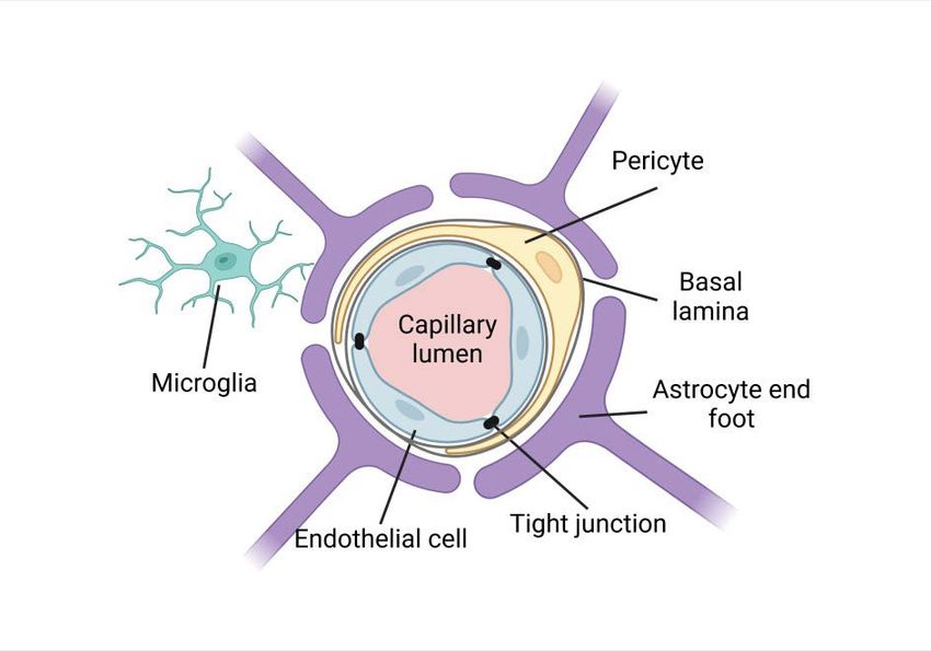
The Blood-brain Barrier
The brain, and the spinal cord, as constituents of the central nervous system
are organs with a highly regulated microenvironment. This regulation is
primarily performed through the function of barriers surrounding the CNS
[1,2]. The BBB is a physiological and metabolic barrier separating the brain
parenchyma from the systemic circulation. It is comprised of the capillary
endothelial cells connected with tight junction proteins, effectively keeping
molecules out of the brain. These connections between the endothelial cells
are built up of various junction proteins, both tight junctions and adherens
junctions [3–5]. Typically these junctions are built up by proteins such as
occludin and claudin. This junctional system provides the primary barrier
function for both macromolecules and polar solutes, as they are generally
prevented from transcellular diffusion. The BBB is part of a wider network
of cells in the CNS referred to as the neurovascular unit (Figure 1). This unit
consists of the capillary endothelium, as well as other cell types such as peri-
cytes, astrocytes, microglia, and the neurons themselves. The interplay be-
tween these cells is crucial for the functioning of the BBB as it involves a
multitude of signalling pathways.
 The capillary network in the brain is extensive, with a surface area of
around 20 m2 in total [6]. The BBB is a highly dynamic unit involved in
many processes, including the maintenance of brain homeostasis, uptake of
nutrients, removal of waste products and the protection of the brain from
potentially toxic compounds. The BBB ensures that the concentration of ions
in the brain is kept at a correct level, with ion channels controlling the exact
concentrations of potassium, sodium, calcium and other ions [7]. Another
function of the BBB is the homeostasis of neurotransmitters, where for ex-
ample the levels of glutamate in blood rise with food intake, but if the BBB
didn’t restrict its entry, severe harm would be caused to the CNS [8,9]. The
BBB is also vital in preventing proteins and other large molecules from en-
tering the brain. Common plasma proteins such as albumin are toxic to the
CNS tissue, and these are effectively kept out of the brain and the cerebro-
spinal fluid (CSF) [10–12]. This also means that the CNS is an immune priv-
ileged site, where neutrophil infiltration is low, and immune cells have lim-
ited access to the brain parenchyma, except post trauma or ischemia or relat-
ed inflammations [13].
 All of these barrier functions pose a problem for drug development, since
the BBB is equipped with various transporters that prevent several therapeu-
tic agents from entering the brain in sufficient concentrations to elicit their
effect. The BBB is not the only barrier within the CNS though. There is also
the blood-cerebrospinal fluid-barrier, separating the blood circulation from
the CSF which is composed of epithelial cells in the choroid plexus and pro-
vides an interface between the CSF and the systemic circulation in which
many transporters can be found [14,15].

12
Figure 1. Schematic view of the blood-brain barrier and the wider neurovascular
unit with pericytes, astrocytes, and microglia. Created with Biorender.

Regional differences in Blood-brain Barrier properties
When developing a therapeutic agent for a CNS disease or disorder, it is
often a specific region of the brain that is targeted, to achieve an effect on
certain receptors in that region of the brain. For example, antipsychotic drugs
target dopamine D2 receptors in the mesocortical and mesolimbic regions of
the brain. On the other hand, certain adverse events can be connected to the
drug eliciting an effect to the same type of receptor in a different region of
the brain, such as the dyskinesia that can appear through interaction with D2
receptors in the striatal area of the brain [16,17]. The antipsychotic drugs
also interact with cortical 5-HT2A receptors, and understanding the differen-
tial distribution of these drugs can help in calculating their receptor occupan-
cy in the specific area. Examples of these antipsychotic drugs are haloperi-
dol, clozapine, olanzapine, quetiapine, risperidone, and paliperidone. By
understanding the regional pharmacokinetic-pharmacodynamic relationship
for these compounds, it is possible to better elucidate their effect and side-
effect profiles to design better treatments.
 It is therefore of interest to understand if there are regional differences in
the BBB transport of drugs, and also if there are regional differences in drug
tissue binding. Regional differences in the BBB might be explained by dif-
ferential expression of transporters in different regions of the brain micro-
vasculature. Another, but less likely difference might be that the tight junc-

 13
tions in the BBB are different in different regions, leading to various levels
of leakiness through the endothelium.

BBB transport
There are several routes through which a compound can cross from the sys-
temic circulation into the brain parenchyma. Small typically lipophilic com-
pounds can cross the BBB through passive diffusion by a transcellular route
through the endothelial cells [18]. This diffusion is only driven by the con-
centration gradient from blood to brain without any need for energy con-
sumption. In other tissues, paracellular diffusion can also occur, but this
pathway is highly restricted at the BBB due to the tight junctions connecting
the endothelial cells. Another pathway, of high importance for the transport
across the BBB, is carrier mediated transport. This pathway is of importance,
not only for the transport of drugs, but also for essential nutrients such as
glucose and amino acids into the brain. Carrier mediated transport is also
essential for the protective qualities of the BBB, with various transporters
hindering the entrance of exogenous compounds into the brain. This pathway
functions through transport proteins located in the membrane of the endothe-
lial cells, with some on the luminal side of the endothelium and others on the
abluminal side. The most common and well-studied of these transport pro-
teins belong to two families of transporters; ATP-binding cassette- (ABC)
and Solute Carrier (SLC) transporters [19]. Within the ABC transporter fam-
ily we can find important transporters such as P-glycoprotein (Pgp) and
breast cancer resistance protein (BCRP). These are responsible for the efflux
of a large portion of drugs at the BBB and do therefore pose a large obstacle
in the development of effective therapeutics for CNS disorders.

Pharmacokinetic concepts in BBB transport
For the evaluation of transport of drugs into the brain two broad processes
are crucial; the rate of transport across the BBB, and the extent of transport
across the BBB [18]. The rate of transport into the brain is often described as
the permeability of a compound across the BBB, and can either be measured
in vivo by methods such as in situ brain perfusion, or in vitro by cell culture
transwell studies [20–23]. This phenomenon can be described by several
different parameters, such as the permeability surface area product, also
called endothelial permeability, CLin, or Kin. Permeability determined from
in vitro studies is often described with the parameter apparent permeability
(Papp), but can also be described as the endothelial permeability (Pe). These
parameters describe the permeability across the cells and the transwell insert
(Papp) or only arcoss the cell layer (Pe). The advantage of calculating Pe is
that the ability of the studied compound to permeate the insert itself can be a
rate limiting step, and by correcting the permeability for this step we can

14
obtain a clearer picture of the actual permeability across the cells them-
selves. When performing studies on BBB permeability with in vitro systems
it is important to consider the unique properties of the BBB. The cell layer
should consist of brain endothelial cells, and if possible the system should
include a co-culture with other cells from the neurovascular unit such as
pericytes or astrocytes. It has been shown that such a co-culture better pro-
motes the correct BBB phenotype in the model system [21,22,24].
 Processes related to the transport of compounds out of the brain, through
passive or active transport can be described as a measurement of the clear-
ance out of the brani with the parameter CLout. Whilst the rate of transport
can be useful for the understanding of how different uptake processes work,
in a clinical setting it is only really useful when discussing acute treatment,
where the initial onset of the effect is crucial. With more chronic treatments,
it is rather the extent of transport which is the more crucial process. The
extent of transport informs us of the ratio of drug that enters the brain rela-
tive to that in plasma. It is most clearly described as the unbound brain-to-
unbound plasma concentration ratio, Kp,uu,brain, at steady state conditions, or
as unbound brain-to-plasma exposure ratio [18,25].

 , ,
 , , = = Eq 1
 , ,

In this equation, Cu,brain is the unbound concentration in brain tissue and
Cu,plasma is the unbound concentration in plasma. AUCu,brain is the unbound
exposure in brain and AUCu,plasma is the unbound exposure in plasma. The
reason for using unbound concentrations is that under the free drug hypothe-
sis, only the unbound drug can cross membranes or elicit an effect on the
target receptor [26,27]. It is thus most relevant to measure the unbound con-
centration whenever possible. This unbound concentration ratio can give us
good estimations of the transport processes at the BBB, where a Kp,uu,brain < 1
indicates net efflux out of the BBB whereas a Kp,uu > 1 indicates net influx at
the BBB. If the Kp,uu value is close to unity, this often reflects a compound
which mainly crosses the BBB through passive diffusion, though the possi-
bility of both influx and efflux of the same magnitude cannot be excluded.
 The direct measurement of unbound concentrations in the brain is typical-
ly performed by microdialysis, a technique where a probe is inserted into the
tissue, and unbound concentrations are sampled. This method is however not
always feasible and other methods are required to access the unbound con-
centrations in brain and plasma. One such method is the Combinatory Map-
ping Approach (CMA), which combines in vivo and in vitro methods to
calculate Kp,uu,brain [28]. Here the total concentrations in brain and plasma are
measured through in vivo experiments, providing the total brain-to-plasma
ratio, Kp,brain, which is a composite parameter that describes both the BBB
transport as well as the binding characteristics of the compound in both brain

 15
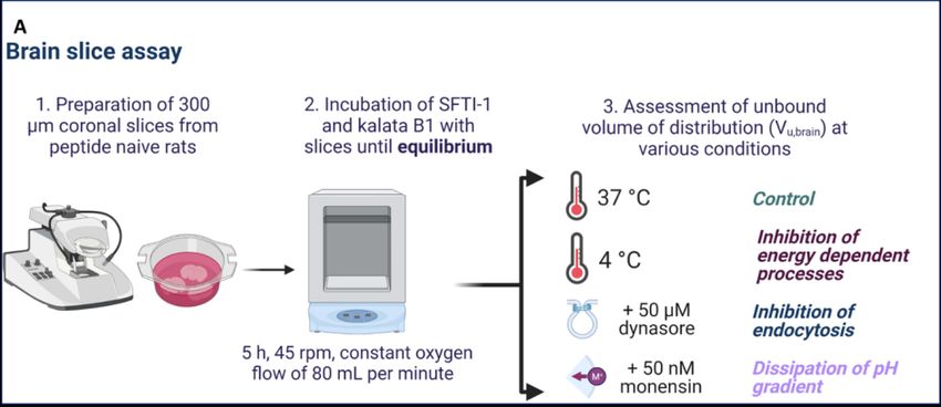
and plasma. To separate the BBB transport from the binding, the Kp,brain val-
ue is then combined with in vitro assessments of the binding of the com-
pound to both plasma proteins and brain tissue (Figure 2). The most classic
way of estimating the binding is via equilibrium dialysis, however, for the
assessment of binding in the brain, the more sophisticated brain slice method
should be preferred since it can capture more processes than simply the non-
specific binding [27,29–31].

Figure 2. Schematic view over the compartments of the brain and plasma with the
BBB and the cellular barrier (CB) are indicated with dashed red lines. Binding equi-
libria in plasma and brain cells are indicated.

Intra-brain distribution
A separate but also important aspect of the pharmacokinetics within the CNS
is the intra-brain distribution of the studied compound. This describes the
binding of the compound to the brain tissue, differential distribution between
the regions of the brain, and the uptake into cells or subcellular compart-
ments. One of the main parameters that describes the intra brain distribution
is Vu,brain, i.e the unbound volume of distribution of the compound in the
brain [31]. This parameter tells us about both specific and non-specific bind-
ing in the brain, as well as any cellular uptake, driven either by active pro-
cesses or by phenomena such as lysosomal trapping, driven by pH differ-
ences [32]. Measuring Vu,brain is commonly done via the brain slice method,
in which thin slices of brains are incubated in artificial extracellular fluid
spiked with the studied compound [31]. If no accumulation into the cells of
the brain parenchyma occurs, the value of Vu,brain inversed should correspond
with the value of fu,brain, describing the non-specific binding to the brain tis-
sue components. A Vu,brain value of 0.2 mL/g brain indicates that the studied
compound does not enter the cells of the brain at all, but rather stays in the

16
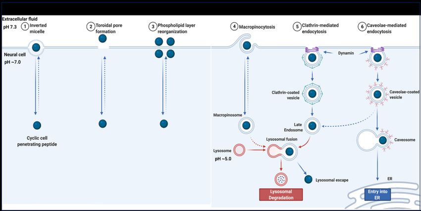
interstitial fluid. No upper limit for Vu,brain exists, and values above 1000
mL/g brain have been observed [30].
 By combining Vu,brain and fu,brain we can calculate the parameter Kp,uu,cell,
describing any potential accumulation into cells [32]. A Kp,uu,cell value > 1
means that the compound is either actively taken up into the cells, or that it
is accumulating by some other means. Examples of this are lysosomal trap-
ping, whereby the pH difference between the cytosol and the lysosome
changes the charge of the molecule, causing it to be trapped within the lyso-
some. Since ionised molecules are incapable of diffusing through mem-
branes, therefore, mainly bases are prone to lysosomal trapping.

Cellular uptake and distribution pathways
Once a compound has entered the brain, there are several pathways by which
it may distribute within the brain or be taken up by different cell types. This
distribution can either take place via passive diffusion, direct translocation
mechanisms, or via energy dependent active uptake (Figure 3). Distribution
can also be driven by binding to membranes or cellular components. Since
many targets for drugs are found within cells, it is highly important to study
this intra-brain distribution, both with regards to cell penetration and accu-
mulation, as well as binding characteristics. When exploring the pathways
that may lead to accumulation of the studied compound within cells, i.e.
those with a Kp,uu,cell value above unity, two main pathways should be con-
sidered; active transport through one or several of many transporters present
on cell membranes or vesicular uptake through the formation of endocytotic
vesicles in the cell membranes [1]. These endocytotic pathways are generally
divided into clathrin- and caveolae-mediated endocytosis, which are both
dependent on dynamin, a protein that is crucial for the budding of the vesi-
cles from the cell membrane [33,34]. These endocytotic processes are initi-
ated either by a molecule binding to a specific receptor on the cell surface, or
by adsorption of the molecule to the cell surface [35]. Examples of such
receptor mediated endocytotic systems are the transferrin, insulin and low
density lipoprotein receptors. The transferrin receptor system has been stud-
ied extensively and several studies have reported success in targeting the
transferrin receptor for delivery of large molecules such as antibodies
[36,37].
 Small molecules are more likely to accumulate in cells through trans-
porter mediated uptake, whereas larger molecules such as peptides and pro-
teins are more likely taken up through vesicular pathways. A third pathway
which has been proposed as a pathway for entry into cells for cell penetrat-
ing peptides in particular, is the formation of transient pores in the cell
membrane [38]. These pores can be formed by the peptides oligomerizing
within the cell membrane, thereby forming an opening through which entry
can occur, so called Barrel-Stave pores [39]. Another type of pore formation

 17
is that of toroidal pores, where the interaction between the peptide and the
cell membrane causes the membrane to fold in on itself, thus forming a pore
[40–42]. All in all, these various pathways for cellular entry leads to a multi-
tude of possibilities for molecules, small and large, to enter into the cells of
the CNS to elicit their effect.

Figure 3. Transcellular uptake pathways for large molecules. The two pathways on
the left, clathrin- and caveloae-mediated endocytosis are dynamin dependent. Creat-
ed with Biorender.

Cell penetrating peptides
A method of overcoming the cellular barrier to deliver therapeutic moieties
to intracellular targets could be the use of so called cell penetrating peptides.
These first came into the spotlight when the transactivating transcriptional
factor (TAT) peptide from the HIV virus was discovered in 1988 [43,44].
TAT is a protein with a high capacity to penetrate cell membranes and enter
into the cell. Later, the protein penetratin was also discovered, and together,
these discoveries laid the foundation for the development of cell penetrating
peptides as system for intracellular delivery [45]. A definition of cell pene-
trating peptides has been given as a peptide that contains less than 40 amino
acids and is able to enter cells by various mechanisms [39]. The amino acid
sequence of CPPs varies considerably, but common features are often posi-
tively charged amino acids in the sequence. One of the simplest and most
used CPPs is just a chain of arginines [39]. It has also been shown that the
secondary structure of the peptide can influence the capacity to penetrate
into cells. In general, CPPs are not considered to target specific cells, but are
assumed to enter cells non-selectively, but certain peptide sequences have

18
been described to target tissues more specifically, with one example being
able to target breast cancer cells [46].
 Cell penetrating peptides are generally entering cells through one of the
pathways mentioned above, with the most prominent ones being direct pene-
tration of the plasma membrane, or through some form of endocytosis. It
should however be noted that the exact mechanisms behind cell penetrating
properties of peptides have not yet been fully understood., There are many
contradictory reports on uptake mechanisms, and it is likely that the mecha-
nisms might vary depending on experimental conditions, where factors such
as pH, cell type, incubation time, and concentration of the peptide can yield
different uptake mechanisms in different experiments [47–53]. This is then
further proof that in order to properly evaluate the usefulness of a CPP it
needs to be studied in the correct system, with conditions matching those of
its intended use, i.e as close as possible to physiological conditions, with
clinically relevant concentrations if such are known.

Cyclic cell penetrating peptides
One specific subgroup of CPPs is cyclic cell penetrating peptides, which are
peptides that have some form cyclic nature, either head-to-tail, head-to-side
chain, or side chain-to-side chain [54,55]. They are typically more stable
than linear peptides, as the cyclic nature prevents proteolytic degradation.
Several cyclic peptides have successfully been developed into medicines,
such as cyclosporine A, vancomycin, and daptomycin, and the interest in
utilising cyclic peptides to target intracellular targets is increasing [56].
Where larger peptides have issues crossing cellular membranes due to their
size and charge of side chains, certain cyclic CPPs are assumed to have a
better chance of entering cells due to various transport mechanisms men-
tioned earlier. Cyclic CPPs are generally considered to enter cells through
one of two pathways; either by endosomal uptake or through a direct trans-
location over the cellular membrane directly into the cytoplasm [54]. A spe-
cific group of cyclic CPPs called cyclotides are of particular interest due to
their high stability [57–60]. These peptides are of plant origin, have a head-
to-tail cyclic structure, and are further stabilized by disulphide bonds be-
tween cysteine groups in the peptides. Cyclotides such as kalata B1 and
MCoTI have been reported to be able to penetrate into cells [41,61]. Fur-
thermore the plant derived cyclic peptide sunflower trypsin inhibitor (SFTI-
1), which shares many characteristics with the cyclotides but is smaller in
size, 15 amino acids vs the 28 amino acids found in kalata B1, has also been
reported to have cell penetrating potential [62].

 19
Aims

The overall aim of this thesis was to further develop a pharmacokinetic
framework for quantifying regional differences in blood-brain barrier
transport, and intra-brain distribution of small molecules and cell penetrating
peptides.

The specific aims were the following:

 1. To investigate possible regional distribution of drugs in the CNS with
 antipsychotics as model drugs

 2. To build a basis for studies of cyclic peptides and their behavior at the
 BBB and in the brain, by developing a quantitative LC-MS/MS
 method for the analysis of biological samples of the cyclic peptide
 kalata B1

 3. To investigate in vivo, ex vivo and in vitro BBB transport and intra-
 brain distribution properties of the model cyclic peptides kalata B1
 and SFTI-1, to increase the knowledge about their possible use as
 scaffolds for CNS drug delivery

 4. To evaluate the intra-brain distribution of the cyclic peptides kalata
 B1 and SFTI-1 in tissues ex vivo in comparison to cells in culture to
 further investigate distribution patterns

20
Materials and Methods Animals Male Sprague-Dawley rats (250-300 g) were used in all experiments (Taconic, Lille Skensved, Denmark). All animal experiments were performed in accord- ance with the guidelines from the Swedish National Board for Laboratory Animals, and were approved by the Animal Ethics Committee of Uppsala, Sweden approval numbers: C16/12, C351/11, C188/14. The animals were housed in groups with ad libitum access to food and water and with a 12 hour light-dark cycle, and were allowed to rest for 7 days before the study start. For in vivo pharmacokinetic and neuropharmacokinetic experiments in papers I, II, and III, catheters made of polyethylene (PE50) were implanted in the femoral veins and arteries of the animals for dosing and sampling. All surgeries were performed the day before the experiments under isoflurane anaesthesia. During the experiments the animals were housed in a CMA120 system (CMA, Solna, Sweden) allowing for free movement and ad libitum access to food and water. Peptide production SFTI-1 was extracted as follows: seeds from Helianthus annuus, common sunflower, were crushed and extracted with 60 % AcN for 3 h. The mixture was filtered and the seeds were re-extracted in fresh solvent for an additional 3 h. Following filtration, the extracts were pooled and the major part of AcN was removed by rotary evaporation. The extract was separated from lipids through partitioning against dichloromethane (1:2, v/v). The aqueous layer was freeze-dried, dissolved in MQ-water with 10 % AcN and 0.05 % TFA, and subjected to solid phase extraction on a C18 Isolute cartridge (Bio- tage, Uppsala, Sweden). The extract was loaded, washed with 0.05 % TFA in MilliQ-water and eluted with increasing concentrations of AcN in MilliQ- water with 0.05 % TFA. The SFTI-1 containing fractions were pooled, dilut- ed and purified on Reverse Phase-HPLC (C18, 250 x 10 mm, 5 µm, 300 Å, Phenomenex, Torrance, CA, USA). Purity and identity of SFTI-1 were ana- lysed by HPLC-UV (Shimadzu, Tokyo, Japan) and UPLC-MS/MS (Waters, Milford, MA). 21
Kalata B1 was extracted and purified from the leaves of Oldenlandia affinis
with the same procedure as SFTI-1 using a similar protocol [63]. The plant
material was homogenized and extracted with dichloromethane/methanol (1:1,
v/v). The extract was then freeze dried and then dissolved in 10 % methanol in
water and separated on a C18 column. The peptide containing fraction was
purified and analysed with LC-MS. Both peptides were > 95 % pure.

Experimental procedures
In vivo pharmacokinetics
In Papers II and III the in vivo pharmacokinetics of SFTI-1 and kalata B1
were assessed, since the information on the PK of cyclic peptides was very
limited. Both peptides were given at a dose of 1 mg/kg body weight to
healthy male Sprague-Dawley rats. The dose was administered as a 10 min
short infusion after which plasma samples were taken for two hours for
SFTI-1 and four hours for kalata B1. All samples were added to heparinized
low binding Eppendorf tubes. They were then centrifuged and the plasma
was transferred to new tubes and frozen. The samples were analysed and the
concentration-time curves were used to determine pharmacokinetic parame-
ters such as half-life, clearance, and volume of distribution with non-
compartmental methods.

Blood-brain barrier transport
In Papers I and III the BBB transport was assayed by measurement of the
parameters Kp,brain and Kp,uu,brain. These were obtained from steady state
measurements of brain and plasma concentrations after a 4 hours constant
rate infusion. Plasma samples were taken repeatedly during the infusion to
ensure that steady state conditions were obtained and maintained. After the
infusion, a heart puncture was performed and the whole brain was collected.
In Paper I the brain was then further dissected into the regions of interest;
hypothalamus, cerebellum, frontal cortex, striatum, hippocampus, and brain-
stem. The spinal cord was also collected. All tissue and plasma samples were
immediately frozen on dry ice and stored pending analysis. To calculate
Kp,brain and Kp,uu,brain the following equations were used:

 , = (Eq. 2)

 ,
 , , = (Eq. 3)
 , ∗ ,

22
The calculation of Kp,uu,brain was performed using the CMA, either for whole
brain, or for the brain region of interest [28]. This methodology utilises a
combination of in vivo and in vitro methods where both BBB transport and
binding and distribution characteristics of the studied compound are assessed
in order to provide the full picture of the extent of the BBB transport.

In vitro blood-brain barrier permeability
In order to measure the permeability of the two peptides, SFTI-1 and kalata
B1, an in vitro BBB model was used. The model was based on CD34+ endo-
thelial cells derived from human hematopoietic stem cells in co-culture with
bovine brain pericytes [21,22]. The experiment was set up in a transwell
system, and incubated for three hours after which the concentrations in both
the donor and receiver compartments were measured. The concentration of
peptide was also measured in the endothelial cells themselves, to understand
if any fraction of the added peptide was able to enter into the endothelium
without crossing to the receiver compartment. From these studies, the per-
meability parameters Pe and Papp were calculated.

 1 1 1
 = − (Eq 4)

 = (Eq 5)

 = (Eq 6)
 ∗ 0

Where Pse is the permeability surface area product across the endothelial cell
layer, Pst is the total permeability surface area product across the endothelial
cell layer and filter insert, and Psf is the permeability surface area product
across the filter insert. S is the surface area of the transwell insert (in this
experiment 1.12 cm2). Papp is the apparent permeability, J is the rate of ap-
pearance of the compound in the receiver compartment (amount/sec), and C0
is the concentration in the donor compartment at the start of the experiment.
 To ensure the integrity of the endothelial monolayer, sodium fluorescein
was used as a marker for paracellular transport. SFTI-1 was studied at two
concentrations, 250 nM and 500 nM whereas kalata B1 was studied at five
concentrations, 250, 500, 1000, 2000, and 4000 nM due to detection issues at
lower concentrations.

In vitro binding assay
In order to determine the binding of both small molecular drugs and peptides
to plasma proteins and brain tissue homogenate, as well as the binding of

 23
peptides to lung tissue homogenate and cell lysate, equilibrium dialysis was
performed. These experiments allow us to measure the parameters unbound
fraction in plasma (fu,plasma) and unbound fraction in tissue (fu,tissue) in plasma
and diluted tissue homogenate from brains or lungs. In these experiments
PBS was spiked with the studied compound and added to one side of the
semipermeable membrane, with the biological matrix being studied on the
other side of the membrane. The membrane in these studies had a molecular
weight cut off of 12-13 kDa allowing for both small molecules and peptides
to cross through, whilst restricting the passage of proteins and tissue compo-
nents. The device used for the equilibrium dialysis was in a high-throughput
96 well format, allowing for measurement of the unbound fraction in differ-
ent matrices at the same time, with multiple replicates. The experiment was
run for 6 h at 37 oC in a MaxQ4450 shaker (Thermo Fisher Scientific,
NinoLab, Sweden) at 200 rpm. The interaction between the studied com-
pound and the binding sites in plasma or tissue are generally assumed to be
reversible and an assumption is that equilibrium is rapidly reached between
the bound and unbound fractions. This allows measuring the unbound frac-
tion as the ratio between the concentrations of the compound in the receiver
side and the donor side at equilibrium. This was calculated as follows:

 , = (Eq 7)

For fu,brain/lung there was a need to compensate for the dilution of the homoge-
nate, and the following equation was used:

 ,ℎ, = (Eq 8)
 ℎ

 1
 , =
 (Eq 9)
 1 1
 (( )−1)+
 ,ℎ,

In these equations fu,h,D is the diluted tissue homogenate, and D is the dilu-
tion factor [27]. The dilution factor used for brain and lung homogenate was
5, whereas the dilution of cell lysate in Paper IV was 35.3.

In vitro unbound tissue volume of distribution assay
The tissue slice method for measuring Vu,brain was used in Papers I, III, and
IV. The methods have been described in full in Fridén et al and Bäckström et
al [30,64]. Here follows a brief summary of the methods. For the brain slic-
es, six coronal slices were cut from the striatal area of drug naïve rats at a

24
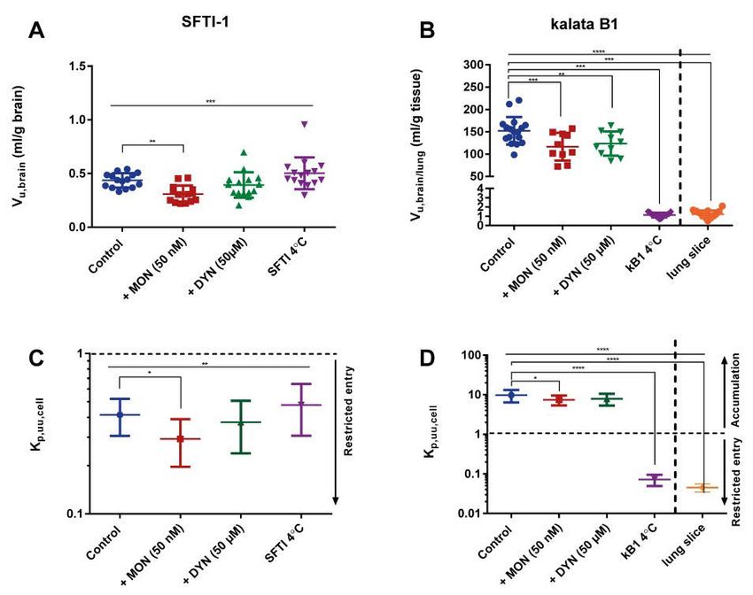
thickness of 300 µm using a Leica VT1200 microtome slicer (Leica Mi-
crosystems, Wetzlar, Germany). These were incubated with 200 nM of the
studied drug in 15 mL of artificial extracellular fluid (aECF) for 5 hours in a
MaxQ4450 shaker incubator (Thermo Fischer Scientific, NinoLab, Sweden).
After the incubation the slices were dried on filter paper, weighed, and ho-
mogenised in nine volumes (w/v) of aECF. The buffer was sampled directly
from the dish in which the experiment was performed. Vu,brain was then de-
termined with the following equation.

 − ∗
 , = (Eq 10)
 (1− )

In this equation, Aslice stands for the measured concentration of compound in
the slice per g tissue, Cbuffer is the concentration in the buffer at the end of the
experiment. Vi is the volume of buffer film coating the slice, which has been
determined to a value of 0.094 mL/g brain [30].
 In Paper IV, brain slice experiments were also run at 4°C to elucidate
whether any active processes were involved in the uptake and distribution of
the peptides (Figure 4). These experiments followed the same procedures as
previously described. Experiments with blockers or modulators of uptake
pathways were also run. In these experiments the slices were pre-incubated
with monensin or dynasore for 30 min prior to the addition of either SFTI-1
or kalata B1. Monensin was chosen as an ionophore modulator of the pH in
the subcellular compartments, decreasing the potential for lysosomal trap-
ping. The chosen concentration of monensin was 50 nM which was the high-
est tolerable concentration in the slices, able to elicit an effect without com-
promising the viability of the slices [32,65,66]. Dynasore is a dynamin inhib-
itor which prevents the formation of clathrin and caveolin coated vesicles in
the cell membrane [67,68]. The concentration used was 50 µM which was
previously shown to provide a 70 % decrease in dynamin activity.

 25
Figure 4. Experimental setup for brain slice assay. Illustration by Irena Loryan.
Created with Biorender.

For the lung slice experiments an in situ perfusion of rat lungs was per-
formed with physiological saline, after which the lungs were filled with aga-
rose type VII-A to facilitate slicing. The slices were prepared with a Leica
VT1200 microtome at a thickness of 500 µm. Three slices from three biolog-
ical replicates were incubated in 15 mL of aECF for 5h with a peptide con-
centration of 200 nM. The slices were then sampled in the same way as the
brain slices, with drying and sampling of the buffer. Vu,lung was calculated in
the same way as Vu,brain, but with a different Vi value, 0.73 mL/g lung, since
the liquid film layer surrounding lung slices differ from the one for brain
slices.

Cellular accumulation assay
In order to assess the cellular accumulation of the cyclic peptides SFTI-1 and
kalata B1, they were incubated with stem cell derived neurons and astrocytes
differentiated from human neural stem cells (hNSCs) [69]. These cells were
seeded at a density of 2.5 x 10⁴ cells/cm2 in triplicates per cell type on six
well plates. The peptides were added to Neurobasal medium to a concentra-
tion of 500 nM and 2 mL of this peptide containing medium was added to
each well. The cells were incubated for 5 hours at 37°C with the peptides,
after which the medium was removed and the cells were lysed with 300 µL
of Pierce lysis buffer per well. Samples were later kept att -20°C pending
analysis.
 The uptake of the peptides into cells was calculated with the concentra-
tion ratio Kp,u,cell, which is the ratio of total cellular-to-unbound medium con-
centrations. This is a partition coefficient between unbound concentrations in
the buffer and total concentrations in the cells, meaning that the partitioning
coefficient is a composite of both the uptake in the cells, and the distribution
driven by binding to cellular components. That means that this parameter is

26
similar in concept to Vu,brain, giving a description of uptake and binding char-
acteristics. Kp,u,cell is calculated using the following equation:

 , , = (Eq 11)

Ccell is the measured concentration in the lysed cells, calculated as the
amount of peptide in the cell lysate divided by the volume of the cells in the
lysate. The cell volume was determined through the measurement of protein
content in the cell lysate. A volume of 6.5 µL/mg protein was then used to
determine the total cell volume [70]. Cmedium is the measured concentration in
the medium. Kp,u,cell can further be used to calculate Kp,uu,cell for the specific
cell type by combining Kp,u,cell with fu,cell measured with equilibrium dialysis.

 , , = , , ∗ , (Eq 12)

This Kp,uu,cell value describe the cellular accumulation in specific cell types,
in contrast to the Kp,uu,cell determined from brain slice experiments where all
cell types present in the brain are involved, and can therefore differ from
values obtained from those experiments.

Bioanalytical setup
For the quantification of both the antipsychotic drugs and the peptides high
quality LC-MS methods were used. In Paper I an LC-MS/MS setup was
used, where the LC system was set up with two LC-10AD pumps coupled to
a SIL-HTc autosampler (Shidmadzu, Kyoto, Japan). The separation was
performed on a HyPurity C18 column (3µm particle size, 50 x 4.6 mm,
Thermo Scientific, Waltham, MA, USA). Quantification was carried out on
a Quattro Ultima Pt (Waters, Milford, MA, USA) using multiple reaction
monitoring in positive electrospray mode.
 In Paper II, the analytical method for kalata B1 was developed. It was
performed with LC-10AD pumps coupled to a SIL-HTc autosampler con-
nected to the aforementioned HyPurity C18 column. Quantification was
performed on the Quattro Ultima Pt. Validation was performed to determine
the accuracy and precision of the method as well as the LLOQ and linear
range. The validation was performed in accordance with the FDA guidance
of validation of bioanalytical methods [71]. Intra- and inter-day accuracy and
precision were determined by the analysis of all standards, as well as six
replicates of each QC during one day. The LLOQ was determined by run-
ning five LLOQ-candidates at the same time as the standards and QC sam-
ples. The accuracy was described as the deviation of the measured concen-
tration from the nominal concentration as a percentage. The precision was

 27
determined with the coefficient of variation (CV) and was obtained by divid-
ing the standard deviation with the mean concentration of the measured
samples. The CV was below 15 % for all QC levels in both plasma and brain
homogenate. The accuracy was also within the threshold of 15 % deviation
from the nominal concentrations.
 In Papers III and IV quantification of kalata B1 and SFTI-1 was per-
formed on either a Quattro Ultima Pt or on a Xevo TQ-S micro triple quad-
rupole mass spectrometer (Waters, Milford, MA, USA). The LC system was
either the LC-10AD pumps with a SIL-HTc autosampler or an Aqcuity
UPLC (Waters, Milford, MA, USA). The columns were a HyPurity C18 and
a Peptide CSH C18 (50 x 2.1 mm, particle size 1.7 µm) (Waters, MA, USA).
For Paper I, the LC separation was performed using a gradient based on two
mobile phases, mobile phase A with 0.1 % formic acid in water, and mobile
phase B with 90 % acetonitrile in water with 0.1 % formic acid. In Papers II-
IV LC separation was performed with the following mobile phases, mobile
phase A with 10 % acetonitrile in water with 0.5 % formic acid, and mobile
phase B with 90 % acetonitrile in water with 0.5 % formic acid. The ob-
served retention times for the two peptides were 1.85 min and 3.47 min for
kalata B1 and SFTI-1 respectively, thereby enabling fast analysis of the pep-
tide samples.

28
Results & Discussion

Regional distribution of antipsychotics (Paper I)
Paper I shows quantitative evidence of differences in BBB transport between
different regions of the brain. It also shows regional differences in brain
tissue binding.
 The paper describes the neuropharmacokinetics of haloperidol, clozapine,
olanzapine, quetiapine, risperidone, and paliperidone, includning their re-
gional BBB transport in the following regions of the CNS; frontal cortex,
striatum, hippocampus, brainstem, cerebellum, hypothalamus, and the spinal
cord. The paper also describes the regional intra-brain distribution and bind-
ing, with fu,brain and Vu,brain being measured for all compounds in cortex, stria-
tum, and coronal brain slices. With these parameters measured, using the
combinatory mapping approach, we could spatially map the differences in
transport and distribution for antipsychotic drugs in the CNS.
 With the exception of clozapine which had a uniform BBB transport
throughout all regions, all the compounds exhibited significant spatial differ-
ences in their BBB transport (Figure 5). The largest differences in BBB
transport were seen for risperidone and paliperidone, which are both quite
strong Pgp substrates. This could signify differences in expression levels of
the efflux proteins, or differences in their functionality between sites. The
studied antipsychotics were effluxed at the BBB with the exception of
haloperidol and olanzapine with Kp,uu,brain values ranging from 1.5 to 0.05
throughout the study. When looking at the different regions, cerebellum
showed the most efficient efflux for all compounds, whereas cortex showed
the least efficient efflux. An example of these differences was seen for
risperidone, with a 5.4-fold difference in Kp,uu,brain between frontal cortex
(0.28 ± 0.11) and cerebellum (0.05 ± 0.02). Another clear example of the
spatial differences was seen for paliperidone, with a 4-fold difference in
Kp,uu,brain between frontal cortex (0.12 ± 0.03) and the spinal cord (0.03 ±
0.009). These findings are clear proofs that assumptions of uniform distribu-
tion to the brain are not valid, and that the BBB characteristics vary between
brain regions. This is therefore something that should be kept in mind when
developing therapeutics with a target in a specific brain region, or side ef-
fects linked to another brain region than that of the target.

 29
When looking at the brain tissue binding of the different antipsychotics,
the binding capacity varied 24-fold between the lowest recorded fu,brain value,
0.007 for clozapine in spinal cord, and the highest fu,brain value of 0.17 pali-
peridone in hypothalamus. Olanzapine and paliperidone had similar binding
in all regions, but the other studied compounds exhibited significant differ-
ences in binding between the different regions. When looking at the Vu,brain
values for the antipsychotics, studied in cortex and striatum, there was a
clear trend towards higher binding and/or uptake in striatum compared to
cortex for all compounds except for risperidone. By calculating the Kp,uu,cell
value for the antipsycotics in order to evaluate their intracellular accumula-
tion, it was found that olanzapine, clozapine and paliperidone all accumulat-
ed in cells, with olanzapine having on average 5-fold higher intracellular
concentrations compared to extracellular concentrations. As binding, both
specific and non-specific, plays a significant role in the distribution of drugs,
and we show that this binding, as well as cellular uptake can vary signifi-
cantly between different regions of the brain, it is clearly not enough to
measure total concentrations in the brain when assessing the potential of
therapeutic agents targeting the CNS. Rather, by combining unbound, re-
gional concentrations with the receptor occupancy needed to elicit and ef-
fect, it is possible to accurately predict the effect of a compound, as well as
any side effect that may arise from the measured concentrations. In fact,
these findings might be part of the explanation as to why newer atypical
antipsychotics have not performed better than their older typical counterparts
[72,73]. Another important lesson from this study is the fact that it is not
only the Kp,uu,brain value of a compound that determines its capability to func-
tion as an effective agent within the CNS, since we show that two of the
studied antipsychotic agents have Kp,uu,brain values below 0.2 indicating major
efflux at the BBB. Rather, one must regard the extent of BBB transport in
conjunction with the potency of the drug, and thereby establish a proper,
site-of-action, PKPD-relationship in order to fully appreciate the potential of
a given compound to elicit an effect within the CNS.

30
Figure 5. Kp,brain and Kp,uu,brain graphs for the six studied antipsychotics in the studied
regions of the brain; spinal cord (SC), brain stem (BS), hippocampus (HPC), stria-
tum (STR), cortex (CRT), cerebellum (CRB), hypothalamus (HPT). Individual val-
ues and averages. *) p < 0.05, **) p < 0.01, ***) p < 0.001, ****) p < 0.0001

 31
Validation of bioanalytical method for kalata B1 (Paper
II)
Paper II describes the validation of the bioanalytical method for kalata B1.
This method was robust, simple, and with sufficient sensitivity for the quan-
tification of kalata B1 in plasma and brain homogenate, matrices essential
for the determination of BBB transport. The linear range in plasma was 2-
10000 ng/mL and in brain tissue it was 5-2000 ng/mL. The LLOQs in plas-
ma and brain homogenate were 2 ng/mL and 5 ng/mL respectively.
 The inter- and intra-day precision and accuracy were assessed and were
found to be low and within the acceptable limits. The precision had a CV <
15 % at both inter- and intra-day assessment, and the accuracy was within ±
12 %, both for the inter- and intra-day validation. These levels of accuracy
and precision were found in both plasma and brain homogenate and provided
a clear starting point for analysis of biological samples of the cyclic peptide.
The method was applied to assess the systemic pharmacokinetics of kalata
B1, describing the half-life of the peptide. When developing the method it
was noticed that interfering peaks appeared. It was found that a fragment of
human keratin has the same mass as the studied ion of kalata B1. This issue
was resolved by preparing the samples in a clean environment. Another issue
discovered in the method development was the sticking of kalata B1 to
glassware. This resulted in high variability in sample concentrations. This
issue was remediated through silanization of the glassware, which eliminated
the sticking issue, as well as using low binding plastic tubes for sample
preparation.

32
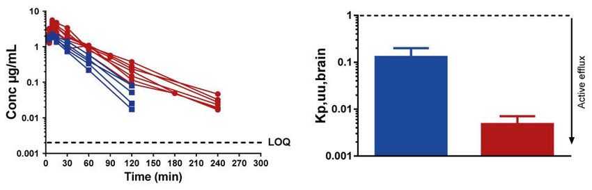
Blood-brain barrier transport of cyclic peptides (Paper
III)
Paper III describes the BBB transport of two cyclic peptides, the smaller
SFTI-1, isolated from sunflower seeds, and the cyclotide kalata B1, isolated
from the plant oldenlandia affinis. The paper describes the systemic pharma-
cokinetics of the peptides, as well as their in vivo extent of BBB transport
and the in vitro permeability across an endothelial cell BBB model.
 The results from the in vivo pharmacokinetics study confirm the in vivo
stability of the cyclic peptides, with relatively long half-lives of 20-30
minutes for both peptides (Figure 6). Both of the peptides had relatively
similar clearance (CL) values of 1.82 and 1.60 mL/min/kg for SFTI-1 and
kalata B1, respectively. These values are close to the renal filtration rate of
rats, which coupled with the high metabolic stability of the cyclic peptides
indicate a primarily renal excretion of these peptides. The plasma protein
binding of both peptides was also assessed, with SFTI-1 being highly un-
bound, whereas kalata B1 exhibiting a higher degree of plasma protein bind-
ing with fu,plasma values of 0.936 ± 0.038 and 0.299 ± 0.095 for SFTI-1 and
kalata B1 respectively.

Figure 6. Plasma pharmacokinetics and BBB transport of SFTI-1 (blue) and kalata
B1 (red).

In vivo blood-brain barrier transport
The assessment of BBB transport of SFTI-1 and kalata B1 was performed
with a constant rate infusion over four hours. Kp,brain was calculated as the
total concentration ration between brain and plasma of the steady state con-
centrations at four hours. By combining the Kp,brain values with fu,plasma and
Vu,brain the extent of BBB transport at equilibrium at steady state, Kp,uu,brain
was assessed and was found to be 0.129 ± 0.0698 for SFTI-1 and 0.00479 ±
0.00229 for kalata B1, indicating efflux or restricted entry across the BBB.
Interestingly, SFTI-1 had a higher extent of BBB transport, more than 25
times that of kalata B1, despite earlier reports on the cell penetrating capabil-
ities of kalata B1 [41,42].

 33
You can also read