Reversible switching of leukemic cells to a drug-resistant, stem-like subset via IL-4 mediated cross-talk with mesenchymal stroma
←
→
Page content transcription
If your browser does not render page correctly, please read the page content below
Reversible switching of leukemic cells to a drug-resistant,
stem-like subset via IL-4 mediated cross-talk with
mesenchymal stroma

by Hae-Ri Lee, Ga-Young Lee, Eung-Won Kim, Hee-Je Kim, Minho Lee, R. Keith Humphries,
and Il-Hoan Oh

Haematologica 2021 [Epub ahead of print]

Citation: Hae-Ri Lee, Ga-Young Lee, Eung-Won Kim, Hee-Je Kim, Minho Lee, R. Keith Humphries,
and Il-Hoan Oh. Reversible switching of leukemic cells to a drug-resistant, stem-like subset via IL-4
mediated cross-talk with mesenchymal stroma.
Haematologica. 2021; 106:xxx
doi:10.3324/haematol.2020.269944

Publisher's Disclaimer.
E-publishing ahead of print is increasingly important for the rapid dissemination of science.
Haematologica is, therefore, E-publishing PDF files of an early version of manuscripts that
have completed a regular peer review and have been accepted for publication. E-publishing
of this PDF file has been approved by the authors. After having E-published Ahead of Print,
manuscripts will then undergo technical and English editing, typesetting, proof correction and
be presented for the authors' final approval; the final version of the manuscript will then
appear in print on a regular issue of the journal. All legal disclaimers that apply to the
journal also pertain to this production process.

Reversible switching of leukemic cells to a drug-resistant, stem-like subset via IL-4 mediated cross-talk

with mesenchymal stroma

Hae-Ri Lee1, Ga-Young Lee1, Eung-Won Kim1, Hee-Je Kim2, Minho Lee3, R. Keith Humphries4,5, Il-Hoan

Oh1

1
    Catholic High-Performance Cell Therapy Center & Department of Medical Life Science, College of

Medicine, The Catholic University, Seoul, Republic of Korea
2
    Division of Hematology, Department of Internal Medicine, St Mary’s Hematology Hospital, College of

Medicine, The Catholic University of Korea
3
    Department of Life Science, Dongguk University-Seoul, Goyang-si, Gyeonggi-do, Republic of Korea
4
    Terry Fox Laboratory, British Columbia Cancer Agency, Vancouver, Canada; 5Department of Medicine,

University of British Columbia, Vancouver, Canada

Running Title: functional switching of leukemic cells by stromal contact

Key words: leukemia, microenvironment, chemoresistance, stemness

Correspondence

         Dr. Il-Hoan Oh, M.D., Ph.D.

         Catholic High-Performance Cell Therapy Center,

         The Catholic University of Korea

         505, Banpo-Dong, Seocho-Ku, Seoul 137-701, Korea

         Phone: 82-2-2258-8268, Fax: 82-2-591-3994

         E-mail: iho@catholic.ac.kr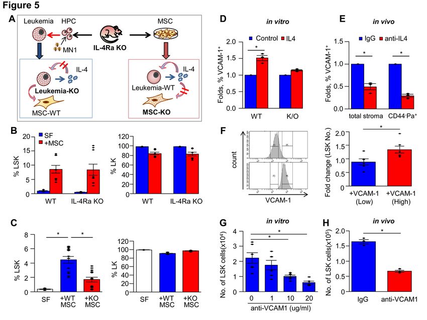
Competing interests statement

This study is supported by the NRF of Korea and funded by Ministry of Science, ICT, & Future Planning

(2017M3A9B3061947)

Key points

     Subset of leukemic cells undergo functional evolution to stem-like, drug-resistant cells in a reversible,
     stochastic manner via IL-4 mediated cross-talks with mesenchymal stroma.

     Development of the new leukemic subsets by stromal interaction represent another class of drug-

     resistance that can cause a non-homologous response to chemotherapy in concert with BM

     microenvironment

Authors’ contribution

HRL, GYL, EWK, HJK: performed experiments and collected data

MHL: performed experiments on genomics, statistical analysis of data.

RHK: conceptualized research, provided study materials, wrote manuscript

IHO: conceptualized idea & research, supervised research, wrote the manuscript, provided financial support

Acknowledgements

We thank Dr. Lee, Jeong-Hwa (College of Medicine, Catholic University of Korea) for thelp kind supply of

bis knock-out mice and Dr. Kang, Chang-Yul (College of Pharmacy, Seoul National University) for the kind

supply of mice lacking the IL-4 receptor and Life Science Editors for manuscript editing. We thank Dr. Jin-A

Kim for help in clinical data processing. We also thank Department of Biostatistics of the Catholic Research

Coordinating Center for statistical support. This study is supported by the NRF of Korea and funded by

Ministry of Science, ICT, & Future Planning (2017M3A9B3061947)

DECLARATION OF INTERESTS

The authors indicated no conflicts of interest.
ABSTRACT

Chemoresistance of leukemic cells has largely been attributed to clonal evolution secondary to accumulating

mutations. Here, we show that a subset of leukemic blasts in contact with the mesenchymal stroma undergo

cellular conversion into a distinct cell type that exhibits a stem cell-like phenotype and chemoresistance.

These stroma-induced changes occurred in a reversible and stochastic manner driven by cross-talk, whereby

stromal contact induces IL-4 in leukemic cells that in turn targets the mesenchymal stroma to facilitate the

development of new subset. This mechanism was dependent on IL-4 mediated up-regulation of vascular cell

adhesion molecule-1 in mesenchymal stroma, causing tight adherence of leukemic cells to mesenchymal

progenitors for generation of new subsets. Together, our study reveals another class of chemoresistance in

leukemic blasts via functional evolution through stromal cross-talk, and demonstrates dynamic switching of

leukemic cell fates that could cause a non-homologous response to chemotherapy in concert with the patient-

specific microenvironment.

INTRODUCTION

Acute myeloid leukemia (AML) is a heterogeneous, clonal hematopoietic disorder characterized by

excessive proliferation of stem cell-like progenitor cells in the bone marrow (BM). AML has a highly

variable prognosis1 and a very high risk of relapse particularly in elderly patients2.

Leukemia progression and relapse are widely viewed to occur via clonal evolution from preleukemic cells to

overt leukemia driven by genetic mutations 3, followed by additional mutations leading to treatment-

resistant, relapsed clone(s) 4. However, in-depth clonal analyses have revealed the persistence of founding

clones 4, and functional heterogeneity among the developed leukemic clones 5, suggesting that other

mechanisms may be involved.

Several studies have highlighted leukemic stem cell (LSC) properties contributing to drug resistance 6: AML

patients whose leukemic blast exhibit higher levels of stem cell signatures are at greater risk of relapse and

have a poorer prognosis 7, 8. However, the specific relationship between stemness and functional

heterogeneity of LSCs related to drug resistance, remain poorly understood 7, 9.

There is increasing awareness that the microenvironment, including growth factors, cytokines and niche

stromal cells, can provide protection to leukemic cells and thereby contribute to the acquisition of

chemoresistance 10, 11. For example, leukemic cell subsets surviving chemotherapy were localized to the

surface of osteoblasts in the BM 12-15. Subsequently, multiple protective signals from stroma have been

shown to enhance leukemic cell survival through activation of receptor tyrosine kinases 16 or interaction with

the extracellular matrix. However, despite these protective signals, a role for the stroma in the clonal

development of leukemic blasts for the acquisition of chemoresistance has not been demonstrated.

Here, we show that subsets of leukemic cells in stromal contact undergo reversible changes associated with a

stem cell-like phenotype and drug-resistant state. These changes are stochastic, and distinct from changes

induced by other mechanisms of chemoresistance, thus representing a new class of drug-resistant cells

developed in the leukemic microenvironment.
METHODS

Human sample collection

Primary leukemic blasts were collected from newly diagnosed AML patients without prior treatment history.

Part of the BM samples are from AML patients who had complete medical records during 5 years of follow-

up in clinical courses. Human MSCs were separated from BMs of normal donor under informed consent.

This study was approved by the Institutional Review Boards of St. Mary’s Hospital and Catholic University

of Korea.

Animals

C58/BL6 mice were obtained from the Jackson Laboratories (Bar Harbor, ME). Bis+/+, Bis+/-, Bis-/- mice 17

were provided by Dr. Jeong-Hwa Lee (Catholic University of Korea). Mice with disruption of IL-4 receptor
18
     were provided by Dr. Chang Yul Kang (Seoul National University).

Mouse and human AML cells and MSCs

Fresh murine or human MSCs were analyzed in the BM using flowcytometry. Cultured MSCs (5-8 passage)

were obtained by serial plating of BM cells in the DMEM containing 10% FBS as described19, 20. For

generation of murine AML cells, 5-FU treated BM cells were transduced with MN-1 or Meis1/HoxA9

through retroviral infection as described21, 22. For co-culture, MSCs were irradiated (15 Gy) 18-24 hours prior

to use and leukemic cells were seeded on the MSCs for co-culture. For co-culture with transwell, MSCs were

seeded into the upper chamber (6-well type, polyethylene terephthalate (PET) membrane with 0.4μm pores;

BD Bioscience, San Diego, USA) and leukemic cells were seeded into the lower well.

Flow cytometry of leukemic cells and MSCs.

Murine leukemic cells were analyzed by flow cytometry using the following antibodies: CD45.1-APC (BD

PharMingen, USA), Lineage cocktail (StemCell Technologies Inc, Canada), Sca-1-PE-Cy7 and c-kit-PE (BD

PharMingen). For human leukemic cells, CD45-APC, CD34-BV421, CD90-FITC (BD PharMingen)

antibodies were used. For MSCs, anti-CD106(VCAM-1)-biotin, CD51-PE (eBioscience, CA, USA.),

CD140a (PDGFR-a)-APC, Sca-1-PE-Cy7 (BD PharMingen) were used.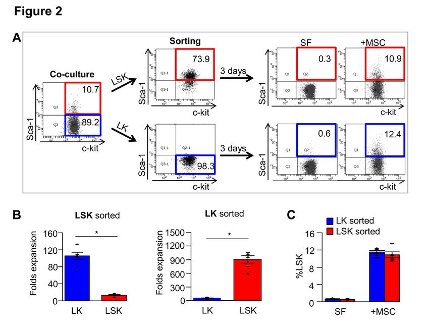
Treatment of antibody and cytotoxic drug

Leukemic cells seeded on irradiated MSCs were treated with anti-IL-4 antibody (R&D Systems Inc., USA),

anti-CD106(VCAM-1) (R&D Systems Inc.) for 3 days. For in-vivo antibody injections, mice received

intraperitoneal injection of anti-IL-4 Ab (1mg/kg) (R&D Systems Inc.) or intravenous injection of anti-

VCAM-1 Ab(10mg/kg) (Bio X cell, USA) along with IgG from rat serum(Bio X cell). Cytotoxicity of in-

vivo leukemic cells was examined by treatment with 100mg/kg of Ara-C(Sigma-Aldrich, MO, USA) and

3mg/kg of doxorubicin hydrochloride (Sigma).

Gene expression analysis

Sequencing libraries of two subjects were prepared according to the TruSeq Stranded Total RNA Sample

Preparation guide. Aligned reads were quantified using HTSeq-count23. Differentially expressed genes, fold

ratio, p-value, and false discovery rate were identified by edgeR algorithm24 for each subject. Enriched

KEGG pathways were identified by GSEA-P25.

Statistical analysis

To compare the generation of CD90+ subsets from individual primary human leukemia patients’ samples, or

the responses of individual patients’ leukemia cells to chemotherapy, we used Mann-Whitney test. To

compare the differences of means in specific experimental settings, we used a standard unpaired, 2-tailed

student t-test. The frequencies of leukemia-initiating cells in limiting dilution analysis were calculated by

applying Poisson statistics with 95% confidence interval representing ± 2 SEM.

RESULTS

A subset of leukemic cells acquires a stem cell-like phenotype by contact with mesenchymal stroma

To investigate the influence of stromal cells on the function of leukemic cells, we employed an in-vitro co-

culture model of murine leukemic cells in contact with BM-derived mesenchymal stromal cells (MSCs).

Murine AML cells were generated by transducing BM mononuclear cells (MNCs) with meningioma-1

(MN1) 21 or HoxA9-Meis1 (H9M1) 22 (Figure 1A). When co-cultured with MSCs, a subset of MN1 leukemic

cells acquired a Sca-1(+) phenotype (Lin-c-kit+sca-1+; LSK) mimicking normal hematopoietic progenitors,

while the majority remained Sca-1(-) (Lin-c-kit+sca-1-) (Figure 1B). The acquisition of Sca-1(+) phenotypes

was similarly observed in other types of leukemic cells (H9M1) or leukemia cell line (C1498) independent of

irradiation (Supplementary Figure S1A, B). The emergence of the Sca-1(+) subset was dependent on direct

contact with the mesenchymal stroma (Supplementary Figure S1C), as these cells were not observed in

stroma-free conditions or in stromal co-culture with a transwell filter (Figure 1B).

To determine if acquisition of the Sca-1(+) phenotype occurs in vivo, MN1 leukemogenic cells (Lin-c-

kit+sca-1-) were transplanted into mice. Consistent with the in vitro results, a subset of leukemic cells (GFP+)

in recipient mice acquired a Sca-1(+) (Lin-c-kit+sca-1+) phenotype (Figure 1C).

To determine whether acquisition of the Sca-1(+) phenotype in leukemic cells originated from their fusion

with stromal cells, as implicated previously 26, we co-cultured MN1 leukemic cells (GFP+) with MSCs

transduced with YFP. None of the GFP+ leukemic cells co-expressed YFP (Figure 1D). Moreover, there was

no difference in cell size between Sca-1(+) and Sca-1(-) cells, as determined by identical forward scatter in

flow cytometry, and no increase in tetraploidy in the Sca-1(+) cells (Figure 1E). Similarly, there was no

evidence of cell fusion in this in-vivo generated Sca-1(+) subset (Supplementary Figure S1D).

A recent study implicated mitochondrial transfer from MSCs to leukemic cells during acquisition of

chemoresistance 27, 28. To examine this, MSCs were labeled with a mitochondrial tracker and co-cultured

with leukemic cells. There was no difference in mitochondrial tracker intensity between the Sca-1(-) and

Sca-1(+) subsets (Figure 1F).

Altogether, this emergence of a new leukemic subset with a stem cell-like phenotype (Sca-1+) represents an

intrinsic cellular evolution of leukemic cells that occurs independently of cell fusion or mitochondrial

transfer during in-vivo leukemogenesis and in-vitro culture with stromal cells.

Switching to the Sca-1(+) phenotype is reversible

To determine if the Sca-1(+) subset is a stable phenotype, we sort-purified Sca-1(+) (LSK) and Sca-1(-) (LK)

leukemic cells generated during co-culture with stroma, and re-plated for a second round of co-culture with

or without stroma. The purified Sca-1(-) cell fraction again generated Sca-1(+) cells during the second round

selectively in the presence of stroma, whereas purified Sca-1(+) cells co-cultured with stroma rapidly

decreased in frequency (Figure 2A, B) with the emergence of a major Sca-1(-) cell population. Thus, final

stable ratios of Sca-1(+) and Sca-1(-) cells were similarly maintained under secondary stromal co-culture

conditions regardless of the phenotype of the initial cell population (Figure 2C). The changes in cell

populations occurred rapidly within 3 days of co-culture suggesting that the conversion between Sca-1(-) and

Sca-1(+) cells occurs by phenotypic switching rather than selective proliferation in the culture.

Thus, the emergence of Sca-1(+) leukemic cells during stromal contact occurs in a reversible manner in any

subsets of leukemic cells without clonal predisposition (stochastic), but with similar probability of each cells

(equipotent) for conversion among the total leukemic cell populations.

Functional heterogeneity acquired in stem cell-like leukemic subsets:

We next determined whether the Sca-1(+) stem cell-like leukemic subset arising by stromal contact was

functionally distinct. When MN1 leukemic cells were treated with the chemotherapeutic Ara-C during co-

culture with mesenchymal cells, Sca-1(-) subsets exhibited significant decrease of cell numbers, but the Sca-

1(+) subset exhibited higher resistance compared to the Sca-1(-) subsets, with no significant changes in cell

numbers (Figure 3A). Drug resistance in the Sca-1(+) subset was similarly reproduced in other leukemia cell

types tested (HoxA9/Meis1-induced leukemic cells or C1498 leukemia cell line) (Figure 3A).

The chemoresistance of the Sca-1(+) (LSK) leukemic population compared to the rest of the Sca-1(-) (LK)

cells was similarly observed in vivo with mice engrafted with MN1 leukemic cells and treated with

chemotherapeutic drug (Ara-C and doxorubicin) 29 (Figure 3B). Thus, enhanced drug resistance is a common
feature of leukemic subsets acquiring a Sca-1(+) phenotype upon stromal contact in a range of leukemic cell

models.

To further investigate the drug resistance of the Sca-1(+) cells, we analyzed their cell cycling in BM and

found that % of quiescent cell population (G0) was higher in LSK cells (Figure 3C). We also compared the

frequency of leukemic initiating cells (LIC), a functional assay for leukemia stem cells 30 in the Sca-1(+)

leukemic subset in comparison to the other subsets. Thus, subsets of Lin(+) cells, Lin-c-kit-, LK(Lin-c-

kit+sca-1-) cells and LSK(Lin-c-kit+ sca-1+) leukemic cells generated in BM of MN1 transplanted mice were

sort purified and transplanted into secondary recipient mice in a limiting dilution assay. However, the LK

and LSK populations exhibited a similar frequency of LICs, while exhibited significantly higher frequencies

than the other cell populations (Figure 3D & Supplementary Figure S2A). These two population (LK and

LSK) also exhibited comparable levels of in-vivo leukemic engraftment or in-vitro leukemia colony

formation (Supplementary Figure S2B, C), indicating that the Sca-1(+) subset developed during in-vivo

leukemogenesis comprise a subset of LICs that does not display significantly different leukemogenic activity

compared to their Sca-1(-) counterparts.

Thus, the Sca-1(+) leukemia subset generated from leukemic cells represents a distinct leukemic cell

population that has acquired drug-resistance without altering their leukemogenic activity.

IL-4 plays a role in the emergence of drug-resistant Sca-1(+) cells

We next sought to identify possible signals from the stroma that induce emergence of Sca-1(+) cells. Given

that altered production of cytokines and/or growth factors are frequently observed in leukemic cells 10, we

examined the cytokine/growth factor gene expression induced by stromal contact of leukemic cells

(Supplementary Figure S3A). Upon contact with stroma, the murine leukemic cells exhibited a notable

induction of cytokines and growth factors implicated in leukemogenic activity, including IL-4, PDGF-A,

PDGF-D, CCL-2, CCL-5, CXCL-1 and stem cell factor31-39, but not in the presence of transwell filters

(Supplementary Figure S3B). Among those cytokines, IL-4 was selectively induced in LSK subsets, but not

in the majority of remaining cells (LK) as determined by its transcript and protein level (Supplementary

Figure S3C, D). Thus, we examined whether IL-4 acts as an autocrine signal for generating Sca-1(+) subsets.
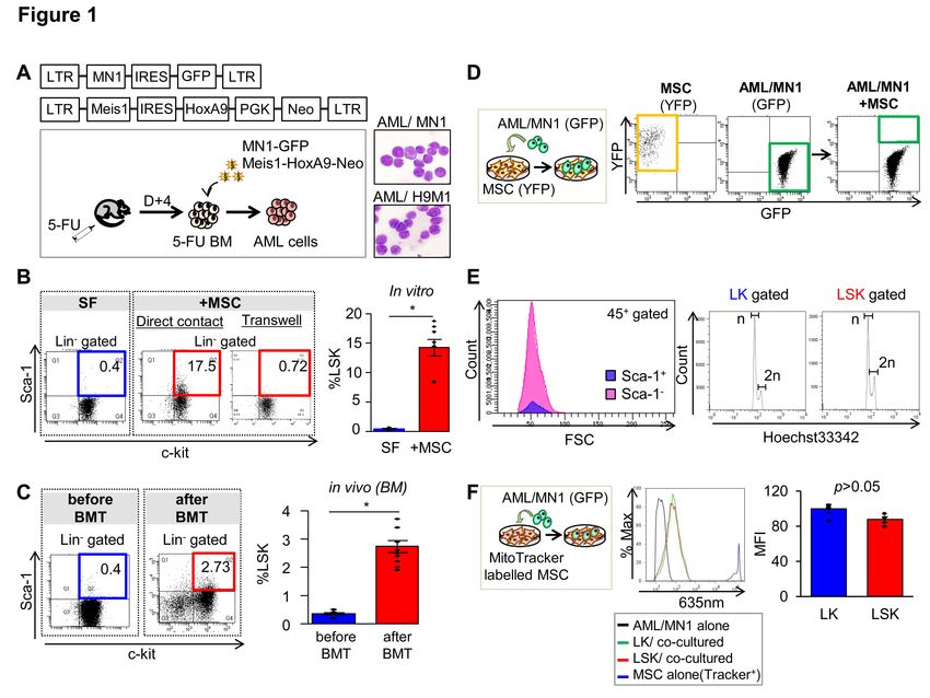
Addition of recombinant IL-4 increased the frequency of Sca-1(+) subsets (LSK) in a dose-dependent

manner (Figure 4A). Conversely, addition of an IL-4 neutralizing antibody significantly decreased the

frequency of LSK during co-culture (Figure 4A). Injection of an antibody against IL-4 into recipient mice

along with MN1 leukemic cells also decreased the LSK population in the BM of recipients without changes

in overall engraftment levels (Figure 4B). Moreover, IL-4 neutralizing antibody abrogated resistance of the

LSK population to chemotherapeutic drugs (Ara-C and doxorubicin), markedly decreasing the LSK

population in recipient BM (Figure 4C), which caused a decrease in the residual burden of surviving LICs

that can initiate leukemogenesis (Supplementary Figure S4).

Together, these results support a key role for IL-4 in the generation of drug resistant Sca-1(+) subset upon

stromal contact.

IL-4 dependent generation of Sca-1(+) leukemic cells is generated by stromal cross-talk

To investigate the mechanisms underlying IL-4-mediated generation of Sca-1(+) subsets, we examined the

cellular target of IL-4 during the co-culture of leukemic cells and stroma (Figure 5A). First, to see if IL-4

acts directly on leukemic cells, we established MN1 leukemic cells from hematopoietic progenitors of mice

lacking IL-4 receptor alpha (IL-4Ra KO). Co-culture of leukemic cells from IL-4Ra KO or wild type (WT)

with stromal cells led to comparable frequencies of LSK or LK subsets in each group (Figure 5B). In

contrast, when mesenchymal stromal cells from IL-4Ra KO mice were co-cultured with MN1 leukemic cells,

significantly lower frequencies of LSK, but not LK subsets, were observed compared to the WT stroma

group (Figure 5C). Thus, IL-4 signals target mesenchymal stromal cells, rather than leukemic cells, to

facilitate stroma-mediated generation of the LSK subset, indicating that IL-4-mediated cross-talk promotes

the functional evolution of leukemic cells.

Next, to investigate the effects of IL-4 on mesenchymal stroma, we examined whether the mode of cellular

interaction between MSCs and leukemic cells is influenced by IL-4. LSK subsets were predominantly

generated among the leukemic cells tightly adherent to the mesenchymal cells, for both MN1 or H9M1

leukemic cells, but seldom among the loosely adherent/suspension leukemic cells (Supplementary Figure

S5A). Supporting the influence of IL-4 on stromal adherence, the level of vascular cell adhesion molecules 1
(VCAM-1) in MSCs, which mediate stromal adherence of leukemic cells40, were up-regulated by IL-4 in

WT-MSCs, but not in IL-4Ra KO MSCs (Figure 5D). Conversely, in-vivo injection of IL-4 neutralizing

antibody caused a significant decrease of VCAM-1 expressions in BM mesenchymal cells including subsets

enriched for mesenchymal progenitors (CD44(-)PDGFR-1(+))41-43 (Figure 5E).

To further examine the influences of stromal VCAM-1 expression level on the generation of LSK subsets,

leukemic cells were co-cultured with sort-purified MSC fractions for different levels of VCAM-1. MSCs

with higher VCAM-1 levels increased LSK generation during co-culture, whereas MSCs expressing lower

levels of VCAM-1 decreased it, in comparison to LSK cells from unsorted MSC co-cultures (Figure 5F).

Similarly, VCAM-1 blocking antibody significantly decreased stromal adherence of leukemic cells

(Supplementary Figure S5B), which led to a concomitant decrease in the generation of the LSK subset

(Figure 5G). Moreover, in-vivo administration of VCAM-1 antibody caused a significant decrease of LSK

numbers in BM (Figure 5H). These data, together with positive expression of VCAM-1 ligands in leukemic

cells (Supplementary Figure S6) indicates that tight adherence of leukemic cells to VCAM-1 in MSCs

facilitates emergence of LSK subsets.

Consistent with these findings, gene expression changes in MSCs induced by IL-4 treatment during culture

revealed 41 differentially expressed genes (DEG), the most profound changes of which were in the gene

ontology group related to the ‘binding’ molecular function, supporting their role in the cellular interaction

with leukemic cells (Supplementary Figure S7A, B). Thus, IL-4 enhances the cellular interaction of stroma

and leukemic cells to facilitate stroma-dependent evolution of the Sca-1(+) leukemic subset exhibiting drug-

resistance.

Stroma-induced changes in Human leukemic cell models

To investigate whether a similar phenomenon can be seen in human leukemic cells, we examined human

AML cells for acquisition of CD90(+) as a phenotype for stem-like subsets based on findings that a subset of

CD90(+) cells amongst CD34(+) cells represent long-term repopulating HSCs 44 and that CD90 expression

in human leukemic cells represents high-risk leukemia with stem cell properties 45, 46. We first examined

human leukemic cell lines, MOLM-14 and MV4-11 (M5 type FAB), and HL-60 (M3 type FAB). For each
leukemic cell line tested, co-culture with human BM-derived MSCs resulted in the emergence of leukemic

subsets with the CD90(+) phenotype, albeit to variable levels (Figure 6A). Moreover, when chemoresistance

was compared between leukemic subsets, significant resistance to Ara-C treatment was observed selectively

for CD90(+) cells in all tested leukemic cells (Figure 6B) similarly exhibiting quiescence in cell cycle

(Supplementary Figure S8)

To examine these findings in primary leukemic cells, we examined the response of primary AML blasts from

5-7 individual patients to mesenchymal stroma. Primary AML blasts exhibited a significant induction of

CD90(+) cells upon stromal contact, which was further increased by IL-4 treatment during co-culture (Figure

6C, D). Sort-purified subsets of CD90(+) and CD90 (-) leukemic cells exhibited similar switching of

phenotypes to maintain constant ratios in CD90(+) subsets in total leukemic cells, as observed for murine

leukemic cells (Figure 6E). Moreover, the CD90 (+) subset generated during stromal contact exhibited

higher resistance to Ara-C (Figure 6F) than the remaining CD90(-) cell population in the same co-culture,

demonstrating a similar drug-resistance of newly emerging leukemic subsets in primary human leukemic

cells.

Gene expression study on two independent patients showed that CD90(+) subsets of primary human AML

cells are significantly enriched with gene sets specific for leukemia stem cell 47 than the remaining CD90(-)

cells (Figure 6G), and enriched with gene sets involved in the interaction with ECM or focal adhesion

(Supplementary Figure S9).

Thus, subsets of human leukemic cells in contact with stroma exhibit a stem-like properties to acquire drug-

resistance through interaction with stroma.

Stromal heterogeneity for generation of stem cell-like leukemic subsets

Extensive heterogeneity has been documented among mesenchymal populations in BM stroma48, 49.

Therefore, we investigated the mesenchymal subpopulations responsible for generation of Sca-1(+) cells.

Given that VCAM-1 expressing MSCs played a role in the generation of drug-resistant subsets, we examined

VCAM-1 expression among stromal cell populations in BM. VCAM-1(+) mesenchymal cells were

predominantly enriched by a CD44(-) population, where colony-forming mesenchymal progenitors (MPCs)
are exclusively localized 41-43, 50 (Figure 7A). Similarly, VCAM-1 is exclusively enriched in subsets for

MPCs as defined by PDGFRa(+)/Sca-1(+) or PDGFRa(+)/CD51(+) subsets41-43, 50, indicating that the

VCAM-1(+) cells that can drive emergence of drug-resistant subsets predominantly overlap with

mesenchymal progenitor cells (Figure 7B).

Supporting this finding, when MN1 leukemic cells were transplanted into the homozygous Bis KO mice,

where self-renewing mesenchymal progenitors are decreased in BM 17, 51, a significant decrease in the Sca-

1(+) subset (LSK) among BM engrafted leukemic cells was observed compared to WT or heterozygote Bis

KO mice (Figure 7C). This decrease was not associated with altered overall engraftment levels (Figure 7C),

consistent with the differences between LSK and LK subsets.

Similarly, supporting the role of mesenchymal progenitors for development of drug resistant leukemic cells,

AML patients wo relapsed after treatment exhibited higher numbers of mesenchymal progenitor subsets

(MPCs: CD146+/166-)19, 43, 52 in BM, i.e, retrospective studies on AML patients who had undergone relapse

within 1 year after complete remission exhibited higher numbers of mesenchymal progenitor subsets (MPCs)

in BM than those who maintained complete remission for 5 years (Figure 7D). Notably, this difference was

observed regardless of the underlying cytogenetic abnormality of the leukemic blasts, indicating that the

heterogeneity of stromal cells could be an additional factor drug-resistance of leukemic cells.

Altogether, this heterogeneity in the BM mesenchymal progenitors can influence the stroma-dependent

generation of stem cell-like leukemic subsets.

DISCUSSION

Leukemic cell evolution has prevented effective management of a diverse spectrum of leukemic

disease.

Here, using a variety of murine and human leukemia cells both in-vitro and in-vivo, we show that subsets of

leukemic cells can undergo a phenotypic conversion into a stem-like phenotype that exhibit a higher

resistance to chemotherapy in the context of stromal contact. This development of chemoresistant subsets by
stromal contact was not dependent on cell fusion or changes in leukemogenic activities observed in

mitochondrial transfer27, 28. The acquisition of the stem cell-like phenotype was reversible, being rapidly

reverted to the non-stem cell phenotype under stroma-free conditions independent of difference in cell cycles

or apoptosis (Supplementary Figure S10), unlike the stable maintenance of chemoresistance in leukemic

clones generated by clonal evolution.

Moreover, the frequencies of LSK cells among leukemic cells in contact with stroma were maintained

constant regardless of the phenotype of the initial cell populations. This suggests that the stroma-mediated

development of the stem cell-like, drug-resistant subpopulation occurs in a stochastic and reversible manner

in leukemic cells with similar probabilities among leukemic cells (equipotent) without clonal predisposition.

Reminiscent of these findings, recent studies showed that non-stem cancer cells can be spontaneously

converted to stem-like state, and these plasticity of cancer cells allows cellular switching between distinct

functional state53 54. Together, these studies raise the possibility that the stochastic development of

chemoresistant clones by stromal contact is an intrinsic process of leukemogenesis that could cause a non-

homogenous response to chemotherapy among the leukemic cell populations.

The mechanisms for dynamic equilibrium among different subsets of leukemic cells remains yet unclear.

One possibility is a feedback control mechanism that maintains a constant ratio of stem-like vs. non-stem-

like leukemic cells, probably through cellular interaction between distinct leukemic subsets, as inferred from

clonal interactions between heterogenous subsets 55. Similarly, studies on cancer stem cells have suggested

that non-tumorigenic cells regulate the maintenance of cancer stem cells influencing their relative

frequencies in the population 56. Since clonal heterogeneity of leukemia or cancer cells underlies the

differential response to chemotherapy and emergence of relapsing clones 55, 57, 58, the kinetics of generating

these stem-like subsets could be a factor for differential response to chemotherapy.

Interestingly, we show that the development of these drug-resistant leukemic subset is facilitated by bi-

directional cross-talk between stroma and leukemic cells mediated by IL-4, exhibiting resistance to apoptosis

(Supplementary Figure S11). While IL-4 was implicated in inhibition of leukemic cells and apoptosis 59, we

did not find increased apoptosis of non-stem-like population precluding the selective enrichment of stem-like

subsets by IL-4 (Supplementary Fig S12). Moreover, rather than acting directly on the leukemic cells, IL-4

targets stromal cells, which facilitate the generation of LSK subsets. How IL-4 acts on stromal cells to
facilitate the generation of a drug-resistant leukemic subset remains unclear. However, we demonstrated a

key role for VCAM-1 downstream of IL-4 in MSCs leading to tight adherence of leukemic cells and MSCs,

which was necessary for generation of the LSK subset. Similarly, we found IL-4 dependent induction of

gene clusters in MSCs whose functions are related to ‘binding function’. This suggests that the mode of

interaction between MSCs and leukemic cells is altered by IL-4 acting on MSCs, which facilitates the

development of the drug-resistant leukemic subset.

Interestingly, we also found functional differences between stromal cells in terms of their capacity to drive

development of drug-resistant leukemic cells. We found that expression of VCAM-1 in stromal cell was

important for adherence-dependent generation of leukemic subsets, while other adhesion molecules we tested

did not influence the process (Supplementary Figure S13). Importantly, the VCAM-1 expressing stromal

cells were selectively enriched in mesenchymal progenitors. Since mesenchymal stromal cells undergo

various degenerative changes during leukemia 19, 60, 61, it is possible that patient-to-patient heterogeneity in

BM mesenchymal progenitor cell content could differentially contribute to the development of drug-resistant

clones. Consistent with this, AML patients whose BM has higher levels of a primitive (CD146+) subset of

mesenchymal cells 19, 43, 52 that express higher levels of VCAM-1 tend to have a higher risk of leukemic

relapse compared to those who maintained complete remission. Thus, independent of oncogenic mutations or

cytogenetic abnormalities in the blasts 62, heterogeneity per se in mesenchymal progenitors in BM could be

another factor for development of drug-resistant leukemic subsets.

In summary, our study reveals an additional mechanism of functional evolution of leukemic cells induced by

contact with the mesenchymal stroma that can cause a reversible switch to a stem cell-like, drug-resistant

subset independent of mutation-driven clonal evolution in leukemic blasts (Supplementary Figure S14).

These findings thus provide further insight into the multiple mechanisms for development of drug-resistance

that could generate leukemic cells with distinct characteristics and chemoresistance, highlighting the

importance of the microenvironment in this process. This supports the need for better defining the

mechanisms of drug resistance in leukemia patients, and could lead to the development of more

comprehensive management of leukemic diseases.
REFERENCES

1.      Estey E, Dohner H. Acute myeloid leukaemia. Lancet. 2006;368(9550):1894-1907.
2.      Craddock C, Tauro S, Moss P, Grimwade D. Biology and management of relapsed acute myeloid
leukaemia. Br J Haematol. 2005;129(1):18-34.
3.      Welch JS, Ley TJ, Link DC, et al. The origin and evolution of mutations in acute myeloid leukemia.
Cell. 2012;150(2):264-278.
4.      Ding L, Ley TJ, Larson DE, et al. Clonal evolution in relapsed acute myeloid leukaemia revealed by
whole-genome sequencing. Nature. 2012;481(7382):506-510.
5.      Klco JM, Spencer DH, Miller CA, et al. Functional heterogeneity of genetically defined subclones in
acute myeloid leukemia. Cancer Cell. 2014;25(3):379-392.
6.      Dick JE. Acute myeloid leukemia stem cells. Ann N Y Acad Sci. 2005;1044:1-5.
7.      Hope KJ, Jin L, Dick JE. Acute myeloid leukemia originates from a hierarchy of leukemic stem cell
classes that differ in self-renewal capacity. Nat Immunol. 2004;5(7):738-743.
8.      Eppert K, Takenaka K, Lechman ER, et al. Stem cell gene expression programs influence clinical
outcome in human leukemia. Nat Med. 2011;17(9):1086-1093.
9.      Guenechea G, Gan OI, Dorrell C, Dick JE. Distinct classes of human stem cells that differ in
proliferative and self-renewal potential. Nat Immunol. 2001;2(1):75-82.
10.     Ayala F, Dewar R, Kieran M, Kalluri R. Contribution of bone microenvironment to leukemogenesis
and leukemia progression. Leukemia. 2009;23(12):2233-2241.
11.     Konopleva M, Konoplev S, Hu W, Zaritskey AY, Afanasiev BV, Andreeff M. Stromal cells prevent
apoptosis of AML cells by up-regulation of anti-apoptotic proteins. Leukemia. 2002;16(9):1713-1724.
12.     Katsumi A, Kiyoi H, Abe A, et al. FLT3/ ITD regulates leukaemia cell adhesion through
alpha4beta1 integrin and Pyk2 signalling. Eur J Haematol. 2011;86(3):191-198.
13.     Ninomiya M, Abe A, Katsumi A, et al. Homing, proliferation and survival sites of human leukemia
cells in vivo in immunodeficient mice. Leukemia. 2007;21(1):136-142.
14.     Saito Y, Uchida N, Tanaka S, et al. Induction of cell cycle entry eliminates human leukemia stem
cells in a mouse model of AML. Nat Biotechnol. 2010;28(3):275-280.
15.     Ishikawa F, Yoshida S, Saito Y, et al. Chemotherapy-resistant human AML stem cells home to and
engraft within the bone-marrow endosteal region. Nat Biotechnol. 2007;25(11):1315-1321.
16.     Doepfner KT, Boller D, Arcaro A. Targeting receptor tyrosine kinase signaling in acute myeloid
leukemia. Crit Rev Oncol Hematol. 2007;63(3):215-230.
17.     Youn DY, Lee DH, Lim MH, et al. Bis deficiency results in early lethality with metabolic
deterioration and involution of spleen and thymus. Am J Physiol Endocrinol Metab. 2008;295(6):E1349-
1357.
18.     Kim IK, Kim BS, Koh CH, et al. Glucocorticoid-induced tumor necrosis factor receptor-related
protein co-stimulation facilitates tumor regression by inducing IL-9-producing helper T cells. Nat Med.
2015;21(9):1010-1017.
19.     Kim JA, Shim JS, Lee GY, et al. Microenvironmental remodeling as a parameter and prognostic
factor of heterogeneous leukemogenesis in acute myelogenous leukemia. Cancer Res. 2015;75(11):2222-
2231.
20.     Kim JH, Lee HS, Choi HK, et al. Heterogeneous Niche Activity of Ex-Vivo Expanded MSCs as
Factor for Variable Outcomes in Hematopoietic Recovery. PloS One. 2016;11(12):e0168036.
21.     Heuser M, Argiropoulos B, Kuchenbauer F, et al. MN1 overexpression induces acute myeloid
leukemia in mice and predicts ATRA resistance in patients with AML. Blood. 2007;110(5):1639-1647.
22.     Kroon E, Krosl J, Thorsteinsdottir U, Baban S, Buchberg AM, Sauvageau G. Hoxa9 transforms
primary bone marrow cells through specific collaboration with Meis1a but not Pbx1b. EMBO J.
1998;17(13):3714-3725.
23.     Anders S, Pyl PT, Huber W. HTSeq--a Python framework to work with high-throughput sequencing
data. Bioinformatics. 2015;31(2):166-169.
24.     Robinson MD, McCarthy DJ, Smyth GK. edgeR: a Bioconductor package for differential expression
analysis of digital gene expression data. Bioinformatics. 2010;26(1):139-140.
25.     Subramanian A, Kuehn H, Gould J, Tamayo P, Mesirov JP. GSEA-P: a desktop application for Gene
Set Enrichment Analysis. Bioinformatics. 2007;23(23):3251-3253.
26.     Cogle CR, Goldman DC, Madlambayan GJ, et al. Functional integration of acute myeloid leukemia
into the vascular niche. Leukemia. 2014;28(10):1978-1987.
27.     Moschoi R, Imbert V, Nebout M, et al. Protective mitochondrial transfer from bone marrow stromal
cells to acute myeloid leukemic cells during chemotherapy. Blood. 2016;128(2):253-264.
28.     Wang J, Liu X, Qiu Y, et al. Cell adhesion-mediated mitochondria transfer contributes to
mesenchymal stem cell-induced chemoresistance on T cell acute lymphoblastic leukemia cells. J Hematol
Oncol. 2018;11(1):11.
29.     Zuber J, Radtke I, Pardee TS, et al. Mouse models of human AML accurately predict chemotherapy
response. Genes Dev. 2009;23(7):877-889.
30.     Bonnet D, Dick JE. Human acute myeloid leukemia is organized as a hierarchy that originates from
a primitive hematopoietic cell. Nat Med. 1997;3(7):730-737.
31.     Kittang AO, Hatfield K, Sand K, Reikvam H, Bruserud O. The chemokine network in acute
myelogenous leukemia: molecular mechanisms involved in leukemogenesis and therapeutic implications.
Curr Top Microbiol Immunol. 2010;341:149-172.
32.     Hassan HT, Zander A. Stem cell factor as a survival and growth factor in human normal and
malignant hematopoiesis. Acta Haematol. 1996;95(3-4):257-262.
33.     Mazur G, Wrobel T, Butrym A, Kapelko-Slowik K, Poreba R, Kuliczkowski K. Increased monocyte
chemoattractant protein 1 (MCP-1/CCL-2) serum level in acute myeloid leukemia. Neoplasma.
2007;54(4):285-289.
34.     Yang J, Liu X, Nyland SB, et al. Platelet-derived growth factor mediates survival of leukemic large
granular lymphocytes via an autocrine regulatory pathway. Blood. 2010;115(1):51-60.
35.     Akashi K, Harada M, Shibuya T, et al. Effects of interleukin-4 and interleukin-6 on the proliferation
of CD34+ and CD34- blasts from acute myelogenous leukemia. Blood. 1991;78(1):197-204.
36.     Piccaluga PP, Rossi M, Agostinelli C, et al. Platelet-derived growth factor alpha mediates the
proliferation of peripheral T-cell lymphoma cells via an autocrine regulatory pathway. Leukemia.
2014;28(8):1687-1697.
37.     Tsuyada A, Chow A, Wu J, et al. CCL2 mediates cross-talk between cancer cells and stromal
fibroblasts that regulates breast cancer stem cells. Cancer Res. 2012;72(11):2768-2779.
38.     Ding W, Knox TR, Tschumper RC, et al. Platelet-derived growth factor (PDGF)-PDGF receptor
interaction activates bone marrow-derived mesenchymal stromal cells derived from chronic lymphocytic
leukemia: implications for an angiogenic switch. Blood. 2010;116(16):2984-2993.
39.     Schulz A, Toedt G, Zenz T, Stilgenbauer S, Lichter P, Seiffert M. Inflammatory cytokines and
signaling pathways are associated with survival of primary chronic lymphocytic leukemia cells in vitro: a
dominant role of CCL2. Haematologica. 2011;96(3):408-416.
40.     Juneja HS, Schmalsteig FC, Lee S, Chen J. Vascular cell adhesion molecule-1 and VLA-4 are
obligatory adhesion proteins in the heterotypic adherence between human leukemia/lymphoma cells and
marrow stromal cells. Exp Hematol. 1993;21(3):444-450.
41.     Morikawa S, Mabuchi Y, Kubota Y, et al. Prospective identification, isolation, and systemic
transplantation of multipotent mesenchymal stem cells in murine bone marrow. J Exp Med.
2009;206(11):2483-2496.
42.     Pinho S, Lacombe J, Hanoun M, et al. PDGFRalpha and CD51 mark human nestin+ sphere-forming
mesenchymal stem cells capable of hematopoietic progenitor cell expansion. J Exp Med. 2013;210(7):1351-
1367.
43.     Qian H, Le Blanc K, Sigvardsson M. Primary mesenchymal stem and progenitor cells from bone
marrow lack expression of CD44 protein. J Biol Chem. 2012;287(31):25795-25807.
44.     Peault B, Weissman IL, Buckle AM, Tsukamoto A, Baum C. Thy-1-expressing CD34+ human cells
express multiple hematopoietic potentialities in vitro and in SCID-hu mice. Nouv Rev Fr Hematol.
1993;35(1):91-93.
45.     Buccisano F, Rossi FM, Venditti A, et al. CD90/Thy-1 is preferentially expressed on blast cells of
high risk acute myeloid leukaemias. Br J Haematol. 2004;125(2):203-212.
46.     Yamazaki H, Nishida H, Iwata S, Dang NH, Morimoto C. CD90 and CD110 correlate with cancer
stem cell potentials in human T-acute lymphoblastic leukemia cells. Biochem Biophys Res Commun.
2009;383(2):172-177.
47.     Saito Y, Kitamura H, Hijikata A, et al. Identification of therapeutic targets for quiescent,
chemotherapy-resistant human leukemia stem cells. Sci Transl Med. 2010;2(17):17ra19.
48.     Oh IH, Kwon KR. Concise review: multiple niches for hematopoietic stem cell regulations. Stem
Cells. 2010;28(7):1243-1249.
49.     Wei Q, Frenette PS. Niches for Hematopoietic Stem Cells and Their Progeny. Immunity.
2018;48(4):632-648.
50.     Mendez-Ferrer S, Michurina TV, Ferraro F, et al. Mesenchymal and haematopoietic stem cells form
a unique bone marrow niche. Nature. 2010;466(7308):829-834.
51.     Kwon KR, Ahn JY, Kim MS, Jung JY, Lee JH, Oh IH. Disruption of bis leads to the deterioration of
the vascular niche for hematopoietic stem cells. Stem Cells. 2010;28(2):268-278.
52.     Sacchetti B, Funari A, Michienzi S, et al. Self-renewing osteoprogenitors in bone marrow sinusoids
can organize a hematopoietic microenvironment. Cell. 2007;131(2):324-336.
53.     Chaffer CL, Brueckmann I, Scheel C, et al. Normal and neoplastic nonstem cells can spontaneously
convert to a stem-like state. Proc Natl Acad Sci U S A. 2011;108(19):7950-7955.
54.     da Silva-Diz V, Lorenzo-Sanz L, Bernat-Peguera A, Lopez-Cerda M, Munoz P. Cancer cell
plasticity: Impact on tumor progression and therapy response. Semin Cancer Biol. 2018;53:48-58.
55.     McGranahan N, Swanton C. Biological and therapeutic impact of intratumor heterogeneity in cancer
evolution. Cancer Cell. 2015;27(1):15-26.
56.     Sprouffske K, Athena Aktipis C, Radich JP, Carroll M, Nedelcu AM, Maley CC. An evolutionary
explanation for the presence of cancer nonstem cells in neoplasms. Evol Appl. 2013;6(1):92-101.
57.     Jamal-Hanjani M, Quezada SA, Larkin J, Swanton C. Translational implications of tumor
heterogeneity. Clin Cancer Res. 2015;21(6):1258-1266.
58.     Quek L, David MD, Kennedy A, et al. Clonal heterogeneity of acute myeloid leukemia treated with
the IDH2 inhibitor enasidenib. Nat Med. 2018;24(8):1167-1177.
59.     Peña-Martínez P, Eriksson M, Ramakrishnan R, et al. Interleukin 4 induces apoptosis of acute
myeloid leukemia cells in a Stat6-dependent manner. Leukemia. 2018;32(3):588-596.
60.     Schepers K, Pietras EM, Reynaud D, et al. Myeloproliferative neoplasia remodels the endosteal bone
marrow niche into a self-reinforcing leukemic niche. Cell Stem Cell. 2013;13(3):285-299.
61.     Zhang B, Ho YW, Huang Q, et al. Altered microenvironmental regulation of leukemic and normal
stem cells in chronic myelogenous leukemia. Cancer Cell. 2012;21(4):577-592.
62.     Byrd JC, Mrozek K, Dodge RK, et al. Pretreatment cytogenetic abnormalities are predictive of
induction success, cumulative incidence of relapse, and overall survival in adult patients with de novo acute
myeloid leukemia: results from Cancer and Leukemia Group B (CALGB 8461). Blood. 2002;100(13):4325-
4336.
FIGURE LEGENDS

Figure 1. Generation of a stem cell-like phenotype in a subset of leukemic cells.

(A) Schematic illustration of the experiment. Murine acute myeloid leukemia (AML) cells were generated

by transduction of 5-FU treated bone marrow cells with retrovirus encoding oncogene (MN1, or

HoxA9/Meis1). Shown are retroviral vectors, experimental procedure for transplantation into mice, and the

light microscopy morphology of transformed leukemic cells visualized by Giemsa staining.

(B) Generation of Sca-1(+) (Lin-c-kit+sca-1+: LSK) leukemic cells during co-culture with murine

mesenchymal stromal cells (mMSCs). Co-cultures with mMSCs for 3 days were performed in the presence

(transwell) or absence (direct contact) of a transwell membrane between the cells in comparison to stroma-

free (SF) culture. Phenotypes of leukemic cells (CD45+GFP+) from co-cultured MSCs (CD45-GFP-) were

analyzed by flow cytometry. Shown are the representative profile (left) and quantification (right) (mean ±

SEM , n=7, *;p
Figure 2. Reversible and equipotent nature for generation of LSK leukemic subsets. LSK (Sca-1(+)) or

LK (Sca-1(-)) subsets of MN1 leukemic cells generated by co-culture were sort-purified and then re-plated

for 3 days in the absence (SF) or presence of MSCs (+MSCs).

(A) Flow cytometry profiles. (B, C) Quantitative analysis for expansion of cell numbers for LSK or LK

subsets from input numbers of sorted LSK or LK cell populations during the second round of co-culture with

murine MSCs. Shown are the fold increases of cell numbers compared with input in the 2nd co-culture (B)

and final frequencies for LSKs from each set of 2nd co-culture (C) (mean ± SEM, n=6 , *; p
Figure 4. Role of stroma-induced IL-4 in the generation of the stem cell-like leukemic subset.

(A) Effects of IL-4 during co-culture of leukemic cells. Left: Experimental scheme; Middle: % LSK

generated during 3-day co-culture of leukemic cells with stroma supplemented with recombinant IL-4

(mean±SEM, n= 6, 2 expts); Right: Effects of antibody against IL-4 on generation of LSK during stromal co-

culture of leukemic cells. Shown are the mean±SEM for % LSK in leukemic cells (GFP+CD45+) (n= 7, 4

expts. *; p
(D-H) IL-4 targeting of MSCs facilitates generation of leukemic subsets by controlling VCAM-1

expression in MSCs. (D) Effects of IL-4 signals on VCAM-1 expression levels of MSCs. Murine MSCs

from WT or IL-4R KO mice were treated with recombinant IL-4 and the fold increase of % VCAM-1(+)

were analyzed in comparison to the control group (mean±SEM, n=6, *; p
(C-G) Generation of stem-like, drug-resistant leukemic subsets in human leukemic cells from AML

patients. (C) Human leukemia cells from AML patients were co-cultured in the presence or absence of

human MSCs. Shown are the % CD90 (+) cells in total leukemic cells after co-culture for 3 days and

differences were analyzed by Mann-Whitney U test (n=14 from 7 individual patients’ samples, p
(B) Comparisons of frequency of VCAM-1(+) cells in mice BM between the mesenchymal progenitor

and non-progenitor subsets of murine mesenchymal stromal cells. Mesenchymal progenitor subsets in

fresh mice BM were defined by PDGFRa(+)/Sca-1(+) or PDGFRa(+)CD51(+) based on published reports41-
43, 50
         .

(C) Leukemogenesis in the Bis KO mouse model. MN1 leukemic cells were transplanted into Bis KO

mice, where mesenchymal progenitor populations are selectively decreased. Two weeks after transplantation

into neonates of each mice model, engraftment of leukemic cells in BMs and % LSK among engrafted

leukemic cells were analyzed. Shown are the experimental design (upper) and % LSK leukemic subsets

among engrafted leukemic cells for each indicated mice recipient (lower, left) and % engraftment of

leukemic cells (GFP (+)) in BM (lower, right) (mean±SEM, n=6 for WT, n=27 for Hetero, n=10 for KO, 4

expts).

(D) Comparisons of mesenchymal progenitor cell numbers in BMs of AML patients with respect to the

clinical course. (upper) Experimental design. Fresh uncultured BMs of AML patients without prior

treatment were analyzed for cytogenetic abnormalities of leukemic blasts and content of mesenchymal

progenitor cells (MPC; CD45-31-235a-146+166-) in fresh BMs. 5 years after the initial analysis, MPC

numbers in patients’ fresh BMs were compared with subsequent clinical courses (maintenance of complete

remission or relapse) with respect to the karyotype of leukemic blasts. (lower) Mean numbers of MPCs

(CD146+166-) in fresh BMs of AML patients for each indicated clinical course and karyotype (mean±SEM,

n= 14 for normal karyotype, n=5 for MLL, n=10 for others).
Supplementary Figure S1
A MN1 H9M1 C1498
B p>0.05
25 4
*
10 * 20
*
% of LSK cells
% Lin-sca1+
20 3 15
% LSK
% LSK
15
2 5 10
10
5 1 5
0 0 0 0
SF +MSC SF + MSC SF +MSC SF non-IR IR
hMSC mMSC
C
Osteogenic Adipogenic CFU-F Osteogenic Adipogenic CFU-F
Differentiation
Differentiation
LK
D LSK
Count
Donor(GFP+)gated
Sca-1
Lin
FSC
LK gated LSK gated
n n
Count
FSC c-kit
2n 2n
Hoechst33342Supplementary Figure S1. Generation of Sca-1(+) leukemic cells during co-culture with stromal cells. (A) Generation of Sca-1(+) (Lin-c-kit+sca-1+: LSK) cells during stromal co-culture in various murine leukemic cells. The indicated murine leukemic cells were cultured in the presence (+MSC) or absence (stroma-free: SF) of murine MSCs. The phenotype of leukemic cells after 3 days of co-culture was analyzed by flow cytometry by gating the leukemic cell population (CD45 +GFP+) from MSCs (CD45-GFP-). Shown are quantitative analysis for generation of Sca-1(+) subsets (mean SEM , n=7 for MN1, 6 for H9M1, 7 for C1498). (B) Influence of irradiation on MSCs for generation of Sca-1(+) subsets. Leukemic cells were co-cultured with irradiated or non-irradiated murine MSCs. The phenotype of leukemic cells after 3 days of co-culture was analyzed by flow cytometry by gating the leukemic cell population (CD45+GFP+) from MSCs (CD45-GFP-). (C) Characterization of culture-established MSCs. MSCs established by in-vitro culture adherence were subcultured for 5-8 passages. MSCs were examined for colony formation (CFU-F) and differentiation into osteogenic or adipogenic lineages were examined by staining with Alizarin red S or Oil Red O, respectively. Shown are the representative images for differentiation of human (left) and murine (right) MSCs. (D) Lack of evidence for cell fusion between leukemic cells and MSCs for in-vivo generation of LSK subsets. MN1 leukemic cells were transplanted into recipient mice, and LSK and LK subsets generated in the BM of recipient mice were compared for cell size (FSC) and DNA content (Hoechst33342).
Supplementary Figure S2

       A                          input donor    frequency of   frequency of leukemia
                                    cell dose leukemia (+) mice       initiating cells
                                      200             0/5
                                                                         1/28558
                           Lin+       2000            1/4
                                                                    (8707.6-93664)
                                     20000            2/5
                                      200             0/4
                                                                         1/14778
                 Lin-c-kit-           600             0/5
                                                                   (2104.5-103789)
                                      2000            1/6
                                       60             1/3
                                      200             5/5                  1/179
                           LK
                                      600             4/5               (80.2-400)
                                      2000            4/4
                                       60             2/7
                                      200             4/5                  1/302
                           LSK
                                      600             4/6              (144.2-634)
                                      2000            6/6

       B                   100                                               C                  40

                                                                                 No. of CFU-L
           % Engraftment

                                                                                  /1000 cells
                            80                           p>0.05                                 30
                            60                                                                  20
                            40
                                                                                                10
                            20                                                                        †         †
                             0                                                                  0
                                  Lin+   Lin-/c-kit-   LK      LSK                                   Lin+   Lin-/c-kit-   LK   LSK
Supplementary Figure S2. Comparison of leukemogenic activity between leukemic subsets. (A) Comparisons of leukemia-initiating cell (LIC) frequencies for each leukemic subset. MN1 leukemic cells were transplanted into mice and each subset of leukemic cells in BM were sort-purified for transplantation into secondary recipients in a limiting dilution dose. Shown are each leukemic subset analyzed by Poisson statistics, and the resulting LIC frequencies are shown with 95% confidence interval in parenthesis. The plot of limiting dilution analysis for frequencies of LIC are shown in Fig. 3D. (B) MN1 leukemic cells were first engrafted into recipients, and various leukemic subsets generated in the recipient BMs were sort- purified for secondary transplantation. Shown are the mean SEM for % engraftment in secondary recipient mice transplanted with each subset (n= 5 for Lin+, LK, and LSK, n=4 for Lin-c-kit-). (C) Comparisons of leukemic proliferation between each subset. Leukemic subsets generated in recipient BMs were sort-purified and plated to analyze in-vitro leukemia cell proliferation. Shown are the mean numbers of leukemia-proliferating cells from 1000 leukemic cells plated on soft agar. (†; no colony found).
Supplementary Figure S3
A
                         Stroma free
                                             Cytokine
                                             Growth factor
    Leukemic                                 Receptor
      cells                  CD45+
                                                   LK            Cytokine
                                                                 Growth factor
                               Sort purify
                                                   LSK           Receptor

        Direct contact          Transwell
B                                                       C                        D                              MN1
                                                                                                      3         *
                                                                      LK   LSK

                                                                                     mIL-4 (pg/ml)
        IL-4                   IL-4                            IL-4                                   2
        IL-3                   IL-3                          IL-4R
                                                                                                      1
        IL-6                   IL-6
                                                             CCL-2
       IL-10                  IL-10                                                                   0
                                                             CCR2
     PDGF-A                                                                                                SF        +MSC
                           PDGF-A
                                                            PDGF-A
     PDGF-D                PDGF-D
                                                            PDGF-D
        SCF                   SCF                           PDGFRα                                         MOLM-14
                                                            PDGFRβ                                    15
      CCL-2                  CCL-2                                                                               *

                                                                                      hIL-4 (pg/ml)
      CCL5                  CCL5                             GAPDH                                    10
     CXCL-1                CXCL-1
     CXCL12                CXCL12                                                                     5

       IL-4R                 IL-4R                                                                    0
       CCR2                  CCR2                                                                          SF        +MSC
    PDGFRα                PDGFRα
    PDGFRβ                PDGFRβ
     GAPDH                 GAPDH
Supplementary Figure S3. Induction of IL-4 in leukemic subsets by stromal contact (A) Experimental design. Leukemic cells were cultured in the presence or absence of stroma or transwell filters between the cells, and expression of cytokines or growth factors were examined. (B) Expression changes of cytokine and growth factor by co-culture with mesenchymal stroma. Shown are the representative profiles of RT-PCR for each indicated cytokines in the presence or absence of mesenchymal stromal feeder (C) Comparisons for the expression of cytokines in purified leukemic subsets. Leukemic cells co-cultured with MSCs were sort purified for Lin-c-kit+Sca-1- (LK) or Lin-c-kit+Sca-1+ (LSK), and the populations compared for the expression of indicated cytokine/growth factor genes. (D) Detection of IL-4 protein in the supernatant. Leukemic cells were co-cultured with MSCs and enriched for adherent leukemic cells by removing non-adherent leukemic cells with fresh medium change. Co-cultures were continued for 72hrs and supernatants were measured for IL-4 protein by ELISA. IL-4 levels in MSCs or stroma-free leukemic culture were determined in the cultures that had been plated with equivalent numbers of leukemic cells or MSCs for same culture period (n=8, 2 expts, *;p
Supplementary Figure S4
      A
          Leukemia

                                                                IgG+drug
                                    10days                                                     Residual
                                                                                               Leukemogenic
                                                                                               Subsets in BM

                              IgG
                              anti-IL4
                              Drug                            anti-IL4+drug

      B                                      PBS       Drug       C
          Fold reduction of LIC

                                  1.2                                                   35         *
                                                   *
                                  1.0                              % frequency of LIC   30
                                  0.8                                                   25
                                                                                        20
                                  0.6
                                                                                        15
                                  0.4                                                   10
                                  0.2                                                    5
                                  0.0                                                    0
                                         IgG       anti-IL4                                  IgG       anti-IL4
Supplementary Fig S4. Effect of IL-4 blocking on the chemotherapy of leukemia (A) Experimental scheme. Mice engrafted with MN1 leukemic cells (10 days) were injected with either IgG or IL-4 antibody and chemotherapeutic drug (AraC+doxorubicin) for 4 days, and residual leukemia initiating cells (LICs) in BMs were measured. (B, C) Effects IL-4 Ab on the reduction of LICs in BMs of chemotherapy treated mice. Mice treated with chemotherapy in combination of IL-4 Ab or IgG were analyzed for LICs in BM. LICs were counted by sum of LSK and LK subsets of leukemic cells. Shown are the fold redutions of LICs in BMs of mice treated with chemotherapy drugs in combination of IgG or IL-4 Ab (B) and % of LICs in total leukemic cells (GFP+) in BM of mice (C) (meanSEM, n= 5, *; p
Supplementary Figure S5
A Lin- gated B
SF +MSC
adhesion suspension
MN1 120 S A
% adherent cells
100
80
60
Sca-1
40
H9M1 20
0
c-kit
Supplementary Figure S5. Generation of LSK subsets is dependent on tight adherence to stromal cells.
(A) H9M1 or MN1 leukemic cells were co-cultured with stroma. After co-culture, leukemic cells in loose adherence/suspension were
analyzed by taking the culture soup with PBS wash (2X), whereas leukemic cells tightly adherent to mesenchymal stroma were analyzed
by trypsin/EDTA treatment of adherent cells after the PBS wash. Generation of LSK subsets among leukemic cells tightly adherent to
stroma (adhesion) or those loosely adherent (suspension) cells were compared. Blue box shows the area for LSK subsets in each panel.
(B) Effects of blocking antibody against VCAM-1 on stromal adherence of leukemic cells. During co-culture of leukemic cells with
stroma, indicated amount of antibody against VCAM-1 was added and changes in the leukemic cell adherence during the co-culture were
analyzed (meanSEM n= 6, 2 expts). (S; suspension/loose adherent cells, A; adherent leukemic cells).You can also read