The history, current state and perspectives of aerosol therapy
←
→
Page content transcription
If your browser does not render page correctly, please read the page content below
Acta Pharm. 72 (2022) xxx–xxx Review
https://doi.org/10.2478/acph-2022-0017
The history, current state and perspectives of aerosol therapy
BARTŁOMIEJ ROSPOND1 Nebulization is a very effective method of drug administra-
AGATA KRAKOWSKA1,* tion. This technique has been popular since ancient times
BOŻENA MUSZYŃSKA2
WŁODZIMIERZ OPOKA1 when inhalation of plants rich in tropane alkaloids with
spasmolytic and analgesic effects was widely used. Un-
1
Department of Inorganic and Analytical doubtedly, the invention of anasthesia in the 19th century
Chemistry, Faculty of Pharmacy had an influence on the development of this technique. It
Jagiellonian University Medical College resulted in the search for devices that facilitated anasthesia
30-688 Kraków, Poland such as pulveriser or hydronium. From the second half of
the 21st century, when the first DPI and MDI inhalers were
2
Department of Pharmaceutical Botany
launched, the constant development of aerosol therapy has
Faculty of Pharmacy, Jagiellonian
been noticed. This is due to the fact that nebulization, com-
University Medical College, 30-688 pared with other means of medicinal substance application
Kraków, Poland (such as oral and intravenous routes of administration), is
safer and it exhibits a positive dose/efficacy ratio connected
to the reduction of the dose. It enables drugs administration
through the lung and possesses very fast onset action.
Therefore, various drugs prescribed in respiratory diseases
(such as corticosteroids, β-agonists, anticholinergics) are
present on the market in a form of an aerosol.
Accepted June 9, 2021 Keywords: inhalation, nebulization, plant raw materials,
Published online June 9, 2021 metered-dose inhalers and dry-powder inhalers
The history of aerosol therapy use
Aerosol therapy has a long history as a method used for the administration of drugs
(1–3). In ancient times aerosols were obtained mainly from plants rich in tropane alkaloids
(such as hyoscyamine, atropine and scopolamine) (4–8). The first data came from Ayurvedic
medicine about 4,000 years ago. In ancient Egypt (approx. 1500 B.C.), papyrus scrolls con-
taining a drawing presenting a patient inhaling vapours from incinerated Hyoscyamus
niger (henbane) leaves were found in one of the Thebes pyramids (9–13). Anticholinergic
properties of raw plant materials were described by the Indian physicians Charaka and
Sushruta (approx. 600 B.C.), who recommended a method for obtaining a breathlessness
relieving aerosol from D. stramonium herbs (14, 15). Some authors believe that smoking
cigarettes made of D. stramonium leaves has a similar effect to taking bronchodilator drugs
(4).
Another raw material in antiquity was Papaver somniferum, from which balsam and
resin (opium) were obtained (16). In South and Central America (approx. 1500 B.C.) and
* Corresponding author: agata.krakowska@uj.edu.pl
1B. Rospond et al.: The history, current state and perspectives of aerosol therapy, Acta Pharm. 72 (2022) xxx–xxx.
China (1100 B.C.), opium was smoked in pipes and the smoke was aspirated into the lungs.
The medicinal properties of opium were much later described by Avicenna; that is, about
1025 A.D. According to Avicenna, opium had mainly analgesic and antidiarrheal proper-
ties, but it could be toxic and easily overdosed (14, 17).
Ephedra sinica (also called Ma Huang, and the medicinal substance it contained –
ephedrine) was a therapeutic plant popular in ancient China (1100 B.C.). Unlike other sub-
stances, Ma Huang could be taken orally (in the form of a herbal extract or pills) and was
considered one of the most popular asthma therapeutics in the Roman Empire, particu-
larly in combination with red wine (18). The popularity of ephedrine, which was isolated
by Nagayoshi Nagai in 1885, lasted until the 20th century (the 1950s) when it was displaced
Table I. Raw materials used in aerosol therapy
Region Date Plant/raw material used Ref.
India 2000 B.C. Datura stramonium 5
Egypt 1500 B.C. Hyoscyamus niger 10
Central America 1500 B.C. Papaver somniferum 10
China 1100 B.C. Papaver somniferum 10
China 1100 B.C. Ma Huang 4
India 600 B.C. Datura stramonium 10
5
Greece 460 B.C. Rhus coriaria, Anethum spp.
10
Ephedra sinica
Roman empire 23–79 A.D. 10
Apium spp.
Roman empire 400–500 A.D. Seawater 10
Styrax spp.,
5
Greece 7th century A.D. Capsicum spp.,
10
Pistacia lentiscus
D. stramonium,
Arabia 9th century A.D. H. niger 10
Mandragora oficinarum
Papaver somniferum
Iodine, Chlorine
18th century A.D. 10
Conium maculatum
England 19th century A.D. 10
Veratrum vide
19th century A.D. 34
Glycyrrhiza glabra
Phenolic acids
Camellia spp.
Europe 20th century A.D. Atropa belladonna 35
D. stramonium
2B. Rospond et al.: The history, current state and perspectives of aerosol therapy, Acta Pharm. 72 (2022) xxx–xxx.
from medicine by more efficient substances (19). It should be added that attempts at con-
structing inhalation devices were made in ancient times.
In ancient Egypt, Ammi visnaga, rich in khellins, was used for inhalations. These com-
pounds, known for such a long time, are nowadays used as precursors of cromoglycates,
the use of which in asthma therapy began only in the 1960s (4, 20, 21).
There is little information on the use of aerosol therapy from the fall of the Roman
Empire to the Industrial Revolution (4th–18th century). The first mentions concern Caelius
Aurelianus, a Roman physician who lived between the 4th and 5th century, and who
recommended inhalation from steam and seawater for the therapy of numerous diseases,
including asthma (10). In the 7th century, Paulus Egega catalogued numerous components
recommended for inhalation to treat persistent cough and recommend aspiration through
funnel evaporation of a mixture of i.a. resin, pepper, parsley. In the 9th century, the Persian
physician Abu Bakr Muhammad ibn Zakarijja ar-Razi (also known as Rhazes) proposed a
more innovative form of nebulization with the use of sponges which were soaked in a
solution of narcotic drugs (i.a. Papaver somniferum, Hyoscyamus niger, Mandragora officinarum)
and left to dry (10, 22, 23).
Maimonides, a Spanish medic to the Egyptian sultan Saladin, made an undoubted
contribution to the development of nebulization (12th century), he wrote the first book on
asthma (A Treatise on Asthma), in which he recommended aspiration of an aerosol formed
from herbs incinerated in the fire. He found the relationship between exposure to polluted
air and the development of asthma (24, 25). Raw materials used in aerosol therapy are
presented in Table I.
Devices used for nebulization
It should be added that attempts at constructing inhalation devices were made since
ancient times. A highly innovative invention was developed by Hippocrates about 400 B.C.
He invented a device reminiscent of a modern inhaler in terms of its structure. It consisted
of a container and a cover with an opening, to which a reed was attached enabling inhala-
tion (4, 14, 20, 20, 26).
From the 17th century onwards, people began looking for devices for drug nebuliza-
tion. English scholars, including Cristopher Bennet, John Mudge, and Philip Stern, who
can be referred to as pioneers, had a vast impact on the development of inhalation tech-
niques. In his book Theatri Tabidorum, Cristopher Bennet presents an apparatus reminis-
cent of a modern dry powder inhaler (DPI), the Turbohaler® (27). In the 18th century (1778)
in his study A Radical and Expeditious Cure for a recent Catarrhous Cough, John Mudge used
the word inhaler for the first time. On the other hand, Philip Stern recommended inhalation
as the sole efficient manner of administering drugs to the lungs (14, 28).
In the 18th and 19th century there were many events worth noticing. In 1799, Thomas
Beddoes established the Pneumatic Institute in Bristol (Clifton), one of the first units where
patients were treated with inhalation. This is where the anesthetic properties of nitrogen
oxide were demonstrated at the end of the 18th century (18, 29). In 1834, the English physi-
cian Francis Hopkins Ramadge constructed a device (‘Ramadge Inhaling Pipe’), whose
main component was a long glass pipe finished with a combustion chamber. This appara-
tus enabled the aspiration of vapours containing, for example: iodine, turpentine and
3B. Rospond et al.: The history, current state and perspectives of aerosol therapy, Acta Pharm. 72 (2022) xxx–xxx.
other substances. In 1840, Sir Charles Scudamore, an English physician, recommended
inhalation infusions based on iodine and Conium maculatum in lung diseases, including
tuberculosis.
The first inhalational administration of anaesthetics, ethyl ether in 1846 was an impor
tant event that contributed to the development of aerosol therapy methods (30). This event
resulted in anaesthetics being routinely used in surgical procedures. What is more important,
people started looking for devices that would enable the administration of anaesthetic
agents to patients, some of which although modified, are still used up to the present day
(31).
In 1849 dr. Auphon Euget-Les Bain from France constructed a device producing aero-
sol as a result of mechanical disintegration of liquid particles; that is, atomization. Nine
years later, in 1858, Jean Sales-Girons presented a portable atomizer named the ‘pulverisa-
teur’. His invention was characterized by a pump, which collected solution from a con-
tainer and pumped it through a nozzle with a sprayer (5). In 1852, a physician from Boston,
Ira Warren, patented the world’s first inhaler with a design very similar to a dry powder
inhaler (DPI). It comprised of two pipes. The first one was internal (with openings, into
which the drug was placed), surrounded by another, an external pipe connected with a
mouthpiece. With aspiration, the air movement rotated the internal pipe and produced an
aerosol, thanks to which the drug was transported through the openings of the inner pipe
to the respiratory tract. Interestingly, the device could be purchased with vials of inhala-
tion powder (i.a. mercury and silver nitrate, copper sulphate) (10). It is worth adding that
only slightly later, in 1864, Alfred Vincent Newton patented an inhaler that also generated
aerosol from a suspension of dry powder. Although his device did not achieve commercial
success, the constructor’s assumptions in terms of the therapeutic substance placed inside
the apparatus were highly significant. Newton observed that the powder must be fine and
should be maintained in a dry state.
In 1862, the German physician Bergson constructed a device known as the Hydro
konium. It consisted of two pipes. The first was connected to a drug container, while the
second to a set of two rubber blow bulbs that generated an air stream (32, 33). Several years
later, another German scholar, dr. Emil Seigle, constructed a nebulizer which was one of
the few that did not require external work to produce an aerosol. In this device, steam
generated from a small boiler was used to disintegrate liquid into fine droplets, and two
pipes were used through which the aerosol was released from the nebulizer. Siegle’s steam
atomizers were highly popular in the 19th century in Europe, the USA and Japan (33).
An interesting invention patented in 1889 was the Carbolic Smoke Ball, a rubber ball
with a pipe attached to it, filled with a phenolic extract of herbs (Glycyrrhiza glabra, Veratrum
spp.). When under pressure, the rubber ball released aerosol that could be used for inhala-
tion of the upper respiratory tract. It aimed to be the efficient means to combat influenza,
which was spreading in the 19th century. In addition, the advertisement for the Carbolic
Smoke Ball included information that if any patient who underwent therapy with the use
of the described device according to recommendations still became infected with influ-
enza, the manufacturer would pay considerable amount of 100 pounds. Unfortunately,
shortly after the advertisement was published, a female patient issued a claim for payment
because her chasing with the Carbolic Smoke Ball proved ineffective. The case was resolved
in court, where it was determined that offers made through an advertisement constituted
a legal contract (34).
4B. Rospond et al.: The history, current state and perspectives of aerosol therapy, Acta Pharm. 72 (2022) xxx–xxx.
Fig. 1. Devices used for inhalation.
At the turn of the 19th and 20th centuries powders and herbal cigarettes used to treat
asthma and other pulmonary diseases were highly popular. The cigarettes contained
leaves of D. stramonium and other ingredients such as Camellia spp., and Atropa belladonna.
In Poland, medicinal cigarettes known as Astmosan were available for purchase until the
1950s (34, 35).
The beginning of the 20th century also saw the advent of efficient drugs against respi-
ratory tract diseases. One of these was adrenalin, which was first used by an American
professor of medicine, Solomon da Silva Solis-Cohen, in 1900. Ten years later it was used
in aerosolized form. It was available as a bronchodilator in the form of a hydrochloride
solution (commercial name Asthma Nefrin) and in combination with papaverine (Broncho
vydrin®). The use of bronchodilator drugs led to the creation of modern inhalers, which
utilized a compressor to produce an aerosol (Pneumostat®) and portable inhalers were also
developed: Parke-Davis Glaseptic and De Vilbiss Glass Nebulizator. The latter was in use
until the end of the 1950s when it was replaced by more modern devices (36, 5). The figure
below (Fig. 1) presents inhalation devices from antiquity to the present day.
Currently used inhaler types
European Pharmacopoeia 10th Edition (monography Inhalandia precisely) distin-
guishes between nebulizers and inhalers. A nebulizer is a device that produces an aerosol,
a suspension in which the dispersed phase is a liquid (containing a solution of a medicinal
substance) and gas is the dispersion medium. Liquids solution for nebulisation (pH 3–8,5)
could be also prepared from powders. A nebulizer produces aerosol in a continuous man-
ner, it does not require aspiration to produce the aerosol, it is used with the elderly and
small children (32). An inhaler typically has portable dimensions and enables the admini
5B. Rospond et al.: The history, current state and perspectives of aerosol therapy, Acta Pharm. 72 (2022) xxx–xxx.
stration of a precisely measured dose of a therapeutic substance (37–39). What is more,
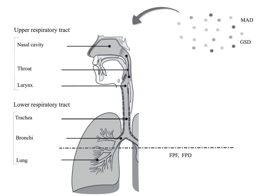
inhalers don’t need propellant liquid or gas to produce an aerosol (43). Independently of
the manner in which aerosol is formed, the main parameters characterizing the aerosol-
cloud (Fig. 2) are:
MAD – mass median aerodynamic diameter (µm)
FPF – fine particle fraction; the respirable fraction of particles with diameter < 5 µm
FPD – fine particle dose; a dose of the drug with diameter < 5 µm, dose of the
deposited drug
GSD – geometric standard deviation; particle size distribution of aerosol (41–43).
One of the most commonly used inhalers is the dry powder inhaler (DPI). The dry
powder inhaler was first used in 1948 for the inhalation of an antibiotic, penicillin (Aero-
haler by Abott®) (47). The principle of its operation consists of producing aerosol from
powder. The patient’s aspiration provides energy, producing shear forces and turbulence
that disperse the drug in the form of powder. This phenomenon is also known as deagglo
meration. Subsequently, the drug is transported to the lungs. The majority of DPIs contain
a micronized drug (size 1–5 µm) mixed with carrier particles, such as (most commonly)
lactose, but also mannitol or sorbitol that prevent aggregation (45–48). Depending on the
dosing system, DPIs can be divided into inhalers dosing a single dose of a drug (capsules
made of hydroxypropyl methylcellulose or gelatin, such as Spiriva®, HandiHaler®, Aerolizer®),
devices dosing several single doses (blister packs, Anoro Ellipta®) or multi-dose inhalers
(Foradil®) (49–53). Another highly important trait differentiation of dry powder inhalers is
Fig. 2. Parameters characterizing an aerosol.
6B. Rospond et al.: The history, current state and perspectives of aerosol therapy, Acta Pharm. 72 (2022) xxx–xxx.
Table II. Dry powder inhaler drug products
Brand name of Brand name of the Active pharmaceutical
Dose (µg)
DPI inhalator medicinal product ingredient
Aerolizer Foradil formoterol fumarate 12
(Cyklohaler) Miflonide budesonide 200, 400
Seebri Breezhaler glycopyrronium 44
Miflonide Breezhaler budesonide 200, 400,
Breezhaler
Onbrez Breezhaler indacaterol 150, 300
Ultibro Breezhaler indacaterol + glycopyrronium 85 + 43
fluticasone propionate 50, 100, 250, 500
Flixotide Dysk
fluticasone propionate + 100 + 50
Seretide Dysk
Dysk salmeterol 250 + 50
Serevent Dysk
salmeterol 500 + 50
Ventolin Dysk
salbutamol 50, 200
budesonide 100, 200, 400
Budesonide Easyhaler
budesonide + formoterol 160 + 4.5
Bufomix Easyhaler
Easyhaler fumarate dihydrate 320 + 9
Buventol Easyhaler
salbutamol 100, 200
Formoterol Easyhaler
formoterol 12
Relvar Ellipta fluticasone furoate + vilanterol 92 + 22, 184 + 22
Ellipta Incruse Ellipta umeclidinum 55
Anoro Ellipta umeclidinum + vilanterol 55 + 25
fluticasone propionate + 250 + 50
Forspiro AirFluSal Forspiro
salmeterol 500 + 50
HandiHaler Spiriva tiotropium bromide 18
beclomethasone dipropionate,
NEXThaler Fostex 100 + 6
formoterol fumarate dehydrate
Budelin Novolizer budesonide 200
Novolizer
Ventilastin Novolizer salbutamol 100
Podhaler Tobi Podhaler Tobramycin 28000
budesonide + formoterol 160 + 4.5
Spiromax DuoResp Spiromax
fumarate dehydrate 320 + 9
Twisthaler Asmanex Twisthaler mometasone furoate 400
formoterol fumarate dihydrate 4.5
Oxis Turbuhaler
budesonide 200
Turbuhaler Pulmicort Turbuhaler
budesonide + formoterol 80 + 4.5
Symbicort Turbuhaler
fumarate dehydrate 160 + 4.5
Zonda Braltus Tiotropium 10
7B. Rospond et al.: The history, current state and perspectives of aerosol therapy, Acta Pharm. 72 (2022) xxx–xxx.
the inhaler’s own resistance to the flowing air. Thus, DPIs differ in the values of aspiration
flow interval values necessary to activate the drug dose (54, 55). In these terms, two types
of dry powder inhalers can be distinguished: high resistance (optimum aspiration flow
> 60 L/min, i.a. Turbohaler®) and low resistance (aspiration flow 20–30 L min–1, i.a.
HandiHaler®, Easyhaler®) (56–59). It should be added that dry powder inhalers can admini
ster large doses of the active pharmaceutical ingredient at a level higher than 25 mg
(PulmoSphere® or Tobi Podhaler® technology) (60–62). The characteristic of substances
available with dry powder inhaler are presented in Table II.
There are many substances available with DPI inhalers. It can be divided into few
pharmacological groups:
– short-acting β-adrenoreceptor agonists (salbutamol), acting up to 6 hours, usually
used several times a day,
– long-acting β-adrenoreceptor agonists (salmeterol, formoterol), usually applied
twice daily,
– ultra-long-acting β-adrenoreceptor agonists (indacaterol, vilanterol), acting 24
hours, allowing dosing once a day,
– steroids with anti-inflammatory properties (budesonide, fluticasone, beclometha-
sone), where dose depends on the severity of the disease and the individual re-
sponse of the patient to treatment,
– short-acting muscarinic antagonist (ipratropium), which dosage depends on exacer-
bations of COPD,
– long-acting muscarinic antagonist (tiotropium, glycopyrronium) that are applied
once daily (63, 64).
Another type of inhalers, metered-dose inhalers (MDI), were introduced to medicine
at the end of the 1960s when DPI inhalers had already been known. Epinephrine (Medi-
haler Epi®, Riker Laboratories) and isoprenaline (Medihaler Iso®, Riker Laboratories) were
the first drugs administered with this device type (10). MDIs comprise an approx. 100 mL
container, in which the active substance is suspended or dissolved in a propellant, usually
hydrofluoroalkanes (HFA-134a or HFA-227). Hydrofluoralkanes replaced more harmful to
the environment chlorofluorocarbons more commonly known as the freons, which can be
found in the older device. The outflow of the container consists of a triggering and dose
metering mechanism, as well as a nozzle. Typically, the container is filled with pressurized
gas (up to > 4 atm) containing 20–500 µg of a drug at doses between 25–100 µL (65, 66). The
use of gasses that are harmless to the atmosphere, such as hydrofluoroalkanes (on the
basis of the Montreal Protocol on substances that deplete the ozone layer) has resulted in
a change to the formulation of the whole preparation. Apart from the drug and carrier gas,
it is necessary to add surfactants and elastomer materials that would seal the valve releas-
ing the drug dose (70). The use of pressurized gasses is the reason for the very high velo
city of the aerosol stream (approx. 10 m s–1) released from the container. This may result in
adverse events, primarily related to the deposition of the drug in the upper respiratory
tract (throat, larynx), and thus produce hoarseness, local fungal infections (after glucocor-
ticoid use) and the cold freon-effect; that is, spontaneous spasm of the upper airway mus-
cles (mostly of the larynx) as a result of local cooling. This effect was more perceptible in
the case of those inhalers containing freons (chlorofluorocarbons) as carrier gasses (68–70).
Research shows that over half of patients are insufficiently informed about the proper use
of inhalers (71–73). What is more, MDI misuse is observed in about 40–45 % of patients (74,
8B. Rospond et al.: The history, current state and perspectives of aerosol therapy, Acta Pharm. 72 (2022) xxx–xxx.
Table III. Pressurized metered-dose inhaler drug products
Brand name of the Active pharmaceutical
Dose Medical indication
medicinal product ingredient
Alvesco ciclesonide 80 µg, 160 µg Asthma
Atimos formoterol fumarate dihydrate 12 µg Asthma, COPD
Aspulmo, Ventolin, salbutamol sulfate 100 µg Asthma, Dyspnoea
Antrovent N tiotropium bromide 20 µg Asthma, COPD
Berotec N fenoterol hydrobromide 100 µg Asthma, COPD
fenoterol hydrobromide +
Berodual N 50 µg + 20 µg Asthma, COPD
ipratropium bromide
Budiar, Ribuspir budesonide 200 µg Asthma
fluticasone furoate + 125 µg+ 25 µg
Comboterol Asthma
salmeterol xinafoate 250 µg + 25 µg
Flixotide fluticasone propionate 50 µg, 125 µg Asthma
beclometasone dipropionate +
Fostex 100 µg + 6 µg Asthma, COPD
formoterol fumarate dihydrate
50 µg + 25 µg
fluticasone furoate +
Seretide 125 µg+ 25 µg Asthma, COPD
salmeterol xinafoate
250 µg + 25 µg
Serevent salmeterol xinafoate 25 µg Asthma, COPD
75). Typical errors include the lack of coordination between aspiration and dose applica-
tion, too shallow or rapid aspiration and the absence of a several-second holding of breath
after drug administration (76–78). Errors during drug application considerably reduce the
efficacy of therapy. These problems can be counteracted by means of proper patient educa-
tion and through the use of spacers (79–81). When used, such spacers separate the aerosol
cloud from its source, eliminating the above adverse events associated with the use of
metered-dose inhalers (85). What is more, they may result in the aerosol being suspended
for several seconds. Spacers are divided into: low-volume (i.a. Optimiser®, Syncroner®),
medium-volume (Optichamber®, Aerochamber®, Aerochamber Plus®, Nebuchamber®,
Jet-spacer®), and high-volume (Volumatic®, Fissonair®) (83–85). It has been assessed that
MDI therapy with the use of spacers is equally efficient to nebulizer therapy (89). Characte
ristics of a drug available with metered-dose inhalers were listed in Table III.
There are few drugs that are available only with MDI inhalers. To this group belong
short-acting b-agonists fenoterol, steroid ciclesonide and a drug used in exacerbations of
asthma ipratropium bromide.
A metered-dose liquid inhaler (MDLI), also known as a soft mist inhaler (SMI), is used
for the treatment of chronic obstructive pulmonary disease. In this device, an aerosol is
formed by the pressure caused by a specially designed spring mechanism and the micro-
channels in the dosing chamber. MDLI, although utilizing a pressure mechanism for dose
release, does not use carrier gasses (in contrast to MDI), and it also negates the patient’s
9B. Rospond et al.: The history, current state and perspectives of aerosol therapy, Acta Pharm. 72 (2022) xxx–xxx.
Table IV. Soft-mist inhaler drug products
Brand name of the
Active pharmaceutical ingredient Dose (µg) Medical indication
medicinal product
Spiriva Respimat tiotropium 2.5 Asthma, COPD
tiotropium bromide monohydrate +
Spiolto Respimat 2.5 + 2.5 COPD
olodaterol chloride
need for aspiration effort. Furthermore, it offers higher lung deposition and a lower dose
of the nominal drug than MDI.
During the dose application, the drug in the form of aqueous or ethanol solution is
transported under high pressure of about 250 bar through the device head, creating an
aerosol with a very high contribution of the deposited fraction (over 60 % of aerosol par-
ticles are of < 5 µm size) (87, 88). What is more, the water mist produced is released at a low
velocity, which enhances drug deposition in the lungs (92). There are only few drugs
equipped with MDLI, which are presenting in Table IV.
Nebulizers (jet, ultrasonic, mesh)
Nebulizers are used to produce aerosol composed of liquid dispersion of drug in a
gas. In contrast, inhalers usually produce solid/gas dispersion. What differentiates nebu-
lizers from inhalers is also the need for the electricity supply connection and the ability of
passive drug application that does not require aspiration coordination. Nebulization is in
particular used with small children, as well as patients who are in life-threatening condi-
tion (93). As a result of their different means of producing aerosol, they are divided into
three main subtypes: pneumatic, ultrasonic and mesh nebulizers. In terms of design, the
oldest nebulizers are jet and air-blast (pneumatic) nebulizers. A nebulizer is a term used
for the entire device, that is the compressor, head (vessel for inhalation) and mouthpiece.
The air that is compressed in the compressor is then transported through the nozzle and
head and eventually forms an aerosol that can be emitted (47, 91, 92). Theoretically, there is
potential for combining any compressor with any nebulizer head; however, these elements
should be precisely designed for one another. If these elements are not fully compatible,
the formation of an aerosol with an incorrect particle size may result. What should also be
emphasized is that the emitted aerosol has a polydisperse nature, meaning that it is char-
acterized by particles of different shapes and sizes. Unfavourable distribution of the size
of the liquid and gas suspension emitted is observed at the end of the effective aerosol
formation when drops of considerable size start to appear and the phenomenon of sputter-
ing occurs. It should be mentioned that not all of the available liquid undergoes atomiza-
tion in compressor nebulizers. A certain amount of liquid always remains in the nebulizer
head, which is not used in the nebulization process and this is referred to as dead volume
(96). This may account for up to 40 % of the initial fluid volume and primarily stems from
the design of the head (86). What is more, due to the rapid expansion of gas particles at the
end of the nozzle, a considerable solution temperature drop is observed (approx. 10–15 °C),
which may result in a bronchospasm. Therefore, for most patients with lower respiratory
tract hyperresponsiveness it is recommended to use a thermostat that would enable an
aerosol with constant temperature to be obtained (97).
10B. Rospond et al.: The history, current state and perspectives of aerosol therapy, Acta Pharm. 72 (2022) xxx–xxx.
Table V. Advantages and disadvantages of different types of inhalers and nebulizers
Type of inhaler Advantages Disadvantages
– small size – activation of the device by the
– simple construction patient’s inhalation
– acoustic and visual control of the dose taken – not all types of medication are
Dry powder
– always ready for use available
inhalers (DPI)
– no carrier gases – susceptibility to moisture
– easy to clean – cannot be used by small children,
– fixed dose amount aged less than 4 years
– requires the patient to inhale upon actuation
– easy to use
– compact size – correct inhalation technique
– multi-dose packaging required (coordination of inhalation
– resistance to weather conditions with drug application)
– reliable – significant drug deposition in the
Metered-dose upper respiratory tract
– no carrier gases
inhalers (MDI)
–high pulmonary deposition – no dose counter
– requires no effort from – “cold freon effect”
respiratory system – intended only for drugs soluble
– low speed of escaping aerosol in water or ethanol
– small size
– dose counter
– no carrier gases
– high pulmonary deposition
Metered-dose
– requires the patient to inhale upon actuation – intended only for water or ethanol
liquid inhalers
– low speed of escaping aerosol drug solutions
(MDLI)
– small size
– dose counter
– large dead volume
– continuous operation – lowering of the temperature of the
– suitable for all age groups of patients solution during aerosol therapy
Pneumatic/Jet
– proper for all drugs – device sterilization required
nebulizers
– insensitive to change of viscosity and – large starting volume of nebuliser
osmolarity of the liquid solution
(> 2 mL)
– high aerosol production capacity – only selected drugs can be used
Ultrasonic
nebulizers – high sphericality and small size of the – thermal degradation of sensitive
produced droplets drugs
– portable
– high efficiency of aerosol respiratory
fraction production
– very low dead volume – high cost
Mesh
nebulizers – work quietly – unsuitable for high–density
solutions
– low power consumption
– can be used in a horizontal position
– short inhalation time
11B. Rospond et al.: The history, current state and perspectives of aerosol therapy, Acta Pharm. 72 (2022) xxx–xxx.
Another type of these devices are ultrasonic nebulizers. The principle of their opera-
tion is based on the formation of a high-frequency acoustic wave (approx. 20,000 Hz) by
means of a piezoelectric crystal. The high-energy acoustic wave reaches the liquid creating
its waving and resulting in aerosol production (98). This is a polydisperse aerosol with a
smaller and more spheral particle size than those obtained by pneumatic nebulizers (99).
However, the generation of a high-energy wave often results in increased liquid tempera-
ture and drug inactivation (i.a. antibiotics, mucolytics). Therefore, this nebulizer type is
only used to administer physiological saline solutions (100).
One of the latest introductions to medicine are vibrating-mesh nebulizers. The aerosol
produced by these nebulizers is formed as a result of the passage of low-frequency waves
(about 100 kHz, generated by a piezoelectric crystal) through a set of multiple micronozzles,
openings (approx. 4,000) located on a common membrane (101). An undisputed advantage
of this device is the production of a high-quality aerosol (fine particles, monodispersion)
with a low initial velocity and high pulmonary deposition (99, 100). Furthermore, these
devices are very small, have low energy consumption, and can be used in the horizontal
position, without the need for a continuous power supply (99, 101–104). Vibrating-mesh
nebulizers are expensive, and their cleaning is rather inconvenient (102). The advantages
and disadvantages of the above devices are listed in the table below.
New perspectives
Nebulizers utilizing vibrating mesh technology are among the state-of-the-art com-
mercially used devices. This type of device can be divided into passive (Omron® and Res-
pironics®, their principle of operation has been discussed in section 3) and active (Airon®
and PARI®) (111). In the latter, a set of micropumps is connected to a membrane, thanks to
which the fluid is pressed through openings and transformed into an aerosol (112). Apart
from the advantages of this type discussed in section 3, special attention should be paid to
the especially high efficiency of aerosol production, reduced drug use and possibility for
performing different applications. In contrast to conventional nebulizers, vibrating mesh
nebulizers enable the administration to patients of interferon-gamma (a technology utiliz-
ing double membranes, Photo-defined aperture plate, PDAP®), aerosol application in pa-
tients subject to mechanical ventilation and in infants (Aerogen Pro® and Aerogen Solo®),
and administration of a series of other drugs, including antibiotics (Amikacin Bayer
HealthCare Pharmaceuticals®), mucolytics and prostaglandins (106, 110).
Another direction for the development of aerosol therapy seems to be the inhalation
administration of drugs, for which the application is associated with patient discomfort,
such as insulin. In 2014, FDA approved a product called Afrezza®, which is a fast-acting,
recombinant insulin, used to treat both types I and II diabetes in patients without the
pulmonary disease (asthma, COPD). It is available on the market as Afrezza Inhalation
System with an inhaler and with three cartridges which differentiates according to colour
and dosage: 4 U (blue), 8 U (green), and 12 U (yellow). Afrezza® is a form of dry powder
consisting of insulin in a form of technosphere (fumaryl diketopiperazine) microparticles
that are absorbed in the lung and rapidly reach the bloodstream. It lowers blood glucose
level faster than other injectable rapid-acting insulin (within 12–15 minutes) therefore it
can be applied at mealtime. It has a short duration of action (up to 3 hours) therefore it can
be easily combined with other types of insulin. In comparison with other inhalational
12B. Rospond et al.: The history, current state and perspectives of aerosol therapy, Acta Pharm. 72 (2022) xxx–xxx.
insulin (e.g. Exubera®), this preparation is characterized by considerably higher bioavail-
ability and a more convenient inhaler and it does not require the calculation of the dose
(mg per units/mL) (114, 115).
CONCLUSIONS
An efficient method for inhalational drug administration has been sought since an-
cient times. From antiquity to the modern era, the main raw materials used have been
plants containing tropane alkaloids (such as Datura stramonium, Hyoscyamus niger and
opium) with spasmolytic and analgesic effects. Rapid progress in aerosol therapy tech-
niques has been observed since the 18th century when the word inhaler was first used. The
invention of anaesthesia had an undoubted impact on the use of nebulization because this
marked the start of the search for devices enabling efficient anaesthesia. It should be em-
phasized that the inhalation technique is one of the safest methods for drug administra-
tion and in some cases the only efficient one. The main adverse reactions stem from incor-
rect drug application techniques or inappropriate selection of device for the patient’s
needs. Therefore, from the second half of the 20th century, from the introduction of dry
powder inhalers up to this day, the constant development of aerosol therapy methods has
been noticed. Numerous modern inhalers have been implemented in therapy, such as
metered-dose inhalers and liquid metered-dose inhalers. Moreover, vast improvement of
nebulizers has been observed. These devices enable passive drug application in a continuous
manner. It is very convenient especially for elderly people and small children. Currently,
there are jet, ultrasonic and mesh nebulizers available on the market. Mesh nebulizers
differ from other devices by producing aerosol with higher efficiency and the possibility
of administration of selected drugs (antibiotics, mucolytics and prostaglandins). The
f uture direction of aerosol therapy development seems to enable the administration of
drugs seemingly impossible to apply or those whose application is associated with patient
discomfort, such as insulin.
REFERENCES
1. A. Weiss, Symposium papers, Agron. J. 95 (2003) 1–3; https://doi.org/10.1016/j.niox.2004.07.002
2. L. Pitance, G. Reychler, T. Leal, H. Reychler, G. Liistro, J. Montharu, P. Diot and L. Vecellio, Aero-
sol delivery to the lung is more efficient using an extension with a standard jet nebulizer than an
open-vent jet nebulizer, J. Aerosol Med. Pulmon. Drug Del. 26 (2013) 208–214; https://doi.org/10.1089/
jamp.2012.0994
3. G. Crompton, A brief history of inhaled asthma therapy over the last fifty years, Prim. Care Respir. J.
15 (2006) 326–331; https://doi.org/10.1016/j.pcrj.2006.09.002
4. S. D. Anderson, Repurposing drugs as inhaled therapies in asthma, Adv. Drug Del. Rev. 133 (2018)
19–33; https://doi.org/10.1016/j.addr.2018.06.006
5. P. J. Anderson, History of aerosol therapy: Liquid nebulization to MDIs to DPIs ceramic inhalers,
Respir. Care 50 (2005) 1139–1149.
6. K. Fatur and S. Kreft, Common anticholinergic solanaceaous plants of temperate Europe – A re-
view of intoxications from the literature (1966–2018), Toxicon 177 (2020) 52–88; https://doi.
org/10.1016/j.toxicon.2020.02.005
13B. Rospond et al.: The history, current state and perspectives of aerosol therapy, Acta Pharm. 72 (2022) xxx–xxx.
7. P. Soni, A. A. Siddiqui, J. Dwivedi and V. Soni, Pharmacological properties of Datura stramonium
L. as a potential medicinal tree: An overview, Asian Pac. J. Trop. Biomed. 2 (2012) 1002–1008; https://
doi.org/10.1016/S2221-1691(13)60014-3
8. A. G. S. Pigatto, C. C. Blanco, L. A. Mentz and G. L. G. Soares, Tropane alkaloids and calystegines
as chemotaxonomic markers in the Solanaceae, Anais Acad. Brasil. Cienc. 87 (2015) 2139–2149;
https://doi.org/10.1590/0001-3765201520140231
9. P. K. Mukherjee, R. K. Harwansh, S. Bahadur, S. Banerjee, A. Kar, J. Chanda, S. Biswas, S. M.
Ahmmed and C. K. Katiyar, Development of Ayurveda – Tradition to trend, J. Ethnopharm. 197
(2017) 10–24; https://doi.org/10.1016/j.jep.2016.09.024
10. S. W. Stein and C. G. Thiel, The history of therapeutic aerosols: A chronological review, J. Aerosol
Med. Pulmon. Drug Del. 30 (2017) 20–41; https://doi.org/10.1089/jamp.2016.1297
11. S. Schläger and B. Dräger, Exploiting plant alkaloids, Curr. Opin. Biotechnol. 37 (2016) 155–164;
https://doi.org/10.1016/j.copbio.2015.12.003
12. A. Alizadeh, M. Moshiri, J. Alizadeh and M. Balali-Mood, Black henbane and its toxicity – a de-
scriptive review, Avicenna J. Phytomed. 4 (2014) 297–311; https://doi.org/10.22038/ajp.2014.3187
13. K. Fatur and S. Kreft, Common anticholinergic solanaceaous plants of temperate Europe – A re-
view of intoxications from the literature (1966–2018), Toxicon 177 (2020) 52–88; https://doi.
org/10.1016/j.toxicon.2020.02.005
14. M. Sanders, Inhalation therapy: An historical review, Prim. Care Respir. J. 16 (2007) 71–81; https://
doi.org/10.3132/pcrj.2007.00017
15. R. Arya, Ancient Indian concepts about phenomenology, biology, and therapeutics of epilepsy,
J. Hist. Neurosci. 27 (2018) 56–71; https://doi.org/10.1080/0964704X.2017.1358039
16. M. A. Martínez and S. Ballesteros, Opium poisoning in modern times. An overview, Forensic Sci.
Int. 302 (2019) 109848; https://doi.org/10.1016/j.forsciint.2019.06.006
17. K. Laios, K. Manes, M. Kontaxaki, M. Karamanou and G. Androutsos, Special Article, JAMA 87
(1926) 755; https://doi.org/10.1001/jama.1926.02680100039010
18. W. Thiesen, The letters of John Dastin, Ambix 55 (2008) 153–168; https://doi.org/10.1179/
174582308X255389
19. M. G. Soni, I. G. Carabin, J. C. Griffiths and G. A. Burdock, Safety of ephedra: Lessons learned,
Toxicol. Lett. 150 (2004) 97–110; https://doi.org/10.1016/j.toxlet.2003.07.006
20. A. E. Al-Snafi, A review of medicinal plants with broncho-dilatory effect – Part 1, Schol. Acad. J.
Pharm. 5 (2016) 297–304; https://doi.org/10.21276/sajp.2016.5.7.6
21. N. Khalil, M. Bishr, S. Desouky and O. Salama, Ammi visnaga L., a potential medicinal plant: A
review, Molecules 25 (2020) 1–18; https://doi.org/10.3390/molecules25020301
22. G. S. Mahmoudi Nezhad and B. Dalfardi, Rhazes (865–925 AD), the icon of Persian cardiology, Int.
J. Cardiol. 177 (2014) 744–747; https://doi.org/10.1016/j.ijcard.2014.11.045
23. H. D. Modanlou, Medical care of children during the golden age of Islamic medicine, Arch. Iran.
Med. 18 (2015) 263–265.
24. R. Beyar, K. Skorecki and S. Blazer, The Maimonides heritage: Discovery and propagation of
medical knowledge, Rambam Maimonides Med. J. 9 (2018) 1–3; https://doi.org/10.5041/rmmj.10340
25. B. Gesundheit, Maimonides’ appreciation for medicine, Rambam Maimonides Med. J. 2 (2011) 1–8;
https://doi.org/10.5041/rmmj.10018
26. G. Tsoucalas and M. Sgantzos, Hippocrates, on the infection of the lower respiratory tract among
the general population in ancient Greece, Gen. Med. Open Access 04 (2016) 1–5; https://doi.
org/10.4172/2327-5146.1000272
27. K. Gourd, John Mudge, Lancet Respir. Med. 4 (2016) 16–17; https://doi.org/10.1016/S2213-2600(15)00422-1
14B. Rospond et al.: The history, current state and perspectives of aerosol therapy, Acta Pharm. 72 (2022) xxx–xxx.
28. K. Nikander, C. Nicholls, J. Denyer and J. Pritchard, The evolution of spacers and valved holding
chambers, J. Aerosol Med. Pulmon. Drug Del. 27 (2014) 4–23; https://doi.org/10.1089/jamp.2013.1076
29. T. H. Levere, T. H. Dr Thomas Beddoes: chemistry, medicine, and the perils of democracy, Notes
and Records of the Royal Society of London, 63 (2009) 215–229; https://doi.org/10.1098/rsnr.2009.0032
30. D. H. Robinson and A. H. Toledo, Historical development of modern anesthesia, J. Invest. Surg. 25
(2012) 141–149; https://doi.org/10.3109/08941939.2012.690328
31. C. L. Mai and C. J. Coté, A history of pediatric anesthesia: A tale of pioneers and equipment, Paed.
Anaesthesia 22 (2012) 511–520; https://doi.org/10.1111/j.1460-9592.2012.03828.x
32. H. M. Janssens and H. A. W. M. Tiddens, Aerosol therapy: The special needs of young children,
Paediatr. Respir. Rev. 7 (2006) 83–85; https://doi.org/10.1016/j.prrv.2006.04.167
33. K. Nikander and M. Sanders, The early evolution of nebulizers, MedicaMundi 54 (2010) 47–53.
34. N. J. C. Snell, The carbolic smoke ball, Int. J. Pharm. Med. 15 (2001) 195–196; https://doi.org/10.1097/
00124363-200108000-00006
35. M. Jackson, “Divine stramonium”: The rise and fall of smoking for asthma, Med. Hist. 54 (2010)
171–194; https://doi.org/10.1017/S0025727300000235
36. C. Sorino, S. Negri, A. Spanevello, D. Visca and N. Scichilone, Inhalation therapy devices for the
treatment of obstructive lung diseases: the history of inhalers towards the ideal inhaler, Eur. J.
Inter. Med. 75 (2020) 15–18; https://doi.org/10.1016/j.ejim.2020.02.023
37. H. Chrystyn, D. B. Price, M. Molimard, J. Haughney, S. Bosnic-Anticevich, F. Lavorini, J. Efthimiou,
D. Shan, E. Sims, A. Burden, C. Hutton and N. Roche, Comparison of serious inhaler technique
errors made by device-naïve patients using three different dry powder inhalers: A randomised,
crossover, open-label study, BMC Pulmon. Med. 16 (2016) 1–14; https://doi.org/10.1186/s12890-016-
0169-5
38. L. Vecellio, The mesh nebuliser, Breathe 2 (2006) 252–260; https://doi.org/ 10.1183/18106838.0203.252
39. D. P. Tashkin, A review of nebulized drug delivery in COPD, Int. J. COPD 11 (2016) 2585–2596;
https://doi.org/10.2147/COPD.S114034
40. N. Islam and E. Gladki, Dry powder inhalers (DPIs) – A review of device reliability and innova-
tion, Int. J. Pharm. 360 (2008) 1–11; https://doi.org/10.1016/j.ijpharm.2008.04.044
41. A. S. Melani, P. Canessa, I. Coloretti, G. Deangelis, R. Detullio, M. Del Donno, R. Giacobbe, I.
Scarlato, A. Serafini, N. Barbato, A. Vaghi and P. Sestini, Inhaler mishandling is very common in
patients with chronic airflow obstruction and long-term home nebuliser use, Respir. Med. 106
(2012) 668–676; https://doi.org/10.1016/j.rmed.2011.11.016
42. M. V. C. de Oliveira, E. Pizzichini, C. H. da Costa, C. C. Fritscher, E. O. Vianna, P. J. Z. Teixeira, R.
Stirbulov, M. F. Rabahi and N. C. de Pinho, Evaluation of the preference, satisfaction and correct
use of Breezhaler® and Respimat ® inhalers in patients with chronic obstructive pulmonary dis-
ease – INHALATOR study, Respir. Med. 144 (2018) 61–67; https://doi.org/10.1016/j.rmed.2018.10.006
43. A. R. Clark, Medical aerosol inhalers: Past, present, and future, Aerosol Sci. Technol. 22 (1995)
374–391; https://doi.org/10.1080/02786829408959755
44. J. O. H. Sham, Y. Zhang, W. H. Finlay, W. H. Roa and R. Löbenberg, Formulation and characteri-
zation of spray-dried powders containing nanoparticles for aerosol delivery to the lung, Int. J.
Pharmac. 269 (2004) 457–467; https://doi.org/10.1016/j.ijpharm.2003.09.041
45. P. Sheth, S. W. Stein and P. B. Myrdal, Factors influencing aerodynamic particle size distribution
of suspension pressurized metered dose inhalers, AAPS PharmSciTech 16 (2014) 192–201; https://
doi.org/10.1208/s12249-014-0210-z
46. M. Yousefi, K. Inthavong and J. Tu, Effect of pressurized metered dose inhaler spray characteris-
tics and particle size distribution on drug delivery efficiency, J. Aerosol Med. Pulmon. Drug Del. 30
(2017) 359–372; https://doi.or g/10.1089/jamp.2016.1299
15B. Rospond et al.: The history, current state and perspectives of aerosol therapy, Acta Pharm. 72 (2022) xxx–xxx.
47. A. H. de Boer, P. Hagedoorn, M. Hoppentocht, F. Buttini, F. Grasmeijer and H. W. Frijlink, Dry
powder inhalation: past, present and future, Exp. Opin. Drug Deliv. 14 (2017) 499–512; https://doi.
org/10.1080/17425247.2016.1224846
48. M. L. Levy, W. Carroll, J. L. Izquierdo Alonso, C. Keller, F. Lavorini and L. Lehtimäki, Understand-
ing dry powder inhalers: Key technical and patient preference attributes, Advan. Ther. 36 (2019)
02547–2557; https://doi.org/10.1007/s12325-019-01066-6
49. P. Mehta, Dry Powder Inhalers: A focus on advancements in novel drug delivery systems, J. Drug
Deliv. 2016 (2016) 1–17; https://doi.org/10.1155/2016/8290963
50. M. J. Telko and A. J. Hickey, Dry powder inhaler formulation, Respir. Care 50 (2005) 1209–1227.
51. M. Ibrahim, R. Verma and L. Garcia-Contreras, Inhalation drug delivery devices: Technology
update, Med. Devic. Evid. Res. 8 (2015) 131–139; https://doi.org/10.2147/MDER.S48888
52. P. Muralidharan, D. Hayes and H. M. Mansour, Dry powder inhalers in COPD, lung inflammation
and pulmonary infections, Exp. Opin. Drug Deliv. 12 (2015) 947–962; https://doi.org/10.1517/174252
47.2015.977783
53. J. Perry, B. Trautman, J. Takher-Smith, S. Kramer, K. Kane, M. Silverman, L. Tan, S. Haughie, W.
Richter, V. Kirkov, S. Arsova, J. Ward and D. L. Hava, Particle size and gastrointestinal absorption
influence tiotropium pharmacokinetics: a pilot bioequivalence study of PUR0200 and Spiriva
HandiHaler, Br. J. Clin. Pharmacol. 85 (2019) 580–589; https://doi.org/10.1111/bcp.13831
54. A. C. Grant, R. Walker, M. Hamilton and K. Garrill, The ELLIPTA® dry powder inhaler: Design,
functionality, in vitro dosing performance and critical task compliance by patients and caregivers,
J. Aerosol Med. Pulmon. Drug Del. 28 (2015) 474–485; https://doi.org/10.1089/jamp.2015.1223
55. D. Cada, K. Ingram, J. Leonard and D. Baker, Umeclidinium bromide and vilanterol trifenatate
inhalation powder, Hosp. Pharm. 49 (2014) 554–562; https://doi.org/10.1310/hpj4906-554
56. N. J. Gross and J. F. Donohue, Nebulized formoterol: A review of clinical efficacy and safety in
COPD, Int. J. Chron. Obstr. Pulmon. Dis. 5 (2010) 223–232; https://doi.org/10.2147/copd.s11006
57. D. R. Hess, Aerosol delivery devices in the treatment of asthma, Respir. Care 53 (2008) 699–723.
58. S. Ghosh, J. A. Ohar and M. B. Drummond, Peak inspiratory flow rate in chronic obstructive pul-
monary disease: implications for dry powder inhalers, J. Aerosol Med. Pulmon. Drug Del. 30 (2017)
381–387; https://doi.org/10.1089/jamp.2017.1416
59. H. Mohammed, J. Arp, F. Chambers, M. Copley, V. Glaab, M. Hammond, D. Solomon, K. Bradford,
T. Russell, Y. Sizer, S. C. Nichols, D. L. Roberts, C. Shelton, R. Greguletz and J. P. Mitchell, Investi-
gation of Dry Powder Inhaler (DPI) resistance and aerosol dispersion timing on emitted aerosol
aerodynamic particle sizing by multistage cascade impactor when sampled volume is reduced
from compendial value of 4 L, AAPS PharmSciTech 15 (2014) 1126–1137; https://doi.org/10.1208/
s12249-014-0111-1
60. F. Lavorini, Easyhaler®: An overview of an inhaler device for day-to-day use in patients with
asthma and chronic obstructive pulmonary disease, Drugs Context 8 (2019) 1–8; https://doi.
org/10.7573/dic.212596
61. J. Haikarainen, P. Rytilä, S. Roos, S. Metsärinne and A. Happonen, Dose uniformity of budesonide
Easyhaler® under simulated real-life conditions and with low inspiration flow rates, Chron. Respir.
Dis. 15 (2018) 265–271; https://doi.org/10.1177/1479972317745733
62. H. Chrystyn, Closer to an ‘Ideal Inhaler’ with the Easyhaler: an innovative dry powder inhaler,
Clin. Drug Invest. 26 (2006) 175–183; https://doi.org/10.2165/00044011-200626040-00001
63. R. Scherließ and C. Etschmann, DPI formulations for high dose applications – Challenges and
opportunities, Int. J. Pharm. 548 (2018) 49–53; https://doi.org/10.1016/j.ijpharm.2018.06.038
64. D. R. VanDevanter and D. E. Geller, Tobramycin administered by the TOBI® Podhaler® for persons
with cystic fibrosis: A review, Med. Devices: Evid. Res. 4 (2011) 179–188; https://doi.org/10.2147/
MDER.S16360
16B. Rospond et al.: The history, current state and perspectives of aerosol therapy, Acta Pharm. 72 (2022) xxx–xxx.
65. D. E. Geller, S. Z. Nasr, S. Piggott, E. He, G. Angyalosi and M. Higgins, Tobramycin inhalation
powder in cystic fibrosis patients: Response by age group, Respir. Care 59 (2014) 388–398; https://
doi.org/10.4187/respcare.02264
66. L. Jacobson, Y. Okuda and S. A. Godwin, SimWars Simulation Case Book: Emergency Med., Cambridge
University Press, 2015 pp. 89–92; https://doi.org/10.1017/CBO9781107111011.023
67. G. M. Keating, Tiotropium bromide inhalation powder, Drugs 72 (2012) 273–300; https://doi.
org/10.2165/11208620-000000000-00000
68. S. P. Newman, Principles of metered-dose inhaler design, Respir. Care 50 (2005) 1177–1188.
69. C. Kleinstreuer, H. Shi and Z. Zhe, Computational analyses of a pressurized metered dose inhal-
er and a new drug-aerosol targeting methodology, J. Aerosol Med. 20 (2007) 294–309; https://doi.
org/10.1089/jam.2006.0617
70. N. Roche and P. N. R. Dekhuijzen, The evolution of pressurized metered-dose inhalers from early
to modern devices, J. Aerosol Med. Pulmon. Drug Del. 29 (2016) 311–327; https://doi.org/10.1089/
jamp.2015.1232
71. J. Sanchis, C. Corrigan, M. L. Levy and J. L. Viejo, Inhaler devices – From theory to practice, Respir.
Med. 107 (2013) 495–502; https://doi.org/10.1016/j.rmed.2012.12.007
72. J. Goldberg, W. Böhning, P. Schmidt and E. Freund, Fenoterol hydrobromide delivered via HFA-
MDI or CFC-MDI in patients with asthma: A safety and efficacy comparison, Respir. Med. 94 (2000)
948–953; https://doi.org/10.1053/rmed.2000.0864
73. K. J. McDonald and G. P. Martin, Transition to CFC-free metered dose inhalers – Into the new
millennium, Int. J. Pharm. 201 (2000) 89–107; https://doi.org/10.1016/S0378-5173(00)00401-4
74. M. Capanoglu, E. Dibek Misirlioglu, M. Toyran, E. Civelek and C. N. Kocabas, Evaluation of in-
haler technique, adherence to therapy and their effect on disease control among children with
asthma using metered dose or dry powder inhalers, J. Asthma 52 (2015) 838–845; https://doi.org/1
0.3109/02770903.2015.1028075
75. S. Barbara, V. Kritikos and S. B. Anticevich, Inhaler technique: Does age matter? A systematic
review, Eur. Respir. Rev. 26 (2017) 1–10; https://doi.org/10.1183/16000617.0055-2017
76. G. Huchon, P. Hofbauer, G. Cannizzaro, P. Iacono and F. Wald, Comparison of the safety of drug
delivery via HFA- and CFC-metered dose inhalers in CAO, Eur. Respir. J. 15 (2000) 663–669; https://
doi.org/10.1034/j.1399-3003.2000.15d07.x
77. R. J. Scarfone, G. A. Capraro, J. J. Zorc and H. Zhao, Demonstrated use of metered-dose inhalers
and peak flow meters by children and adolescents with acute asthma exacerbations, Arch. Pediatr.
Adol. Med. 156 (2002) 378–383; https://doi.org/10.1001/archpedi.156.4.378
78. S. L. Klijn, M. Hiligsmann, S. M. A. A. Evers, M. Román-Rodríguez, T. Van Der Molen and J. F. M.
Van Boven, Effectiveness and success factors of educational inhaler technique interventions in
asthma & COPD patients: A systematic review, NPJ Prim. Care Respir. Med. 27 (2017) 1–10; https://
doi.org/10.1038/s41533-017-0022-1
79. J. Sanchis, I. Gich and S. Pedersen, Systematic review of errors in inhaler use: Has patient tech-
nique improved over time?, Chest 150 (2016) 394–406; https://doi.org/10.1016/j.chest.2016.03.041
80. J. W. H. Kocks, H. Chrystyn, J. van der Palen, M. Thomas, L. Yates, S. H. Landis, M. T. Driessen,
M. Gokhale, R. Sharma and M. Molimard, Systematic review of association between critical errors
in inhalation and health outcomes in asthma and COPD, npj Prim. Care Respir. Med. 28 (2018) 1–6;
https://doi.org/10.1038/s41533-018-0110-x
81. H. AL-Jahdali, A. Ahmed, A. AL-Harbi, M. Khan, S. Baharoon, S. Bin Salih, R. Halwani and S.
Al-Muhsen, Improper inhaler technique is associated with poor asthma control and frequent
emergency department visits, Allergy Asthma Clin. Immunol. 9 (2013) 1–7; https://doi.org/10.1186/1710-
1492-9-8
17B. Rospond et al.: The history, current state and perspectives of aerosol therapy, Acta Pharm. 72 (2022) xxx–xxx.
82. J. P. Mitchell and M. W. Nagel, Valved holding chambers (VHCs) for use with pressurised me-
tered-dose inhalers (pMDIs): A review of causes of inconsistent medication delivery, Prim. Care
Respir. J. 16 (2007) 207–214; https://doi.org/10.3132/pcrj.2007.00034
83. M. Reznik, E. J. Silver and Y. Cao, Evaluation of MDI-spacer utilization and technique in caregiv-
ers of urban minority children with persistent asthma, J. Asthma 51 (2014) 149–154; https://doi.org
/10.3109/02770903.2013.854379
84. S. D. Scott, M. H. Osmond, K. A. O’Leary, I. D. Graham, J. Grimshaw and T. Klassen, Barriers and
supports to implementation of MDI/spacer use in nine Canadian pediatric emergency depart-
ments: A qualitative study, Implement. Sci. 4 (2009) 1–10; https://doi.org/10.1186/1748-5908-4-65
85. A. S. Melani, M. Bonavia, V. Cilenti, C. Cinti, M. Lodi, P. Martucci, M. Serra, N. Scichilone,
P. Sestini, M. Aliani and M. Neri, Inhaler mishandling remains common in real life and is
associated with reduced disease control, Respir. Med. 105 (2011) 930–938; https://doi.org/10.1016/j.
rmed.2011.01.005
86. T. R. Sosnowski, Aerozole wziewne i inhalatory, Oficyna Wydawnicza Politechniki Warszawskiej,
Warszawa 2010, pp. 73–111.
87. S. Dissanayake, M. Nagel, E. Falaschetti and J. Suggett, Are valved holding chambers (VHCs) in-
terchangeable? An in vitro evaluation of VHC equivalence, Pulmon. Pharmacol. Ther. 48 (2018)
179–184; https://doi.org/10.1016/j.pupt.2017.10.005
88. D. Singh, S. Collarini, G. Poli, D. Acerbi, A. Amadasi and A. Rusca, Effect of AeroChamber PlusTM
on the lung and systemic bioavailability of beclometasone dipropionate/formoterol pMDI, Br. J.
Clin. Pharmacol. 72 (2011) 932–939; https://doi.org/10.1111/j.1365-2125.2011.04024.x
89. C. J. Cates, E. J. Welsh and B. H. Rowe, Holding chambers (spacers) versus nebulisers for beta-
-agonist treatment of acute asthma, Cochrane Database System. Rev. 9 (2013) 1–94; https://doi.
org/10.1002/ 14651858.CD000052
90. T. Iwanaga, Y. Tohda, S. Nakamura and Y. Suga, The Respimat® soft mist inhaler: Implications of
drug delivery characteristics for patients, Clin. Drug Invest. 39 (2019) 1021–1030; https://doi.
org/10.1007/s40261-019-00835-z
91. H. A. Blair, Tiotropium/olodaterol: A review in COPD, Drugs 79 (2019) 997–1008; https://doi.
org/10.1007/s40265-019-01133-w
92. P. Brand, B. Hederer, G. Austen, H. Dewberry and T. Meyer, Higher lung deposition with Respi-
mat® Soft MistTM Inhaler than HFA-MDI in COPD patients with poor technique, Int. J. COPD 3
(2008) 763–770; https://doi.org/10.2147/COPD.S3930
93. C. Smith and R. D. Goldman, Nebulizers versus pressurized metered-dose inhalers in preschool
children with wheezing, Can. Fam. Physic. 58 (2012) 528–530.
94. A. Ari, Aerosol therapy in pulmonary critical care, Respir. Care 60 (2015) 858–874; https://doi.
org/10.4187/respcare.03790
95. M. Najlah, A. Vali, M. Taylor, B. T. Arafat, W. Ahmed, D. A. Phoenix, K. M. G. Taylor and A. Elhissi,
A study of the effects of sodium halides on the performance of air-jet and vibrating-mesh nebu-
lizers, Int. J. Pharm. 456 (2013) 520–527; https://doi.org/10.1016/j.ijpharm.2013.08.023
96. N. Collins, Nebulizer therapy in cystic fibrosis: An overview, J. Roy. Soc. Med., Suppl. 102 (2009)
11–17; https://doi.org/10.1258/jrsm.2009.s19003
97. M. Ochowiak, A. Kasperkowiak, M. Doligalski, T. R. Sosnowski, M. Matuszak, S. Włodarczak, M.
Markowska, A. Krupińska and K. Jabłczyńska, The thermostated medical jet nebulizer: Aerosol
characteristics, Int. J. Pharm. 567 (2019) 1–10; https://doi.org/10.1016/j.ijpharm.2019.118475
98. M. P. Flament, P. Leterme and A. Gayot, Study of the technological parameters of ultrasonic nebu-
lization, Drug Develop. Ind. Pharm. 27 (2001) 643–649; https://doi.org/10.1081/DDC-100107320
99. L. Broniarz-Press, T. R. Sosnowski, M. Matuszak, M. Ochowiak and K. Jabłczyńska, The effect of
shear and extensional viscosities on atomization of Newtonian and non-Newtonian fluids in
ultrasonic inhaler, Int. J. Pharm. 485 (2015) 41–49; https://doi.org/10.1016/j.ijpharm.2015.02.065
18You can also read