The Physcomitrium patens egg cell expresses several distinct epigenetic components and utilizes homologues of BONOBO genes for cell specification
←
→
Page content transcription
If your browser does not render page correctly, please read the page content below

Zurich Open Repository and
                                                                          Archive
                                                                          University of Zurich
                                                                          University Library
                                                                          Strickhofstrasse 39
                                                                          CH-8057 Zurich
                                                                          www.zora.uzh.ch

 Year: 2022

  The Physcomitrium patens egg cell expresses several distinct epigenetic
components and utilizes homologues of BONOBO genes for cell specification

    Sanchez‐Vera, Victoria ; Landberg, Katarina ; Lopez‐Obando, Mauricio ; Thelander, Mattias ;
     Lagercrantz, Ulf ; Muñoz‐Viana, Rafael ; Schmidt, Anja ; Grossniklaus, Ueli ; Sundberg, Eva

Abstract: Although land plant germ cells have received much attention, knowledge about their specifi-
cation is still limited. We thus identified transcripts enriched in egg cells of the bryophyte model species
Physcomitrium patens, compared the results with angiosperm egg cells, and selected important candidate
genes for functional analysis. We used laser-assisted microdissection to perform a cell-type-specific tran-
scriptome analysis on egg cells for comparison with available expression profiles of vegetative tissues and
male reproductive organs. We made reporter lines and knockout mutants of the two BONOBO (PbBNB)
genes and studied their role in reproduction. We observed an overlap in gene activity between bryophyte
and angiosperm egg cells, but also clear differences. Strikingly, several processes that are male-germline
specific in Arabidopsis are active in the P. patens egg cell. Among those were the moss PbBNB genes,
which control proliferation and identity of both female and male germlines. Pathways shared between
male and female germlines were most likely present in the common ancestors of land plants, besides sex-
specifying factors. A set of genes may also be involved in the switches between the diploid and haploid
moss generations. Nonangiosperm gene networks also contribute to the specification of the P. patens egg
cell.

DOI: https://doi.org/10.1111/nph.17938

Posted at the Zurich Open Repository and Archive, University of Zurich
ZORA URL: https://doi.org/10.5167/uzh-214419
Journal Article
Published Version

The following work is licensed under a Creative Commons: Attribution-NonCommercial-NoDerivatives
4.0 International (CC BY-NC-ND 4.0) License.

Originally published at:
Sanchez‐Vera, Victoria; Landberg, Katarina; Lopez‐Obando, Mauricio; Thelander, Mattias; Lagercrantz,
Ulf; Muñoz‐Viana, Rafael; Schmidt, Anja; Grossniklaus, Ueli; Sundberg, Eva (2022). The Physcomitrium
patens egg cell expresses several distinct epigenetic components and utilizes homologues of BONOBO
genes for cell specification. New Phytologist, 233(6):2614-2628.
DOI: https://doi.org/10.1111/nph.17938
Research

The Physcomitrium patens egg cell expresses several distinct
epigenetic components and utilizes homologues of BONOBO
genes for cell specification
Victoria Sanchez-Vera1* , Katarina Landberg1* , Mauricio Lopez-Obando1* , Mattias Thelander1 ,
Ulf Lagercrantz2 , Rafael Mu~
                            noz-Viana1 , Anja Schmidt3 , Ueli Grossniklaus3 and Eva Sundberg1
1
Department of Plant Biology, The Linnean Centre of Plant Biology in Uppsala, Swedish University of Agricultural Sciences, PO Box 7080, Uppsala SE-75007, Sweden; 2Department of
Ecology and Genetics, Uppsala University, Norbyv€agen 18 D, Uppsala SE-752 36, Sweden; 3Department of Plant and Microbial Biology & Zurich-Basel Plant Science Center, University of
Zurich, Zollikerstrasse 107, Zurich CH-8008, Switzerland

                                                            Summary
Author for correspondence:                                   Although land plant germ cells have received much attention, knowledge about their speci-
Eva Sundberg                                                fication is still limited. We thus identified transcripts enriched in egg cells of the bryophyte
Email: Eva.Sundberg@slu.se
                                                            model species Physcomitrium patens, compared the results with angiosperm egg cells, and
                                                            selected important candidate genes for functional analysis.
Received: 11 August 2021                                     We used laser-assisted microdissection to perform a cell-type-specific transcriptome analy-
Accepted: 14 December 2021
                                                            sis on egg cells for comparison with available expression profiles of vegetative tissues and
                                                            male reproductive organs. We made reporter lines and knockout mutants of the two
New Phytologist (2022)                                      BONOBO (PbBNB) genes and studied their role in reproduction.
doi: 10.1111/nph.17938                                       We observed an overlap in gene activity between bryophyte and angiosperm egg cells, but
                                                            also clear differences. Strikingly, several processes that are male-germline specific in Arabidop-
Key words: BONOBO, bryophytes, egg cell,                    sis are active in the P. patens egg cell. Among those were the moss PbBNB genes, which con-
gametes, moss, Physcomitrium patens,                        trol proliferation and identity of both female and male germlines.
transcriptome.                                               Pathways shared between male and female germlines were most likely present in the com-
                                                            mon ancestors of land plants, besides sex-specifying factors. A set of genes may also be
                                                            involved in the switches between the diploid and haploid moss generations. Nonangiosperm
                                                            gene networks also contribute to the specification of the P. patens egg cell.

                                                                                                  2011). A complex network including DAZ1/2, GCS1/HAP2, and
Introduction
                                                                                                  GEX2 has been suggested to act downstream of DUO1 (see
Whereas animal gametes are directly produced by meiosis, land                                     review Hackenberg & Twell, 2019). Interestingly, transcriptome
plant gametes form by mitotic divisions and reprogramming of                                      analysis of male antheridia of the bryophyte Marchantia polymor-
somatic cells in the haploid gametophyte generation. In                                           pha revealed the enrichment of transcripts homologous to
bryophytes, a sister phylum to vascular plants, the gametophytic                                  DUO1, DAZ2, GCS1/HAP2, and GEX2 (Higo et al., 2016), sug-
phase dominates the life cycle, and gametes are produced by hap-                                  gesting a conservation of their function in male germ cell devel-
loid male (antheridia) and female (archegonia) reproductive                                       opment throughout land plant evolution. This is further
organs. In the vascular plant angiosperm lineage, the gameto-                                     supported by the fact that MpDUO1 is specifically expressed in
phytic phase has become dramatically reduced and consists of                                      spermatid mother cells and spermatids, controls MpDAZ1, and
only a few cells, with both female (embryo sac) and male gameto-                                  is necessary for the production of fertile sperm cells (Higo et al.,
phytes (pollen) producing a pair of gametes (see reviews Hater                                    2018).
et al., 2020; Hafidh & Honys, 2021). Although land plant germ                                        Recently, the first candidates for egg cell specification and fate
cells have received much attention, knowledge about the genetic                                   determination were identified. The RKD TF-clade of the non-
components that specify plant germ cell identity is limited.                                      metazoan RWP-RK family appears to have a conserved function
   In the angiosperm model Arabidopsis thaliana, the MYB tran-                                    in germ cell fate regulation throughout land plant evolution. In
scription factor (TF) DUO POLLEN1 (DUO1) is required for                                          Arabidopsis, two RKD genes are preferentially expressed in the
cell cycle progression of sperm precursor cells and the promotion                                 egg cell, and ectopic AtRKD2 expression induces egg cell-like
of sperm cell differentiation (Brownfield et al., 2009; Borg et al.,                              transcription (K}oszegi et al., 2011). In addition, the single
                                                                                                  M. polymorpha RKD gene is expressed in developing egg and
*Joint first authors.                                                                             sperm precursors and maintained in the egg until fertilization

Ó 2021 The Authors                                                                                                                                        New Phytologist (2022) 1
New Phytologist Ó 2021 New Phytologist Foundation                                                                                                      www.newphytologist.com
This is an open access article under the terms of the Creative Commons Attribution-NonCommercial-NoDerivs License, which permits use and
distribution in any medium, provided the original work is properly cited, the use is non-commercial and no modifications or adaptations are made.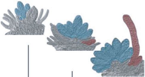
New
2 Research                                                                                                                        Phytologist

  (Koi et al., 2016; R€ovekamp et al., 2016). The egg cells of lines     instrument (MMI). Egg cells at stage 8, egg cells at stage 9, cells
  with reduced MpRKD expression failed to fully differentiate and        forming the cavity wall, and cells from the gametophore shoot
  maintain quiescence, resulting in ectopic cell divisions prior to      were harvested separately (on average, 250 cells per cell type;
  fertilization, suggesting that MpRKD may also regulate the             Fig. S1a–e). Microdissected material was stored at 80°C. RNA
  gametophyte-to-sporophyte transition by suppressing partheno-          was extracted using the Picopure RNA extraction kit from Arc-
  genesis (Koi et al., 2016; R€ovekamp et al., 2016). MpRKD also         turus (Thermo Fisher Scientific, Waltham, MA, USA) and
  affects cell division of the sperm precursors in liverwort             amplified using the SMARTer Ultra Low Input RNA kit for
  antheridia (Koi et al., 2016).                                         Sequencing v.3 (Clontech Laboratories, Mountain View, CA,
     To further our understanding of plant egg cell specification,       USA) using polyT primers and following the manufacturer’s pro-
  we used laser-assisted microdissection (LAM) for cell-type-            tocol applying 16 amplification cycles.
  specific transcriptome analysis of unfertilized egg cells of another
  bryophyte model species, the moss Physcomitrium patens. Tran-
                                                                         Library preparation, sequencing, and RNA-sequencing
  scription of genes involved in DNA and RNA-processing were
                                                                         analysis
  overrepresented in the moss egg, as was previously found in Ara-
  bidopsis (Wuest et al., 2010). Apart from genes shown to be            Libraries were created with the ThruPLEX® DNA-seq Kit (Rubi-
  important in egg cells of other plant species, we also identified      con Genomics, Ann Arbor, MI, USA) and 50 bp single-end reads
  expression of genes related to male germ cell differentiation in       (high output run SR50) were generated using a HiSeq2500
  Arabidopsis. Among those were two homologues of the class              machine using HiSeq SBS Kit v4 chemistry (Illumina, San
  VIIIa basic helix–loop–helix (bHLH) TFs BONOBO (BNB)                   Diego, CA, USA). FASTQC v.0.10.1 (Andrews, 2010) was used
  (Yamaoka et al., 2018). Knockout of PpBNB1 and PpBNB2 did              to check read quality. Reads were filtered through FASTP (Chen
  not affect reproductive organ initiation, as loss of the M. polymor-   et al., 2018) and BBDUK (Bushnell, 2014) to remove low-quality
  pha homologue does (Yamaoka et al., 2018), but disturbs prolif-        sequences, adapters, runs of polyA, polyT, and sequences match-
  eration and identity of both the female and male germlines,            ing ribosomal RNA (rRNA; using BBDUK and rRNA sequences
  suggesting that the moss BNBs play important roles in germ cell        from www.arb-silva.de). Read counts before and after filtering
  specification.                                                         are shown in Table S1. Filtered reads were pseudoaligned to
                                                                         P. patens complementary DNA sequences v.3.3 (https://
                                                                         peatmoss.online.uni-marburg.de/downloads/Annotations/P.
  Materials and Methods
                                                                         patens_v3.3/P.patens_v3.3_cDNA.fasta) and quantified per gene
                                                                         using KALLISTO (Bray et al., 2016). The resulting count table is
  Plant material and growth conditions
                                                                         available through Gene Expression Omnibus (Series XX), and
  Plant material, growth conditions, harvesting, and methods for         the corresponding reads per kilobase of transcript, per million
  crosses are described in Supporting Information Methods S1.            mapped reads (RPKM) are presented in Table S1. We used the
                                                                         zFPKM transformation (Hart et al., 2013; Ammar & Thompson,
                                                                         2021) and a cutoff of at zFPKM ≥ 3 as the threshold for biolog-
  Sample preparation and RNA extraction for RNA-
                                                                         ically relevant expression as opposed to noise.
  sequencing
                                                                            Identification of differentially expressed genes in egg compared
  Samples were prepared as described previously (Wuest et al.,           with other tissues was conducted using the glmQLFit function in
  2010; Schmidt et al., 2011; Florez-Rueda et al., 2016). Shoots         EDGER (Robinson et al., 2010). Processed read count data from
  with reproductive organs were fixed in 90% ethanol (EtOH)–             additional tissues (Table S5; Perroud et al., 2018) were kindly
  10% acetic acid on ice with 2 9 15 min of vacuum followed by           supplied by Professor Stefan Rensing. Genes with at least 0.8
  an overnight incubation at 4°C. Samples were transferred to            counts per million in at least two samples were kept for statistical
  90% EtOH and stored until embedding. Embedding was per-                analysis. To illustrate consistency of tissue replicates, we built a
  formed using the automatic system Leica ASP200 (Leica                  multidimensional scaling (MDS) plot for the top 1000 genes
  Microsystems, Heerbrugg, Switzerland): 1 h 70% EtOH, 3 9 1 h           with large fold-changes using function plotMDS from the EDGER
  90% EtOH, 3 9 1 h 100% EtOH, 2 9 1 h 100% xylol,                       package (Fig. S1f). Normalization was performed by trimmed
  1 9 15 min 100% xylol, all at room temperature, followed by            mean of M values (TMM) (Robinson & Oshlack, 2010), and a
  2 9 1 h and 1 9 3 h Paraplast X-tra at 56°C. Samples were              model contrasting egg samples (egg stage 8, egg stage 9) to all
  mounted on paraffin blocks and stored at 4°C before they were          other samples (samples from green sporophytes excluded) was
  sectioned on a Leica microtome RM2145 (Leica Microsystems)             used to find genes differentially expressed in egg cells. The P-
  to 6 µm thickness and mounted onto a drop of RNAase-free               values were adjusted for multiple testing using the Benjamini–
  MilliQ water on RNAase-free polyethylene naphthalate mem-              Hochberg false discovery rate (FDR) method (Benjamini &
  brane metal frame slides (Molecular Machines & Industries              Hochberg, 1995). The FDR was set to 0.01 if not stated other-
  (MMI), Glattbrugg, Switzerland). Slides were dried overnight at        wise. A volcano plot was produced by plotting log2(fold change)
  42°C on a heating table before they were deparaffinized in 100%        against log10(FDR) (Fig. S1g).
  xylol for 2 9 10 min at room temperature and air-dried for                Patterns of gene expression among differentially expressed genes
  10 min. Then, single cells were dissected using an SL lCut             were identified through clustering of genes with the function hclust

  New Phytologist (2022)                                                                                                     Ó 2021 The Authors
  www.newphytologist.com                                                                        New Phytologist Ó 2021 New Phytologist Foundation
New
Phytologist Research 3
in R. RPKM values for antheridia of the Reute ecotype were added later, Fig. 3c). At archegonium stage 8, the egg is specified, has
from Meyberg et al. (2020). All samples, including those of green detached from surrounding cells, but is not yet accessible for fertil-
sporophytes, were normalized using the function scale, Euclidean ization. At stage 9, the archegonium has opened, connecting the
distance calculated with the function dist, and clustering per- egg cavity to the exterior by a canal formed by degradation of a
formed using the method complete in hclust. central cell file (Landberg et al., 2013; see later, Fig. 3c). As our
For comparison with the egg transcriptome of A. thaliana, we strain of the moss ecotype Gransden is male sterile (Landberg et al.,
downloaded normalized expression values of egg, central, and 2020; Meyberg et al., 2020), we could harvest egg cells from stages
synergid cells from Song et al. (2020). 8 and 9 using single-cell LAM to study the transcriptome of the
Overrepresentation of biological functions among differen- unfertilized egg (Fig. S1). As internal controls, LAM-captured cells
tially expressed genes was performed using the TOPGO package from the shoot apex and the cavity wall were used (Fig. S1a,e).
(Alexa & Rahnenfuhrer, 2021) in R. Mapping of Owing to the detachment of the egg from the surrounding cells,
genes to gene ontology (GO) terms was taken from the annota- cross-contamination between samples was negligible.
tion file ‘P.patens_lookup_table_20181029.xlsx’ obtained from After Illumina short-read sequencing and filtering of the two
https://peatmoss.online.uni-marburg.de/ppatens_db/downloads. egg samples and that of the cell wall, between 11 million and
php. Tests were performed with TOPGO using Fisher’s exact test 22 million reads remained for mapping to the 32 458 predicted
statistic and the weight01 algorithm. Separate tests were per- P. patens genes (genome v.3.3; https://peatmoss.online.uni-
formed for (1) all genes with a fold-change > 2 comparing egg marburg.de/downloads/Annotations/P.patens_v3.3/P.patens_v3.
cells with all other samples (6622 genes); (2) the subset of those 3_cDNA.fasta). Alignment of c. 70% was achieved for all sam-
genes also expressed in A. thaliana egg cells (891 genes); (3) the ples (Table S1). To identify genes preferentially expressed in egg
subset not expressed in A. thaliana egg cells (891 genes); and (4) cells, a statistical analysis was performed that included previously
those in each of the four clusters identified in the cluster analysis published RNA-sequencing (RNA-seq) data from other P. patens
(1745, 630, 340, and 95 genes). tissues (Perroud et al., 2018; Table S5). Preliminary analyses indi-
cated a considerable overlap between egg samples and those from
the green sporophyte. Thus, we excluded this early sporophyte
Generation and verification of transgenic lines
stage from the analysis to avoid overlooking important egg genes.
The generation and verification of new transgenic lines is To identify genes preferentially expressed in egg cells, we consid-
described in Methods S2. An overview of reporter constructs is ered stage 8 and stage 9 egg cells as replicates and contrasted those
given in Fig. S2, characteristics of crispr RNAs (crRNAs) in with replicates of protonema, gametophores, leaflets, brown
guide-RNA-expressing plasmids in Table S2 and of CRISPR- sporophytes, spores, and finally our LAM samples from the cavity
CAS9 gene edited mutants in Table S3. For lists of primers, see wall and shoot apex as replicates of nonegg cell samples. An
Table S4. MDS plot visualizes the relationships among samples (MDSplot;
Fig. S1f). The proximity of replicate samples indicated low tech-
nical and biological variation and that egg stages 8 and 9 can be
Reverse transcription quantitative PCR to determine
used as replicates.
PpBNB transcript abundance
A general linear-model approach in EDGER identified differen-
Methods for reverse transcription quantitative PCR (RT-qPCR) tially expressed genes. At an FDR of 0.01, 7693 genes were found
to determine PpBNB transcript abundance is described in Meth- differentially expressed in the comparison egg cells vs nonegg
ods S3. For primer sequences, see Table S4. cells. Of those genes, 3432 were upregulated and 4261 downreg-
ulated in the egg cell (Table S6). A volcano plot shows that differ-
entially expressed genes (FDR < 0.01), with a few exceptions,
Light microscopy and Ppbnb mutant phenotyping
have an absolute fold change > 2 (Fig. S1g).
Methods for tissue harvesting and light microscopy are described To visualize expression patterns of egg-expressed genes we per-
in Methods S4. formed a cluster analysis of the 6622 genes with a fold change of
at least 2. In this analysis, we also included normalized expression
data from antheridia, obtained from Meyberg et al. (2020), and
Confocal microscopy and PpBNB reporter gene analysis
green sporophytes. Choosing four clusters, one gathered genes
Methods for tissue harvesting and confocal imaging are described highly expressed mainly in egg cells (cluster 1), two showed genes
in Methods S5. preferentially expressed in egg cells and antheridia (clusters 2 and
3), and one cluster contained genes highly expressed both in egg
cells and the green sporophyte (cluster 4; Fig. 1).
Results
Transcriptome analysis Transcripts known to be expressed in egg cells are present
in the laser-assisted microdissection egg transcriptome
Reproductive organs develop at the P. patens gametophore shoot
apex upon inductive conditions. The egg cell forms by an asym- To validate the egg transcriptome, the literature was surveyed for
metric mitotic division of a pre-egg cell (Landberg et al., 2013; see P. patens genes expressed in the egg. Those include the class-1
Ó 2021 The Authors New Phytologist (2022)
New Phytologist Ó 2021 New Phytologist Foundation www.newphytologist.comNew
4 Research Phytologist
Fig. 1 Cluster analysis of preferentially egg-expressed genes in Physcomitrium patens. Expression patterns for genes with a high expression in egg cells
(greater than two-fold compared with other tissues) visualized through cluster analysis. Expression in egg cells (Egg) was compared with gametophores
(Gam), protonema (Prot), leaf-like structures (Leaf), brown sporophyte (SpB), spores, and the LAM control that were included in statistical analysis plus
those of green sporophyte (SpG) and antheridia (Ant). See the Materials and Methods section for details. RPKM, reads per kilobase of transcript per million
mapped reads.
KNOX gene PpMKN2 and the homeobox genes PpBELL1 and M. polymorpha (K}oszegi et al., 2011; Koi et al., 2016; R€ovekamp
PpBELL2. Knock-in translational reporters of these genes are et al., 2016). The P. patens homologue of the RKD-clade,
highly active in the egg, although not restricted to it (Sakakibara PpRKD, and two additional RWP-RK genes belonging to the
et al., 2008; Horst et al., 2016). Our data show that the tran- RPW-clade were expressed in the egg but not significantly
scripts of these genes are significantly enriched in the egg enriched compared with other tissues, whereas the transcripts of
(Table S7). Egg cell activity of members of five additional TF three NLP-clade genes were (Table S7).
families (PpWOX13LA, PpCLF, PpFIE, PpLFY1, PpLFY2, Finally, four members of the EGG CELL-SPECIFIC (ECS)
PpSHI1, and PpSHI2), one auxin efflux carrier (PpPINA), and gene family encoding eukaryotic A1 aspartic endopeptidases were
one auxin biosynthesis gene (PpTARA), all expressed but not strongly upregulated in the P. patens egg cell (Table S7). Two
enriched in the egg cell (Table S7), was demonstrated previously members of this family (AtECS1 and AtECS2) are exclusively
through reporters (Tanahashi et al., 2005; Mosquna et al., 2009; expressed in the Arabidopsis egg from which they are secreted to
Okano et al., 2009; Landberg et al., 2013, 2020; Sakakibara et al., the extracellular space after fertilization to prevent polytubey
2014). Loss of function of several of these genes affects reproduc- (Sprunck et al., 2012; Yu et al., 2021).
tive development (Table S7).
In addition, PpEXO70.3d, encoding an exocyst subunit
Gene ontology analysis suggest similarities in enriched
important for cell elongation and differentiation, was recently
processes in egg cells across kingdoms
shown to be essential for normal egg cell development (Rawat
et al., 2017). Accordingly, we found this gene to be expressed We used the R package TOPGO to search for processes and func-
in the egg cell (Table S7). Moreover, we identified egg expres- tions enriched in the P. patens egg cell. Using a P-value cutoff at
sion of PpMSH2 (Table S7), encoding a central component of 0.01, we identified 15 biological process GO terms (Table 1). In
the mismatch DNA repair pathway essential for reproductive accordance with studies of Arabidopsis and human egg transcrip-
organ development as well as male and female fertility tomes Wuest et al., 2010), those include terms related to DNA
(Trouiller et al., 2006). replication, DNA repair, translation, ribosome biogenesis, chro-
We also looked for egg expression of genes belonging to the matin silencing, and histone acetylation (Table 1). We also tested
RWP-RK family, which includes members important for egg cell subsets of genes overexpressed in the moss egg for enrichment of
fate determination and differentiation in Arabidopsis and GO terms (6622 genes with fold change > 2). For genes having
New Phytologist (2022) Ó 2021 The Authors
www.newphytologist.com New Phytologist Ó 2021 New Phytologist FoundationNew
Phytologist Research 5
Table 1 Gene ontology (GO) term enrichment table showing significantly enriched biological processes among significantly overexpressed genes in the egg
cell.
GO ID Term Annotated Significant Expected Weight/Fischer
GO: 0006412 Translation 291 95 52.37 3.7e-09
GO: 0042254 Ribosome biogenesis 229 49 41.21 7.4e-07
GO: 0001510 RNA methylation 92 34 16.56 2.9e-05
GO: 0007018 Microtubule-based movement 101 37 18.18 0.00015
GO: 0080129 Proteasome core complex assembly 67 24 12.06 0.00038
GO: 0051788 Response to misfolded protein 83 27 14.94 0.00095
GO: 0043966 Histone H3 acetylation 4 4 0.72 0.00104
GO: 1903047 Mitotic cell cycle process 53 21 9.54 0.00201
GO: 0000724 Double-strand break repair via homologous recombination 32 12 5.76 0.00336
GO: 0044458 Motile cilium assembly 5 4 0.90 0.00447
GO: 0006094 Gluconeogenesis 118 33 21.24 0.00471
GO: 0006275 Regulation of DNA replication 52 16 9.36 0.00609
GO: 0009826 Unidimensional cell growth 69 17 12.42 0.00634
GO: 0006342 Chromatin silencing 36 13 6.48 0.00746
GO: 0009555 Pollen development 87 25 15.66 0.00915
The subset of genes analyzed is increased in expression at least two-fold in Physcomitrium patens egg cells compared with other tissues.
Arabidopsis homologues also expressed in egg cells (1745 genes; transcripts of AtFDM4 are enriched in the Arabidopsis egg
data obtained from Song et al. (2020)), 24 GO terms, including (Wuest et al., 2010; Sprunck et al., 2019; Song et al., 2020), it
the aforementioned, were identified (Table S8). For P. patens appears likely that DNA methylation is important to control the
genes with Arabidopsis homologues not expressed in the Ara- egg cell transcriptome in land plants by keeping transposons
bidopsis egg (891 genes), 15 biological GO terms were identified, silent and egg cells quiescent prior to fertilization. Accordingly,
including ‘membrane fusion’ and ‘microtubule-based move- transcripts of DEMETER, encoding a DNA glycosylase involved
ment’, suggesting that these processes may be specifically upregu- in DNA demethylation, were not detected in the Arabidopsis egg
lated in the P. patens egg (Table S9). Finally, we tested genes (Choi et al., 2002; Morales-Ruiz et al., 2006; Schoft et al., 2011)
from the four clusters in Fig. 1 and found that, for example, the and, in the P. patens egg, a homologous gene was strongly down-
terms ‘unidimensional cell growth’, ‘regulation of DNA replica- regulated (Table S11).
tion’, and ‘ammonium transport’ were specific to the egg (cluster Other important players in RdDM are clade III
1), whereas terms like ‘chromatin silencing’, ‘floral organ mor- ARGOUNAUTE (AGO) proteins that, in Arabidopsis, include
phogenesis’, ‘sperm individualization’, ‘microtubule-based pro- AtAGO4/6/9/8 that interact with Pol V transcripts to target DNA
cesses’, and ‘karyogamy’ were shared with antheridia (clusters 2 methylation (Mallory & Vaucheret, 2010; Matzke & Mosher,
and 3), and ‘regulation of telomere maintenance’, ‘mitotic 2014). AtAGO9 was reported to restrict megasporocyte fate to a
recombination’, ‘cell wall modification’ and ‘sporopollenin single cell in the ovule and to silence transposons in the female
biosynthetic process’ were shared with the green sporophyte gametophyte (Olmedo-Monfil et al., 2010); correspondingly, its
(cluster 4) (Table S10). transcripts and translational products were found to be enriched in
the egg (Wuest et al., 2010; Sprunck et al., 2019; Jullien et al.,
2020; Song et al., 2020). Two genes encoding clade III AGOs were
Transcripts of genes involved in RNA-based silencing
expressed in the P. patens egg, one (homologous to AtAGO9) with
mechanisms are enriched in the P. patens egg
24-fold enrichment and another (homologous to AtAGO4) with
The microRNA (miRNA) and RNA-directed DNA methylation similar expression levels as in other tissues (Table S11).
(RdDM) pathways are required for the establishment of tran- Among Arabidopsis clade I AGO proteins (AtAGO1/5/10), at
scriptionally repressive DNA methylation and include the activity least AGO1 is important for executing miRNA function to regu-
of many genes (Matzke & Mosher, 2014; Erdmann & Picard, late developmental programs and repress transposons (Mallory &
2020). Both pathways are important for the regulation of repro- Vaucheret, 2010; Matzke & Mosher, 2014). Both AGO1 and
duction in plants at different levels (Vashisht & Nodine, 2014; AGO5 are expressed in the Arabidopsis egg (Wuest et al., 2010;
Martinez & K€ohler, 2017; Gehring, 2019; Paro et al., 2021). Jullien et al., 2020; Song et al., 2020), and a clade I AGO homo-
Three highly enriched transcripts in the P. patens egg belong to a logue also showed significant expression in the P. patens stage 8 egg
family that encodes double-stranded RNA-binding proteins with (Table S11). DICER-LIKE (DCL) proteins function upstream of
conserved XH/XS domains and implicated in the regulation of the AGOs in small RNA biogenesis involved in RNA-based silenc-
RdDM at chromatin targets. These are similar to the Arabidopsis ing (Finnegan et al., 2003; Margis et al., 2006; Nagano et al.,
genes FACTOR OF DNA METHYLATION1 (AtFDM1) and 2014; Paro et al., 2021), and one member, DCL2, is preferentially
AtFDM2 (Table S11; Xie et al., 2012; Butt et al., 2014). As expressed in the Arabidopsis egg (Takanashi et al., 2011). Given
Ó 2021 The Authors New Phytologist (2022)
New Phytologist Ó 2021 New Phytologist Foundation www.newphytologist.comNew
6 Research Phytologist
this, it is not surprising that the P. patens DCL homologue et al., 2010; Julca et al., 2021), our data suggest a specific role of
PpmDCL (Coruh et al., 2015) also showed five-fold enriched ammonium transport in early land plant reproduction.
expression in the egg, although the adjusted P-value comparing
egg cells with other tissues was 0.02 (Table S11).
Unexpectedly, homologues of several genes important to
In addition, the expression of two histone-lysine N-
male reproduction in angiosperms are active in the P.
methyltransferase (HKMTase) homologues were significantly
patens egg
enriched in the moss egg (Table S11). HKMTases are implicated
in the epigenetic regulation of gene expression and catalyze the Interestingly, the GO term ‘pollen development’ was overrepre-
methylation of histone lysine (K) residues through their SET sented among egg-enriched genes (Table 1), and we identified
domains. The egg-enriched moss HKMTases are homologous to enriched expression of homologues of several genes implicated in
AtSUVH4 and AtSUVR4, and a third gene, similar to AtSUVR3, male reproductive development in Arabidopsis, primarily in the
was also expressed in the egg. Computational analysis of Ara- process of pollen and pollen wall development. These include
bidopsis HKMTases suggests that several SUVH proteins and homologues of the Arabidopsis gene MS1, encoding a PHD-
SUVR3 catalyze mono or dimethylation of H3K9 (Satish et al., finger TF expressed in the sporophytic tapetum layer surround-
2018), whereas SUVR4 trimethylates H3K9 to suppress trans- ing developing pollen, where it activates the production of pollen
poson activity in Arabidopsis (Veiseth et al., 2010). In the Ara- coat proteins required for pollen wall development and pollen
bidopsis gametophyte, the egg showed a stronger H3K9me2 viability (Wilson et al., 2001; Ito & Shinozaki, 2002; Ito et al.,
signal compared with neighboring cells, and this signal persisted 2007; Yang et al., 2007; Lu et al., 2020). Though AtMS1 is not
after fertilization (Pillot et al., 2010), suggesting that the active in the egg (Julca et al., 2021), transcripts of the two
H3K9me2 status plays a specific role in land plant egg cells. P. patens MS1 homologues (PpMS1A and PpMS1B) are signifi-
cantly enriched in the egg. As these genes are also expressed in
antheridia of the male fertile ecotype Reute, they may be involved
Homologues of genes involved in karyogamy are active in
in processes common to both male and female germlines
the P. patens egg
(Tables 2, S14; Meyberg et al., 2020). The single M. polymorpha
Our data also revealed the enrichment of transcripts homologous MS1 homologue, MpMS1, is also expressed in male spermatoge-
to genes important for female reproductive success in Arabidopsis. nous tissues (Higo et al., 2016), but its activity in female repro-
The GO term ‘karyogamy’ came up in cluster 3, mainly consisting ductive tissues has not yet been studied.
of genes highly expressed in both egg and antheridia (Table S12). In an attempt to determine if the unexpected expression of the
This includes GAMETE EXPRESSED1 (GEX1), encoding a mem- two moss MS1 homologues in the egg cell is indeed due to sense
brane protein homologous to the yeast karyogamy protein Kar5 transcripts, we analyzed how RNA-seq reads from egg cells, cavity
(Beh et al., 1997; Alandete-Saez et al., 2011; Ning et al., 2013; wall, antheridia, and gametophores mapped to these genes, as
Speijer et al., 2015; Nishikawa et al., 2020). GEX1 is localized to well as to all other genes listed in Table S14 (Fig. S3). We also
the nuclear membrane of the egg and central cell, and, upon dou- included a reference gene expressed in all four tissue types
ble fertilization, it is required for sperm nuclear fusion in both (Fig. S3a). For this reference gene, the strong overrepresentation
cells. The enriched activity of a GEX1 homologue in the moss egg of RNA-seq reads mapping only in the sense direction in the
suggests that members of this family may represent a conserved gametophore library shows that this library, unlike the others,
karyogamy factor in land plants. Three Arabidopsis genes encoding was prepared to retain strand information. However, the LAM-
BINDING IMMUNOGLOBULIN PROTEINs of the heat captured samples display a strong 30 bias in read mapping, most
shock protein 70 family are also involved in the fusion of polar likely as a combinatorial effect of partial RNA degradation and
nuclei together with their regulatory partners, ER-resident J- polyA-based messenger RNA amplification prior to library prepa-
domain-containing proteins (ERDJ3A/B) (Yamamoto et al., 2008; ration. This bias, which is also present in the antheridia library,
Maruyama et al., 2014a,b, 2015, 2020). Homologues potentially suggests that sense transcripts are formed from the reference gene
encoding a corresponding complex in moss are also active or even in all samples. Correct split mapping of reads across introns in
enriched in the moss egg cell (Table S12). many instances further supports sense rather than antisense tran-
scription. To further test our interpretation, we also analyzed the
RNA-seq reads from egg cells of genes known to express sense
Ammonium transporters are expressed in the P. patens egg
transcripts in the egg cell (i.e. the first 15 genes listed in Table S7)
One of the most strongly upregulated genes in the P. patens egg and found the expected 30 bias in read mapping (Fig. S4).
encodes a conserved membrane-bound ammonium transporter Based on the aforementioned reasoning, biased read mapping
homologous to Arabidopsis AMT2;1 (Hao et al., 2020), and the of antheridia and egg cell samples towards the annotated 30 end
transcripts of two additional homologues are also significantly of PpMS1A and PpMS1B suggests that they represent sense tran-
enriched (Table S13). Ammonium transport also came up as an scripts (Fig. S3b,c). Additional smaller clusters of egg-cell-derived
enriched GO term in cluster 1, containing genes mainly reads also mapped closer to the 50 end of PpMS1B. As several
expressed in the egg (Table S13). Although AtAMT2;1 expression alternative splice variants have been identified with exons other
is not elevated in the Arabidopsis female gametophyte (Wuest than the three in the primary transcript (Fig. S3c), it is difficult
New Phytologist (2022) Ó 2021 The Authors
www.newphytologist.com New Phytologist Ó 2021 New Phytologist FoundationNew
Phytologist Research 7
Table 2 Expression levels (transcripts per million normalized) of PpMS1, PpBNB, and homologs to TPD and DAZ genes in vegetative tissue, egg cells, and
antheridia of Physcomitrium patens.
TPD1 hom. TPD1 hom. TPD1 hom. DAZ hom.
Tissue PpMS1A PpMS1B PpBNB1 PpBNB2 Pp3c19_9830 Pp3c5_9010 Pp3c22_22416 Pp3c18_22010
Egg 15.1 9.1 4.9 11.7 133 40.8 22.5 17.7
Antheridia 10.9 13.7 8.2 13.5 5.8 9.3 25 8.9
Cavity 0.1 0 0 0 3.5 0.5 0 1.3
Shoot tip 0 0 0 0 0.1 0 2.5 0
Gametophore 0 0 0 0 1.2 2.5 3.5 5.3
to determine if these reads represent sense or low level of addi- PpDUO1B protein production is likely silenced in the egg cell.
tional antisense transcripts. Similarly, PpDUO1A is likely silenced in the gametophore, as the
Furthermore, predicted sense transcripts of three P. patens retained read strand orientation reveals antisense transcription
genes encoding TAPETUM DETERMINANT1 (TPD1)-like (Fig. S3k). Both AtDUO1 and MpDUO1 directly control the
cysteine-rich ligands (Huang et al., 2016) are significantly expression of DUO1-ACTIVATED ZINC FINGER (DAZ) genes
enriched in the egg and also expressed in antheridia (Tables 2, in the male germline (Borg et al., 2011, 2014; Higo et al., 2018),
S14; Fig. S3d–f; Meyberg et al., 2020). In Arabidopsis, TPD1 is and although PpDUO1 protein production appears repressed in
produced in, and secreted from, the microsporocytes, which are the moss egg, three P. patens DAZ homologues produce predicted
diploid precursors of the male gametophytes. Differentiation of sense transcripts there, one of which is significantly enriched
the surrounding tapetum layer depends on the perception of the (Tables 2, S14; Fig. S3l–n), suggesting a role of these components
TPD1 signal peptide via the EXCESS MICROSPOROCYTES1 in the egg.
(EMS1) receptor kinase (Canales et al., 2002; Zhao et al., 2002; In contrast to PpDUO1B, sense transcripts homologous to the
Huang et al., 2016). Although mutations in TPD1 and EMS1 BNB genes, which are important for male generative cell specifi-
have no effect on female reproductive development in Arabidop- cation in Arabidopsis, accumulated in the P. patens egg cell
sis, homologues in rice (Oryza sativa) and maize (Zea mays) are (Fig. S3o,p; Table S14). The BONOBO genes encode class VIIIa
required to limit the number of megasporocytes in the ovules, bHLH TFs (Yamaoka et al., 2018; Zhang et al., 2020). In
besides their conserved roles in anther development (Sheridan M. polymorpha, the single BNB gene MpBNB is expressed in the
et al., 1996; Nonomura et al., 2003; Zhao et al., 2008; Hong initial cells of both male and female reproductive organs, as well
et al., 2012; Wang et al., 2012). Though two EMS1-like genes as in the egg cell of immature archegonia and in sperm progeni-
produce predicted sense transcripts in the P. patens egg, none of tors (Yamaoka et al., 2018). Because Mpbnb knockout mutants
them appear significantly enriched, opening the possibility that fail to develop reproductive organs, the role of MpBNB in
the egg-produced TPD1 may be exported to activate receptors M. polymorpha gamete development could not be elucidated.
expressed elsewhere (Fig. S3g,h; Table S14). However, MpBNB could, at least partially, replace the AtBNB1
DUO1 was identified as a male-germline-specific TF in Ara- and AtBNB2 function in Arabidopsis male generative cell specifi-
bidopsis and M. polymorpha (Durbarry et al., 2005; Rotman cation (Yamaoka et al., 2018). We found that the expression of
et al., 2005; Borg et al., 2011; Higo et al., 2018). As expected, the two BNB homologues in P. patens (Yamaoka et al., 2018),
both P. patens DUO1 homologues are expressed in antheridia, here called PpBNB1 (PpbHLH081) and PpBNB2 (PpbHLH074),
but to our surprise we also found a strong enrichment of tran- are significantly enriched in the egg and that both are also
scripts of one of them, PpDUO1B (Higo et al., 2018), in the egg expressed in antheridia (Tables 2, S14; Meyberg et al., 2020). To
(Table S14; Meyberg et al., 2020). As the previous annotation of elucidate the potential role of BNB in the generative cells of
PpDUO1B to Pp3c24_11770 disappeared during a recent bryophytes, we analyzed their expression profiles and the conse-
genome curation, we used the RNA-seq reads of antheridia to quence of their loss of function.
map the PpDUO1B gene in the genomic region around the cur-
rent Pp3c24_11770V.3 gene (Fig. S3i). This revealed a gene
PpBNBs are expressed in reproductive organ apical stem
structure for PpDUO1B with four exons, the last of which coin-
cells and in the immature female and male germ cells
cides with the single exon of the predicted Pp3c24_11770V.3
gene (Fig. S3i), encoding a protein with an almost identical We could confirm that both PpBNB genes are expressed in
sequence to the uncharacterized protein XP_024363644 at the archegonia and antheridia of the P. patens Reute ecotype using
National Center for Biotechnology Information, and high simi- RT-qPCR, and we made reporter lines in the same ecotype to
larity to that of Pp3c8_16720, designated as PpDUO1A (Higo determine their expression in more detail (Figs 2, S4). The activ-
et al., 2018) (Fig. S3j). The read mapping suggests that ity of the translational knock-in PpBNB1 reporter and the tran-
PpDUO1B is producing sense transcripts in antheridia, whereas scriptional PpBNB2 reporter lines strongly overlapped during
predicted antisense transcripts are enriched in the egg cell reproductive organ development, indicating that the PpBNB
(Fig. S3i). The egg cell reads thus accumulate in the 50 end of the proteins may have redundant functions (Figs 3a,b, 4a,b). Expres-
PpDUO1B gene and also over the last intron. This suggests that sion initiated as soon as the respective reproductive organ
Ó 2021 The Authors New Phytologist (2022)
New Phytologist Ó 2021 New Phytologist Foundation www.newphytologist.comNew
8 Research Phytologist
10 dpi
(a) (b)
13 dpi Fig. 2 Relative PpBNB1 and PpBNB2
2 2 transcript abundance in Physcomitrium
16 dpi PpBNB1 patens assayed by quantitative PCR. (a)
Relative expression
Relative expression
Expression in detached shoot apices
1.5 1.5 harvested at different time points after
PpBNB1 PpBNB2 transfer to inductive conditions. Image insets
demonstrate that apices possess young
PpBNB2
1 1 antheridia (Anth.) at 10 days post-induction
(dpi), apices possess mid-stage antheridia
and young archegonia (Arch.) at 13 dpi, and
0.5 apices possess mature antheridia and mid-
0.5
stage archegonia (antheridia and archegonia
are false colored in blue and red,
respectively) at 16 dpi. (b) Expression in
0 0
isolated antheridia and archegonia bundles.
0 dpi 2 dpi 10 dpi 13 dpi 16 dpi Anth. Arch. Each data point represents an average of
three independent biological replicates, and
Shoot apices Isolated organs error bars indicate SD.
primordia were defined in the initial apical stem cells (defined in pre-egg divided into two similarly sized cells, which either
Landberg et al. (2020)) and remained in all daughter cells formed arrested or divided once more, either asymmetrically to form a
in two cell files during the first developmental stages, with the smaller UBC and a pre-egg as in WT, or into two equally sized
strongest signal in the apicalmost cells. When inner cells formed cells resembling immature canal cells. In rare cases, the pre-egg or
and further divided during stages 3–6 (stages as in Landberg et al. UBC divided longitudinally. The extra divisions blocked further
(2013)), PpBNB1/2 expression became restricted to these cells development of the inner cells; the pre-egg never matured and
and the apical tip cells. In archegonia, a total of six inner cells the extra UBC and canal cells did not degrade properly. With a
forms in a single row. The basalmost of these cells matures into few exceptions, the apical part of the neck developed more nor-
the egg cell at stages 7–9, whereas the remaining inner cells mally, the apical canal cells degraded to a large extent, and the
degrade to leave an empty canal connecting the egg to the outside apex opened. The double mutant archegonia were even further
as the organ tip opens. PpBNB1/2 remained active in the canal affected (Fig. 3f). Instead of the strict transversal inner cell divi-
cells until they degraded and, in accordance with the egg tran- sion of the WT, the Ppbnb1bnb2 inner cells divided in all direc-
scriptome data, in the pre-egg up to stage 8, but the reporter sig- tions, forming an almost globular organ without signs of a
nals faded in the mature egg cell at stage 9. The antheridial inner specified egg cell. In addition, neck development arrested in the
cells constitute the spermatogenous cell initials that undergo sper- majority of archegonia (76%, n = 33), and although some
matogenesis during stages 7–9; at the initiation of this process, showed partial neck elongation, they never fully developed and
PpBNB1/2 expression declined. always lacked a proper single-celled canal that degraded and
opened at the tip.
Though antheridia developed normally in the single mutants
Loss of PpBNB function results in aberrant inner cell
(Fig. 4c,d), the number of antheridia was reduced in Ppbnb2 and
division and gamete differentiation
even further so in Ppbnb1bnb2 double mutant lines (Table S15).
Using the highly self-fertile ecotype Reute (Hiss et al., 2017), we In addition, double mutant antheridia were severely affected
produced several independent single and double loss-of-function (Fig. 4e). Although the first cell divisions appeared normal, the
Ppbnb mutants by CRISPR-Cas9 genome editing, using crRNAs number of inner cells formed was dramatically reduced (on aver-
specifically targeting the two genes (Table S2). By sequencing the age, 1.3 cells compared with six in WT) (Landberg et al., 2020).
two loci in the resulting lines, we could identify three Ppbnb1, two The majority arrested and appeared morphologically similar to
Ppbnb2, and two Ppbnb1Ppbnb2 mutants, in which the targeted the outer vegetative cells. Occasionally, the inner cell(s) divided
gene(s) were destroyed (Table S3). As putative off-targets of all once more and, rarely, one or two of them produced a few small
crRNAs used had at least four mismatches, it is extremely unlikely and round spermatogenous cells. In contrast to the inner cell
that off-target editing events occur (Table S2; Modrzejewski et al., defects, the development of the organ’s tip and base appeared nor-
2020), and sequencing of the putative off-targets in the double mal, indicating that the regulation of these tissues is independent
mutant lines revealed no deviations from the wild-type (WT). of PpBNBs and of the progression of inner cell development.
Archegonia initiated at approximately the same time in the As expected, the Ppbnb1 and Ppbnb1bnb2 mutants did not
mutants and the WT. Whereas the Ppbnb2 single mutant develop sporophytes, and even though the reproductive organs of
archegonia were indistinguishable from the WT, some Ppbnb1 Ppbnb2 single mutants appeared normal, the frequency of sporo-
inner cells (Fig. 3c,d), most commonly the pre-egg, the upper phyte formation was clearly reduced, suggesting that gametes of
basal cell (UBC), and the two basal canal cells, underwent one or one or both sexes were affected (Table 3). When the self-sterile
two extra transverse cell divisions (Fig. 3e). Already at stage 5, the Ppbnb1 mutant was used as a sperm donor in crossings with the
New Phytologist (2022) Ó 2021 The Authors
www.newphytologist.com New Phytologist Ó 2021 New Phytologist FoundationNew
Phytologist Research 9
(a) (b)
(c) (d) (e)
(f)
Fig. 3 PpBNB expression pattern and Ppbnb mutant phenotype during Physcomitrium patens archegonial development. Expression of the (a) translational
knock-in reporter PpBNB1pro::PpBNB1-GFPGUS-1 and (b) transcriptional reporter PpBNB2pro::GFPGUS integrated in locus P108 at the stages of organ
development indicated. For each archegonium stage in (a) and (b) a merge of confocal channels detecting green fluorescent protein (green) and
chloroplast autofluorescence (magenta) is shown. (c–f) Differential interference contrast images of representative stages of (c) wild-type (WT), (d) Ppbnb2,
(e) Ppbnb1, and (f) Ppbnb1bnb2 archegonia. Organs at stages marked with a thin black line show examples of the phenotypic variation. It is not possible
to distinguish individual stages 4–6 and 7–8 in the double mutant. Numbers 1–9 indicate developmental stages as defined in Landberg et al. (2013, 2020).
Cell types are indicated in (c) as follows: A, apical canal cell; B, basal canal cell; PE, pre-egg; E, egg; UBC, upper basal cell. * and + in (e) indicate extra cell
division of the pre-egg and the basalmost canal cell, respectively. Inner cells and their borders have been traced in black for clarity at some stages. Bar,
20 µm.
Ó 2021 The Authors New Phytologist (2022)
New Phytologist Ó 2021 New Phytologist Foundation www.newphytologist.comNew
10 Research Phytologist
Table 3 Analysis of number of sporophytes formed per shoot after selfing or after crossing between indicated PpBNB mutant line and the wild-type (WT)
strains Gransden (Gd) or Reute (R) of Physcomitrium patens.
Frequency of shoots with initiated No. shoots with at least one
Female genotype Male genotype sporophyte development (%) No. shoots analyzed sporophyte formed
bnb1bnb2-1 bnb1bnb2-1 0 793 0
bnb1bnb2-2 bnb1bnb2-2 0 786 0
bnb1-1 bnb1-1 0 290 0
bnb1-2 bnb1-2 0 259 0
bnb2-1 bnb2-1 65 455 295
bnb2-2 bnb2-2 71 506 358
R WT R WT 98 300 294
Gd WT Gd WT 1 448 4
Gd WT R WT 71 48 34
Gd WT bnb1-1 42 146 62
Gd WT bnb1-2 33 75 25
bnb1-1 R WT 0 253 0
largely male sterile WT ecotype Gransden, the frequency of which is the most obvious potential function, it can also act as a
sporophytes on Gransden shoots clearly increased, although not signaling molecule to trigger physiological and morphological
as much as when the WT ecotype Reute was used as sperm changes in plants (Liu & von Wiren, 2017). In the green unicel-
donor, revealing some Ppbnb1 male fertility. However, when lular flagellated algae Chlamydomonas reinhardtii, ammonium is
adding sperm cells from the highly fertile WT ecotype Reute to used as a chemoattractant (Ermilova et al., 2007). It would be
Ppbnb1 mutants, no sporophytes formed, indicating that Ppbnb1 interesting to know if ammonium also acts as a chemoattractant
mutant egg cells are nonfunctional (Table 3). for sperm cells. Autophagy is highly active in the P. patens egg
(Sanchez-Vera et al., 2017) and may contribute to ammonium
accumulation from, for example, protein and amino acid degra-
Discussion
dation. If so, the PpAMT2;1 proteins could potentially act as
Comparisons of egg cell expression from different plant lineages exporters of ammonium out of the egg cell instead of into it.
help to highlight commonly expressed genes for the specification Strikingly, processes that are male germline specific in Arabidop-
of this key reproductive cell type. Processes shared between sis are active not only in antheridia but also in the P. patens egg.
bryophyte and angiosperm egg cells include epigenetic modifica- This may suggest that pathways shared between the two germlines
tions, DNA repair, and the activity of genes important for egg cell were present in the common ancestors of all land plants besides the
specification and function, such as the RWP-RK TFs of the RKD, sex-specifying factors. Work on volvocine algae suggests that the
RWP, and NLP clades and ECS-like endopeptidases (Wuest et al., on–off shift of a single sex-determining factor controls the gender of
2010; K}oszegi et al., 2011; Sprunck et al., 2012; Koi et al., 2016; a gamete precursor (Geng et al., 2014), revealing that a separation
R€ovekamp et al., 2016; Yu et al., 2021). In M. polymorpha, Schmid of gamete and sex determination is also theoretically possible in land
et al. (2018) showed that epigenetic reprogramming occurs by sim- plants. This assumption is supported by the fact that the germ-cell-
ilar but (compared with the sporophytic tissues) distinct processes specifying BNB gene is also active in both germlines in M. polymor-
in male and female reproductive organs. They also revealed that pha. However, unlike the MpBNB, the two moss PpBNBs are dis-
the level of DNA methylation increased in reproductive organs pensable for reproductive organ initiation from apical stem cells but
compared with the vegetative tissues from which they were derived. are needed for the continued formation of antheridia. The main
Whether there is a similar increase in the P. patens egg is not possi- function of the PpBNBs is inner cell specification, an apparent
ble to elucidate from our work, but it appears that the genetic pro- requirement for proper cell divisions and gamete differentiation.
gram driving RdDM is largely distinct from vegetative tissues. The changes in inner cell identity, and thus division, resemble that
Despite a clear overlap in gene activity between bryophyte and of auxin biosynthesis mutants: reduced division combined with
angiosperm egg cells, there are also several differences revealed by developmental arrest in antheridia and ectopic cell division com-
our transcriptome analysis. The upregulation of genes involved bined with loss of cell identity in archegonia (Landberg et al., 2020).
in, for example, microtubule-based movement and membrane Although this may not necessarily imply that auxin sensing is
fusion appears specific to the P. patens egg. This may correlate affected in Ppbnb mutant reproductive organs, it clearly demon-
with the extensive number of small vesicles with electron-dense strates the importance of strictly controlled cell division patterns for
material that form in the moss egg. These eventually fuse with proper gamete differentiation. Albeit MpBNB function at these
the plasma membrane to release the contents to the surrounding stages could not be analyzed, the fact that MpBNB is also expressed
cavity matrix to form a proper environment for the egg and the in immature egg and sperm cell progenitors (Yamaoka et al., 2018)
entering sperm cells (Sanchez-Vera et al., 2017). indicates that the BNBs have a conserved function in both male
Another process that appears specific to the moss egg cell is and female germ cell specification in bryophytes, whereas their func-
ammonium transport. Besides the nutritious role of ammonium, tion in flowering plants is restricted to the male germ cells.
New Phytologist (2022) Ó 2021 The Authors
www.newphytologist.com New Phytologist Ó 2021 New Phytologist FoundationNew
Phytologist Research 11
(a) (b)
(d)
(c)
(e)
Fig. 4 PpBNB expression and mutant phenotype relating to antheridia development in Physcomitrium patens. Expression of the (a) translational knock-in
reporter PpBNB1pro::PpBNB1-GFPGUS-1 and (b) transcriptional reporter PpBNB2pro::GFPGUS integrated in locus P108 at indicated stages of antheridia
development. The images in (a) and (b) are a merge of confocal channels detecting green fluorescent protein (green) and chloroplast autofluorescence
(magenta). (c) Differential interference contrast (DIC) images of representative stages from wild-type (WT) antheridial development. (d) DIC images of
stage 9 antheridia of the single mutants Ppbnb1 and Ppbnb2. (e) DIC images of representative organs from Ppbnb1bnb2 double mutant. For stages 3–9,
three organs are shown exemplifying the phenotypic variation. It is not possible to distinguish individual stages 4–6 and 7–8 in the double mutant.
Numbers 0–9 indicate antheridia stages according to Landberg et al. (2013, 2020). In (c, e) the inner cells have been traced in black for clarity. Bar, 20 µm.
In (a) the left scale bar represents stages 0–3 and the right scale bar represents stages 4–6.
A subset of the genes upregulated in the P. patens egg is also Therefore, the transcriptome data suggest that nonangiosperm
highly active in the green sporophyte stage. We know little about gene networks widely contribute to the specification and differen-
cell-specific expression patterns in the embryo/sporophyte, but it is tiation of the P. patens egg. This is not surprising given the dis-
tempting to speculate that several of these genes are involved in the tinct environment in which the egg cell develops and the distinct
preparation of upcoming phase changes: the transitions between mechanisms of fertilization (Sharma et al., 2021).
the generations of the plant life cycle from diploid to haploid in
the sporophyte and from haploid to diploid in the egg.
Acknowledgements
It is important to note that the proportion of largely egg-
specific genes (the 524 genes with a fold change > 256) without This work was supported by grants from the Knut and Alice
clear homology to Arabidopsis genes reached more than 72%, Wallenberg Foundation (KAW; 2012.0087 to ES), the
whereas the corresponding figure for all genes was only 31%. Swedish Research Council (VR; 621-2014-4941; 2018-04068
Ó 2021 The Authors New Phytologist (2022)
New Phytologist Ó 2021 New Phytologist Foundation www.newphytologist.comNew
12 Research Phytologist
to ES), the University of Zurich, and a grant from the Beh CT, Brizzio V, Rose MD. 1997. KAR5 encodes a novel pheromone-
‘Staatssekretariat f€
ur Bildung und Forschung’ in the framework inducible protein required for homotypic nuclear fusion. Journal of Cell Biology
139: 1063–1076.
of COST action FA0903 (to UG and AS). Sequencing was Benjamini Y, Hochberg Y. 1995. Controlling the false discovery rate: a practical
performed by the SNP&SEQ Technology Platform in Upp- and powerful approach to multiple testing. Journal of the Royal Statistical
sala, which is part of the National Genomics Infrastructure Society. Series B: Methodological 57: 289–300.
(NGI) Sweden and Science for Life Laboratory. The platform Borg M, Brownfield L, Khatab H, Sidorova A, Lingaya M, Twell D. 2011. The
is also supported by VR and KAW. We thank Dr German R2R3 MYB transcription factor DUO1 activates a male germline-specific
regulon essential for sperm cell differentiation in Arabidopsis. Plant Cell 23:
Martinez Arias and two anonymous reviewers for helpful com- 534–549.
ments on the manuscript and Prof. Stefan Rensing for kindly Borg M, Rutley N, Kagale S, Hamamura Y, Gherghinoiu M, Kumar S, Sari U,
sharing RNA-seq data. Esparza-Franco MA, Sakamoto W, Rozwadowski K et al. 2014. An EAR-
dependent regulatory module promotes male germ cell division and sperm
fertility in Arabidopsis. Plant Cell 26: 2098–2113.
Author contributions Bray NL, Pimentel H, Melsted P, Pachter L. 2016. Near-optimal probabilistic
RNA-seq quantification. Nature Biotechnology 34: 525–527.
VS-V, KL, ML-O and MT conducted the experiments and ana- Brownfield L, Hafidh S, Borg M, Sidorova A, Mori T, Twell D. 2009. A plant
lyzed the data together with RM-V, UL and ES; VS-V, AS, UG, germline-specific integrator of sperm specification and cell cycle progression.
ML-O, KL, MT and ES designed the experiments; ES and UG PLoS Genetics 5: e1000430.
provided materials and resources; VS-V, ML-O, KL, MT, UL Bushnell B. 2014. BBMap: a fast, accurate, splice-aware aligner. https://
sourceforge.net/projects/bbmap/ [accessed 3 February 2014].
and ES interpreted the results and wrote the manuscript. All Butt H, Graner S, Luschnig C. 2014. Expression analysis of Arabidopsis XH/XS
authors reviewed and commented on the manuscript. VS-V, KL domain proteins indicates overlapping and distinct functions for members of
and ML-O have shared first authorship. this gene family. Journal of Experimental Botany 65: 1217–1227.
Canales C, Bhatt AM, Scott R, Dickinson H. 2002. EXS, a putative LRR
receptor kinase, regulates male germline cell number and tapetal identity and
ORCID promotes seed development in Arabidopsis. Current Biology 12: 1718–27.
Chen S, Zhou Y, Chen Y, Gu J. 2018. FASTP: an ultra-fast all-in-one FASTQ
Ueli Grossniklaus https://orcid.org/0000-0002-0522-8974 preprocessor. Bioinformatics 34: i884–i890.
Ulf Lagercrantz https://orcid.org/0000-0003-2440-0677 Choi YH, Gehring M, Johnson L, Hannon M, Harada JJ, Goldberg RB,
Katarina Landberg https://orcid.org/0000-0002-2945-8571 Jacobsen SE, Fisher RL. 2002. DEMETER, a DNA glycosylase domain
Mauricio Lopez-Obando https://orcid.org/0000-0002-1380- protein, is required for endosperm gene imprinting and seed viability in
Arabidopsis. Cell 110: 33–42.
0643 Coruh C, Cho SH, Shahid S, Liu Q, Wierzbicki A, Axtell MJ. 2015.
Rafael Mu~ noz-Viana https://orcid.org/0000-0002-1363-6978 Comprehensive annotation of Physcomitrella patens small RNA loci reveals that
Victoria Sanchez-Vera https://orcid.org/0000-0001-8615- the heterochromatic short interfering RNA pathway is largely conserved in land
5270 plants. Plant Cell 27: 2148–2162.
Anja Schmidt https://orcid.org/0000-0002-3276-3243 Durbarry A, Vizir I, Twell D. 2005. Male germ line development in Arabidopsis.
duo pollen mutants reveal gametophytic regulators of generative cell cycle
Eva Sundberg https://orcid.org/0000-0003-4228-434X progression. Plant Physiology 137: 297–307.
Mattias Thelander https://orcid.org/0000-0002-6663-7405 Erdmann RM, Picard CL. 2020. RNA-directed DNA methylation. PLoS Genetics
16: e1009034.
Ermilova EV, Nikitin MM, Ferna ndez E. 2007. Chemotaxis to ammonium/
Data availability methylammonium in Chlamydomonas reinhardtii: the role of transport systems
for ammonium/methylammonium. Planta 226: 1323–1332.
The data that support the findings of this study are available Finnegan J, Margis R, Waterhouse PM. 2003. Posttranscriptional gene silencing
from the corresponding author upon reasonable request. is not compromised in the Arabidopsis CARPEL FACTORY (DICER-LIKE1)
The RNA-seq data are openly available in the Gene Expression mutant, a homolog of Dicer-1 from Drosophila. Current Biology 13: 236–240.
Omnibus at https://www.ncbi.nlm.nih.gov/geo, reference num- Florez Rueda AM, Grossniklaus U, Schmidt A. 2016. Laser-assisted
ber GSE182112. microdissection (LAM) as a tool for transcriptional profiling of individual cell
types. Journal of Visualized Experiments 10: e53916.
Gehring M. 2019. Epigenetic dynamics during flowering plant reproduction:
References evidence for reprogramming? New Phytologist 224: 91–96.
Geng S, De Hoff P, Umen JG. 2014. Evolution of sexes from an ancestral
Alandete-Saez M, Ron M, Leiboff S, McCormick S. 2011. Arabidopsis thaliana mating-type specification pathway. PLoS Biology 12: e1001904.
GEX1 has dual functions in gametophyte development and early Hackenberg D, Twell D. 2019. The evolution and patterning of male
embryogenesis. The Plant Journal 68: 620–632. gametophyte development. Current Topics in Developmental Biology 131: 257–
Alexa A, Rahnenfuhrer J. 2021. TOPGO: enrichment analysis for gene ontology. R 298.
package v.2.44.0. [WWW document] URL https://bioconductor.org/packages/ Hafidh S, Honys D. 2021. Reproduction multitasking: the male gametophyte.
release/bioc/html/topGO.html [accessed 28 April 2021]. Annual Review of Plant Biology 72: 581–614.
Ammar R, Thompson J. 2021. ZFPKM: a suite of functions to facilitate zFPKM Hao DL, Zhou JY, Yang SY, Qi W, Yang KJ, Su YH. 2020. Function and
transformations. R package v.1.14.0. [WWW document] URL https://github. regulation of ammonium transporters in plants. International Journal of
com/ronammar/zFPKM/ [accessed 28 April 2021]. Molecular Science 18: e3557.
Andrews S. 2010. FASTQC: a quality control tool for high throughput sequence data. Hart T, Komori HK, LaMere S, Podshivalova K, Salomon DR. 2013. Finding
[WWW document] URL http://www.bioinformatics.babraham.ac.uk/projects/ the active genes in deep RNA-seq gene expression studies. BMC Genomics 14:
fastqc [accessed 29 April 2021]. e778.
New Phytologist (2022) Ó 2021 The Authors
www.newphytologist.com New Phytologist Ó 2021 New Phytologist FoundationYou can also read