Epstein-Barr Virus in Inborn Immunodeficiency-More Than Infection
←
→
Page content transcription
If your browser does not render page correctly, please read the page content below
cancers
Review
Epstein–Barr Virus in Inborn Immunodeficiency—More
Than Infection
Ciro Novaes Rosa Lino and Sujal Ghosh *

                                          Department of Pediatric Oncology, Hematology and Clinical Immunology, Center of Child and Adolescent
                                          Health, Medical Faculty, Heinrich-Heine-University, 40225 Duesseldorf, Germany;
                                          ciro.novaesrosalino@med.uni-duesseldorf.de
                                          * Correspondence: sujal.ghosh@med.uni-duesseldorf.de; Tel.: +49-211-811-6224; Fax: +49-211-811-6191

                                          Simple Summary: Epstein–Barr Virus (EBV) is a common virus that is readily controlled by a healthy
                                          immune system and rarely causes serious problems in infected people. However, patients with
                                          certain genetic defects of their immune system might have difficulties controlling EBV and often
                                          develop severe and life-threatening conditions, such as severe inflammation and malignancies. In
                                          this review, we provide a summary of inherited immune diseases that lead to a high susceptibility to
                                          EBV infection and discuss how this infection is associated with cancer development.

                                          Abstract: Epstein–Barr Virus (EBV) is a ubiquitous virus affecting more than 90% of the world’s
                                          population. Upon infection, it establishes latency in B cells. It is a rather benign virus for immune-
                                          competent individuals, in whom infections usually go unnoticed. Nevertheless, EBV has been
                                          extensively associated with tumorigenesis. Patients suffering from certain inborn errors of immunity
                                are at high risk of developing malignancies, while infection in the majority of immune-competent in-
                                   dividuals does not seem to lead to immune dysregulation. Herein, we discuss how inborn mutations
Citation: Lino, C.N.R.; Ghosh, S.         in TNFRSF9, CD27, CD70, CORO1A, CTPS1, ITK, MAGT1, RASGRP1, STK4, CARMIL2, SH2D1A, and
Epstein–Barr Virus in Inborn              XIAP affect the development, differentiation, and function of key factors involved in the immunity
Immunodeficiency—More Than                against EBV, leading to increased susceptibility to lymphoproliferative disease and lymphoma.
Infection. Cancers 2021, 13, 4752.
https://doi.org/10.3390/                  Keywords: Epstein–Barr Virus; EBV; inborn errors of immunity; cancer; lymphoma; immunodefi-
cancers13194752                           ciency

Academic Editors: Lorenzo Leoncini
and Lucia Mundo

                                          1. Introduction
Received: 8 August 2021
Accepted: 6 September 2021                      Epstein–Barr Virus (EBV) is a gammaherpesvirus with a prevalence of over 90% in
Published: 23 September 2021              the adult population. In immune-competent patients, EBV establishes a life-long latent
                                          infection [1–3]. Most individuals are infected during childhood with few or no overt
Publisher’s Note: MDPI stays neutral      symptoms. Adolescents and young adults usually develop infectious mononucleosis (IM),
with regard to jurisdictional claims in   a self-limiting illness with fever, sore throat, lymphadenopathy, hepatosplenomegaly, and
published maps and institutional affil-   fatigue, caused by acute inflammation and hyperactivation of CD8+ T cells [4,5].
iations.                                        Primary EBV infection occurs mainly through the oropharyngeal epithelium transmit-
                                          ted by saliva [6]. The lytic infection of the epithelium is followed by a high tropism of the
                                          virus towards B cells in which it switches to its latent program. Naïve B cells are driven by
                                          EBV into full latency (stage III, during which all latency genes are expressed Epstein–Barr
Copyright: © 2021 by the authors.         nuclear antigen (EBNA)-1, 2, 3A, 3B, 3C, and LP, Latent membrane protein (LMP)-1, 2A
Licensee MDPI, Basel, Switzerland.        and 2B, EBV-encoded small RNAs (EBERs), and Bam-HI A rightward transcripts (BARTs)).
This article is an open access article    During further progression, EBV gradually reduces the number of encoded genes. Naïve
distributed under the terms and           B cells migrate to the germinal center and undergo further expansion. At the germinal
conditions of the Creative Commons        center stage, B cells show a restricted gene expression profile (EBNA-1, LMP-1, 2A and
Attribution (CC BY) license (https://     2B, EBERs, and BARTs), known as latency II, mediating survival and differentiation of
creativecommons.org/licenses/by/          EBV-infected B cells into memory cells. Finally, EBV-infected memory B cells, the site
4.0/).

Cancers 2021, 13, 4752. https://doi.org/10.3390/cancers13194752                                           https://www.mdpi.com/journal/cancers
Cancers 2021, 13, 4752 2 of 16
of virus persistence, further restrict their expression program to EBERs and BARTs only
(latency 0), or additionally EBNA-1 (latency I) during homeostatic proliferation.
Occasionally, EBV turns to its lytic program in plasma cells, leading to the produc-
tion of new virions, repeated epithelial infection, and shedding of viral particles into the
saliva [7–10]. Viral antigens expressed by EBV during its lytic and latent stages are highly
immunogenic and induce a strong response against infected cells. Hence, the downreg-
ulation of such molecules is essential to escape immune surveillance and provide virus
persistence [11–13].
Natural killer (NK) and T cells play a major role in controlling EBV. Viral infection
decreases MHC class I expression, but natural killer (NK) cells can recognize this state and
destroy the cells. EBV lytic infection causes suppression of MHC class I expression and
induction of expression of CD112 and UL16 binding protein 1, NK cell activation receptor
ligands [14]. Thus, lytic infected cells are eliminated by NK cells, but most EBV infections
evade NK cell attack by shifting to latent infection [14–16]. In humanized mice, which were
challenged with EBV, depletion of NK cells caused exacerbated IM symptoms, with higher
viral loads, larger spleens, increased weight loss, and more tumor burden [15].
Cytotoxic CD8+ T cell responses play an even bigger part in the immune response to
EBV, addressing both lytic and latent stages of infection [17]. During IM, EBV-specific CD8+
T cells targeting mainly lytic proteins can expand up to 50% of the circulating CD8+ T cell
pool. [18,19]. CD4+ T cells recognize a variety of EBV epitopes; however, their expansion
is much less [20,21]. Interestingly, some CD4+ T cells develop a cytotoxic phenotype,
with expression of granzyme and perforin, and are able to lyse lymphoblastoid cell lines
(LCLs) and EBV loaded peripheral blood mononuclear cells (PBMCs) [22–24]. Recently,
the impact of γδ and natural killer T (NKT) cells on immunity against EBV could be
partially delineated. A comprehensive review of the T cell response to EBV, including
unconventional populations, was conducted by Long et al. [25].
The importance of T cells to control EBV can be observed in several conditions in
which effector cells are compromised, such as aging, human immunodeficiency virus
(HIV) infection, transplantation, or as reviewed here, inborn errors. In those individuals,
persistent reactivation and proliferation of EBV-infected cells are associated with severe
pathologies that can have lethal outcomes [26–29]. In this review, we will discuss genetic
diseases, which lead to uncontrolled EBV-associated immune dysregulation.
2. Inborn Errors of Immunity (IEI)
IEI (also known as primary immunodeficiencies) are a heterogeneous group of dis-
eases, in which patients manifest with increased susceptibility to infections or other im-
munological disturbances such as autoimmunity, autoinflammation, or immune dysregu-
lation [30]. These conditions result from germline mutations affecting the development,
differentiation, and/or function of the immune system. More than 430 genes have been
associated with specific diseases; due to next-generation sequencing technologies, this
number is constantly growing [30]. IEI are expected to affect 1/1000 to 1/5000 births [31].
Interestingly, while many IEI show a broad susceptibility to several pathogens includ-
ing EBV, few have a restricted vulnerability to EBV only [17]. Mutations in genes involving
non-redundant mechanisms of immunity against EBV lead to this EBV predisposition
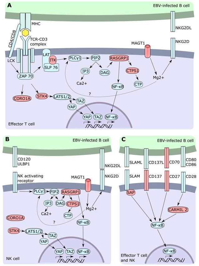
syndrome (Figure 1).
Although EBV has been associated with various malignancies [32–34], a healthy
immune system is usually capable of controlling the infection. Most individuals remain
asymptomatic, and EBV-associated cancer in immunocompetent individuals is relatively
rare [35]. Disturbances of host immunity can tilt this balance to favor the virus, allowing
its full oncogenic potential. Besides the persistent inflammatory environment caused by
EBV viremia and the expression of oncogenic EBV proteins and nucleic acids, there is an
inability to kill transformed cells due to defects in cytotoxicity in certain IEI [36]. In the IEI
discussed below, the mechanisms involved in the immunity against EBV are dysfunctional
leading to immune dysregulation and malignancies.Cancers 2021, 13, 4752 3 of 16
Cancers 2021, 13, x FOR PEER REVIEW 3 of 17
Figure
Figure 1. T cell and NK cell signaling
signaling following
following EBV-infected cell
cell recognition.
recognition. Cascade associated
with
with TCR
TCR (A),
(A), NK
NK activating
activating receptor
receptor (B),
(B), and
and co-stimulatory (C) stimulation.
co-stimulatory (C) stimulation. Red
Red color
color describes
describes aa
gene
gene with
with mutations
mutations associated
associated with
with EBV.
EBV.
3. CD27-CD70
Although EBV Deficiency
has been associated with various malignancies [32–34], a healthy im-
muneCD27 system
is a is usually capable
co-stimulatory of controlling
receptor the infection.inMost
expressed constitutively individuals
a variety remain
of lymphocytes,
asymptomatic,
such as NK andand EBV-associated
T cells [37]. It bindscancer
to CD70in resulting
immunocompetent individuals
in nuclear factor is relatively
kappa-light chain-
rare [35]. Disturbances
enhancer of activated Bofcells
host(NF-kB)
immunity can tiltactivation
pathway this balance to CD70,
[38]. favor the virus,
on the allowing
other hand,
its full oncogenic potential. Besides the persistent inflammatory environment
is only transiently inducible upon stimulation on T, B, NK, and dendritic cells [39–42]. caused by
EBV viremia
However, andinfection
upon the expression of oncogenic
with EBV, EBV proteinson
CD70 is upregulated and nucleic
B cells acids,
[43]. High there is an
levels of
inability
CD70 aretoalsokill observed
transformed in Bcells
anddue to defects
T cell lymphomain cytotoxicity
and manyinsolid
certain IEI [36].
tumors [44].InMurine
the IEI
discussed
models have below,
shed the
lightmechanisms involved
on the CD27–CD70 in the immunity
interaction. against EBV induces
CD27 co-stimulation are dysfunc-
T cell
tional leading to immune dysregulation and malignancies.Cancers 2021, 13, 4752 4 of 16
development [45], increases CD8+ T cell activation [46–48] and cell survival, and further
contributes to the differentiation of CD8+ T cells into memory cells [37,47,49].
Hypogammaglobulinemia clinical features of CD27- and CD70-deficient patients
mainly result from EBV-associated immune dysregulation. They present with severe IM,
lymphoproliferative disease (LPD), lymphoma, and hemophagocytic lymphohistiocytosis
(HLH). In a retrospective study, nearly half (11/21) of CD70-deficient and 36% (12/33) of
CD27-deficient patients developed lymphomas, with Hodgkin’s lymphoma (HL) being
the most common malignancy (Table 1) [50,51]. The exact mechanisms involved in the
defective immune response are still unknown. However, T cells from those patients show
an altered phenotype, decreased EBV-specific expansion, and reduced cytotoxicity towards
EBV-transformed B cells [43,50,52]. Additionally, CD27 and CD70 might play an important
role in immune control of malignancies, even irrespective of EBV infection. In fact, the
axis has the potential to induce expansion of effector T cells, break tolerance, and activate
response in non-immunogenic tumors; several drugs—such as Varlilumab, SGN-CD70,
SGN-7, and MDX-1203—targeting the CD27–CD70 axis are currently being tested in cancer
therapy [44,53].
Table 1. Inborn errors of immunity with high EBV susceptibility and disease.
Number of Types and Number of Malignancies
Gene Common Clinical Features
Reported Cases in Reported IEI Patients
EBV viremia, LPD, HLH
HL (9/33) [50]
Recurrent infections
BL (1/33) [50]
CD27 Autoimmunity 33 [50]
NHL (1/33) [50]
Lymphadenopathy
DLBCL (3/33) [50]
Hypogammaglobulinemia
EBV viremia, LPD, HLH
Recurrent infections HL (9/21) [50,51]
CD70 Autoimmunity 21 [50,51] BL (1/21) [50]
Lymphadenopathy NHL (1/21) [51]
Hypogammaglobulinemia
HL (8/21) [56,57]
EBV viremia, LPD HL-like (2/21) [57]
Recurrent infections DLBCL (2/21) [57,58]
ITK CD4+ T cell lymphopenia 21 [54–58] DLBCL-like (1/21) [57]
↓ iNKT BL (1/21) [57]
Hypogammaglobulinemia NHL (1/21) [57]
SMT (1/21) [57]
HL (7/37) [59,60]
Chronic EBV viremia, LPD
BL (2/37) [59,60]
Recurrent infections
Unclassified lymphoma (1/37) [59,60]
↓ NKG2D
MAGT1 37 [59,60] Liposarcoma (1/37) [59,60]
Inverted CD4+ :CD8+ ratio
DLBCL (1/37) [59,60]
Hypogammaglobulinemia
EMZL (1/37) [59,60]
Autoimmunity
Kaposi sarcoma (1/37) [59,60]
EBV viremia, LPD
HL (2/8) [62,63]
TNFRSF9 Recurrent infections
8 [61–63] DLBCL (1/8) [63]
(4-1BB/CD137) Lymphadenopathy
BL (1/8) [62]
Hypogammaglobulinemia
DLBCL (3/10) [67,68]
EBV viremia, LPD
Unclassified lymphoma (1/10) [65]
CORO1A Recurrent infections 10 [64–69]
Intracranial B cell lymphoma (1/10)
Lymphopenia
[69]Cancers 2021, 13, 4752 5 of 16
Table 1. Cont.
Number of Types and Number of Malignancies
Gene Common Clinical Features
Reported Cases in Reported IEI Patients
HL (2/29) [78,80]
DLBCL (1/29) [72]
EBV viremia, LPD
STK4 29 [69–80] BL (1/29) [79]
Recurrent infections
NHL (1/29) [80]
PCTL (1/29) [81]
EBV viremia, LPD
CARMIL2 Recurrent infections 44 [82–95] SMT (8/44) [84–86,89]
Inflammatory bowel disease
EBV viremia, LPD
B-NHL (2/28) [98]
CTPS1 Recurrent infections 28 [96–100]
CNSL (12/28) [100]
Hypogammaglobulinemia
DLBCL (2/9) [101]
EBV viremia, LPD
HL (2/9) [104]
Recurrent infections
low grade unclassified lymphoma (1/9)
RASGRP1 CD4+ T cell lymphopenia 9 [101–105]
[105]
Lymphadenopathy
PBCL (1/9) [103]
Autoimmunity
SMT (1/9) [104]
Total lymphomas (25–30%) [106,107]
EBV viremia, LPD, HLH DLBCL 30–40% [106,107]
SH2D1A (XLP1) >100 [106,107]
Hypogammaglobulinemia BL 40–60% [106,107]
NHL 20–30% [106,107]
HL, Hodgkin’s lymphoma; BL, Burkitt’s lymphoma; DLBCL, diffuse large B-cell lymphoma; EMZL, extranodal subtype of marginal
zone lymphoma; PBCL, polymorphic B-cell lymphoma; NHL, unclassified B cell non-Hodgkin lymphoma; CNSL, central nervous
system lymphomas; SMT, smooth muscle tumor; PCTL, primary cardiac T cell lymphoma; LPD, lymphoproliferative disease; HLH,
hemophagocytic lymphohistiocytosis.
4. CD137 (TNFRSF9, 4-1BB) Deficiency
CD137 (also known as 4-1BB and TNFRSF9) shows many similarities with CD27. Both
receptors are part of the TNFR superfamily and act as co-stimulatory receptors, increasing
T cell proliferation, survival, cytokine production, and cytotoxicity. Unlike CD27, which is
constitutively expressed by resting T cells, CD137 is induced after cell activation [47,108].
The expression of those receptors at different stages could explain why CD27 engagement
favors the formation of effector T cells, while CD137 induces a more robust long-term
immunity and secondary response [46]. CD137 ligand is expressed by dendritic cells,
macrophages, and activated T and B cells, including EBV-infected B cells [47,108].
EBV-specific T cells from CD137-deficient patients presented lower interferon (IFN)-γ
and perforin expression and showed impaired expansion in response to EBV-infected
B cells compared to healthy cells. Similar results were also observed following CD137
blockage in T cells from healthy donors, highlighting its non-redundant role in the immune
response against EBV [62,63]. Susceptibility to EBV was a common clinical feature among
the patients described. Chronic EBV viremia. EBV-associated HLH and lymphoma were
present in the majority of patients (Table 1) [61–63]. Interestingly, Rodriguez et al. suggested
an incomplete clinical penetrance in one of two siblings described. Though both were
carrying the same mutation in TNFRSF9 and were EBV viremic, only one sibling developed
symptoms. Importantly, specific CD8+ T cell responses towards LCL were impaired in both
kindreds. The symptomatic sibling further showed digenic mutations in the PIK3CD gene
(causative of activated PI3 kinase delta syndrome) that might have further contributed to
the EBV-related clinical phenotype [61].
5. ITK Deficiency
Interleukin-2 inducible T cell kinase (ITK) is a member of the Tec family tyrosine
kinases with a crucial role in mediating antigen receptor signaling in T cells. FollowingCancers 2021, 13, 4752 6 of 16
T cell receptor (TCR) engagement, the CD3 immunoreceptor tyrosine-based activation
motifs (ITAMs) are phosphorylated by lymphocyte-specific protein tyrosine kinase (Lck). It
allows zeta-chain-associated protein kinase 70 (Zap-70) to bind to phosphorylated ITAMs
and subsequently to phosphorylate adapters of linker for activation of T cells (LAT) and
the SH2 domain-containing leukocyte protein of 76kDa (SLP-76). ITK is recruited to the
phosphorylated LAT/SLP-76 adapter complex, and together they activate phospholipase
Cγ1 (PLCγ1). Activated PLCγ1 hydrolyzes phosphatidylinositol 4,5-bisphosphate (PIP2)
to produce the second messenger molecules inositol 1,4,5-trisphosphate (IP3) and diacyl-
glycerol (DAG). IP3 induces intracellular Ca2+ release, while DAG induces NF-κB and
MAPK/ERK pathways [57,109].
ITK is not indispensable for TCR downstream signaling, it rather acts as an amplifier.
Therefore, although some processes are barely affected, the development and differentia-
tion of T cells might follow abnormal paths. ITK deficiency leads to a skewed Th1 response
in detriment of the Th2 response, favors Treg differentiation over Th17, induces develop-
ment of “innate like” CD8+ T cells, and abrogates development of NKT cells [54,110–116].
ITK-deficient CD8+ T cells show delayed effector function upon activation, decreased prolif-
eration, and intrinsic defects in degranulation. Interestingly, those defects could be rescued
by increasing costimulatory signals, such as prolonged IL-2 stimulation or the addition of
IL-12 [117]. Intraperitoneal infection of ITK−/− mice with murine gammaherpesvirus-68
(MHV-68) leads to latent intestinal infection, which develops into lethal colitis [118].
T cells from ITK-deficient patients were also reported to have low or delayed Ca2+
flux upon TCR stimulation with anti-CD3 [119]. Clinical features include hypogamma-
globulinemia, EBV viremia, EBV-induced LPD, and lymphoma. Most commonly, HL has
been observed in the reported patients (38%, 8/21). To date, no asymptomatic and/or
EBV-naïve patient has been identified; therefore, it is still not clear whether the increased
risk of developing lymphomas is also present in the absence of EBV. Nevertheless, the
high incidence of HL, together with the fact that all HL and HL-like patients were EBV
seropositive and expressing latency II proteins, suggest that ITK is involved in the immune
control of EBV-associated oncogenesis [120].
6. RASGRP1 Deficiency
Similar to ITK, the nucleotide exchange factor RAS guanyl-releasing protein 1 (RAS-
GRP1) is a secondary TCR messenger. Following increased DAG production by PLCγ1,
RASGRP1 is recruited to the membrane and activates the small G protein RAS that in
turn activates the cascade of MAP kinase (also known as Raf-MEK-ERK kinases) [121].
RASGRP1 is expressed on lymphocytes and its deficiency in NK and CD8+ T cells leads to
defective proliferation and cytotoxic function [104,105]. Salzer et al. showed that although
those cells had an increased expression of perforin and granzyme B, the release of cyto-
toxic granules was impaired [105]. However, this was not observed in other studies [104].
CD27/CD70-induced proliferation was also disturbed in RASGRP1-deficient T cells [104].
Given the importance of this pathway to control EBV-infected and transformed cells (dis-
cussed above), the same mechanism could lead to EBV susceptibility in RASGRP1-deficient
patients. T cells from these individuals show a reduced cytidine triphosphate (CTP) syn-
thase 1 (CTPS1) expression, an enzyme with a key role in DNA replication (discussed
below). Deficiency of CTPS1 expression has been attributed to defective T cell proliferation
in those individuals [104].
RASGRP1-deficient patients commonly show recurrent infections, inverted CD4+ :
CD8+ T cell ratio, poor T cell proliferation, defective NK cell function, autoimmunity, and
EBV-associated lymphoma. Six out of nine patients developed EBV-associate malignancies,
among them two were diagnosed with diffuse large B cell lymphoma (DLBCL), two with
HL, one with low-grade lymphoma, and one with polymorphic B cell lymphoma [101–105].Cancers 2021, 13, 4752 7 of 16
7. CTPS1 Deficiency
CTPS1 is a key enzyme for de novo synthesis of CTP, a limiting nucleotide in cells, and
therefore, essential for DNA replication. Resting T cells express rather low levels of CTPS1,
but it is readily upregulated after TCR stimulation in accordance with its requirement for
DNA synthesis and proliferation [98,122,123]. As expected, CTPS1-deficient cells exhibit
impaired proliferation in response to TCR engagement (IL-2-induced proliferation remains
unaffected), but other T cell functions, such as cytokine production and cytotoxicity, are
not affected [98]. In light of the massive T cell proliferation required in EBV control [18,19],
it is not surprising that CTPS1-deficient patients manifest with EBV-associated immune
dysregulation. Common clinical features include severe IM and EBV-induced LPD, three
out of 28 patients also developed EBV-driven lymphomas. Recurrent viral infections with
other viruses, such as varicella-zoster virus (VZV) and human herpesvirus 6 (HHV6), were
also common [96–100].
8. MAGT1 Deficiency (XMEN Syndrome)
Mutations in the gene encoding the magnesium transporter protein 1 (MAGT1) are
causative of XMEN (X-linked immunodeficiency with magnesium defect, EBV infection,
and neoplasia) syndrome. This disease was initially described in 2011 in two males
showing recurrent pulmonary infections, low CD4+ T cells, and EBV-induced LPD and
lymphoma [124]. Following TCR activation, T cells show a rapid Mg2+ influx that was
abrogated in MAGT1 deficiency. It was thought that Mg2+ was a second intracellular
messenger of the TCR linked with PLCγ1 activation and subsequently Ca2+ influx upon
TCR activation [124–126]. Recently, Ravell et al. revealed another function of MAGT1,
which could not yet be clearly attributed to intracellular Mg2+ transport. It was shown that
defects in MAGT1 cause glycosylation errors in specific subsets of glycoproteins, including
NKG2D and CD70 expressed by immune cells [59]. NKG2D expressed in NK and CD8+ T
cells plays a crucial role in killing EBV-infected and transformed cells, and its decreased
expression makes it a perfect biomarker [127–129]. If poorly glycosylated, these receptors
are prematurely degraded, leading to a low surface expression and subsequent disturbed
effector function against EBV-infected targets [59,130]. Hence, besides recurrent infections,
CD4+ T cell lymphopenia, hypogammaglobulinemia, and lymphadenopathy, the clinical
phenotype of patients with MAGT1 deficiency includes high susceptibility to EBV-induced
LPD and malignancies (14 of 37 reported patients). Again, HL was the most prevalent
lymphoma (7 of 14 patients) (Table 1). Interestingly, EBV-naïve patients also frequently
suffered from lymphadenopathy.
9. Coronin 1A Deficiency
Coronin 1A (coded by CORO1A) belongs to a family of coronins that is highly ex-
pressed in leukocytes. They are actin-binding proteins, which regulate cytoskeletal re-
modeling in response to extracellular signals. They modulate processes such as migration,
phagocytosis, and cell polarization [131,132]. One of the most striking phenotypes of
coronin 1A deficiency is the lack of naïve T cells. Interestingly, effector and memory T
cell survival is barely affected and the intact thymus in these patients suggests normal T
cell development [64–68]. It was initially believed that the accumulation of F-actin due
to lack of coronin 1A activity and subsequent apoptosis was responsible for naïve T cell
reduction [132]. Further studies did not confirm this but associated this finding with poor
Ca2+ mobilization [133,134]. Finally, T cell lymphopenia was also thought to be a conse-
quence of impaired thymic egress. Nevertheless, this hypothesis was based on defective
egress observed in a murine model with a gain-of-function mutation (E26K) [64], while
most patients have been harboring loss-of-function mutations in CORO1A.
Besides some individuals who manifest with a profound T cell reduction, i.e., complete
SCID (severe combined immunodeficiency) phenotype, most coronin 1A-deficient patients
reported to date suffered from recurrent (viral) infections, and an inability to control EBV,
leading to EBV-associated LPD and lymphomas (Table 1). Although the mechanisms whichCancers 2021, 13, 4752 8 of 16
make coronin 1A-deficient patients prone to EBV infection are still not clear, it is likely that
a reduced EBV-specific CD8+ T cell expansion due to T cell lymphopenia plays a major role.
Additionally, coronin 1A-deficient NK cells show diminished cytotoxicity and impaired
degranulation caused by the accumulation of F-actin at the immunological synapse [135].
10. STK4 (MST1) Deficiency
Serine-threonine kinase 4 (STK4, also known as mammalian sterile 20-like 1, MST1), is
a key kinase involved in the signaling of the canonical and non-canonical Hippo pathway.
In the canonical path, STK4 phosphorylates large tumor suppressor kinases (LATS1/2),
which further activate the yes-associated protein (YAP) and transcriptional co-activator
PDZ-binding motif (TAZ). Activated YAP and TAZ act as transcription factors and induce
the expression of various genes controlling cell growth, proliferation, and differentia-
tion [136]. Through the non-canonical Hippo pathway, STK4 exerts a variety of other func-
tions on immune cells, such as extravasation and vesicle trafficking of neutrophils [74,137],
humoral immunity [77] and T cell migration, development, and function [76,138,139]. T
cells from STK4-deficient patients show reduced proliferation upon stimulation [69–80].
Nehme et al. could link decreased T cell proliferation with elevated T cell apoptosis due to
increased FAS expression on the T cell surface [78]. T cells from STK4-deficient patients
also exhibit defective transwell migration in response to the chemokines CCL19, CCL20,
and CXCL11, which is linked to lower expression of CCR7 and L-selectin in T cells [74,78].
STK4-deficient patients suffer from recurrent bacterial, fungal, and/or viral infections,
including EBV-associated LPD, intermittent neutropenia, T and B cell lymphopenia, and
increased risk of autoimmune diseases and lymphoma. Although around half of reported
STK4-deficient patients have manifested with EBV-LPD and viremia, there is a further EBV
independent risk of developing malignancies. Out of six lymphomas reported in five of
the 28 patients, three were tested EBV-negative [79,80]. Several studies have associated
STK4 with tumorigenesis in mice [138]. Kim et al. showed that chromosomal instability
present in STK4 knockout mice accelerated lymphoma development following mutagen
treatment or p53 deletion [140]. Additionally, analysis of publicly available datasets of
B, T, and NK cell lymphoma showed a significant decrease in STK4 expression in those
malignancies [80]. Therefore, the lack of the antitumor capacities of STK4 should be
considered as an additional risk factor in lymphoma development.
11. CARMIL2 (RLTPR) Deficiency
Capping protein regulator and myosin 1 linker 2 (CARMIL2, also known as RLTPR) is
a protein expressed in many cell types, including lymphoid tissue and the gastrointestinal
tract. It controls actin polymerization; hence it regulates a variety of functions as cell
polarization and migration [141]. Despite its functions associated with actin, CARMIL2
acts as a messenger downstream of CD28, bridging the co-stimulatory receptor to the NF-
kB pathway. CARMIL2-defective T cells demonstrate reduced proliferation, differentiation,
and effector function following TCR-dependent CD28 co-stimulation [87,89]. The clinical
features of CARMIL2 deficiency include recurrent and/or chronic bacterial, viral, and
fungal infections, inflammatory bowel disease, and cutaneous manifestations. Patients
present with low-level EBV-viremia. Interestingly, EBV-LPD or lymphoma has never been
observed, instead 20% of the patients (8/44) developed EBV-associated smooth muscle
tumors (SMT) [82–95]. The mechanisms are still unknown.
12. SH2D1A (XLP1 Syndrome) and XIAP Deficiency (XLP2 Syndrome)
X-linked lymphoproliferative disease type 1 (XLP1) is caused by mutations in SH2D1A,
which encodes the signaling lymphocyte activation molecular (SLAM)-associated protein
(SAP) [142–144]. SAP binds to the cytoplasmic domain of SLAM family receptors and
regulates downstream intracellular signaling pathways following activation of SLAM
receptors to their cognate ligands [145,146]. Engagement of the SLAM receptors 2B4 and
NTB-A on SAP-sufficient CD8+ T and NK cells increases their cytotoxic effect. However, inCancers 2021, 13, 4752 9 of 16
SAP-deficient cells, stimulation of those receptors showed an inhibitory effect [147–149].
Furthermore, SAP signaling was only indispensable in response to B cells, as SAP-deficient
CD8+ T cells were still able to kill other cell types, i.e., fibroblasts, monocytes, or dendritic
cells [147,150]. This might explain why individuals with XLP1 do not show any susceptibil-
ity to other common viruses, such as cytomegalovirus (CMV), varicella-zoster virus (VZV),
and human papillomavirus (HPV). While EBV shows a high tropism towards B cells, other
viruses infect different cell types that are unaffected by the loss of SAP.
SAP−/− mice infected with MHV-68 develop hypogammaglobulinemia and chronic
inflammation with exacerbated proliferation of virus-specific CD8+ T cells and conse-
quently increased tissue damage [151,152]. Similar symptoms were observed in XLP1
patients, who often manifest with severe EBV-induced IM and HLH, B cell lymphoma, and
hypogammaglobulinemia. Surprisingly, although 25% of the cases of XLP1 develop B cell
lymphoma, no significant difference between EBV-negative and EBV-positive individuals
was observed. It is suggested that defects on NK and NKT cells, in addition to poor
responsiveness of CD8+ T cells against B cells, play a pivotal role in the development of B
cell lymphoma, rather than the ability of EBV to induce transformation [106].
In X-linked inhibitor of apoptosis protein (XIAP)-deficient patients, cytotoxicity of NK
and CD8+ T cells are unaffected but CD8+ T cells lacking XIAP show increased apoptosis
followed stimulation [153]. XIAP patients show high frequencies of EBV-related HLH;
however, in contrast to SAP deficiency, inflammatory bowel disease manifestations are
common, while B cell lymphomas are rare [154,155].
13. Conclusions
Since EBV was firstly identified 60 years ago, the immunological sequelae of EBV
infection in immunocompetent and immunocompromised individuals has to a large extent
been revealed [7,156]. Its association with malignancies, especially in numerous EBV-
susceptible IEIs, is undisputed.
A shared characteristic observed in most IEIs susceptible to EBV is a CD8+ T cell
dysfunction to various degrees (Figure 1). Although NK cells are also commonly affected,
this feature was not observed in all genetic entities.
Furthermore, although the study of these IEIs contributed immensely to the knowl-
edge of the interaction between the immune system and EBV, the exact mechanisms
underlying lymphoma development are still not completely understood. Further stud-
ies will elucidate whether the high frequency of malignancies observed in some IEIs are
linked to: (1) the uncontrolled EBV-infection, (2) the inability of the organism to control
transformed cells independently of EBV, or most likely (3) a combination of both factors.
As the primary target of EBV is the B cell, it is not surprising that lymphoproliferative
diseases in patients with IEIs are usually of B cell origin. However, our review did not
include the rare but equally important as well as often fatal clinical manifestations of T/NK
cell proliferative diseases. Fujiwara and Nakamura provide an in-depth review of the
unique characteristics of chronic active EBV infection in IEIs with EBV-positive T/NK cell
LPDs in a recent special issue of this journal [157].
The discovery of “new” IEIs have rapidly increased in the past years due to the in-
creased application of next-generation sequences [158]. Novel discoveries should continue
to rise as this technique becomes widely applied and new enhanced diagnoses are devel-
oped. As new cases arise, IEIs will remain a unique source of information to understand
non-redundant pathways involved in the immunity against EBV and EBV-associated tu-
mors. These studies will contribute to the development of better therapies, not only for
individuals presenting those rare genetic diseases but also for more common diseases, such
as severe IM, HLH, and cancer in immunocompetent people.Cancers 2021, 13, 4752 10 of 16
Author Contributions: Conceptualization, S.G.; writing—original draft, C.N.R.L.; writing—review
and editing, S.G. and C.N.R.L. All authors have read and agreed to the published version of the
manuscript.
Funding: C.N.R.L. and S.G. are generously supported by a grant of the Elterninitiative Kinderkreb-
sklinik e.V.
Acknowledgments: We thank all laboratory and clinical members of the Department of Pediatric
Oncology, Hematology, and Clinical Immunology at the University Hospital Düsseldorf for their
dedicated work with children and adolescents with inborn errors of immunity and malignancies. We
thank all patients and their families for their endless willingness to support research in order to help
other patients with these rare diseases.
Conflicts of Interest: The authors declare no conflict of interest.
References
1. Pereira, M.S.; Blake, J.M.; Macrae, A.D. EB Virus Antibody at Different Ages. BMJ 1969, 4, 526–527. [CrossRef] [PubMed]
2. Higgins, C.D.; Swerdlow, A.J.; Macsween, K.F.; Harrison, N.; Williams, H.; McAulay, K.; Thomas, R.; Reid, S.; Conacher, M.;
Britton, K.; et al. A Study of Risk Factors for Acquisition of Epstein-Barr Virus and Its Subtypes. J. Infect. Dis. 2007, 195, 474–482.
[CrossRef] [PubMed]
3. de-Thé, G.; Geser, A.; Day, N.E.; Tukei, P.M.; Williams, E.H.; Beri, D.P.; Smith, P.G.; Dean, A.G.; Bornkamm, G.W.; Feorino, P.;
et al. Epidemiological Evidence for Causal Relationship between Epstein-Barr Virus and Burkitt’s Lymphoma from Ugandan
Prospective Study. Nature 1978, 274, 756–761. [CrossRef]
4. Dunmire, S.K.; Hogquist, K.A.; Balfour, H.H. Infectious Mononucleosis. In Current Topics in Microbiology and Immunology; Springer:
Berlin/Heidelberg, Germany, 2015; Volume 390, pp. 211–240.
5. Dunnet, W.N. Infectious Mononucleosis. BMJ 1963, 1, 1187–1191. [CrossRef] [PubMed]
6. Temple, R.M.; Zhu, J.; Budgeon, L.; Christensen, N.D.; Meyers, C.; Sample, C.E. Efficient Replication of Epstein–Barr Virus in
Stratified Epithelium In Vitro. Proc. Natl. Acad. Sci. USA 2014, 111, 16544–16549. [CrossRef] [PubMed]
7. Young, L.S.; Yap, L.F.; Murray, P.G. Epstein–Barr Virus: More than 50 Years Old and Still Providing Surprises. Nat. Rev. Cancer
2016, 16, 789–802. [CrossRef]
8. Rezk, S.A.; Zhao, X.; Weiss, L.M. Epstein-Barr Virus (EBV)–Associated Lymphoid Proliferations, a 2018 Update. Hum. Pathol.
2018, 79, 18–41. [CrossRef]
9. Kang, M.-S.; Kieff, E. Epstein–Barr Virus Latent Genes. Exp. Mol. Med. 2015, 47, e131. [CrossRef]
10. Babcock, G.J.; Hochberg, D.; Thorley-Lawson, D.A. The Expression Pattern of Epstein-Barr Virus Latent Genes In Vivo Is
Dependent upon the Differentiation Stage of the Infected B Cell. Immunity 2000, 13, 497–506. [CrossRef]
11. Murray, R.J.; Kurilla, M.G.; Brooks, J.M.; Thomas, W.A.; Rowe, M.; Kieff, E.; Rickinson, A.B. Identification of Target Antigens
for the Human Cytotoxic T Cell Response to Epstein-Barr Virus (EBV): Implications for the Immune Control of EBV-Positive
Malignancies. J. Exp. Med. 1992, 176, 157–168. [CrossRef]
12. Blake, N.; Haigh, T.; Shaka’a, G.; Croom-Carter, D.; Rickinson, A. The Importance of Exogenous Antigen in Priming the Human
CD8+ T Cell Response: Lessons from the EBV Nuclear Antigen EBNA1. J. Immunol. 2000, 165, 7078–7087. [CrossRef] [PubMed]
13. Khanna, R.; Burrows, S.R.; Kurilla, M.G.; Jacob, C.A.; Misko, I.S.; Sculley, T.B.; Kieff, E.; Moss, D.J. Localization of Epstein-Barr
Virus Cytotoxic T Cell Epitopes Using Recombinant Vaccinia: Implications for Vaccine Development. J. Exp. Med. 1992, 176,
169–176. [CrossRef]
14. Pappworth, I.Y.; Wang, E.C.; Rowe, M. The Switch from Latent to Productive Infection in Epstein-Barr Virus-Infected B Cells Is
Associated with Sensitization to NK Cell Killing. J. Virol. 2007, 81, 474–482. [CrossRef]
15. Chijioke, O.; Müller, A.; Feederle, R.; Barros, M.H.M.; Krieg, C.; Emmel, V.; Marcenaro, E.; Leung, C.S.; Antsiferova, O.; Landtwing,
V.; et al. Human Natural Killer Cells Prevent Infectious Mononucleosis Features by Targeting Lytic Epstein-Barr Virus Infection.
Cell Rep. 2013, 5, 1489–1498. [CrossRef]
16. Azzi, T.; Lünemann, A.; Murer, A.; Ueda, S.; Béziat, V.; Malmberg, K.-J.; Staubli, G.; Gysin, C.; Berger, C.; Münz, C.; et al. Role for
Early-Differentiated Natural Killer Cells in Infectious Mononucleosis. Blood 2014, 124, 2533–2543. [CrossRef]
17. Latour, S.; Fischer, A. Signaling Pathways Involved in the T-cell-mediated Immunity against Epstein-Barr Virus: Lessons from
Genetic Diseases. Immunol. Rev. 2019, 291, 174–189. [CrossRef]
18. Taylor, G.S.; Long, H.M.; Brooks, J.M.; Rickinson, A.B.; Hislop, A.D. The Immunology of Epstein-Barr Virus–Induced Disease.
Annu. Rev. Immunol. 2015, 33, 787–821. [CrossRef]
19. Hislop, A.D. Tonsillar Homing of Epstein-Barr Virus-Specific CD8+ T Cells and the Virus-Host Balance. J. Clin. Investig. 2005, 115,
2546–2555. [CrossRef]
20. Long, H.M.; Chagoury, O.L.; Leese, A.M.; Ryan, G.B.; James, E.; Morton, L.T.; Abbott, R.J.M.; Sabbah, S.; Kwok, W.; Rickinson,
A.B. MHC II Tetramers Visualize Human CD4+ T Cell Responses to Epstein–Barr Virus Infection and Demonstrate Atypical
Kinetics of the Nuclear Antigen EBNA1 Response. J. Exp. Med. 2013, 210, 933–949. [CrossRef]Cancers 2021, 13, 4752 11 of 16
21. Long, H.M.; Haigh, T.A.; Gudgeon, N.H.; Leen, A.M.; Tsang, C.-W.; Brooks, J.; Landais, E.; Houssaint, E.; Lee, S.P.; Rickinson,
A.B.; et al. CD4+ T-Cell Responses to Epstein-Barr Virus (EBV) Latent-Cycle Antigens and the Recognition of EBV-Transformed
Lymphoblastoid Cell Lines. J. Virol. 2005, 79, 4896–4907. [CrossRef] [PubMed]
22. Lam, J.K.P.; Hui, K.F.; Ning, R.J.; Xu, X.Q.; Chan, K.H.; Chiang, A.K.S. Emergence of CD4+ and CD8+ Polyfunctional T Cell
Responses against Immunodominant Lytic and Latent EBV Antigens in Children with Primary EBV Infection. Front. Microbiol.
2018, 9, 416. [CrossRef] [PubMed]
23. Meckiff, B.J.; Ladell, K.; McLaren, J.E.; Ryan, G.B.; Leese, A.M.; James, E.A.; Price, D.A.; Long, H.M. Primary EBV Infection
Induces an Acute Wave of Activated Antigen-Specific Cytotoxic CD4+ T Cells. J. Immunol. 2019, 203, 1276–1287. [CrossRef]
[PubMed]
24. Long, H.M.; Leese, A.M.; Chagoury, O.L.; Connerty, S.R.; Quarcoopome, J.; Quinn, L.L.; Shannon-Lowe, C.; Rickinson, A.B.
Cytotoxic CD4+ T Cell Responses to EBV Contrast with CD8 Responses in Breadth of Lytic Cycle Antigen Choice and in Lytic
Cycle Recognition. J. Immunol. 2011, 187, 92–101. [CrossRef]
25. Long, H.M.; Meckiff, B.J.; Taylor, G.S. The T-Cell Response to Epstein-Barr Virus–New Tricks from an Old Dog. Front. Immunol.
2019, 10, 2193. [CrossRef]
26. Ru, Y.; Chen, J.; Wu, D. Epstein-Barr Virus Post-Transplant Lymphoproliferative Disease (PTLD) after Hematopoietic Stem Cell
Transplantation. Eur. J. Haematol. 2018, 101, 283–290. [CrossRef]
27. Shindiapina, P.; Ahmed, E.H.; Mozhenkova, A.; Abebe, T.; Baiocchi, R.A. Immunology of EBV-Related Lymphoproliferative
Disease in HIV-Positive Individuals. Front. Oncol. 2020, 10, 1723. [CrossRef]
28. Castillo, J.J.; Beltran, B.E.; Miranda, R.N.; Young, K.H.; Chavez, J.C.; Sotomayor, E.M. EBV-Positive Diffuse Large B-Cell
Lymphoma of the Elderly: 2016 Update on Diagnosis, Risk-Stratification, and Management. Am. J. Hematol. 2016, 91, 529–537.
[CrossRef] [PubMed]
29. Tangye, S.G. Genetic Susceptibility to EBV Infection: Insights from Inborn Errors of Immunity. Hum. Genet. 2020, 139, 885–901.
[CrossRef]
30. Tangye, S.G.; Al-Herz, W.; Bousfiha, A.; Chatila, T.; Cunningham-Rundles, C.; Etzioni, A.; Franco, J.L.; Holland, S.M.; Klein,
C.; Morio, T.; et al. Human Inborn Errors of Immunity: 2019 Update on the Classification from the International Union of
Immunological Societies Expert Committee. J. Clin. Immunol. 2020, 40, 24–64. [CrossRef] [PubMed]
31. Zhang, Q.; Frange, P.; Blanche, S.; Casanova, J.L. Pathogenesis of Infections in HIV-Infected Individuals: Insights from Primary
Immunodeficiencies. Curr. Opin. Immunol. 2017, 48, 122–133. [CrossRef]
32. Luo, Y.; Liu, Y.; Wang, C.; Gan, R. Signaling Pathways of EBV-Induced Oncogenesis. Cancer Cell Int. 2021, 21, 93. [CrossRef]
33. Yin, H.; Qu, J.; Peng, Q.; Gan, R. Molecular Mechanisms of EBV-Driven Cell Cycle Progression and Oncogenesis. Med. Microbiol.
Immunol. 2019, 208, 573–583. [CrossRef]
34. Leong, M.M.L.; Lung, M.L. The Impact of Epstein-Barr Virus Infection on Epigenetic Regulation of Host Cell Gene Expression in
Epithelial and Lymphocytic Malignancies. Front. Oncol. 2021, 11, 201. [CrossRef]
35. Cohen, J.I.; Kimura, H.; Nakamura, S.; Ko, Y.-H.; Jaffe, E.S. Epstein–Barr Virus-Associated Lymphoproliferative Disease in
Non-Immunocompromised Hosts: A Status Report and Summary of an International Meeting, 8–9 September 2008. Ann. Oncol.
2009, 20, 1472–1482. [CrossRef]
36. Kebudi, R.; Kiykim, A.; Sahin, M.K. Primary Immunodeficiency and Cancer in Children; A Review of the Literature. Curr. Pediatr.
Rev. 2019, 15, 245–250. [CrossRef]
37. Grant, E.J.; Nüssing, S.; Sant, S.; Clemens, E.B.; Kedzierska, K. The Role of CD27 in Anti-Viral T-Cell Immunity. Curr. Opin. Virol.
2017, 22, 77–88. [CrossRef] [PubMed]
38. Akiba, H.; Nakano, H.; Nishinaka, S.; Shindo, M.; Kobata, T.; Atsuta, M.; Morimoto, C.; Ware, C.F.; Malinin, N.L.; Wallach, D.; et al.
CD27, a Member of the Tumor Necrosis Factor Receptor Superfamily, Activates NF-KB and Stress-Activated Protein Kinase/c-Jun
N-Terminal Kinase via TRAF2, TRAF5, and NF-KB-Inducing Kinase. J. Biol. Chem. 1998, 273, 13353–13358. [CrossRef] [PubMed]
39. Hintzen, R.Q.; Lens, S.M.; Beckmann, M.P.; Goodwin, R.G.; Lynch, D.; van Lier, R.A. Characterization of the Human CD27 Ligand,
a Novel Member of the TNF Gene Family. J. Immunol. 1994, 152, 1762–1773.
40. Orengo, A.M.; Cantoni, C.; Neglia, F.; Biassoni, R.; Ferrini, S. Reciprocal Expression of CD70 and of Its Receptor, CD27, in Human
Long Term-Activated T and Natural Killer (NK) Cells: Inverse Regulation by Cytokines and Role in Induction of Cytotoxicity.
Clin. Exp. Immunol. 1997, 107, 608–613. [CrossRef] [PubMed]
41. Tesselaar, K.; Xiao, Y.; Arens, R.; van Schijndel, G.M.W.; Schuurhuis, D.H.; Mebius, R.E.; Borst, J.; van Lier, R.A.W. Expression of
the Murine CD27 Ligand CD70 In Vitro and In Vivo. J. Immunol. 2003, 170, 33–40. [CrossRef]
42. Nolte, M.A.; van Olffen, R.W.; van Gisbergen, K.P.J.M.; van Lier, R.A.W. Timing and Tuning of CD27-CD70 Interactions: The
Impact of Signal Strength in Setting the Balance between Adaptive Responses and Immunopathology. Immunol. Rev. 2009, 229,
216–231. [CrossRef] [PubMed]
43. Izawa, K.; Martin, E.; Soudais, C.; Bruneau, J.; Boutboul, D.; Rodriguez, R.; Lenoir, C.; Hislop, A.D.; Besson, C.; Touzot, F.; et al.
Inherited CD70 Deficiency in Humans Reveals a Critical Role for the CD70-CD27 Pathway in Immunity to Epstein-Barr Virus
Infection. J. Exp. Med. 2017, 214, 73–89. [CrossRef] [PubMed]
44. Jacobs, J.; Deschoolmeester, V.; Zwaenepoel, K.; Rolfo, C.; Silence, K.; Rottey, S.; Lardon, F.; Smits, E.; Pauwels, P. CD70: An
Emerging Target in Cancer Immunotherapy. Pharmacol. Ther. 2015, 155, 1–10. [CrossRef]Cancers 2021, 13, 4752 12 of 16
45. Gravestein, L.A.; van Ewijk, W.; Ossendorp, F.; Borst, J. CD27 Cooperates with the Pre-T Cell Receptor in the Regulation of
Murine T Cell Development. J. Exp. Med. 1996, 184, 675–685. [CrossRef] [PubMed]
46. Willoughby, J.E.; Kerr, J.P.; Rogel, A.; Taraban, V.Y.; Buchan, S.L.; Johnson, P.W.M.; Al-Shamkhani, A. Differential Impact of CD27
and 4-1BB Costimulation on Effector and Memory CD8 T Cell Generation Following Peptide Immunization. J. Immunol. 2014,
193, 244–251. [CrossRef] [PubMed]
47. Croft, M. The Role of TNF Superfamily Members in T-Cell Function and Diseases. Nat. Rev. Immunol. 2009, 9, 271–285. [CrossRef]
48. Rowley, T.F.; Al-Shamkhani, A. Stimulation by Soluble CD70 Promotes Strong Primary and Secondary CD8+ Cytotoxic T Cell
Responses In Vivo. J. Immunol. 2004, 172, 6039–6046. [CrossRef]
49. Hendriks, J.; Gravestein, L.A.; Tesselaar, K.; van Lier, R.A.W.; Schumacher, T.N.M.; Borst, J. CD27 Is Required for Generation and
Long-Term Maintenance of T Cell Immunity. Nat. Immunol. 2000, 1, 433–440. [CrossRef]
50. Ghosh, S.; Köstel Bal, S.; Edwards, E.S.J.; Pillay, B.; Jiménez Heredia, R.; Erol Cipe, F.; Rao, G.; Salzer, E.; Zoghi, S.; Abolhassani,
H.; et al. Extended Clinical and Immunological Phenotype and Transplant Outcome in CD27 and CD70 Deficiency. Blood 2020,
136, 2638–2655. [CrossRef]
51. Khodzhaev, K.; Bay, S.B.; Kebudi, R.; Altindirek, D.; Kaya, A.; Erbilgin, Y.; Ng, O.H.; Kiykim, A.; Erol, F.C.; Zengin, F.S.; et al.
Lymphoma Predisposing Gene in an Extended Family: CD70 Signaling Defect. J. Clin. Immunol. 2020, 40, 883–892. [CrossRef]
52. Abolhassani, H.; Edwards, E.S.J.; Ikinciogullari, A.; Jing, H.; Borte, S.; Buggert, M.; Du, L.; Matsuda-Lennikov, M.; Romano, R.;
Caridha, R.; et al. Combined Immunodeficiency and Epstein-Barr Virus–Induced B Cell Malignancy in Humans with Inherited
CD70 Deficiency. J. Exp. Med. 2017, 214, 91–106. [CrossRef] [PubMed]
53. Wajant, H. Therapeutic Targeting of CD70 and CD27. Expert Opin. Ther. Targets 2016, 20, 959–973. [CrossRef]
54. Eken, A.; Cansever, M.; Somekh, I.; Mizoguchi, Y.; Zietara, N.; Okus, F.Z.; Erdem, S.; Canatan, H.; Akyol, S.; Ozcan, A.; et al.
Genetic Deficiency and Biochemical Inhibition of ITK Affect Human Th17, Treg, and Innate Lymphoid Cells. J. Clin. Immunol.
2019, 39, 391–400. [CrossRef] [PubMed]
55. Howe, M.K.; Dowdell, K.; Roy, A.; Niemela, J.E.; Wilson, W.; McElwee, J.J.; Hughes, J.D.; Cohen, J.I. Magnesium Restores Activity
to Peripheral Blood Cells in a Patient with Functionally Impaired Interleukin-2-Inducible T Cell Kinase. Front. Immunol. 2019, 10,
2000. [CrossRef] [PubMed]
56. Youssefian, L.; Vahidnezhad, H.; Yousefi, M.; Saeidian, A.H.; Azizpour, A.; Touati, A.; Nikbakht, N.; Hesari, K.K.; Adib-Sereshki,
M.M.; Zeinali, S.; et al. Inherited Interleukin 2–Inducible T-Cell (ITK) Kinase Deficiency in Siblings with Epidermodysplasia
Verruciformis and Hodgkin Lymphoma. Clin. Infect. Dis. 2019, 68, 1938–1941. [CrossRef]
57. Ghosh, S.; Drexler, I.; Bhatia, S.; Gennery, A.R.; Borkhardt, A. Interleukin-2-Inducible T-Cell Kinase Deficiency-New Patients,
New Insight? Front. Immunol. 2018, 9, 979. [CrossRef]
58. Fang, M.; Abolhassani, H.; Pan-Hammarström, Q.; Sandholm, E.; Liu, X.; Hammarström, L. Compound Heterozygous Mutations
of IL2-Inducible T Cell Kinase in a Swedish Patient: The Importance of Early Genetic Diagnosis. J. Clin. Immunol. 2019, 39,
131–134. [CrossRef] [PubMed]
59. Ravell, J.C.; Matsuda-Lennikov, M.; Chauvin, S.D.; Zou, J.; Biancalana, M.; Deeb, S.J.; Price, S.; Su, H.C.; Notarangelo, G.; Jiang, P.;
et al. Defective Glycosylation and Multisystem Abnormalities Characterize the Primary Immunodeficiency XMEN Disease. J.
Clin. Investig. 2020, 130, 507–522. [CrossRef]
60. Ravell, J.C.; Chauvin, S.D.; He, T.; Lenardo, M. An Update on XMEN Disease. J. Clin. Immunol. 2020, 40, 671–681. [CrossRef]
[PubMed]
61. Rodriguez, R.; Fournier, B.; Cordeiro, D.J.; Winter, S.; Izawa, K.; Martin, E.; Boutboul, D.; Lenoir, C.; Fraitag, S.; Kracker, S.; et al.
Concomitant PIK3CD and TNFRSF9 Deficiencies Cause Chronic Active Epstein-Barr Virus Infection of T Cells. J. Exp. Med. 2019,
216, 2800–2818. [CrossRef]
62. Somekh, I.; Thian, M.; Medgyesi, D.; Gülez, N.; Magg, T.; Duque, A.G.; Stauber, T.; Lev, A.; Genel, F.; Unal, E.; et al. CD137
Deficiency Causes Immune Dysregulation with Predisposition to Lymphomagenesis. Blood 2019, 134, 1510–1516. [CrossRef]
[PubMed]
63. Alosaimi, M.F.; Hoenig, M.; Jaber, F.; Platt, C.D.; Jones, J.; Wallace, J.; Debatin, K.; Schulz, A.; Jacobsen, E.; Möller, P.; et al.
Immunodeficiency and EBV-Induced Lymphoproliferation Caused by 4-1BB Deficiency. J. Allergy Clin. Immunol. 2019, 144,
574–583.e5. [CrossRef]
64. Shiow, L.R.; Roadcap, D.W.; Paris, K.; Watson, S.R.; Grigorova, I.L.; Lebet, T.; An, J.; Xu, Y.; Jenne, C.N.; Föger, N.; et al. The
Actin Regulator Coronin 1A Is Mutant in a Thymic Egress–Deficient Mouse Strain and in a Patient with Severe Combined
Immunodeficiency. Nat. Immunol. 2008, 9, 1307–1315. [CrossRef]
65. Stray-Pedersen, A.; Jouanguy, E.; Crequer, A.; Bertuch, A.A.; Brown, B.S.; Jhangiani, S.N.; Muzny, D.M.; Gambin, T.; Sorte, H.;
Sasa, G.; et al. Compound Heterozygous CORO1A Mutations in Siblings with a Mucocutaneous-Immunodeficiency Syndrome of
Epidermodysplasia Verruciformis-HPV, Molluscum Contagiosum and Granulomatous Tuberculoid Leprosy. J. Clin. Immunol.
2014, 34, 871–890. [CrossRef] [PubMed]
66. Yee, C.S.; Massaad, M.J.; Bainter, W.; Ohsumi, T.K.; Föger, N.; Chan, A.C.; Akarsu, N.A.; Aytekin, C.; Ayvaz, D.Ç.; Tezcan, I.; et al.
Recurrent Viral Infections Associated with a Homozygous CORO1A Mutation That Disrupts Oligomerization and Cytoskeletal
Association. J. Allergy Clin. Immunol. 2016, 137, 879–888.e2. [CrossRef] [PubMed]
67. Punwani, D.; Pelz, B.; Yu, J.; Arva, N.C.; Schafernak, K.; Kondratowicz, K.; Makhija, M.; Puck, J.M. Coronin-1A: Immune
Deficiency in Humans and Mice. J. Clin. Immunol. 2015, 35, 100–107. [CrossRef] [PubMed]Cancers 2021, 13, 4752 13 of 16
68. Moshous, D.; Martin, E.; Carpentier, W.; Lim, A.; Callebaut, I.; Canioni, D.; Hauck, F.; Majewski, J.; Schwartzentruber, J.; Nitschke,
P.; et al. Whole-Exome Sequencing Identifies Coronin-1A Deficiency in 3 Siblings with Immunodeficiency and EBV-Associated
B-Cell Lymphoproliferation. J. Allergy Clin. Immunol. 2013, 131, 1594–1603.e9. [CrossRef] [PubMed]
69. Vignesh, P.; Rawat, A.; Kumrah, R.; Singh, A.; Gummadi, A.; Sharma, M.; Kaur, A.; Nameirakpam, J.; Jindal, A.; Suri, D.; et al.
Clinical, Immunological, and Molecular Features of Severe Combined Immune Deficiency: A Multi-Institutional Experience from
India. Front. Immunol. 2021, 11, 3747. [CrossRef] [PubMed]
70. Abdollahpour, H.; Appaswamy, G.; Kotlarz, D.; Diestelhorst, J.; Beier, R.; Schäffer, A.A.; Gertz, E.M.; Schambach, A.; Kreipe, H.H.;
Pfeifer, D.; et al. The Phenotype of Human STK4 Deficiency. Blood 2012, 119, 3450–3457. [CrossRef] [PubMed]
71. Al-Saud, B.; Alajlan, H.; Sabar, H.; Anwar, S.; Alruwaili, H.; Al-Hussain, T.; Alamri, N.; Alazami, A.M. STK4 Deficiency in a
Patient with Immune Complex Glomerulonephritis, Salt-Losing Tubulopathy, and Castleman’s-Like Disease. J. Clin. Immunol.
2019, 39, 823–826. [CrossRef] [PubMed]
72. Ashrafi, F.; Klein, C.; Poorpooneh, M.; Sherkat, R.; Khoshnevisan, R. A Case Report of Sinusoidal Diffuse Large B-Cell Lymphoma
in a STK4 Deficient Patient. Medicine 2020, 99, e18601. [CrossRef]
73. Crequer, A.; Picard, C.; Patin, E.; D’Amico, A.; Abhyankar, A.; Munzer, M.; Debré, M.; Zhang, S.-Y.; de Saint-Basile, G.; Fischer, A.;
et al. Inherited MST1 Deficiency Underlies Susceptibility to EV-HPV Infections. PLoS ONE 2012, 7, e44010. [CrossRef]
74. Dang, T.S.; Willet, J.D.; Griffin, H.R.; Morgan, N.V.; O’Boyle, G.; Arkwright, P.D.; Hughes, S.M.; Abinun, M.; Tee, L.J.; Barge, D.;
et al. Defective Leukocyte Adhesion and Chemotaxis Contributes to Combined Immunodeficiency in Humans with Autosomal
Recessive MST1 Deficiency. J. Clin. Immunol. 2016, 36, 117–122. [CrossRef] [PubMed]
75. Halacli, S.O.; Ayvaz, D.C.; Sun-Tan, C.; Erman, B.; Uz, E.; Yilmaz, D.Y.; Ozgul, K.; Tezcan, İ.; Sanal, O. STK4 (MST1) Deficiency in
Two Siblings with Autoimmune Cytopenias: A Novel Mutation. Clin. Immunol. 2015, 161, 316–323. [CrossRef]
76. Jørgensen, S.E.; Al-Mousawi, A.; Assing, K.; Hartling, U.; Grosen, D.; Fisker, N.; Nielsen, C.; Jakobsen, M.A.; Mogensen, T.H.
STK4 Deficiency Impairs Innate Immunity and Interferon Production through Negative Regulation of TBK1-IRF3 Signaling. J.
Clin. Immunol. 2021, 41, 109–124. [CrossRef]
77. Moran, I.; Avery, D.T.; Payne, K.; Lenthall, H.; Davies, E.G.; Burns, S.; Ip, W.; Oleastro, M.M.; Reisli, I.; Guner, S.; et al. B
Cell–Intrinsic Requirement for STK4 in Humoral Immunity in Mice and Human Subjects. J. Allergy Clin. Immunol. 2019, 143,
2302–2305. [CrossRef] [PubMed]
78. Nehme, N.T.; Schmid, J.P.; Debeurme, F.; André-Schmutz, I.; Lim, A.; Nitschke, P.; Rieux-Laucat, F.; Lutz, P.; Picard, C.; Mahlaoui,
N.; et al. MST1 Mutations in Autosomal Recessive Primary Immunodeficiency Characterized by Defective Naive T-Cell Survival.
Blood 2012, 119, 3458–3468. [CrossRef] [PubMed]
79. Radwan, N.; El-Owaidy, R.; El-Sayed, Z.A.; Abdel-Baky, A.; El-Haddad, A.; Rashad, H.; Khorshed, E.N.; Platt, C.D.; Wallace,
J.G.; Chou, J.; et al. A Case of STK4 Deficiency with Complications Evoking Mycobacterial Infection. J. Clin. Immunol. 2020, 40,
665–669. [CrossRef]
80. Schipp, C.; Schlütermann, D.; Hönscheid, A.; Nabhani, S.; Höll, J.; Oommen, P.T.; Ginzel, S.; Fleckenstein, B.; Stork, B.; Borkhardt,
A.; et al. EBV Negative Lymphoma and Autoimmune Lymphoproliferative Syndrome Like Phenotype Extend the Clinical
Spectrum of Primary Immunodeficiency Caused by STK4 Deficiency. Front. Immunol. 2018, 9, 2400. [CrossRef]
81. Sherkat, R.; Sabri, M.R.; Dehghan, B.; Bigdelian, H.; Reisi, N.; Afsharmoghadam, N.; Rahimi, H.; Rahmanian, N.; Klein, C. EBV
Lymphoproliferative-Associated Disease and Primary Cardiac T-Cell Lymphoma in a STK4 Deficient Patient. Medicine 2017,
96, e8852. [CrossRef] [PubMed]
82. Marangi, G.; Garcovich, S.; Sante, G.; Orteschi, D.; Frangella, S.; Scaldaferri, F.; Genuardi, M.; Peris, K.; Gurrieri, F.; Zollino, M.
Complex Muco-Cutaneous Manifestations of CARMIL2-Associated Combined Immunodeficiency: A Novel Presentation of
Dysfunctional Epithelial Barriers. Acta Derm. Venereol. 2020, 100, 1–2. [CrossRef] [PubMed]
83. Bosa, L.; Batura, V.; Colavito, D.; Fiedler, K.; Gaio, P.; Guo, C.; Li, Q.; Marzollo, A.; Mescoli, C.; Nambu, R.; et al. Novel CARMIL2
Loss-of-Function Variants Are Associated with Pediatric Inflammatory Bowel Disease. Sci. Rep. 2021, 11, 5945. [CrossRef]
[PubMed]
84. Maccari, M.E.; Speckmann, C.; Heeg, M.; Reimer, A.; Casetti, F.; Has, C.; Ehl, S.; Castro, C.N. Profound Immunodeficiency with
Severe Skin Disease Explained by Concomitant Novel CARMIL2 and PLEC1 Loss-of-Function Mutations. Clin. Immunol. 2019,
208, 108228. [CrossRef]
85. Yonkof, J.R.; Gupta, A.; Rueda, C.M.; Mangray, S.; Prince, B.T.; Rangarajan, H.G.; Alshahrani, M.; Varga, E.; Cripe, T.P.; Abraham,
R.S. A Novel Pathogenic Variant in CARMIL2 (RLTPR) Causing CARMIL2 Deficiency and EBV-Associated Smooth Muscle
Tumors. Front. Immunol. 2020, 11, 884. [CrossRef] [PubMed]
86. Kim, D.; Uner, A.; Saglam, A.; Chadburn, A.; Crane, G.M. Peripheral Eosinophilia in Primary Immunodeficiencies of Actin
Dysregulation: A Case Series of Wiskott-Aldrich Syndrome, CARMIL2 and DOCK8 Deficiency and Review of the Literature.
Ann. Diagn. Pathol. 2019, 43, 151413. [CrossRef]
87. Wang, Y.; Ma, C.S.; Ling, Y.; Bousfiha, A.; Camcioglu, Y.; Jacquot, S.; Payne, K.; Crestani, E.; Roncagalli, R.; Belkadi, A.; et al. Dual
T Cell- and B Cell-Intrinsic Deficiency in Humans with Biallelic RLTPR Mutations. J. Exp. Med. 2016, 213, 2413–2435. [CrossRef]
[PubMed]
88. Alazami, A.M.; Al-Helale, M.; Alhissi, S.; Al-Saud, B.; Alajlan, H.; Monies, D.; Shah, Z.; Abouelhoda, M.; Arnaout, R.; Al-Dhekri,
H.; et al. Novel CARMIL2 Mutations in Patients with Variable Clinical Dermatitis, Infections, and Combined Immunodeficiency.
Front. Immunol. 2018, 9, 203. [CrossRef]You can also read