TOPICAL TMPRSS2 INHIBITION PREVENTS SARS-COV-2 INFECTION IN DIFFERENTIATED HUMAN AIRWAY CULTURES
←
→
Page content transcription
If your browser does not render page correctly, please read the page content below

Published Online: 2 February, 2022 | Supp Info: http://doi.org/10.26508/lsa.202101116
Downloaded from life-science-alliance.org on 18 February, 2022

Research Article

Topical TMPRSS2 inhibition prevents SARS-CoV-2
infection in differentiated human airway cultures
Wenrui Guo1,*, Linsey M Porter1,*, Thomas WM Crozier2,*, Matthew Coates1, Akhilesh Jha1, Mikel McKie3,
James A Nathan2 , Paul J Lehner2, Edward JD Greenwood2 , Frank McCaughan1

Background: There are limited effective prophylactic/early treat-                   a serine protease that cleaves the spike protein thus priming
ments for severe acute respiratory syndrome coronavirus 2 (SARS-                    membrane fusion and cellular entry (Heurich et al, 2014).
CoV-2) infection. Viral entry requires spike protein binding to                        A critical question is whether ACE2 and TMPRSS2 are suitable
the angiotensin-converting enzyme-2 receptor and cleavage by                        targets for the prevention or treatment of SARS-CoV-2. Any useful
transmembrane serine protease 2 (TMPRSS2), a cell surface serine                    intervention targeting viral entry would need to be administered at
protease. Targeting of TMPRSS2 by either androgen blockade or                       the early stage of viral infection to target viral replication rather
direct inhibition is in clinical trials in early SARS-CoV-2 infection.              than later phase COVID-19 disease which is thought to be immune-
Methods: We used differentiated primary human airway epithelial                     mediated. The main entry point for SARS-CoV-2 infection are the
cells at the air–liquid interface to test the impact of targeting                   TMPRSS2 and ACE2 expressing ciliated epithelial cells within the
TMPRSS2 on the prevention of SARS-CoV-2 infection. Results: We                      nasal cavity and bronchial airways (Ruiz Garcı́a et al, 2019; Martines
first modelled the systemic delivery of compounds. Enzalutamide,                     et al, 2020; Sungnak et al, 2020 Preprint). It follows that for pre-
an oral androgen receptor antagonist, had no impact on SARS-CoV-2                   ventative therapies, the use of differentiated airway cells is critical
infection. By contrast, camostat mesylate, an orally available                      to modelling therapeutic efficacy in vitro.
serine protease inhibitor, blocked SARS-CoV-2 entry. However, oral                     Prior work suggests that soluble ACE2 could act as a decoy re-
camostat is rapidly metabolised in the circulation, with poor airway                ceptor for spike protein and reduce SARS-CoV-2 viral load (Chan
bioavailability. We therefore modelled local airway adminis-                        et al, 2020), and clinical trials testing this strategy are underway
tration by applying camostat to the apical surface of differen-                     (Zoufaly et al, 2020). An alternative approach is to modulate the
tiated airway cultures. We demonstrated that a brief exposure to                    cleavage of spike protein by inhibiting TMPRSS2 activity, by (1) using
topical camostat effectively restricts SARS-CoV-2 infection. Con-                   androgen receptor (AR) antagonists to suppress androgen-sensitive
clusion: These experiments demonstrate a potential therapeutic                      TMPRSS2 expression (Samuel et al, 2020; Ziegler et al, 2020), or direct
role for topical camostat for pre- or post-exposure prophylaxis of                  inhibition of the serine protease activity of TMPRSS2 (Hoffmann et al,
SARS-CoV-2, which can now be evaluated in a clinical trial.                         2020b; Stopsack et al, 2020).
                                                                                       TMPRSS2 expression is regulated by androgen signalling via AR
DOI 10.26508/lsa.202101116 | Received 9 May 2021 | Revised 4 January                binding at 13 and 60 kb upstream of the TMPRSS2 transcriptional
2022 | Accepted 5 January 2022 | Published online 2 February 2022
                                                                                    start site (Wang et al, 2007). Translocation of this AR binding site is
                                                                                    both common and pathogenic in androgen-dependent prostate
                                                                                    cancer (Rahim & Uren, 2013). Androgen signalling may underpin
                                                                                    the epidemiological evidence linking male gender with worse
Introduction                                                                        clinical outcomes in COVID-19 (Pradhan & Olsson, 2020). Further-
                                                                                    more, a retrospective survey of cancer patients (Chakravarty et al,
A novel subtype of coronavirus, severe acute respiratory syndrome                   2020; Montopoli et al, 2020) reported a potential impact of androgen
coronavirus 2 (SARS-CoV-2) was first reported in December 2019 and                   deprivation therapy on COVID-19 severity. As a result, clinical trials
has led to a global pandemic. There is compelling in vitro and in                   of short-course androgen deprivation in early, non-hospitalised
vivo evidence that angiotensin-converting enzyme-2 (ACE2) and                       male COVID-19 patients have started recruitment (ClinicalTrials.gov
transmembrane serine protease 2 (TMPRSS2) are required for SARS-                    Identifier: NCT04446429; ClinicalTrials.gov Identifier: NCT04475601).
CoV-2 entry (Breining et al, 2021; Hoffmann et al, 2020b). SARS-CoV-2                  Camostat mesylate, a serine protease inhibitor, directly inhibits
binds ACE2 at the cell surface through its spike protein. TMPRSS2 is                TMPRSS2 (Shirato et al, 2013). Experiments mimicking the systemic

1
 Department of Medicine, Addenbrookes Hospital, University of Cambridge, Cambridge, UK 2Department of Medicine, Cambridge Institute of Therapeutic Immunology
and Infectious Disease, University of Cambridge, Cambridge, UK 3Medical Research Council, Biostatistic Unit, Cambridge, UK

Correspondence: ejdg2@cam.ac.uk; fm319@cam.ac.uk
*Wenrui Guo, Linsey M Porter, and Thomas WM Crozier contributed equally to this work.

© 2022 Guo et al.                                                                       https://doi.org/10.26508/lsa.202101116   vol 5 | no 4 | e202101116   1 of 12
delivery of camostat have shown that it is effective in reducing viral        2 d in the absence of androgen stimulation before 24-h exposure to
entry. Camostat reduced pseudotyped SARS-CoV-2 viral entry in dif-            5-dihydroxytestosterone (DHT) and/or enzalutamide (Fig 1A). DHT is
ferentiated airway epithelial cells (Hoffmann et al, 2020b) and reduced       the 5-α-reductase metabolite of testosterone and is a more potent
the already modest levels of wild type viral entry in distal airway           agonist of the AR than testosterone. Enzalutamide is an androgen
organoids (Samuel et al, 2020; Youk et al, 2020). A potential benefit of       signalling antagonist. Both DHT and enzalutamide had the antic-
camostat is that it is orally delivered as well as being well-tolerated and   ipated impact on canonical AR genes (FKBP5 and NDRG1) in an
inexpensive (Breining et al, 2020). It has been licensed in Japan since       androgen-sensitive prostate cancer cell line (Fig S2) and in dif-
1985 and is in regular use in Japan and South Korea for patients with         ferentiated hBECs (Fig 1D, Donor B1).
chronic pancreatitis. Several clinical trials in patients with COVID-19          On treatment of Donor B1 cells with DHT there was a modest, but not
have now been undertaken (ClinicalTrials.gov Identifier: NCT04455815;          significant, induction of TMPRSS2. Treatment with enzalutamide alone
ClinicalTrials.gov Identifier: NCT04608266) (Gunst et al, 2021).               had no impact on TMPRSS2 mRNA expression, although it reverted any
   Given the potential therapeutic role for targeting TMPRSS2 in SARS-        impact of DHT to baseline. Enzalutamide, in combination with DHT, also
CoV-2 infection, we assessed TMPRSS2 regulation by androgens in               caused a modest reduction of ACE2 mRNA expression (Fig 1E).
primary airway cells and how androgen antagonism or camostat
treatment affected SARS-CoV-2 infection in this clinically relevant           AR antagonism does not significantly impact on SARS-CoV-2
system. We show that direct inhibition of TMPRSS2 with camostat is            infection in human airway epithelial cells (hAECs)
effective in reducing SARS-CoV-2 infection, but similar findings were
not observed with androgen antagonism. Because camostat is rapidly            Next, we infected differentiated hBECs (Donor B1) with SARS-CoV-2,
metabolised in the circulation, we predicted it would have poor airway        with or without prior treatment with DHT and/or enzalutamide.
bioavailability when administered orally. While this manuscript was in        These compounds were added to the basal chamber of the
revision, clinical trials have confirmed the lack of clinical impact of the    Transwell mimicking their systemic delivery. 72 h post inoculation,
oral preparation. We therefore tested whether topically delivered             infection was assessed using a quantitative flow cytometric assay
camostat would be effective. This mimics the local delivery of                for cells expressing nucleoprotein protein, as a measure of viable
camostat to the airway. We were aware that the nasal transit time is          infected cells (Fig 1F). The well-to-well variation in infection in
estimated at 20 min (Englender et al, 1990; Plaza Valı́a et al, 2008) and     primary cells has been reported by multiple groups (Purkayastha
therefore limited the topical exposure to periods of 15 min only. Our         et al, 2020; Hoffmann et al, 2020b; Mulay et al, 2021). We found no
results show that brief exposure to topical camostat in primary hu-           significant impact of either DHT or enzalutamide on the proportion
man airway cells at the air–liquid interface (ALI) effectively reduces        of cells infected in this assay (Fig 1F).
SARS-CoV-2 infection, providing a potentially safe and effective means
to deliver TMPRSS2 inhibition to the site of viral entry.                     Basal camostat is effective at blocking SARS-CoV-2 infection

                                                                              Although manipulating AR signalling was ineffective in modulating
Results                                                                       SARS-CoV-2 infection, we considered that direct inhibition of
                                                                              TMPRSS2 may have more profound effects. We therefore first
ACE2/TMPRSS2 expression in differentiated human airway cells in               treated differentiated hBECs at ALI with camostat for 48 h in the
response to DHT and enzalutamide                                              basal chamber (Fig 2A), as a surrogate for the potential impact of
                                                                              systemic therapy. This was a proof-of-principle experiment in our
Ciliated airway epithelial cells are considered the main initial cell of      system—we were aware that the maximal reported concentration of the
entry for SARS-CoV-2, although goblet and secretory (Club) cells can          primary camostat metabolite is
Figure 1. Modulation of androgen signalling does not impair severe acute respiratory syndrome coronavirus 2 infection of air–liquid interface (ALI) differentiated
human bronchial epithelial cells (hBECs).
Scale bar: 50 μm. (A) Schematic representation of the hBEC ALI experimental setup. (B) H&E staining of fully differentiated pseudostratified epithelium hBECs at the ALI.
Ciliated cells, goblet cells, and basal cells are indicated. H&E image was taken from an ALI culture of cells from Donor B1. (C) Transmembrane serine protease 2 (TMPRSS2)
and angiotensin-converting enzyme-2 (ACE2) expression (mRNA) both increase on culture at the ALI compared to submerged cell culture. P-values were calculated
using an unpaired Mann–Whitney U test. P = 0.0002 and 0.0002 for TMPRSS2 and ACE2 on ALI compared with submerged, respectively. Each dot represents the mean of

Topical camostat for SARS-CoV-2 Guo et al.                                               https://doi.org/10.26508/lsa.202101116    vol 5 | no 4 | e202101116      3 of 12
Ono Pharmaceuticals, 2021). We therefore hypothesised that topically                  the sustained basal treatment was more effective that the topical
administered camostat, mimicking nasal or inhaled/nebulised                           treatment but as noted before the pharmacokinetics means these
camostat, may prevent SARS-CoV-2 infection.                                           concentrations are unachievable. Topical camostat, was sufficient
                                                                                      to significantly reduce cellular infection for all donors (B1 and N3-5)
Topical camostat retains efficacy in preventing SARS-CoV-2                             (Fig 4B–D). Therefore, topical TMPRSS2 inhibition when delivered in
infection                                                                             a clinically relevant and achievable dose to differentiated airways
                                                                                      cells markedly restricts SARC-CoV-2 cellular infection.
We mimicked local therapy by applying topical 50 μM camostat to
the air-exposed side of differentiated hBECs (Donor B1) at ALI for
either two “doses” separated by 24 h or a single “dose” ~4 h before
infection (Fig 3A). Each topical dose comprised application of
                                                                                      Discussion
camostat in solution to the apical surface for 15 min before removal
                                                                                      AR antagonism is unlikely to be effective at limiting viral entry to
by aspiration. 15 min was chosen because the nasal transit time in
                                                                                      airway cells
humans is ~20 min so that 15-min exposure is an accurate model of
topical therapeutics in vivo (Englender et al, 1990; Plaza Valı́a et al,
                                                                                      We have used in vitro systems to model the likely impact of an-
2008). Cells were then infected with SARS-CoV-2 and infection
                                                                                      drogen antagonism as a means of preventing early SARS-CoV-2
analysed at 72 h by flow cytometry (Fig 3B). The proportion of cells
                                                                                      infection (Bennani & Bennani-Baiti, 2020; Samuel et al, 2020). Our
infected with SARS-CoV-2 showed a clear dose-dependent re-
                                                                                      experiments suggest that enzalutamide is unlikely to be useful for
duction. Consistent with the flow cytometry data, immunofluo-
                                                                                      preventing TMPRSS2-mediated cleavage and subsequent SARS-
rescence analysis showed that topical treatment with camostat
                                                                                      CoV-2 infection in cells of the conducting airways. A recent com-
markedly reduced spike protein expression (Fig S3A). Cross-
                                                                                      mentary questioned the potential use of enzalutamide to prevent
sections of the epithelium stained using nucleoprotein antibody
                                                                                      COVID-19 (O’Callaghan et al, 2020). The authors calculated that 434
(Fig 3C) corroborated this result. Apical camostat treatment was
                                                                                      individuals would need to be treated with enzalutamide to prevent
also effective at reducing wild type SARS-CoV-2 infection in nasal
                                                                                      a single case of COVID-19 and noted the unpleasant side effect
ALI cultures from a different individual–Donor N3 (Fig S3B). We
                                                                                      profile of enzalutamide (O’Callaghan et al, 2020). Our data are in
concluded that two short exposures to topical camostat were ef-
                                                                                      contrast to a recent report suggesting anti-androgens (5-α re-
fective in preventing SARS-CoV-2 infection.
                                                                                      ductase inhibitors) may reduce SARS-CoV-2 infection in alveolar
   We then performed a series of experiments to test whether
                                                                                      organoids (Samuel et al, 2020). However, there is a respiratory
topical camostat was effective in preventing infection by the SARS-
                                                                                      epithelial infectivity gradient with proximal airway cells signifi-
CoV-2 B.1.1.7 variant. Bronchial epithelial cells from two donors were
                                                                                      cantly more susceptible than the distal airway (Hou et al, 2020), and
infected after pretreatment with SARS-CoV-2. Quantitative immu-
                                                                                      our quantitative assays of airway infection suggest that compared
nofluorescence of viral nucleoprotein imaging of whole Transwell
                                                                                      with camostat mesylate, androgen antagonism is ineffective at
scans was used to assess the percentage of cells infected (Fig 3D).
                                                                                      preventing proximal airway infection.
There was an attenuated impact of 50 μM with the B.1.1.7 variant
compared to the earlier experiments, but 300 μM was sufficient to
effectively block infection (Fig 3E). Similar results were obtained in                Topical camostat restricts SARS-CoV-2 infection
replicate experiments and using primary nasal ALI cultures from
three donors (Figs 3E and S4). Furthermore, topical doses of camostat                 The pharmacokinetics of systemic camostat render the standard
up to 1 mM did not cause any gross histological toxicity (Fig S5).                    oral preparation unlikely to be effective outside the gastrointestinal
Sustained treatment with basal camostat at both 50 and 300 μM                         tract. Camostat mesylate is almost instantaneously metabolised in
concentrations was sufficient to prevent infection (Fig S6), but as                    the circulation to an intermediate metabolite with significantly less
noted above, these concentrations are not achievable in vivo.                         activity (Breining et al, 2020; Hoffmann et al, 2020a Preprint). Al-
   Finally, we undertook a series of post-infection intervention                      though well tolerated at high oral doses, the maximum concentration
experiments in which we treated with camostat after viral infection                   of the metabolite attained in the circulation at standard dosing is 0.18
either supplementing basal media (at 24 h and refreshed at 48 h)                      μM (Bittmann et al, 2020), significantly lower than the sustained
and topical (two 15 min doses at 24 and 48 h) (Fig 4A). As expected,                  “basal” concentrations used in this and other studies to prevent

three technical replicates from one individual experiment. All replicates were performed using hBECs from Donor B1. (D) mRNA expression of canonical androgen
receptor canonical genes, FKBP5 and NDRG1 after 48 h treatment with 10 nM DHT with or without 10 μM enzalutamide. P-values were calculated using a Kruskal–Wallis test
with Dunn’s correction for multiple comparisons (between all conditions). P = 0.0061 for FKBP5 in HBEC-ALI on DHT + enz arm versus DHT; P = 0.0183 and P = 0.0015 for
NDRG1 in HBEC-ALI on DHT arm versus DMSO and DHT arm versus DHT + enz arm, respectively. Each dot represents the mean of three technical replicates from one
individual experiment. All replicates were performed using cells from Donor B1. (E) mRNA expression of ACE2 and TMPRSS2 in HBEC-ALI after 48-h treatment with 10 nM DHT
with or without 10 μM enzalutamide. P-values were calculated using a Kruskal-Wallis test with Dunn’s correction for multiple comparisons (between all conditions). P =
0.0212 for TMPRSS2 on DHT + enz arm versus DHT; P = 0.0268 for ACE2 on DHT + enz arm versus DMSO. Each dot represents the mean of three technical replicates from one
individual experiment. All replicates were performed using cells from Donor B1. (F) Violin plots showing the quantification of severe acute respiratory syndrome
coronavirus 2 infected total cells post treating with 10 nM DHT, 10 μM enzalutamide along or in the combination with DHT. All compounds were added to the basal
chamber. Each symbol represents one Transwell from two independent experiments. P-values were calculated using a Kruskal–Wallis test with Dunn’s correction for
multiple comparisons (between all conditions). All P-values > 0.5. All replicates were performed using cells from Donor B1.

Topical camostat for SARS-CoV-2 Guo et al.                                              https://doi.org/10.26508/lsa.202101116   vol 5 | no 4 | e202101116      4 of 12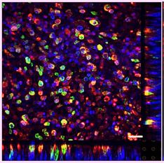
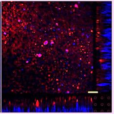
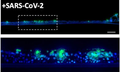
Figure 2. Basal Camostat blocks severe acute respiratory syndrome coronavirus 2 (SARS-CoV-2) infection.
(A) Schematic of experiment. Camostat was added to the basal media 48 h before SARS-CoV-2 infection. (B) Violin plots showing the quantification of SARS-CoV-2
infected total cells after treatment with 50 μM Camostat in the basal chamber with or without DHT. Each dot represents a Transwell. Experiment was performed twice with wells
exposed to either 2 × 10³ or 8 × 10³ TCID50 SARS-CoV-2 preparations in 50 µl. There was a significant reduction in viral infection in the wells treated with camostat. Samples also
treated with DHT are shown in red. P-values were calculated using a Mann–Whitney U test, P = 0.0286 at 2 × 10³ TCID50 and P = 0.0286 at 8 × 10³ TCID50. All replicates were
performed using cells from Donor B1. (C) Representative immunofluorescent image of human bronchial epithelial cell-air–liquid interface (ALI) after SARS-CoV-2 infection

Topical camostat for SARS-CoV-2 Guo et al.                                                   https://doi.org/10.26508/lsa.202101116      vol 5 | no 4 | e202101116        5 of 12
SARS-CoV-2 infection (Bittmann et al, 2020; Breining et al, 2020; Suzuki                   adverse events were recorded, none were judged to be because of
et al, 2020 Preprint; Youk et al, 2020; Hoffmann et al, 2020a Preprint,                    the study medication. Furthermore, the other adverse events were
2020b). These experiments provide a proof-of-principle for TMPRSS2                         minor and not reported in those administered 200 μg or less.
inhibition being a rational and potentially effective strategy but to                      Camostat was not detected in the systemic circulation after nasal
successfully translate to clinical practice a formulation that avoids the                  dosing. The primary metabolite was detected at 3 h, only in those
rapid metabolism in the circulation is critical.                                           individuals given the highest dose of 1,600 μg. Assuming a per
   Our findings show that early administration of a topical for-                            nostril epithelial lining fluid volume of 800 μl (Kaulbach et al, 1993),
mulation at clinically achievable doses may also be very effective in                      the effective concentration after delivery of 1,600 μg is 4.0 mM,
reducing SARS-CoV-2 infection. Local airway delivery of camostat                           substantially higher than the doses shown to be effective in this
was previously proposed as an inhibitor of a serine protease that                          study against two SARS-CoV-2 variants.
regulates sodium channel flux (Coote et al, 2009; Rowe et al, 2013).                           Importantly, a 200 μg dose would be in excess of 400 μM, with the
The objective was to change mucus characteristics in cystic fibrosis.                       data in Fig 3 suggesting 300 μM would have a greater than 90%
Although TMPRSS2 is the key protease targeted by camostat in                               inhibitory effect on cellular infection. Together, these data suggest
SARS-CoV-2 inhibition, it is not possible to formally exclude a role                       that safe and sustained nasal serine protease inhibition can be
for inhibition of other serine proteases in the observed results.                          achieved in primary hAECs using a nasal camostat spray.
   Coote and colleagues Coote et al (2009) reported preclinical data                          TMPRSS2 is a potentially important target for repurposed medi-
in hBECs at ALI, in which cells were exposed to up to 30 μM camostat                       cations and is an attractive strategy for prophylaxis as the target is
topically for 90 min. Camostat inhibition of sodium channel flux                            the host cell rather than SARS-CoV-2. Both direct inhibition and
persisted at least 6 h after treatment, despite multiple washes with                       androgen deprivation have been suggested as strategies to re-
warmed PBS, demonstrating that topical camostat can have a sus-                            duce TMPRSS2 activity. Our data do not support the use of AR
tained impact. This duration is consistent with the proposed “pseudo-                      antagonists but suggest that direct inhibition of TMPRSS2 will have
irreversible” mechanism of serine protease inhibition by camostat                          activity against multiple SARS-CoV-2 variants including B.1.1.7, re-
mediated by covalent binding to the target (Breining et al, 2020) and is                   cently shown to have increased transmissibility (Davies et al,
consistent with the apparent sustained inhibition in our system                            2021). Consistent with our observations, Hoffmann and colleagues
despite the administration being for 15 min periods only.                                  Hoffmann et al (2021) have also shown that camostat effectively
   A number of in vivo preclinical experiments using airway formu-                         inhibits variant cell entry in the Caco-2 colorectal carcinoma cell
lations of camostat have also been reported, including the admin-                          line. They demonstrated this using pseudotyped viral particles
istration of nebulised camostat to anaesthetised sheep (Coote et al,                       engineered to express a series of key variants (B.1.1.7, B1.351, and P.1)
2009). Up to 60 mg camostat was delivered by nebulisation in 3 ml,                         implicated as variants that may alter virus-host cell interactions
equating to a maximal concentration of 50.2 mM, markedly higher                            and confer resistance to antibodies (Hoffmann et al, 2021).
than we used, without toxicity. Importantly, the duration of activity                         The impact of an oral camostat preparation in COVID-19 has now
using a surrogate of therapeutic activity in vivo, was at least 5 h, again                 been reported and the results were negative (Gunst et al, 2021; Ono
consistent with the proposed model of covalent binding to target                           Pharmaceuticals, 2021). Our data provide a rationale for testing
serine proteases. Further unpublished studies in dogs suggested a                          camostat as a local airway administered prophylaxis or early
mild and reversible inflammatory response in individual animals                             treatment for SARS-CoV-2 infection. When effective, local delivery
(lung parenchyma) to inhaled camostat (Novartis, personal com-                             has the advantages of reducing the systemic dose and associated
munication); but no significant toxicity associated with high-dose                          side effects while delivering the drug to the site of disease.
nasal administration. Therefore, nebulised (or inhaled) camostat to                        Camostat is well tolerated in the upper airway, a plausibly effective
lower airways was deemed to not be a safe route of administration                          dose was readily achieved and the compound is soluble in saline
and further development of this route of delivery terminated. The                          and cheap to manufacture (Coote et al, 2009; Breining et al, 2020).
nasal route, however, appeared safe.                                                          While this paper was in review, studies have been published
   Therefore, a subsequent early phase trial focussed on the nasal                         suggesting that nasal TMPRSS2 inhibition using nafamostat is ef-
route - topical camostat was delivered to the nose in volunteers                           fective in preventing SARS-Co-V2 infection in small animal models (Li
with cystic fibrosis (Rowe et al, 2013). The doses tested were in the                       et al, 2021). These data are complementary to our study and strongly
range 5–1,600 μg and nasal potential difference was used as a                              support the strategy being moved into a clinical trial. Nafamostat is a
surrogate of target engagement. The authors reported target en-                            more potent inhibitor in cell-free biochemical assays and cellular
gagement and therapeutic efficacy with a 50% maximal effect dose                            assays but, unlike camostat, does not yet have Phase 1 safety data and
estimated at 18 μg/ml, equivalent to 45.2 μM. Although three serious                       prior United States Food and Drug Administration (FDA) approval for

using antibodies to angiotensin-converting enzyme-2 and the S2 subunit of spike protein. Uninfected human bronchial epithelial cell-ALI was used to verify specificity. Scale
bar: 100 μm. This image used cells from Donor B1. (D) Impact of 48 h of 50 μM basal camostat on cellular infection. Representative immunofluorescent images of human airway
epithelial cell-ALI post SARS-CoV-2 infection using antibodies to angiotensin-converting enzyme-2 and the S2 subunit of spike protein. Scale bar: 100 μm. This image used cells
from Donor B1. (E) H&E staining showing normal morphology in cells exposed to 50 μM camostat for 48 h. This image used cells from Donor B1. (F) LDH release assay as a
measure of cytotoxicity after 48-h treatment using basal camostat on ALI cultures derived from bronchial or nasal cells. Complete lysis is shown as a positive control. Doses of
up to 300 µM (basal) did not cause increased cytotoxicity. Each dot represents a Transwell and for each condition three biological replicates were performed including at least
three technical replicates. These experiments were performed using cells from Donors B1 and N3. P-values were calculated using a Kruskal–Wallis test with Dunn’s
correction for multiple comparisons (between all conditions). There was no significant difference between 50 and 300 µM camostat treatment and the control wells.

Topical camostat for SARS-CoV-2 Guo et al.                                                  https://doi.org/10.26508/lsa.202101116     vol 5 | no 4 | e202101116        6 of 12
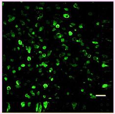
Figure 3. Camostat applied to the apical surface blocks severe acute respiratory syndrome coronavirus 2 (SARS-CoV-2) infection.
(A) Schematic of experimental design for topical camostat treatment before SARS-CoV-2 infection. (B) Violin plots showing the quantification of infected cells when pre-
treated with camostat in the apical chamber. P-values were calculated using a Kruskal–Wallis test with Dunn’s correction for multiple comparisons (apical treatment
conditions compared with control). P = 0.0285 and P = 0.0997 for topical 2× and topical 1× versus control, respectively. Each dot represents a Transwell. Experiments were
performed using cells from Donors B1 and B2. (C) Cross-sectional images of IF staining for nucleoprotein on formalin-fixed paraffin-embedded air–liquid interface
cultures. Scale bars: 100 μm. (D) Air–liquid interface cultures were pre-treated with 2 × 15 min of topical camostat at the indicated dose before infection with 1 × 10⁴ TCID50

Topical camostat for SARS-CoV-2 Guo et al.                                                  https://doi.org/10.26508/lsa.202101116     vol 5 | no 4 | e202101116       7 of 12
use in humans as a nasal preparation. Our study also confirms the the Transwell as indicated and dissolved in DMSO (max final con-
potential for ex vivo differentiated human primary airway model centration 0.1%) (enzalutamide) or PBS (camostat). Differentiated
systems to reduce and replace animal experiments in the context cells were cultured for 2 d in the absence of androgen stimulation
of therapeutic repurposing/reformulation studies where compound before exposure to DHT or enzalutamide for 24 h. For combination
safety has already been established. treatments 24-h enzalutamide pretreatment was applied before DHT
The recent success of the SARS-CoV-2 vaccine trials in protecting exposure.
against COVID-19 disease has had a major impact on the pandemic For topical delivery of camostat mesylate, the apical chamber
(Polack et al, 2020; Baden et al, 2021; Voysey et al, 2021). Furthermore, was washed with PBS before 50 μl of the indicated concentration of
there is now evidence that monoclonal antibodies delivered sub- camostat in PBS (or PBS alone in control wells) was added to the
cutaneously or intravenously are effective in the pre-hospital setting apical chamber for 15 min before being aspirated.
in both the prevention of symptomatic COVID-19 and treatment of
early phase infection (Chen et al, 2021; Weinreich et al, 2021). SARS-CoV-2 infection assays
Despite these critical advances, there remains an urgent unmet
need for a safe, cheap, easily administered, effective therapeutic with The SARS-CoV-2 viruses used in this study are the clinical isolates
potential efficacy in pre- or post-exposure prophylaxis, and in the named “SARS-CoV-2/human/Liverpool/REMRQ0001/2020” (Chu et al,
reduction of transmission, or progression, in asymptomatic/early SARS- 2020; Patterson et al, 2020) and “SARS-CoV-2 England/ATACCC 174/
CoV-2 infection. Our work suggests that topical delivery of camostat 2020” (Lineage B.1.1.7). Stocks were sequenced before use and the
mesylate should now be clinically evaluated for these indications. consensus matched the expected sequence exactly. Viral titre was
determined by 50% tissue culture infectious dose (TCID50) in Huh7-
ACE2 cells.
Materials and Methods For viral infection, the indicated dose was diluted in PBS to a
final volume of 50 μl and added to the apical side of Transwells
Primary hAEC: ALI model containing differentiated HBEC-ALI cultures for 2–3 h, then re-
moved. At 72 h postinfection HBEC-ALI Transwells were washed
hBECs were purchased from Lonza (Donor B1—Bronchial 1) or were once with PBS, dissociated with TrypLE, and fixed in 4% formal-
expanded directly from either bronchial brushings from a main dehyde for 15 min. Fixed cells were washed and incubated for 15
airway at bronchoscopy (Donor B2 – Bronchial 2) or a nasal brushing min at room temperature in Perm/Wash buffer (#554723; BD).
from the inferior turbinate from patients at Cambridge University Permeabilised cells were pelleted, stained for 15 min at room
Hospitals National Health Service Trust (Research Ethics Committee temperature in 100 μl of sheep anti-SARS-CoV-2 nucleoprotein
Reference 19/SW/0152) (Donors N3-N5 – Nasal 3–5). In brief, primary antibody (DA114; MRC-PPU) at a concentration of 0.7 μg/ml, washed,
airway cells at passage 2 were expanded in PneumaCult-Ex Plus and incubated in 100 μl AF488 donkey anti-sheep (#713-545-147;
Medium (Cat. no. #05040; Stemcell) then seeded on collagen (Cat. no. Jackson ImmunoResearch) at a concentration of 2 μg/ml for 15 min
#354236; Corning) coated 24-well Transwell inserts with 0.4-μm pores at room temperature. Stained cells were pelleted and fluorescence
(Cat. no. #353095; Falcon) until fully confluent. Once confluent, the staining analysed on a BD Fortessa flow cytometer. Fluorescent
cells were taken to the ALI and cultured with PneumaCult-ALI Me- images of whole Transwell ALI cultures were captured using a
dium (Cat. no. #05021; Stemcell) for at least 28 d before conducting Cellomics Arrayscan (VTI; Thermo Fisher Scientific). 121 fields of view
experiments. On the day of harvest, adherent cells were washed once per Transwell were captured at 10× magnification. The images were
with PBS and incubated in Accutase (Cat. no. #07920; Stemcell) at 37°C analysed using HCS Studio 2.0 Client Software.
for 15 min. Cells were dissociated by gentle pipetting and then
neutralised with DMEM/10% FBS. Whole cell pellets were collected by qRT-PCR
centrifuge at 300g for 5 min at room temperature. Specific cells used
were hBECs derived from a non-smoking donor (Cat. no. # CC-2540; RNA was extracted using RNeasy Mini Kit (QIAGEN) according to the
Lonza, male, Donor B1); hBECs from a male smoking donor undergoing manufacturer’s recommendation. Gene expression was quantified
bronchoscopy for a non-cancer indication (Donor B2), and nasal using SYBR Green I Dye (Life Technologies) on a QuantStudio 7 Flex
epithelial cells from three female patients (Donors N3, N4, and N5). Real-Time PCR System (Applied Biosystems). Data were analysed
Test compounds: 5-dihydroxytestosterone (DHT) (Cat. no. S4757; using 2−ΔΔCt method and Design and Analysis Software Version 2.5,
Selleckchem), enzalutamide (Cat. no. S1250; Selleckchem), camostat QuantStudio 6/7 Pro systems (Applied Biosystems). The following
mesylate (Cat. no. 3193; Tocris) were added to the basal chamber of primers were used:
of B.1.1.7 SARS-CoV-2. There was a dose-dependent reduction in cellular infection with camostat pretreatment. Images of whole Transwell inserts were captured.
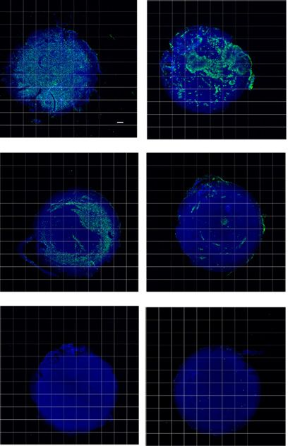
Blue–Hoechst nuclear stain. Green–cells expressing SARS-CoV-2 viral nucleoprotein. Representative microscopy montages of the Transwell inserts after the indicated
treatment. Scale bar: 500 μm. Experiments were performed using cells from Donors B1 and B2. (D, E) Quantitation of infection from (D), pretreatment with 2 × 15 min topical
camostat. All SARS-CoV-2 nucleoprotein positive cells are quantified and presented as percentage of the total number of cells (DAPI) on each Transwell Each dot
represents quantitation from a whole Transwell. Dots are coloured according to the donor. For bronchial cells, Transwells were infected on two separate occasions. For the
nasal cells, this was a single large-scale experiment. For bronchial cells, P-values were calculated using a Kruskal–Wallis test with Dunn’s correction for multiple
comparisons (between both treatment conditions and the control). P = 0.0120 for 50 μM 2× topical treatment versus control. P < 0.0001 for 300 μM versus control. For
nasal cells, P-values were calculated using a Mann–Whitney U test, P < 0.0001. Representative microscopy images for nasal cells are shown in Fig S4.
Topical camostat for SARS-CoV-2 Guo et al. https://doi.org/10.26508/lsa.202101116 vol 5 | no 4 | e202101116 8 of 12Figure 4. Camostat attenuates cellular infection when commenced 24 h after exposure to SARS-CoV-2 virus. (A) Schematic of experiment. A series of experiments was performed with B.1.1.7, this time with camostat administered after viral infection either for 48 h basally or topically for 15 min at 24 and 48 h after viral infection. (B) Images of whole Transwell inserts were captured using immunofluorescence. Blue–Hoechst nuclear stain. Green–cells expressing SARS-CoV-2 viral nucleoprotein. Representative microscopy montages of the Transwell inserts after the indicated treatment. Scale bar: 500 μm. Individual Transwells from four donors–B1, N3-5 are shown. In each case, both topical and basal camostat markedly reduced cellular infection. (C, D) Sustained basal exposure and 2 × 15 min topical exposure to camostat significantly reduced cellular infection in bronchial air–liquid interface cultures from Donor B1 and from nasal donors. Each dot represents quantitation from a whole Topical camostat for SARS-CoV-2 Guo et al. https://doi.org/10.26508/lsa.202101116 vol 5 | no 4 | e202101116 9 of 12
ACE2 Forward (59-39): CGAAGCCGAAGACCTGTTCTA; Reverse (59-39):                           Software). Statistical tests used are detailed in figure legends. P-
GGGCAAGTGTGGACTGTTCC; TMPRSS2 Forward (59-39): CTGCTGGATTTCCGGGTG;                        values were noted as follows: ns, not significant; *P < 0.05; **P < 0.01;
Reverse (59-39) TTCTGAGGTCTTCCCTTTCTCCT; TBP Forward (59-39): AGTGAA-                     ***P < 0.001.
GAACAGTCCAGACTG; Reverse (59-39): CCAGGAAATAACTCTGGCTCAT.

Histology
                                                                                          Data Availability
Transwells were washed three times with PBS, then fixed in 10%
                                                                                          Because of the nature of the data in this study, none has been
Neutral buffered formalin. The membrane was removed from the
                                                                                          deposited in a publicly available database. Request for further
Transwell using a scalpel blade and paraffin embedded. 4-μm
                                                                                          information on the data presented will be honoured by the cor-
sections were cut and stained with haematoxylin and eosin.
                                                                                          responding authors.

Immunofluorescence

Each Transwell was washed three times with PBS and fixed using 4%                          Supplementary Information
PFA for 15 min at room temperature before antibody staining. The
following antibodies were used: anti-ACE2 antibody (Anti-ACE2 anti-                       Supplementary Information is available at https://doi.org/10.26508/lsa.
body)—21115-1-AP; ProteinTech (Du et al, 2021); SARS-CoV/SARS-CoV-2                       202101116.
(COVID-19) spike antibody [1A9] (GTX632604; GeneTex); anti-SARS-CoV-2
nucleoprotein antibody (DA114; MRC-PPU); acetylated tubulin (T7451;
Sigma-Aldrich); Muc5AC (MA5-12178; Invitrogen); Hoechst dye solution
(100 μg/ml) was used for nuclei staining. Confocal images were taken
                                                                                          Acknowledgements
using Nikon Confocal Microscopes C2, magnification 40× oil. Transwell
                                                                                          SARS-CoV-2/human/Liverpool/REMRQ0001/2020 was a kind gift from Lance
inserts were imaged as described above.                                                   Turtle (University of Liverpool) and David Matthews and Andrew Davidson
                                                                                          (University of Bristol). SARS-CoV-2 England/ATACCC 174/2020 was a kind gift
LDH cytotoxicity assay                                                                    from Greg Towers (University College London), and we are also grateful to Ajit
                                                                                          Lalvani, Jake Dunning, Maria Zambon, and colleagues at Public Health En-
                                                                                          gland and Giada Mattiuzzo at the National Institute for Biological Standards
For the positive and negative controls, 50 μl of 10× lysis buffer or                      and Controls and Wendy Barclay and Jonathan Brown and all colleagues in
ultrapure water, respectively, was applied to the apical surface of                       the United Kingdom Research and Innovation funded Genotype to Phe-
HBEC-ALI or nasal ALI Transwells and incubated at 37°C for 45 min.                        notype collaboration (G2PUK). Sheep anti-SARS-CoV-2 nucleoprotein anti-
Then the basal media (500 μl) was used to wash the apical chamber.                        body (DA114) was a kind gift from Paul Davies (obtained from the Medical
                                                                                          Research Council Protein Phosphorylation Unit Reagents and Services,
For each experimental condition, the basal media (500 μl) was used to
                                                                                          University of Dundee). LnCAP cells were a kind gift from Dr Charlie Massie. We
wash the apical surface. Each condition was prepared in triplicate.                       gratefully acknowledge the support from Dr. Ravindra Mahadeva, Dr Jurgen
Each sample was assessed for LDH release using the CyQUANT LDH                            Herre and Ms. Jacqui Galloway in establishing the primary cells from pa-
Cytotoxicity Assay kit (C20300; Invitrogen) according to the manufac-                     tients. We are grateful for the generous support of the United Kingdom
turer’s recommendations. In brief, 50 μl of the final harvested sample                     Research and Innovation COVID Immunology Consortium (MR/V028448/1),
                                                                                          Addenbrooke’s Charitable Trust (15/20A) and the National Institute for Health
was mixed with 50 μl of pre-prepared reaction mix and incubated for                       Research (NIHR) Cambridge Biomedical Research Centre (BRC-1215-20014).
30 min at room temperature. After stopping the reaction, plate ab-                        This work was supported by a Wellcome Trust Principal Research Fellowship
sorbance was measured at 490 and 680 nm.                                                  (084957/Z/08/Z) and Medical Research Council research grant MR/V011561/1
   % Cytotoxicity was calculated as follows:                                              to PJ Lehner. This work was supported by the NC3Rs NC/S001204/1 project
                                                                                          grant and the Roy Castle Lung Cancer Foundation grant (2015/10/McCaughan)
   %Cytotoxicity =                                                                        to F McCaughan. This article presents independent research supported by the
                                                                                        NIHR Cambridge Biomedical Research Centre (BRC). The NIHR Cambridge BRC
     Compound−treated LDH activity − Spontaneous LDH activity                             is a partnership between Cambridge University Hospitals National Health
        Maximum LDH activity − Spontaneous LDH activity                                   Service Foundation Trust and the University of Cambridge, funded by the NIHR.
    × 100:                                                                                The views expressed are those of the author(s) and not necessarily those of
                                                                                          the NIHR or the Department of Health and Social Care.

Quantification and statistical analysis                                                    Author Contributions

Statistical analyses of mRNA expression assays and infection quan-                        W Guo: conceptualization, data curation, formal analysis, investiga-
tification data were performed using Prism 8 software (GraphPad                            tion, methodology, and writing—original draft, review, and editing.

Transwell from a single large-scale experiment. P-values were calculated using a Kruskal–Wallis test with Dunn’s correction for multiple comparisons (between both
treatment conditions and the control). P = 0.0441 and P < 0.0001 for Donor B1 50 and 300 μM versus control, respectively. P = 0.0105 for 300 µM topical versus control;
LM Porter: conceptualization, data curation, formal analysis, in- implications for clinical manifestations, transmissibility, and
vestigation, and methodology. laboratory studies of COVID-19: An observational study. Lancet
Microbe 1: e14–e23. doi:10.1016/S2666-5247(20)30004-5
TWM Crozier: data curation, formal analysis, investigation, and
methodology. Coote K, Atherton-Watson HC, Sugar R, Young A, MacKenzie-Beevor A, Gosling
M, Bhalay G, Bloomfield G, Dunstan A, Bridges RJ, et al (2009) Camostat
M Coates: resources and investigation. attenuates airway epithelial sodium channel function in vivo through
A Jha: resources and investigation. the inhibition of a channel-activating protease. J Pharmacol Exp Ther
M McKie: data curation. 329: 764–774. doi:10.1124/jpet.108.148155
JA Nathan: conceptulisation, methodology, and writing—original Crystal RG, Randell SH, Engelhardt JF, Voynow J, Sunday ME (2008) Airway
draft. epithelial cells: Current concepts and challenges. Proc Am Thorac Soc
PJ Lehner: conceptualization, resources, methodology, and wri- 5: 772–777. doi:10.1513/pats.200805-041HR
ting—original draft. Davies NG, Abbott S, Barnard RC, Jarvis CI, Kucharski AJ, Munday JD, Pearson
EJD Greenwood: conceptualization, formal analysis, investigation, CAB, Russell TW, Tully DC, Washburne AD, et al (2021) Estimated
transmissibility and impact of SARS-CoV-2 lineage B.1.1.7 in England.
methodology, and writing—original draft, review, and editing.
Science 372: eabg3055. doi:10.1126/science.abg3055
F McCaughan: conceptualization, resources, formal analysis, su-
Du Y, Shi R, Zhang Y, Duan X, Li L, Zhang J, Wang F, Zhang R, Shen H, Wang Y,
pervision, funding acquisition, investigation, methodology, project
et al (2021) A broadly neutralizing humanized ACE2-targeting antibody
administration, and writing—original draft, review, and editing. against SARS-CoV-2 variants. Nat Commun 12: 5000. doi:10.1038/
s41467-021-25331-x
Conflict of Interest Statement Englender M, Chamovitz D, Harell M (1990) Nasal transit time in normal
subjects and pathologic conditions. Otolaryngol Head Neck Surg 103:
909–912. doi:10.1177/019459989010300604
The authors declare that they have no conflict of interest.
Gunst JD, Staerke NB, Pahus MH, Kristensen LH, Bodilsen J, Lohse N, Dalgaard
LS, Brønnum D, Fröbert O, Hønge B, et al (2021) Efficacy of the TMPRSS2
inhibitor camostat mesilate in patients hospitalized with Covid-19-a
References double-blind randomized controlled trial. EClinicalMedicine 35:
100849. doi:10.1016/j.eclinm.2021.100849
Baden LR, El Sahly HM, Essink B, Kotloff K, Frey S, Novak R, Diemert D, Spector Heurich A, Hofmann-Winkler H, Gierer S, Liepold T, Jahn O, Pöhlmann S (2014)
SA, Rouphael N, Creech CB, et al (2021) Efficacy and safety of the TMPRSS2 and ADAM17 cleave ACE2 differentially and only proteolysis
mRNA-1273 SARS-CoV-2 vaccine. N Engl J Med 384: 403–416. by TMPRSS2 augments entry driven by the severe acute respiratory
doi:10.1056/NEJMoa2035389 syndrome coronavirus spike protein. J Virol 88: 1293–1307. doi:10.1128/
jvi.02202-13
Bennani NN, Bennani-Baiti IM (2020) Androgen deprivation therapy may
constitute a more effective COVID-19 prophylactic than therapeutic Hoffmann M, Arora P, Gross R, Seidel A, Hörnich BF, Hahn AS, Krüger N,
strategy. Ann Oncol 31: 1585–1586. doi:10.1016/j.annonc.2020.08.2095 Graichen L, Hofmann-Winkler H, Kempf A, et al (2021) SARS-CoV-2
variants B.1.351 and P.1 escape from neutralizing antibodies. Cell 184:
Bittmann S, Weissenstein A, Villalon G, Moschuring-Alieva E, Luchter E (2020)
2384–2393. doi:10.1016/j.cell.2021.03.036
Simultaneous treatment of COVID-19 with serine protease inhibitor
camostat and/or cathepsin l inhibitor? J Clin Med Res 12: 320–322. Hoffmann M, Hofmann-Winkler H, Smith JC, Krüger N, Sørensen LK, Søgaard
doi:10.14740/jocmr4161 OS, Hasselstrøm JB, Winkler M, Hempel T, Raich L, et al (2020a)
Camostat mesylate inhibits SARS-CoV-2 activation by TMPRSS2-
Breining P, Frølund AL, Højen JF, Gunst JD, Staerke NB, Saedder E, Cases-
related proteases and its metabolite GBPA exerts antiviral activity.
Thomas M, Little P, Nielsen LP, Søgaard OS, et al (2021) Camostat
BioRxiv. doi:10.1101/2020.08.05.237651. (Preprint posted August 05, 2020).
mesylate against SARS-CoV-2 and COVID-19-rationale, dosing and
safety. Basic Clin Pharmacol Toxicol 128: 204–212. doi:10.1111/ Hoffmann M, Kleine-Weber H, Schroeder S, Krüger N, Herrler T, Erichsen S,
bcpt.13533 Schiergens TS, Herrler G, Wu N-H, Nitsche A, et al (2020b) SARS-CoV-2
cell entry depends on ACE2 and TMPRSS2 and is blocked by a clinically
Breining P, Frølund AL, Højen JF, Gunst JD, Staerke NB, Saedder E, Cases-
proven protease inhibitor. Cell 181: 271–280.e8. doi:10.1016/
Thomas M, Little P, Nielsen LP, Søgaard OS, et al (2020) Camostat
j.cell.2020.02.052
mesylate against SARS-CoV-2 and COVID-19-Rationale, dosing and
safety. Basic Clin Pharmacol Toxicol 128: 204–212. doi:10.1111/ Hou YJ, Okuda K, Edwards CE, Martinez DR, Asakura T, Dinnon KH 3rd, Kato T,
bcpt.13533 Lee RE, Yount BL, Mascenik TM, et al (2020) SARS-CoV-2 reverse
genetics reveals a variable infection gradient in the respiratory tract.
Chakravarty D, Nair SS, Hammouda N, Ratnani P, Gharib Y, Wagaskar V,
Cell 182: 429–446.e14. doi:10.1016/j.cell.2020.05.042
Mohamed N, Lundon D, Dovey Z, Kyprianou N, et al (2020) Sex
differences in SARS-CoV-2 infection rates and the potential link to Jia HP, Look DC, Shi L, Hickey M, Pewe L, Netland J, Farzan M, Wohlford-Lenane
prostate cancer. Commun Biol 3: 374. doi:10.1038/s42003-020-1088-9 C, Perlman S, McCray PB Jr. (2005) ACE2 receptor expression and severe
acute respiratory syndrome coronavirus infection depend on
Chan KK, Dorosky D, Sharma P, Abbasi SA, Dye JM, Kranz DM, Herbert AS,
differentiation of human airway epithelia. J Virol 79: 14614–14621.
Procko E (2020) Engineering human ACE2 to optimize binding to the
doi:10.1128/JVI.79.23.14614-14621.2005
spike protein of sars coronavirus 2. Science 369: 1261–1265. doi:10.1126/
science.abc0870 Kaulbach HC, White MV, Igarashi Y, Hahn BK, Kaliner MA (1993) Estimation of
nasal epithelial lining fluid using urea as a marker. J Allergy Clin
Chen P, Nirula A, Heller B, Gottlieb RL, Boscia J, Morris J, Huhn G, Cardona J,
Immunol 92: 457–465. doi:10.1016/0091-6749(93)90125-y
Mocherla B, Stosor V, et al (2021) SARS-CoV-2 neutralizing antibody LY-
CoV555 in outpatients with COVID-19. N Engl J Med 384: 229–237. Li K, Meyerholz DK, Bartlett JA, McCray PB Jr. (2021) The TMPRSS2 inhibitor
doi:10.1056/NEJMoa2029849 nafamostat reduces SARS-CoV-2 pulmonary infection in mouse
models of COVID-19. mBio 12: e0097021. doi:10.1128/mBio.00970-21
Chu H, Chan JF-W, Yuen TT-T, Shuai H, Yuan S, Wang Y, Hu B, Yip CC-Y, Tsang
JO-L, Huang X, et al (2020) Comparative tropism, replication kinetics, Martines RB, Ritter JM, Matkovic E, Gary J, Bollweg BC, Bullock H, Goldsmith CS,
and cell damage profiling of SARS-CoV-2 and SARS-CoV with Silva-Flannery L, Seixas JN, Reagan-Steiner S, et al (2020) Pathology
Topical camostat for SARS-CoV-2 Guo et al. https://doi.org/10.26508/lsa.202101116 vol 5 | no 4 | e202101116 11 of 12and pathogenesis of SARS-CoV-2 associated with fatal coronavirus Samuel RM, Majd H, Richter MN, Ghazizadeh Z, Zekavat SM, Navickas A,
disease, United States. Emerg Infect Dis 26: 2005–2015. doi:10.3201/ Ramirez JT, Asgharian H, Simoneau CR, Bonser LR, et al (2020)
eid2609.202095 Androgen signaling regulates SARS-CoV-2 receptor levels and is
associated with severe COVID-19 symptoms in men. Cell Stem Cell 27:
Montopoli M, Zumerle S, Vettor R, Rugge M, Zorzi M, Catapano CV, Carbone GM,
876–889.e12. doi:10.1016/j.stem.2020.11.009
Cavalli A, Pagano F, Ragazzi E, et al (2020) Androgen-deprivation
therapies for prostate cancer and risk of infection by SARS-CoV-2: A Shirato K, Kawase M, Matsuyama S (2013) Middle east respiratory syndrome
population-based study (n = 4532). Ann Oncol 31: 1040–1045. coronavirus infection mediated by the transmembrane serine
doi:10.1016/j.annonc.2020.04.479 protease TMPRSS2. J Virol 87: 12552–12561. doi:10.1128/jvi.01890-13
Mulay A, Konda B, Garcia G, Yao C, Beil S, Villalba JM, Koziol C, Sen C, Stopsack KH, Mucci LA, Antonarakis ES, Nelson PS, Kantoff PW (2020) TMPRSS2
Purkayastha A, Kolls JK, et al (2021) SARS-CoV-2 infection of primary and COVID-19: Serendipity or opportunity for intervention? Cancer
human lung epithelium for COVID-19 modeling and drug discovery. Discov 10: 779–782. doi:10.1158/2159-8290.Cd-20-0451
Cell Rep 35: 109055. doi:10.1016/j.celrep.2021.109055
Sungnak W, Huang N, Bécavin C, Berg M (2020) SARS-COV-2 entry genes are
Ono Pharmaceutical (2021) Phase III study result of foipan® tablets, a most highly expressed in nasal goblet and ciliated cells within human
protease enzyme inhibitor, in japan in patients with novel coronavirus airways. ArXiv doi:10.1038/s41591-020-0868-6
infection (COVID-19). Accessed October 17, 2021. https://www.ono-
Suzuki T, Itoh Y, Sakai Y, Saito A, Okuzaki D, Motooka D, Minami S, Kobayashi T,
pharma.com/news/20210611.html
Yamamoto T, Okamoto T, et al (2020) Generation of human bronchial
O’Callaghan ME, Jay A, Kichenadasse G, Moretti KL (2020) Androgen organoids for SARS-CoV-2 research. BioRxiv. doi:10.1101/
deprivation therapy in unlikely to be effective for treatment of COVID- 2020.05.25.115600. (Preprint posted June 01, 2020).
19. Ann Oncol 31: 1780–1782. doi:10.1016/j.annonc.2020.09.014
Voysey M, Clemens SAC, Madhi SA, Weckx LY, Folegatti PM, Aley PK, Angus B,
Purkayastha A, Sen C, Garcia G Jr., Langerman J, Shia DW, Meneses LK, Vijayaraj Baillie VL, Barnabas SL, Bhorat QE, et al (2021) Safety and efficacy of
P, Durra A, Koloff CR, Freund DR, et al (2020) Direct exposure to SARS- the ChAdOx1 nCoV-19 vaccine (AZD1222) against SARS-CoV-2: An
CoV-2 and cigarette smoke increases infection severity and alters the interim analysis of four randomised controlled trials in Brazil, South
stem cell-derived airway repair response. Cell Stem Cell 27: Africa, and the UK. Lancet 397: 99–111. doi:10.1016/S0140-6736(20)
869–875.e4. doi:10.1016/j.stem.2020.11.010 32661-1
Patterson EI, Prince T, Anderson ER, Casas-Sanchez A, Smith SL, Cansado- Walters MS, Gomi K, Ashbridge B, Moore MAS, Arbelaez V, Heldrich J, Ding B-S,
Utrilla C, Solomon T, Griffiths MJ, Acosta-Serrano Á, Turtle L, et al (2020) Rafii S, Staudt MR, Crystal RG (2013) Generation of a human airway
Methods of inactivation of SARS-CoV-2 for downstream biological epithelium derived basal cell line with multipotent differentiation
assays. J Infect Dis 222: 1462–1467. doi:10.1093/infdis/jiaa507 capacity. Respir Res 14: 135. doi:10.1186/1465-9921-14-135
Plaza Valı́a P, Carrión Valero F, Marı́n Pardo J, Bautista Rentero D, González Wang Q, Li W, Liu XS, Carroll JS, Jänne OA, Keeton EK, Chinnaiyan AM, Pienta KJ,
Monte C (2008) [Saccharin test for the study of mucociliary clearance: Brown M (2007) A hierarchical network of transcription factors
Reference values for a Spanish population]. Arch Bronconeumol 44: governs androgen receptor-dependent prostate cancer growth. Mol
540–545. doi:10.1016/s1579-2129(08)60100-7 Cell 27: 380–392. doi:10.1016/j.molcel.2007.05.041
Polack FP, Thomas SJ, Kitchin N, Absalon J, Gurtman A, Lockhart S, Perez JL, Weinreich DM, Sivapalasingam S, Norton T, Ali S, Gao H, Bhore R, Musser BJ,
Pérez Marc G, Moreira ED, Zerbini C, et al (2020) Safety and efficacy of Soo Y, Rofail D, Im J, et al (2021) Regn-cov2, a neutralizing antibody
the BNT162b2 mRNA Covid-19 vaccine. N Engl J Med 383: 2603–2615. cocktail, in outpatients with COVID-19. N Engl J Med 384: 238–251.
doi:10.1056/NEJMoa2034577 doi:10.1056/NEJMoa2035002
Pradhan A, Olsson PE (2020) Sex differences in severity and mortality from Youk J, Kim T, Evans KV, Jeong YI, Hur Y, Hong SP, Kim JH, Yi K, Kim SY, Na KJ, et al
COVID-19: Are males more vulnerable? Biol Sex Differ 11: 53. (2020) Three-dimensional human alveolar stem cell culture models
doi:10.1186/s13293-020-00330-7 reveal infection response to SARS-CoV-2. Cell Stem Cell 27:
Rahim S, Uren A (2013) Emergence of ETS transcription factors as diagnostic 905–919.e10. doi:10.1016/j.stem.2020.10.004
tools and therapeutic targets in prostate cancer. Am J Transl Res 5: Ziegler CGK, Allon SJ, Nyquist SK, Mbano IM, Miao VN, Tzouanas CN, Cao Y,
254–268. Yousif AS, Bals J, Hauser BM, et al (2020) SARS-CoV-2 receptor ACE2 is
Rowe SM, Reeves G, Hathorne H, Solomon GM, Abbi S, Renard D, Lock R, Zhou an interferon-stimulated gene in human airway epithelial cells and is
P, Danahay H, Clancy JP, et al (2013) Reduced sodium transport with detected in specific cell subsets across tissues. Cell 181: 1016–1035.e19.
nasal administration of the prostasin inhibitor camostat in subjects doi:10.1016/j.cell.2020.04.035
with cystic fibrosis. Chest 144: 200–207. doi:10.1378/chest.12-2431 Zoufaly A, Poglitsch M, Aberle JH, Hoepler W, Seitz T, Traugott M, Grieb A,
Ruiz Garcı́a S, Deprez M, Lebrigand K, Cavard A, Paquet A, Arguel M-J, Magnone Pawelka E, Laferl H, Wenisch C, et al (2020) Human recombinant
V, Truchi M, Caballero I, Leroy S, et al (2019) Novel dynamics of human soluble ACE2 in severe COVID-19. Lancet Respir Med 8: 1154–1158.
mucociliary differentiation revealed by single-cell rna sequencing of doi:10.1016/S2213-2600(20)30418-5
nasal epithelial cultures. Development 146: dev177428. doi:10.1242/
dev.177428
Sachs LA, Finkbeiner WE, Widdicombe JH (2003) Effects of media on License: This article is available under a Creative
differentiation of cultured human tracheal epithelium. In Vitro Cell Commons License (Attribution 4.0 International, as
Dev Biol Anim 39: 56–62. doi:10.1290/1543-706X(2003)0392.0.CO;2 licenses/by/4.0/).
Topical camostat for SARS-CoV-2 Guo et al. https://doi.org/10.26508/lsa.202101116 vol 5 | no 4 | e202101116 12 of 12You can also read