Emerging Quantitative Contrasts: Quantitative Susceptibility Mapping (QSM) - Saskia and ...
←
→
Page content transcription
If your browser does not render page correctly, please read the page content below
Emerging Quantitative Contrasts:
Quantitative Susceptibility Mapping (QSM)
 Theory & Methods

 Steffen Bollmann

 Research Fellow - School of Information Technology and Electrical Engineering
 Affiliate Fellow – Centre for Advanced Imaging
 CI - Centre for Innovation in Biomedical Imaging Technology
 The University of Queensland, Australia
Steffen Bollmann | @sbollmann_MRI | www.mri.sbollmann.net

 Declaration of
 Financial Interests or Relationships
Speaker Name: Steffen Bollmann

I have the following financial interest or relationship(s) to disclose with regard to the subject
matter of this presentation:
• Grant/research support: Siemens Healthineers
• Other: Patent Applications on Deep Learning QSM (US 2019/0204401 A1) and Masking
 for QSM (US 2019/0302200 A1)

 2
Steffen Bollmann | @sbollmann_MRI | www.mri.sbollmann.net

QSM-Acronym soup: Ingredient list
COMPOSER - Combining phase images from array coils SHARP - sophisticated harmonic artifact reduction for phase
using a short echo time reference scan data
COSMOS - Calculation of susceptibility through multiple SHARQnet - Sophisticated Harmonic Artifact Reduction in
orientation sampling Quantitative Susceptibility Mapping using a Deep
EPI – Echo Planar Imaging Convolutional Neural Network
FINE - Fidelity imposed network edit SS – Single Step
GRE – GRadient Echo STI – Susceptibility Tensor Imaging
mIP – minimum Intensity Projection SVD – Singular Value Decomposition
LBV - Laplacian boundary value background field removal SWI – Susceptibility Weighted Imaging
MEDI – Morphology Enabled Dipole Inversion TA – Acquisition Time
ppm – parts per million TR – Repetition Time
PRELUDE – Phase Region Expanding Labeler for TE – Echo Time
Unwrapping Discrete Estimates TFI – Total Field Inversion
QSM – Quantitative Susceptibility Mapping TGV – Total Generalized Variation
SEGUE - A Speedy rEgion-Growing Algorithm for TKD – Truncated K-Space Division
Unwrapping Estimated Phase V-SHARP - variable-radius sophisticated harmonic artifact
SENSE - Sensitivity encoding reduction for phase data

 https://www.chefkoch.de/rezepte/2166951347805623/Buchstabensuppe-mit-frischem-Gemuese.html 3
Steffen Bollmann | @sbollmann_MRI | www.mri.sbollmann.net

Full Reference List
Abdul-Rahman et al. AO 2007: ‘Fast and Robust Three-Dimensional Best Path Phase Unwrapping Algorithm’. Applied Optics 46, no. 26
(10 September 2007): 6623–35. https://doi.org/10.1364/AO.46.006623.
Bernstein et al. MRM 1994: ‘Reconstructions of Phase Contrast, Phased Array Multicoil Data’. Magnetic Resonance in Medicine 32, no.
3 (1994): 330–34.
Bollmann et al. Z Med Phys 2019: ‘SHARQnet – Sophisticated Harmonic Artifact Reduction in Quantitative Susceptibility Mapping Using
a Deep Convolutional Neural Network’. Zeitschrift Für Medizinische Physik, 14 February 2019.
https://doi.org/10.1016/j.zemedi.2019.01.001.
Bollmann et al. NI 2019: ‘DeepQSM - Using Deep Learning to Solve the Dipole Inversion for Quantitative Susceptibility Mapping’.
NeuroImage 195 (15 July 2019): 373–83. https://doi.org/10.1016/j.neuroimage.2019.03.060.
Chatnuntawech et al. NMR Biomed 2017: ‘Single-Step Quantitative Susceptibility Mapping with Variational Penalties: Single-Step Qsm
with Variational Penalties’. NMR in Biomedicine 30, no. 4 (April 2017): e3570. https://doi.org/10.1002/nbm.3570.
Deistung et al. PLoS ONE 2013: ‘Quantitative Susceptibility Mapping Differentiates between Blood Depositions and Calcifications in
Patients with Glioblastoma’. PLoS ONE 8, no. 3 (21 March 2013): e57924. https://doi.org/10.1371/journal.pone.0057924.
Deistung et al. NMR Biomed 2017: ‘Overview of Quantitative Susceptibility Mapping: Overview of Quantitative Susceptibility Mapping’.
NMR in Biomedicine 30, no. 4 (April 2017): e3569. https://doi.org/10.1002/nbm.3569.
Dymerska et al. MRM 2021: ‘Phase Unwrapping with a Rapid Opensource Minimum Spanning Tree Algorithm (ROMEO)’. Magnetic
Resonance in Medicine n/a, no. n/a. Accessed 2 November 2020. https://doi.org/10.1002/mrm.28563.
Eckstein et al MRM 2018: ‘Computationally Efficient Combination of Multi-Channel Phase Data From Multi-Echo Acquisitions (ASPIRE)’.
Magnetic Resonance in Medicine 79, no. 6 (2018): 2996–3006. https://doi.org/10.1002/mrm.26963.
Haacke et al. MRM 2004: ‘Susceptibility Weighted Imaging (SWI)’. Magnetic Resonance in Medicine 52, no. 3 (1 September 2004): 612–
18. https://doi.org/10.1002/mrm.20198.
 4
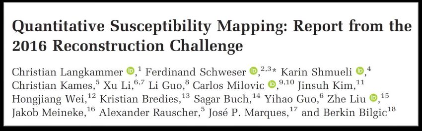
Steffen Bollmann | @sbollmann_MRI | www.mri.sbollmann.net

Full Reference List
Haacke et al. MRI 2014: ‘Quantitative Susceptibility Mapping: Current Status and Future Directions’. Magnetic Resonance Imaging, 2014.
https://doi.org/10.1016/j.mri.2014.09.004.
Hammond et al. NI 2008: ‘Development of a Robust Method for Generating 7.0 T Multichannel Phase Images of the Brain with Application to Normal Volunteers and Patients
with Neurological Diseases’. NeuroImage 39, no. 4 (15 February 2008): 1682–92. https://doi.org/10.1016/j.neuroimage.2007.10.037.
Jenkinson MRM 2003: ‘Fast, Automated, N-Dimensional Phase-Unwrapping Algorithm’. Magnetic Resonance in Medicine 49, no. 1 (2003): 193–97.
https://doi.org/10.1002/mrm.10354.
Yoon et al. NI 2018: ‘Quantitative Susceptibility Mapping Using Deep Neural Network: QSMnet’. NeuroImage 179 (1 October 2018): 199–206.
https://doi.org/10.1016/j.neuroimage.2018.06.030.
Jung et al. NMR Biomed 2020: ‘Overview of Quantitative Susceptibility Mapping Using Deep Learning: Current Status, Challenges and Opportunities’. NMR in Biomedicine,
23 March 2020, e4292. https://doi.org/10.1002/nbm.4292.
Karsa et al. TMI 2019: ‘SEGUE: A Speedy REgion-Growing Algorithm for Unwrapping Estimated Phase’. IEEE Transactions on Medical Imaging 38, no. 6 (June 2019): 1347–
57. https://doi.org/10.1109/TMI.2018.2884093.
Khabipova et al. NI 2015:‘A Modulated Closed Form Solution for Quantitative Susceptibility Mapping — A Thorough Evaluation and Comparison to Iterative Methods Based
on Edge Prior Knowledge’. NeuroImage 107 (15 February 2015): 163–74. https://doi.org/10.1016/j.neuroimage.2014.11.038.
Lai et al. ArXiv 2020:‘Learned Proximal Networks for Quantitative Susceptibility Mapping’. ArXiv:2008.05024 [Cs, Eess], 11 August 2020. http://arxiv.org/abs/2008.05024.
Langkammer et al. NI 2015 ‘Fast Quantitative Susceptibility Mapping Using 3D EPI and Total Generalized Variation’. NeuroImage 111 (1 May 2015): 622–30.
https://doi.org/10.1016/j.neuroimage.2015.02.041.
Langkammer et al. MRM 2018: ‘Quantitative Susceptibility Mapping: Report from the 2016 Reconstruction Challenge: QSM Reconstruction Challenge 2016’. Magnetic
Resonance in Medicine 79, no. 3 (March 2018): 1661–73. https://doi.org/10.1002/mrm.26830.
Li et al. NI 2011: ‘Quantitative Susceptibility Mapping of Human Brain Reflects Spatial Variation in Tissue Composition’. NeuroImage 55, no. 4 (15 April 2011): 1645–56.
https://doi.org/10.1016/j.neuroimage.2010.11.088.

 5
Steffen Bollmann | @sbollmann_MRI | www.mri.sbollmann.net

Full Reference List
Li et al. NI 2015: ‘A Method for Estimating and Removing Streaking Artifacts in Quantitative Susceptibility Mapping’. NeuroImage 108 (March 2015): 111–22.
https://doi.org/10.1016/j.neuroimage.2014.12.043.
Liu et al. MRM 2009: ‘Calculation of Susceptibility through Multiple Orientation Sampling (COSMOS): A Method for Conditioning the Inverse Problem from Measured Magnetic
Field Map to Susceptibility Source Image in MRI’. Magnetic Resonance in Medicine 61, no. 1 (1 January 2009): 196–204. https://doi.org/10.1002/mrm.21828.
Liu MRM 2010: ‘Susceptibility Tensor Imaging’. Magnetic Resonance in Medicine : Official Journal of the Society of Magnetic Resonance in Medicine / Society of Magnetic
Resonance in Medicine 63, no. 6 (June 2010): 1471–77. https://doi.org/10.1002/mrm.22482. https://doi.org/10.1002/mrm.22482.
Liu et al. MRM 2013: ‘Nonlinear Formulation of the Magnetic Field to Source Relationship for Robust Quantitative Susceptibility Mapping’. Magnetic Resonance in Medicine
69, no. 2 (1 February 2013): 467–76. https://doi.org/10.1002/mrm.24272.
Liu et al. MRM 2017: ‘Preconditioned Total Field Inversion (TFI) Method for Quantitative Susceptibility Mapping: QSM Using Preconditioned Total Field Inversion’. Magnetic
Resonance in Medicine 78, no. 1 (July 2017): 303–15. https://doi.org/10.1002/mrm.26331.
Marques et al. MRM 2021: ‘QSM Reconstruction Challenge 2.0: A Realistic in Silico Head Phantom for MRI Data Simulation and Evaluation of Susceptibility Mapping
Procedures’. Magnetic Resonance in Medicine, 26 February 2021. https://doi.org/10.1002/mrm.28716.
Parker et al. MRM 2014: ‘Phase Reconstruction from Multiple Coil Data Using a Virtual Reference Coil’. Magnetic Resonance in Medicine 72, no. 2 (1 August 2014): 563–69.
https://doi.org/10.1002/mrm.24932.
Robinson et al. NMR Biomed 2017: ‘An Illustrated Comparison of Processing Methods for MR Phase Imaging and QSM: Combining Array Coil Signals and Phase
Unwrapping: Phase Image Combination and Unwrapping’. NMR in Biomedicine 30, no. 4 (April 2017): e3601. https://doi.org/10.1002/nbm.3601.
Robinson et al. MRM 2017: ‘Combining Phase Images from Array Coils Using a Short Echo Time Reference Scan (COMPOSER)’. Magnetic Resonance in Medicine 77, no. 1
(January 2017): 318–27. https://doi.org/10.1002/mrm.26093.
Roemer et al MRM 1990: ‘The NMR Phased Array’. Magnetic Resonance in Medicine 16, no. 2 (1990): 192–225. https://doi.org/10.1002/mrm.1910160203.
Schenck et al. MP 1996: ‘The Role of Magnetic Susceptibility in Magnetic Resonance Imaging: MRI Magnetic Compatibility of the First and Second Kinds’. Medical Physics
23, no. 6 (1 June 1996): 815–50. https://doi.org/10.1118/1.597854.
Shmueli et al. MRM 2009: ‘Magnetic Susceptibility Mapping of Brain Tissue in Vivo Using MRI Phase Data’. Magnetic Resonance in Medicine 62, no. 6 (1 December 2009):
1510–22. https://doi.org/10.1002/mrm.22135.

 6
Steffen Bollmann | @sbollmann_MRI | www.mri.sbollmann.net

Full Reference List
Schofield et al. Opt. Lett. 2003: ‘Fast Phase Unwrapping Algorithm for Interferometric Applications’. Optics Letters 28, no. 14 (15 July 2003):
1194–96. https://doi.org/10.1364/OL.28.001194.
Schweser et al. NI 2011: ‘Quantitative Imaging of Intrinsic Magnetic Tissue Properties Using MRI Signal Phase: An Approach to in Vivo Brain Iron
Metabolism?’ NeuroImage 54, no. 4 (14 February 2011): 2789–2807. https://doi.org/10.1016/j.neuroimage.2010.10.070.
Schweser et al. Z Med Phys 2016: ‘Foundations of MRI Phase Imaging and Processing for Quantitative Susceptibility Mapping (QSM)’. Zeitschrift
Für Medizinische Physik 26, no. 1 (March 2016): 6–34. https://doi.org/10.1016/j.zemedi.2015.10.002.
Schweser et al. NMR Biomed 2017: ‘An Illustrated Comparison of Processing Methods for Phase MRI and QSM: Removal of Background Field
Contributions from Sources Outside the Region of Interest: Background Field Elimination’. NMR in Biomedicine 30, no. 4 (April 2017): e3604.
https://doi.org/10.1002/nbm.3604.
Stüber et al. IJoMS 2016: ‘Iron in Multiple Sclerosis and Its Noninvasive Imaging with Quantitative Susceptibility Mapping’. International Journal of
Molecular Sciences 17, no. 1 (14 January 2016): 100. https://doi.org/10.3390/ijms17010100.
Sun et al. MRM 2014: ‘Background Field Removal Using Spherical Mean Value Filtering and Tikhonov Regularization’. Magnetic Resonance in
Medicine 71, no. 3 (1 March 2014): 1151–57. https://doi.org/10.1002/mrm.24765.
Walsh et al. MRM 2000: ‘Adaptive Reconstruction of Phased Array MR Imagery’. Magnetic Resonance in Medicine 43, no. 5 (1 May 2000): 682–
90. https://doi.org/10.1002/(SICI)1522-2594(200005)43:53.0.CO;2-G.
Wei et al. NI 2019: ‘Learning-Based Single-Step Quantitative Susceptibility Mapping Reconstruction without Brain Extraction’. NeuroImage 202 (15
November 2019): 116064. https://doi.org/10.1016/j.neuroimage.2019.116064.
Zhang et al. NI 2020: ‘Fidelity Imposed Network Edit (FINE) for Solving Ill-Posed Image Reconstruction’. NeuroImage, 22 January 2020, 116579.
https://doi.org/10.1016/j.neuroimage.2020.116579.
Zhou et al. NMR Biomed 2014: ‘Background Field Removal by Solving the Laplacian Boundary Value Problem’. NMR in Biomedicine 27, no. 3
(March 2014): 312–19. https://doi.org/10.1002/nbm.3064.

 7
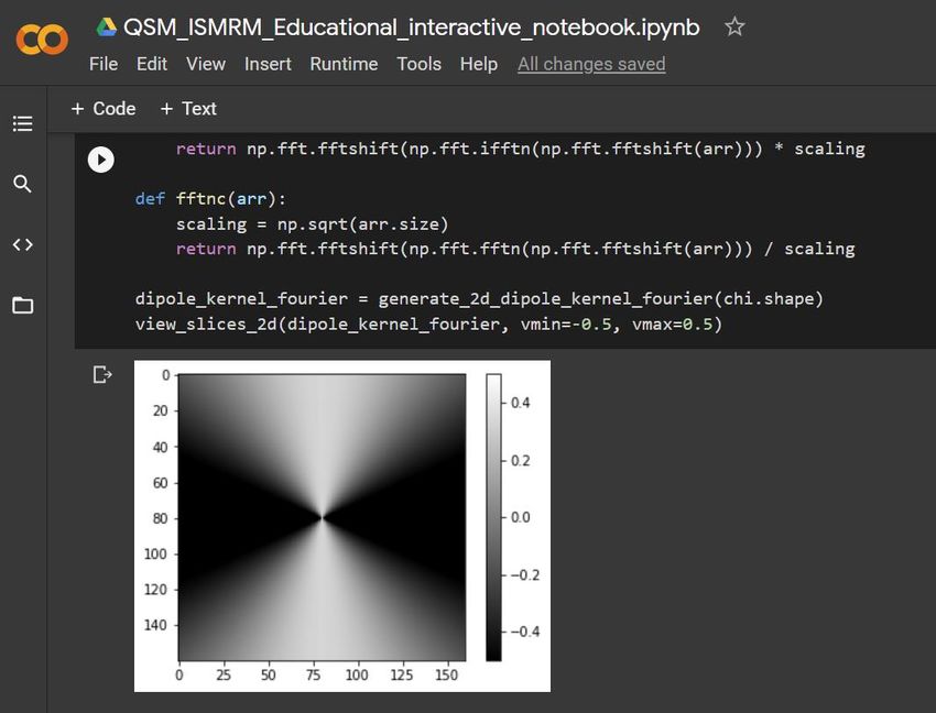
Steffen Bollmann | @sbollmann_MRI | www.mri.sbollmann.net

Interactive Computational Notebook to learn about QSM

• http://bit.ly/ISMRM-QSM-2021

 8
Steffen Bollmann | @sbollmann_MRI | www.mri.sbollmann.net

 From Susceptibility Weighted Imaging (SWI) …
GRE acquisition

 mIP SWI

 Coil
 combination

 minimum
 Magnitude
 Intensity
 weighting
 Projection
 Haacke et al. MRM 2004 9
Steffen Bollmann | @sbollmann_MRI | www.mri.sbollmann.net

Why bother with QSM?
• Is this lesion calcified or haemorrhaging?

 Magnitude SWI filtered Phase SWI MIP SWI

 Images courtesy of Fatima Nasrallah and Abdalla Mohamed with support from Kieran O’Brien & Ashley Stewart 10
Steffen Bollmann | @sbollmann_MRI | www.mri.sbollmann.net

Why bother with QSM?
 SWI Magnitude SWI Filtered Phase QSM QSM differentiates between blood
 Calcification Hypointense (-) Ambiguous Hypointense (-) products (Hyperintense) and calcifications
 Blood products Hypointense (-) Ambiguous Hyperintense (+) (Hypointense).

 Magnitude SWI filtered Phase SWI MIP SWI QSM

 Images courtesy of Fatima Nasrallah and Abdalla Mohamed with support from Kieran O’Brien & Ashley Stewart 11
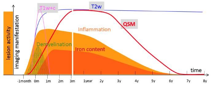
Steffen Bollmann | @sbollmann_MRI | www.mri.sbollmann.net

Why bother with QSM?

• QSM is sensitive to bio-metals - e.g. iron in Multiple Sclerosis:

 • iron accumulates after demyelination in microglia

 • slow iron-depletion from normal appearing white matter

 Stüber et al. IJoMS 2016 12
Steffen Bollmann | @sbollmann_MRI | www.mri.sbollmann.net

 Acquisition Coil combination Unwrapping Masking Background Field Dipole Inversion

 … to Quantitative Susceptibility Mapping (QSM)
GRE acquisition

 Background
 Field
 QSM

 Coil
 combination

 Thank you for slides and material: Simon Robinson, Ashley Stewart, Markus Barth, Francesco Cognolato 13
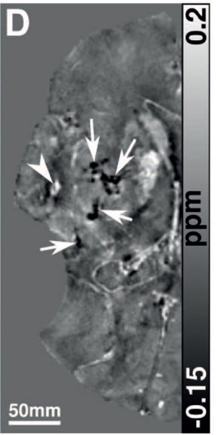
Steffen Bollmann | @sbollmann_MRI | www.mri.sbollmann.net

 What is magnetic susceptibility?
 the degree (χ) that a material can be magnetised (M) by an external magnetic field (H).
 Calcium
 More More deposits
 diagmagnetic paramagnetic

Calcified Lesion Hemorrhagic
 QSM: Susceptibility
 - 0.3ppm to Lesion 0.6ppm
 water to water relative to water
 Deistung et al.
 PLoS ONE 2013

 absolute
 -12 -10 -8 -6 -4 -2 0 2
 susceptibility
 Deoxygenated Liver with heavy in ppm
 Water Air
 Red Blood Cell iron overload
 -9.05 ppm 0.36 ppm Blood
 -6.52 ppm 0 ppm
 products

 Schenck et al. MP 1996 14
Steffen Bollmann | @sbollmann_MRI | www.mri.sbollmann.net

 Acquisition Coil combination Unwrapping Masking Background Field Dipole Inversion

 … to Quantitative Susceptibility Mapping (QSM)
GRE acquisition

 Background
 Field
 QSM

 Coil
 combination

 Thank you for slides and material: Simon Robinson, Ashley Stewart, Markus Barth, Francesco Cognolato 15
Steffen Bollmann | @sbollmann_MRI | www.mri.sbollmann.net

Acquisition Coil combination Unwrapping Masking Background Field Dipole Inversion

 Image acquisition

 Magnetic field inhomogeneities cause
 dephasing due to:

 ● Imperfect static magnetic field
 ● Object susceptibility

 We need the signal phase of a
 gradient echo scan

 Complex MRI Signal

 16
Steffen Bollmann | @sbollmann_MRI | www.mri.sbollmann.net

 Acquisition Coil combination Unwrapping Masking Background Field Dipole Inversion

 Sequence considerations for QSM
 • GRE sequence (e.g. single echo GRE, multi-echo GRE, EPI, Wave-CAIPI GRE)
 • Isotropic acquisition ideal for inverse solution
 • High-resolution (e.g. 1mm or sub-millimetre)
 • Multi-echo is efficient and can compensate signal loss + T2* fit possible, but not absolutely necessary
 • flow compensation is a good idea, especially for first echo

Examples
 Sequence Resolution TE (ms) TR (ms) Acceleration TA (min:sec)
 3D GRE @ 3T 0.8x0.8x0.8 5, 10, 15, 20, 25 31 GRAPPA 1x2 9:20
 3D GRE @ 3T 1x1x1 20 25 GRAPPA 1x3 4:52
 3D EPI @ 3T 0.8x0.8x0.8 31 56 CAIPI 1x2 1:56

 17
Steffen Bollmann | @sbollmann_MRI | www.mri.sbollmann.net

Acquisition Coil combination Unwrapping Masking Background Field Dipole Inversion

 0.65mm 3D EPI with minimal distortion
 compared to standard GRE (yellow outline)

 ISMRM 2021 18
Steffen Bollmann | @sbollmann_MRI | www.mri.sbollmann.net

 Acquisition Coil combination Unwrapping Masking Background Field Dipole Inversion

 … to Quantitative Susceptibility Mapping (QSM)
GRE acquisition

 Background
 Field
 QSM

 Coil
 combination

 Thank you for slides and material: Simon Robinson, Ashley Stewart, Markus Barth, Francesco Cognolato 19
Steffen Bollmann | @sbollmann_MRI | www.mri.sbollmann.net Acquisition Coil combination Unwrapping Masking Background Field Dipole Inversion Coil combination via complex sum (no phase correction) = − Zero signal in magnitude = ∠ − Open-ended fringe line 20
Steffen Bollmann | @sbollmann_MRI | www.mri.sbollmann.net

Acquisition Coil combination Unwrapping Masking Background Field Dipole Inversion

No simple solution

 Unique to each channels

 • Delays in RF chain (scalar)
 • Imposed phase shifts (scalar)
 Each RF coil element • Coil sensitivity (3D)
 has its own spatial
 0 Same for all channels
 varying phase offset
 • Transmit phase 1+ / ETP (3D)
 • Readout gradient – ADC mistiming (1D)
 = 0 + ∆ ∙ • Eddy current effects
 + (wrongly, any non-linear ∆ effects)

 = coil phase at TE,
 0 = coil phase at time 0 (a.k.a phase offset or initial phase)
 = gyromagnetic ratio
 ∆ = deviation from 0
 = echo time 21
Steffen Bollmann | @sbollmann_MRI | www.mri.sbollmann.net

Acquisition Coil combination Unwrapping Masking Background Field Dipole Inversion

Phase combination approaches
 • Scalar phase matching [1]
 1 echo,
 • Adaptive combine [2]
 No reference scan • Virtual Reference Coil [3]

 1 echo, • Roemer/SENSE [4]
 Reference scan • COMPOSER [5]

 • SVD [6]
 • Solve for ∆ 0 via phase difference [7]
 Multiple echoes
 • Solve for 0 : ASPIRE [8]

 [1] Hammond et al. NI 2008 [4] Roemer et al. NI 1990 [6] Khabipova et al. NI 2015
 [2] Walsh et al. MRM 2000 [5] Robinson et al. MRM 2017 [7] Bernstein et al. MRM 1994
 [3] Parker et al. MRM 2014 [8] Eckstein et al. MRM 2018
 Review: Robinson et al. NMR Biomed 2017 22
Steffen Bollmann | @sbollmann_MRI | www.mri.sbollmann.net

Acquisition Coil combination Unwrapping Masking Background Field Dipole Inversion

Coil combination considerations for QSM
• check for phase combination artifacts and signal cancellations:

 Haacke et al.
 MRI
 2014

ask local support for phase optimal combination method, but to get started:
• less of a problem on coils with few channels (e.g. Bruker animal systems)
• SENSE works well (e.g. Philips/GE)
• Siemens: ASPIRE C2P from Simon Robinson available for VB17, VE11, VE12U
• adaptive combine works well, not yet out of the box for systems older than VD or VE12U
 23
Steffen Bollmann | @sbollmann_MRI | www.mri.sbollmann.net

 Acquisition Coil combination Unwrapping Masking Background Field Dipole Inversion

 … to Quantitative Susceptibility Mapping (QSM)
GRE acquisition

 Background
 Field
 QSM

 Coil
 combination

 Thank you for slides and material: Simon Robinson, Ashley Stewart, Markus Barth, Francesco Cognolato 24
Steffen Bollmann | @sbollmann_MRI | www.mri.sbollmann.net

Acquisition Coil combination Unwrapping Masking Background Field Dipole Inversion

Phase unwrapping problem
 Real = + 2

 wrapped unwrapped

 Imaginary

 Phase
 unwrapping
 [rad]

 2

 -π (rad) π -6π (rad) 6π
 0

 0 TE or space
 25
Steffen Bollmann | @sbollmann_MRI | www.mri.sbollmann.net

Acquisition Coil combination Unwrapping Masking Background Field Dipole Inversion

Unwrapping techniques

 Laplacian

 Region-
 growing

 Path
 Based
 26
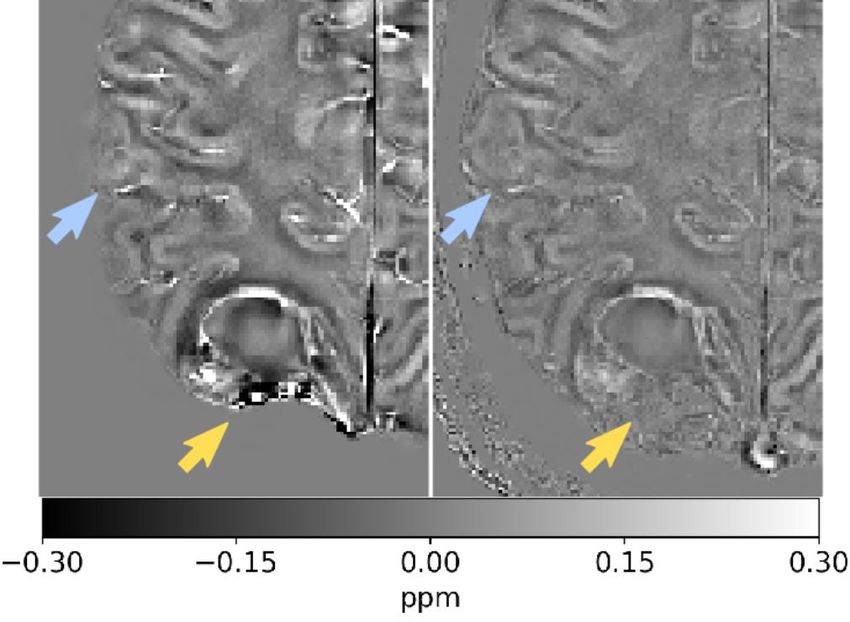
Steffen Bollmann | @sbollmann_MRI | www.mri.sbollmann.net

Acquisition Coil combination Unwrapping Masking Background Field Dipole Inversion

Unwrapping techniques
 • differentiable operator applied to the unwrapped phase can produce the
 same result on the wrapped phase -> Laplacian (Schofield and Zhu,

 Laplacian Opt. Lett. 2003)
 • + fast & robust
 • - introduces background phase

 Wrapped Phase Laplacian-unwrapped phase Difference from true phase

 27
Steffen Bollmann | @sbollmann_MRI | www.mri.sbollmann.net

Acquisition Coil combination Unwrapping Masking Background Field Dipole Inversion

Unwrapping techniques
 • differentiable operator applied to the unwrapped phase can produce
 the same result on the wrapped phase -> Laplacian (Schofield and
 Laplacian Zhu, Opt. Lett. 2003)
 • + fast & robust
 • - introduces background phase

 Region- • Identify discontinuities between regions
 • PRELUDE (Jenkinson MRM 2003) can take a while to compute for
 highly wrapped data

 growing • SEGUE (Karsa et al. TMI 2019) similar accuracy to PRELUDE, but
 faster

 28
Steffen Bollmann | @sbollmann_MRI | www.mri.sbollmann.net

Acquisition Coil combination Unwrapping Masking Background Field Dipole Inversion

Unwrapping techniques
 • differentiable operator applied to the unwrapped phase can produce the same result

 Laplacian on the wrapped phase -> Laplacian (Schofield and Zhu, Opt. Lett. 2003)
 • + fast & robust
 • - introduces background phase

 Region- • Identify discontinuities between regions
 • PRELUDE (Jenkinson MRM 2003) can take a while to compute for highly wrapped
 data
 growing • SEGUE (Karsa et al. TMI 2019) similar accuracy to PRELUDE, but faster

 • 3D voxel-by-voxel unwrapping guided by the quality of voxel connections
 Path Based • BEST PATH (Abdul-Rahman et al. AO 2007)
 • ROMEO (Dymerska et al. MRM 2021)

 Review: Robinson et al., NMR Biomed 2017 29
Steffen Bollmann | @sbollmann_MRI | www.mri.sbollmann.net

 Acquisition Coil combination Unwrapping Masking Background Field Dipole Inversion

 … to Quantitative Susceptibility Mapping (QSM)
GRE acquisition

 Background
 Field
 QSM

 Coil
 combination

 Thank you for slides and material: Simon Robinson, Ashley Stewart, Markus Barth, Francesco Cognolato 30
Steffen Bollmann | @sbollmann_MRI | www.mri.sbollmann.net

Acquisition Coil combination Unwrapping Masking Background Field Dipole Inversion

Why do we need to mask the object of interest?
• required for most background field correction algorithms to define inside/outside object of interest

• including unreliable phase values in the dipole inversion results in artifacts

 Haacke et al. MRI 2014 31
Steffen Bollmann | @sbollmann_MRI | www.mri.sbollmann.net

Acquisition Coil combination Unwrapping Masking Background Field Dipole Inversion

Why do we need to mask the object of interest?

• tradeoff between artifacts and

 masking out regions of interest

 32
Steffen Bollmann | @sbollmann_MRI | www.mri.sbollmann.net Acquisition Coil combination Unwrapping Masking Background Field Dipole Inversion So, masking isn’t trivial? • most QSM toolkits do not bring a masking procedure • commonly used for brain data: BET (Smith et al, HBM 2002) dedicated methods crucial for e.g. abdominal QSM improved masking for QSM (Straub et al., Tomography 2017) (Stewart et al., ISMRM 2021, #0725, #3971) BET QSMxT 33
Steffen Bollmann | @sbollmann_MRI | www.mri.sbollmann.net

 Acquisition Coil combination Unwrapping Masking Background Field Dipole Inversion

 … to Quantitative Susceptibility Mapping (QSM)
GRE acquisition

 Background
 Field
 QSM

 Coil
 combination

 Thank you for slides and material: Simon Robinson, Ashley Stewart, Markus Barth, Francesco Cognolato 34
Steffen Bollmann | @sbollmann_MRI | www.mri.sbollmann.net

Acquisition Coil combination Unwrapping Masking Background Field Dipole Inversion

Background Field Removal
 Unwrapped masked phase Internal tissue field background field

 Static field
 Susceptibility Shim coil inhomogeneities
 difference between fields
 tissue and air
 35
Steffen Bollmann | @sbollmann_MRI | www.mri.sbollmann.net

Acquisition Coil combination Unwrapping Masking Background Field Dipole Inversion

Background field correction methods & assumptions

 no sources close to • SHARP (Schweser et al. NI 2011)
 boundaries • V-SHARP (Li et al. NI 2011)

 no harmonic internal • LBV (Zhou et al. NMR Biomed 2014)
 and boundary fields

 no implicit boundary • RESHARP (Sun et al. MRM 2014)
 assumption • SHARQnet (Bollmann et al. Z Med Phys 2018)

 Review: Schweser et al. NMR Biomed 2017 36
Steffen Bollmann | @sbollmann_MRI | www.mri.sbollmann.net

 Acquisition Coil combination Unwrapping Masking Background Field Dipole Inversion

 … to Quantitative Susceptibility Mapping (QSM)
GRE acquisition

 Background
 Field
 QSM

 Coil
 combination

 Thank you for slides and material: Simon Robinson, Ashley Stewart, Markus Barth, Francesco Cognolato 37
Steffen Bollmann | @sbollmann_MRI | www.mri.sbollmann.net

Acquisition Coil combination Unwrapping Masking Background Field Dipole Inversion

QSM Dipole inversion

 field magnetic susceptibility
 Field-to-source
 perturbation distribution
 inversion

 Forward Model

 Review: Deistung et al. NMR Biomed 2017; Schweser et al. Z Med Phys 2016 38
Steffen Bollmann | @sbollmann_MRI | www.mri.sbollmann.net

Acquisition Coil combination Unwrapping Masking Background Field Dipole Inversion

QSM Dipole inversion

 magnetic
 field susceptibility
 Field-to-source
 perturbation distribution
 inversion

 Forward Model

 Review: Deistung et al. NMR Biomed 2017; Schweser et al. Z Med Phys 2016 39
Steffen Bollmann | @sbollmann_MRI | www.mri.sbollmann.net

Acquisition Coil combination Unwrapping Masking Background Field Dipole Inversion

Dipole inversion methods & assumptions
 • COSMOS (Liu et al. MRM 2009)
 multiple orientations • STI (Liu MRM 2010)
 • analytical solutions, but not practical

 • TKD (Shmueli et al., MRM 2009)
 inverse filtering • fast, but need parameter tweaking

 • LSQR (Li et al., Neuroimage 2015)
 iterative methods • MEDI (Liu et al., MRM 2013)
 • slow, need parameter tweaking

 • DeepQSM (Bollmann et al., NeuroImage 2019)
 agnostic deep learning • QSMnet (Yoon et al., NeuroImage 2018)
 • fast, but fragile

 • FINE (Zhang et al., Neuroimage 2020)
 hybrid methods • Variational Networks (Lai et al., arXiv 2020)
 • Deep learning priors + data consistency constraints

 Reviews: Schweser et al. NMR Biomed 2017; Jung et al. NMR Biomed 2020 40
Steffen Bollmann | @sbollmann_MRI | www.mri.sbollmann.net

Acquisition Coil combination Unwrapping Masking Background Field Dipole Inversion

Dipole inversion methods & assumptions
 • COSMOS (Liu et al. MRM 2009)
 multiple orientations • STI (Liu MRM 2010)
 • analytical solutions, but not practical

 • TKD (Shmueli et al. MRM 2009)
 inverse filtering • fast, but need parameter tweaking

 • LSQR (Li et al., Neuroimage 2015)
 iterative methods • MEDI (Liu et al., MRM 2013)
 • slow, need parameter tweaking

 • DeepQSM (Bollmann et al., TKD: effect2019)
 NeuroImage of various threshold choices
 agnostic deep learning • QSMnet (Yoon et al., NeuroImage 2018)
 • fast, but fragile (Deistung et al. NMR Biomed 2017)

 • FINE (Zhang et al., Neuroimage 2020)
 hybrid methods • Variational Networks (Lai et al., arXiv 2020)
 • Deep learning priors + data consistency constraints

 Reviews: Schweser et al. NMR Biomed 2017; Jung et al. NMR Biomed 2020 41
Steffen Bollmann | @sbollmann_MRI | www.mri.sbollmann.net

Acquisition Coil combination Unwrapping Masking Background Field Dipole Inversion

Dipole inversion methods & assumptions
 • COSMOS (Liu et al. MRM 2009)
 multiple orientations • STI (Liu MRM 2010)
 • analytical solutions, but not practical

 • TKD (Shmueli et al. MRM 2009)
 inverse filtering • fast, but need parameter tweaking

 • LSQR (Li et al. NI 2015)
 iterative methods • MEDI (Liu et al. MRM 2013)
 • slow, need parameter tweaking

 Error function Regularisation
 • DeepQSM (Bollmann et al., NeuroImage 2019)
 agnostic deep learning • QSMnet (Yoon et al., NeuroImage 2018) term
 • fast, but fragile

 • FINE (Zhang et al., Neuroimage 2020)
 hybrid methods • Variational Networks (Lai et al., arXiv 2020)
 Measured tissue
 • Deep learningEstimated
 priors + data tissue perturbation
 consistency constraints
 perturbation (forward model)
 Reviews: Schweser et al. NMR Biomed 2017; Jung et al. NMR Biomed 2020 42
Steffen Bollmann | @sbollmann_MRI | www.mri.sbollmann.net

Acquisition Coil combination Unwrapping Masking Background Field Dipole Inversion

Dipole inversion methods & assumptions
 • COSMOS (Liu et al. MRM 2009)
 multiple orientations • STI (Liu MRM 2010)
 • analytical solutions, but not practical

 • TKD (Shmueli et al. MRM 2009)
 inverse filtering • fast, but need parameter tweaking

 • LSQR (Li et al. NI 2015)
 iterative methods • MEDI (Liu et al. MRM 2013)
 • slow, need parameter tweaking

 • QSMnet (Yoon et al. NI 2018)
 agnostic deep learning • DeepQSM (Bollmann et al. NI 2019)
 • fast, but fragile

 • FINE (Zhang et al., Neuroimage 2020)
 hybrid methods • Variational Networks (Lai et al., arXiv 2020)
 • Deep learning priors + data consistency constraints

 Reviews: Schweser et al. NMR Biomed 2017; Jung et al. NMR Biomed 2020 43
Steffen Bollmann | @sbollmann_MRI | www.mri.sbollmann.net

Acquisition Coil combination Unwrapping Masking Background Field Dipole Inversion

Dipole inversion methods & assumptions
 • COSMOS (Liu et al. MRM 2009)
 multiple orientations • STI (Liu MRM 2010)
 • analytical solutions, but not practical

 • TKD (Shmueli et al. MRM 2009)
 inverse filtering • fast, but need parameter tweaking

 • LSQR (Li et al. NI 2015)
 iterative methods • MEDI (Liu et al. MRM 2013)
 • slow, need parameter tweaking

 • QSMnet (Yoon et al. NI 2018)
 agnostic deep learning • DeepQSM (Bollmann et al. NI 2019)
 • fast, but fragile

 • FINE (Zhang et al. NI 2020)
 hybrid methods • Variational Networks (Lai et al. arXiv 2020)
 • Deep learning priors + data consistency constraints

 Reviews: Schweser et al. NMR Biomed 2017; Jung et al. NMR Biomed 2020 44
Steffen Bollmann | @sbollmann_MRI | www.mri.sbollmann.net

 Acquisition Coil combination Unwrapping Masking Background Field Dipole Inversion

 … to Quantitative Susceptibility Mapping (QSM)
GRE acquisition

 Background
 Field
 QSM

 Coil
 combination

 Thank you for slides and material: Simon Robinson, Ashley Stewart, Markus Barth, Francesco Cognolato 45
Steffen Bollmann | @sbollmann_MRI | www.mri.sbollmann.net

Acquisition Coil combination Unwrapping Masking Background Field Dipole Inversion

Combined approaches (Background field + Dipole inversion)

 iterative • TGV-QSM (Langkammer et al. NI 2015)
 • SS-QSM (Chatnuntawech et al. NMR Biomed 2017)
 methods • TFI (Liu et al. MRM 2017)

 agnostic • AutoQSM (Wei et al. NI 2019)
 deep learning
 hybrid • NeXtQSM (Cognolato et al. ISMRM 2021,
 Submission 122)
 methods
 46
Steffen Bollmann | @sbollmann_MRI | www.mri.sbollmann.net

 QSM – The big picture
GRE acquisition

 Background
 Field
 QSM

 Coil
 combination

 Thank you for slides and material: Simon Robinson, Ashley Stewart, Markus Barth, Francesco Cognolato 47
Steffen Bollmann | @sbollmann_MRI | www.mri.sbollmann.net

 Some Processing Packages for QSM
STI Suite (Matlab) - https://people.eecs.berkeley.edu/~chunlei.liu/software.html
 • Laplacian Phase Unwrapping, Background field correction (vSHARP, iHARPERELLA)
 • Dipole inversion (iLSRQ + STAR QSM)
MEDI toolkit (Matlab) - http://pre.weill.cornell.edu/mri/pages/qsm.html
 • from DICOM to QSM using MEDI framework

FANSI Toolbox (Matlab) - https://gitlab.com/cmilovic/FANSI-toolbox
 • various unwrapping, background field and Dipole inversion methods

SEPIA (Matlab) - https://github.com/kschan0214/sepia
 • GUI for MEDI, STI Suite, FANSI, SEGUE, NDI

QSMxT (Python) - https://github.com/QSMxT
 • DICOM/BIDS, robust masking, NiPype + TGV QSM, integrated anatomical segmentation, optimized for high throughput
 processing on HPCs

 48
Steffen Bollmann | @sbollmann_MRI | www.mri.sbollmann.net

Referencing in QSM
 QSM values are relative to the water centre frequency of the scan-session
 -> consider re-referencing for group studies?
 The ideal reference tissue is a debated Reference region candidates:
 topic.
 ● Cerebrospinal fluid in the
 Challenges: ventricles
 ● The tissue should have very low ● Whole-brain average
 inter-subject variance in age and susceptibility
 pathology ● Red nucleus
 ● The tissue should be easy to ● Cortical gray matter
 segment ● Superior frontal white matter
 ● Quantification should be reliable ● Splenium of the corpus callosum
 ● Other white matter structures

 Straub et al. MRM 2016 49
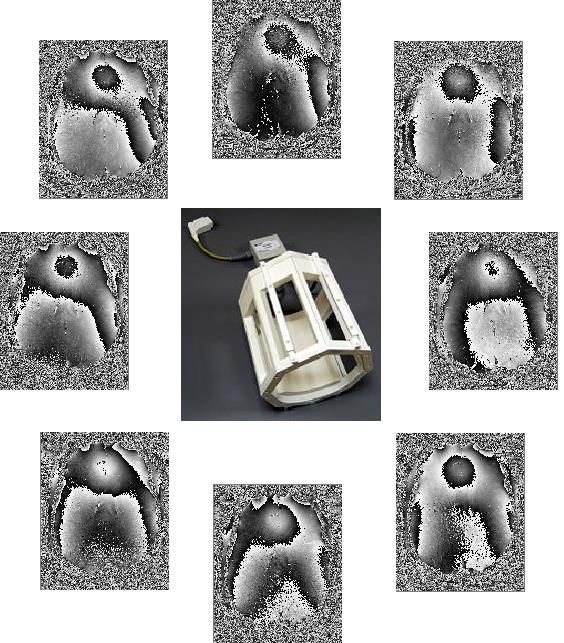
Steffen Bollmann | @sbollmann_MRI | www.mri.sbollmann.net

QSM - The bigger picture ….
• Why are there so many methods to compute QSM and which of them is correct?
 • COSMOS?
 • STI33?
 • The winners of the reconstruction challenges?

 50
Steffen Bollmann | @sbollmann_MRI | www.mri.sbollmann.net

QSM - The bigger picture ….
• Why are there so many methods to compute QSM and which of them is correct?

 ... all models are approximations.
 Essentially, all models are wrong,
 but some are useful. However, the
 approximate nature of the model
 must always be borne in mind....

 George Box & Norman
 Draper, Empirical Model-
 Building and Response
 Surfaces, 1987
 https://en.wikipedia.org/wiki/All_models_are_wrong 51
Steffen Bollmann | @sbollmann_MRI | www.mri.sbollmann.net

How could a useful QSM method look like?
 Fast, robust, no-parameter tweaking (deep learning + data consistency?)

 sensitive to clinical questions?

 developed in an open & high performance language (Julia?)

 automatic and robust masking in brain, joints, body, animal models?

 from DICOMS to results without conversion hassles?

 integrated in the scanner platforms?

 integrated referencing and segmentation to extract values from regions of interest?

 52
Thank you

 s.bollmann@uq.edu.au

 www.mri.sbollmann.net
 @sbollmann_MRI
 github.com/sbollmannmri
CRICOS code 00025B

 https://www.emtphub.org/
You can also read