Improvement Effect of Metformin on Female and Male Reproduction in Endocrine Pathologies and Its Mechanisms
←
→
Page content transcription
If your browser does not render page correctly, please read the page content below
pharmaceuticals

Review
Improvement Effect of Metformin on Female and Male
Reproduction in Endocrine Pathologies and Its Mechanisms
Alexander O. Shpakov

                                           I.M. Sechenov Institute of Evolutionary Physiology and Biochemistry of Russian Academy of Sciences,
                                           194223 Saint Petersburg, Russia; alex_shpakov@list.ru; Tel.: +7-812-5523117

                                           Abstract: Metformin (MF), a first-line drug to treat type 2 diabetes mellitus (T2DM), alone and
                                           in combination with other drugs, restores the ovarian function in women with polycystic ovary
                                           syndrome (PCOS) and improves fetal development, pregnancy outcomes and offspring health in
                                           gestational diabetes mellitus (GDM) and T2DM. MF treatment is demonstrated to improve the
                                           efficiency of in vitro fertilization and is considered a supplementary drug in assisted reproductive
                                           technologies. MF administration shows positive effect on steroidogenesis and spermatogenesis
                                           in men with metabolic disorders, thus MF treatment indicates prospective use for improvement
                                           of male reproductive functions and fertility. MF lacks teratogenic effects and has positive health
                                           effect in newborns. The review is focused on use of MF therapy for restoration of female and
                                           male reproductive functions and improvement of pregnancy outcomes in metabolic and endocrine
                                           disorders. The mechanisms of MF action are discussed, including normalization of metabolic and
                                           hormonal status in PCOS, GDM, T2DM and metabolic syndrome and restoration of functional activity
                                           and hormonal regulation of the gonadal axis.

                                           Keywords: metformin; diabetes mellitus; gestational diabetes mellitus; polycystic ovary syndrome;
                                           in vitro fertilization; ovary; testes; insulin; gonadotropin; folliculogenesis; steroidogenesis


Citation: Shpakov, A.O.
Improvement Effect of Metformin on         1. Introduction
Female and Male Reproduction in
                                                 Metformin (1,1-dimethyl biguanide hydrochloride) (MF), an orally administered
Endocrine Pathologies and Its
Mechanisms. Pharmaceuticals 2021, 14,
                                           biguanide, is a first-line drug for the treatment of type 2 diabetes mellitus (T2DM). It
42. https://doi.org/10.3390/ph
                                           reduces the adipose tissue mass and increases the tissue sensitivity to insulin, thereby
14010042                                   reducing hyperglycemia, normalizing carbohydrate and lipid metabolism and preventing
                                           inflammation and oxidative stress in the tissues [1,2]. MF is also used to treat non-alcoholic
Received: 11 November 2020                 fatty liver disease [3], coronary artery disease [4,5], acute kidney injury and chronic kidney
Accepted: 6 January 2021                   disease [6], in patients with T2DM, metabolic syndrome (MetS) and obesity, and in patients
Published: 8 January 2021                  without apparent symptoms of metabolic disorders [7]. There are numerous clinical
                                           and experimental studies indicating the effectiveness of MF as an anticancer drug, used
Publisher’s Note: MDPI stays neu-          to prevent the growth and metastasis in breast cancer [8,9], endometrial cancer [10–12],
tral with regard to jurisdictional clai-   colorectal cancer [9,13], prostate cancer [14] and in a number of the other tumors [15,16].
ms in published maps and institutio-             Currently, there is a large body of evidence for the effectiveness of MF therapy in
nal affiliations.                          restoration of reproductive functions and fertility in women with polycystic ovary syn-
                                           drome (PCOS), gestational diabetes mellitus (GDM) and T2DM, as well as to improve the
                                           effectiveness of the assisted reproductive technologies (ART), such as in vitro fertilization
Copyright: © 2021 by the author. Li-
                                           (IVF) and intracytoplasmic sperm injection (ICSI). Important attributes of MF use in the
censee MDPI, Basel, Switzerland.
                                           treatment of pregnant women with PCOS and T2DM include its lack of teratogenic effect
This article is an open access article
                                           and established positive effect on fetal development, pregnancy outcomes and newborn
distributed under the terms and con-       health. Moreover, convincing evidence has been obtained for the restorative effects of
ditions of the Creative Commons At-        MF on steroidogenic and spermatogenic functions in men with diabetes mellitus (DM)
tribution (CC BY) license (https://        and MetS. This review offers an overview of problems when utilizing MF therapy for the
creativecommons.org/licenses/by/           correction of reproductive dysfunctions in women and men and includes the analysis of
4.0/).                                     possible mechanisms for positive effects of MF on reproduction. The review also includes

Pharmaceuticals 2021, 14, 42. https://doi.org/10.3390/ph14010042                                    https://www.mdpi.com/journal/pharmaceuticals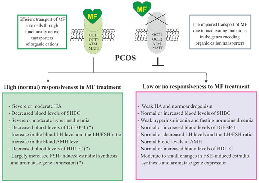
Pharmaceuticals 2021, 14, 42                                                                                          2 of 45

                               only a brief description of the molecular mechanisms of MF action in target cells; these
                               mechanisms are the focus of other review articles [17–24].

                               2. Summary of Cell Targets and Molecular Mechanisms of Action of Metformin
                                    The signaling pathways of MF in cells of human and mammals are still not fully
                               understood, they seem to be dependent on species and cell type, as well as doses and
                               routes of administration, along with metabolic and hormonal status of subjects [22,24–27].
                                    The molecule of MF, a small hydrophilic cation, is transported from the extracellular
                               space to the cytoplasm of the target cell through organic cation transporters-1 and -2 (OCT1,
                               OCT2), multidrug and toxin extrusion transporters (MATE), and ATM (ataxia telangiectasia
                               mutated) transporter, and OCT1 and OCT2 are considered as the main functional units of
                               MF transmembrane transport [28]. The transfer of MF across the placental barrier during
                               pregnancy is largely dependent on the transporter OCT3 [29]. The ultimate intracellular
                               target for MF is the 50 -adenosine monophosphate-activated protein kinase (AMPK), the key
                               energy sensor of the cell, although MF does not interact directly with the enzyme [22,30–32].
                               In pathological conditions, like T2DM and MetS, the activity of AMPK is reduced. MF’s
                               action increases the activity of AMPK, and consequently normalizes the energy metabolism
                               of the target cell. The AMPK consists of a catalytic α-subunit and the regulatory β- and
                               γ-subunits that form a functionally active αβγ-heterotrimeric complex, and is widely
                               distributed in all subcellular compartments (cytoplasmic, lysosomal, mitochondrial, and
                               nuclear). AMPK is activated by increasing levels of AMP, a positive allosteric regulator
                               of the enzyme [31,33,34]. The interaction of AMP with the adenine nucleotides-binding
                               sites located in the γ subunit leads to stabilization of the αβγ heterotrimeric complex
                               and enables phosphorylation of the α-subunit by liver kinase B1 (LKB1), which leads
                               to the increase in AMPK activity [31,32,35] (Figure 1). Activating phosphorylation of
                               AMPK may be also mediated by Ca2+ -calmodulin-dependent protein kinase kinase 2
                               (CaMKK2) [36,37] and transforming growth factor β activated kinase-1 (TAK1) [38–40], but
                               LKB1 is most important for AMPK activation [31,34,41–43]. Allosteric binding of AMP and
                               ADP to γ-subunit of AMPK increases the ability of LKB1 and CaMKK2 to phosphorylate
                               AMPK α-subunit at the Thr172 [44–46]. In the lysosomes, the “non-canonical” pathway
                               of LKB1-mediated AMPK activation is carried out through dissociation of fructose 1,6-
                               bisphosphate from aldolase. At the lysosomal surface, free aldolase promotes the formation
                               of a multiprotein complex, including the vacuolar H+ -ATPase and the scaffold protein
                               AXIN, and this complex ensures the effective binding between AMPK and LKB1, thereby
                               activating AMPK [47,48]. A negative regulator of AMPK is the protein phosphatase
                               2C (PP2C), which dephosphorylates and inactivates the α-subunit of AMPK, causing
                               the dissociation of the αβγ-heterotrimeric complex. Elevated levels of AMP lead to an
                               inhibition of PP2C activity, which allows AMPK to remain stable in the active Thr172 -
                               phosphorylated state [49,50].
                                    MF penetrates into the mitochondria through intracellular space and accumulates in
                               them. While in the mitochondria, MF inhibits the mitochondrial ETC complex I, which
                               leads to decrease in ATP production and increase in the [AMP]i /[ATP]i and [ADP]i /[ATP]i
                               ratios [51–54]. Moreover, MF decreases the activity of the enzyme AMP-deaminase (AMPD),
                               which converts AMP to inosine monophosphate, inducing the accumulation of AMP
                               within the cell [55]. The MF-induced increase in the intracellular AMP level leads to
                               the activation of AMPK as described above [41,56]. The MF effect on AMPK activity is
                               observed at drug concentrations below 80 µM, which are achieved with oral administration
                               of therapeutic doses of MF [57]. The MF-induced activation of AMPK results in the
                               stimulation of energy-producing catabolic pathways that mediate the increased glucose
                               uptake by cells, the increased expression and activity of the membrane glucose transporters,
                               the activated metabolic processes such as glycolysis and oxidative phosphorylation, and
                               the normalization of mitochondrial biogenesis [20,24,58,59]. The MF-induced AMPK
                               stimulation leads to phosphorylation of types 1 and 2 acetyl-CoA carboxylases (ACC1 and
                               ACC2), inducing an inhibition of lipogenesis and stimulation of the β-oxidation of free
Pharmaceuticals 2021, 14, x FOR PEER REVIEW 3 of 46
Pharmaceuticals 2021, 14, 42 3 of 45
and the normalization of mitochondrial biogenesis [20,24,58,59]. The MF‐induced AMPK
stimulation leads to phosphorylation of types 1 and 2 acetyl‐CoA carboxylases (ACC1 and
ACC2), inducing an inhibition of lipogenesis and stimulation of the β‐oxidation of free
fatty acids [60–62] (Figure 1). The ultimate results of this metabolic cascade is the decrease
of T2DM-
T2DM‐ andand MetS-produced
MetS‐produceddyslipidemia,
dyslipidemia,and andthe
the normalization
normalization of of lipid
lipid metabolism.
metabolism. In
In addition,
addition, thethe AMPK
AMPK activation
activation induces
induces a plethora
a plethora of cellular
of cellular events,
events, including
including regula‐
regulation
of autophagy
tion and and
of autophagy apoptotic processes,
apoptotic a decrease
processes, in the
a decrease in activity of inflammatory
the activity of inflammatoryfactors,
fac‐
including nuclear factor κB (NF-κB) and interleukin 1β, an inhibition of the ROS
tors, including nuclear factor κB (NF‐κB) and interleukin 1β, an inhibition of the ROS pro‐production,
a decrease
duction, in the ERin
a decrease stress,
the ERas stress,
well asas
a decrease
well as a in insulin/IGF-1-induced
decrease activationactiva‐
in insulin/IGF‐1‐induced of the
mTORC1/2
tion complexescomplexes
of the mTORC1/2 and a decrease
and aindecrease
the protein synthesis
in the protein[60,62–66].
synthesis [60,62–66].
Figure
Figure 1.1. The
The cellular
cellularmechanisms
mechanismsof ofmetformin
metforminaction
actionwhich
whichare arecarried
carriedout
outbyby activation
activationofof
thethe
AMP‐activated protein kinase and inhibition of the mitochondrial electron transport
AMP-activated protein kinase and inhibition of the mitochondrial electron transport chain complex chain com‐
plex I. Abbreviations: AC, adenylyl cyclase; ACC1/2, acetyl‐CoA carboxylases 1 and 2; AMPD,
I. Abbreviations: AC, adenylyl cyclase; ACC1/2, acetyl-CoA carboxylases 1 and 2; AMPD, AMP
AMP deaminase; AMPK, the heterotrimeric AMP‐activated protein kinase consisting of the α1/2
deaminase; AMPK, the heterotrimeric AMP-activated172 protein kinase consisting of the α1/2 (the target
(the target for activation phosphorylation 172 at the Thr ), β1/2 and γ1/2/3 subunits; CREB, cAMP‐
for activation
activated phosphorylation
transcription at theresponse
factor (cAMP Thr ), β1/2 and γ1/2/3protein);
element‐binding subunits; CREB,
ETC cAMP-activated
complex I, the mito‐
transcription factor (cAMP response element-binding protein); ETC
chondrial NADH‐dehydrogenase complex, the first complex of the respiratory electron complex I, the mitochondrial
transport
NADH-dehydrogenase
chain; FA, fatty acids; LKB1,complex, the firstB1;
liver kinase complex
mG3PDH, of the respiratory electron
mitochondrial transport chain;
glycerol‐3‐phosphate dehy‐FA,
fatty acids; mTORC2,
drogenase; LKB1, liverthe kinase
mTOR B1;complex
mG3PDH, mitochondrial
2; NFκB, glycerol-3-phosphate
nuclear factor dehydrogenase;
κB; OCT1/2, the organic cations
transporters
mTORC2, the1 mTORand 2; complex
pCBP, the 2; Ser 436‐phosphorylated form of CREB‐binding protein with acetyl‐
NFκB, nuclear factor κB; OCT1/2, the organic cations transporters 1
transferase activity, a
436co‐activator
and 2; pCBP, the Ser -phosphorylated form of the factor
of CREB; PDE4B, protein
CREB-binding cAMP‐specific 3′,5′‐cyclic phos‐
with acetyltransferase activity,
phodiesterase 4B; PKA, cAMP‐dependent protein kinase; PP2C,
0 0 protein
a co-activator of the factor CREB; PDE4B, cAMP-specific 3 ,5 -cyclic phosphodiesterase phosphatase 2C; ROS,
4B; PKA,
reactive oxygen species.
cAMP-dependent protein kinase; PP2C, protein phosphatase 2C; ROS, reactive oxygen species.
The
The MF
MF is
is aa functional
functional antagonist
antagonist of of cAMP‐dependent signaling cascades,
cAMP-dependent signaling cascades, which
which are
are
stimulated by hormones, glucagon in particular, through the G protein‐coupled
stimulated by hormones, glucagon in particular, through the Gs protein-coupled receptors
s receptors
and the membrane-bound
and the membrane‐bound forms forms ofof adenylyl
adenylyl cyclase
cyclase (AC)
(AC) [67,68].
[67,68]. The
The stimulation
stimulation of of AC
AC
results in
in an
anincrease
increaseininthetheintracellular
intracellular cAMP
cAMP level
level andand
thethe activation
activation of the
of the protein
protein ki‐
kinase
nase
A A (PKA)
(PKA) and theandcAMP-activated
the cAMP‐activated transcription
transcription factorfactor
CREBCREB (cAMP(cAMP response
response ele‐
element-
ment‐binding
binding protein).protein). The MF‐induced
The MF-induced activation
activation of AMPK
of AMPK promotes
promotes phosphorylation
phosphorylation and
and activation of cAMP‐specific 0 03′,5′‐cyclic phosphodiesterase 4B (PDE4B),
activation of cAMP-specific 3 ,5 -cyclic phosphodiesterase 4B (PDE4B), thereby reducing thereby reduc‐
ing the intracellular
the intracellular levellevel of cAMP
of cAMP [68].[68]. Moreover,
Moreover, MF causes
MF causes an increase
an increase in intracellular
in the the intracel‐
lular
level level of AMP,
of AMP, a negative
a negative regulator
regulator of theof the catalytic
catalytic site ofsite
AC,ofwhich
AC, which leads
leads to to inhibi‐
inhibition of
tionactivity
AC of AC activity and a decrease
and a decrease in cAMP in production.
cAMP production. An increase
An increase in the
in the level oflevel
AMPofcan AMPbe
the result of both inhibition of the mitochondrial ETC complex I, and suppression of the
activity of AMP deaminase [55,69] (Figure 1). A decrease in the activity of cAMP-dependent
pathways in the liver, like activation of AMPK, leads to the inhibition of glucose synthesisPharmaceuticals 2021, 14, 42 4 of 45
in hepatocytes. Furthermore, MF-induced AMPK activation induces the protein kinase
ι/λ-mediated phosphorylation of cyclic AMP response element binding (CREB)-binding
protein (CBP or CREBBP) at the Ser436 , which leads to the inability of the phospho-CBP to
form a functionally active complex with the factor CREB and thereby inhibits the cAMP-
dependent gene transcription [70].
Along with AMPK-dependent, there are also AMPK-independent pathways of MF
action on the intracellular effector systems and gene expression. High-dose MF inhibits the
activity of the mitochondrial glycerol-3-phosphate dehydrogenase (mG3PDH) [71]. The
inhibition of mG3PDH leads to an increase in NADH levels and decreases NAD+ levels,
and this causes a deficiency in NAD+ , which is involved in the conversion of lactate to
pyruvate (Figure 1). Since a decrease in mG3PDH activity inhibits the conversion of lactate
to glucose, the result of impaired gluconeogenesis in hepatocytes is an accumulation of
lactate, which can cause lactic acidosis in the conditions of high-dose MF treatment [71,72].
Another target of MF is the enzyme H3K27me3-demethylase KDM6A/UTX, which is
responsible for the transcriptional activity of a large number of genes [73].
The antidiabetic effects of MF may be due to the changes in the gut microbiota,
due to stimulation of the growth of bacteria that produce short-chain fatty acids [74]. By
modulating the composition of the microbiota in rodents with T2DM and MetS, MF reduces
the levels of bacterial lipopolysaccharides in the blood [75], and activates AMPK-dependent
pathways in the mucosal layer of the intestine, reducing glucose absorption [76].
The most important mechanism of action of MF on target cells is the enhancement of
the insulin signaling pathways and the decrease in insulin resistance (IR). This may be due
to inhibition of hyperactivated nuclear factor κB (NF-κB), a transcription factor that pro-
vokes the development of IR, as well as a decrease in the expression of the phosphatase and
tensin homolog (PTEN), which dephosphorylates phosphatidylinositol-3,4,5-triphosphate
and thereby prevents insulin-induced stimulation of Akt kinase, a key effector component
in the 3-phosphoinositide signaling pathway. The inhibitory effect of MF on the activity
of NF-κB-dependent signaling pathways is carried out mainly through the stimulation of
AMPK [25,77,78]. Since NF-κB plays a key role in inflammatory reactions, its inhibition
by MF promotes the weakening of inflammation and increases the cell survival, and these
effects of MF are prevented by AMPK inhibitors [25,79,80].
3. Metformin and Polycystic Ovary Syndrome
3.1. Pathophysiology of Polycystic Ovary Syndrome
The PCOS occurs in average from 9% to 18% of women of reproductive age and
includes a number of metabolic and endocrine dysfunctions [81]. Some of them are: (i) the
ovarian dysfunction, characterized by irregular or no ovulation (oligo- or amenorrhea), the
increased secretion of androgens (hyperandrogenism, HA) and estrogens, the endometrial
hyperplasia and the increased size of the ovaries, (ii) the pancreatic dysfunction leading
to insulin hypersecretion and, as a result, to insulin resistance (IR) development, (iii) the
adrenal dysfunction, which leads to hyperproduction of androgens, and (iv) the functional
changes in the hypothalamic and pituitary links of the female hypothalamic-pituitary-
gonadal (HPG) axis [82–85]. Since these dysfunctions and changes are usually associated
with obesity, MetS and T2DM, the PCOS is much more common in women with these
metabolic disorders (on average in 30% of cases), with a significant proportion of PCOS
patients having IR with accompanying compensatory hyperinsulinemia [86–91]. Accord-
ing to the Rotterdam criteria (2003), the main diagnostic criteria for PCOS are clinical or
biochemical HA, oligo- or amenorrhea associated with chronic anovulation, and morpho-
logical features of PCOS, which include 12 or more follicles (2 to 9 mm) in each ovary
and/or an increase in ovarian volume over 10 mL [92–94]. It should be noted that about
80% of women with anovulatory infertility have typical signs of PCOS [81].
The etiology and clinical manifestations of PCOS depend on many factors, as well
as combinations and interactions between them. The genetic predisposition [95–98] and
epigenetic factors, including an increased level of gene methylation, histone modification,Pharmaceuticals 2021, 14, 42 5 of 45
and microRNA pattern variation [99–101], are important for the development of PCOS.
Environmental and socioeconomic factors are also of great importance, including ethnic
characteristics, nutrition, and adverse environmental factors (toxins, xenobiotics, chemical
mutagens, and ionizing radiation) [102–104]. The development of PCOS in women largely
depends on the effects of maternal hormones during the prenatal period, as well as on their
metabolic and hormonal status in the early childhood [84,99,105–107].
3.2. The Use of Metformin in PCOS Women
In recent years, MF therapy has become widely used for correction of the metabolic and
hormonal impairments in women with PCOS and for restoration of their reproductive func-
tions [85,108–110], including the improvement of IVF/ICSI outcomes in PCOS [111–114].
MF is most effective in treating PCOS patients with the metabolic disorders such as T2DM,
obesity, dyslipidemia, and severe IR [85,115,116]. This is majorly attributed to the allevi-
ation of negative effects of these disorders on the female reproduction by MF, increased
tissues sensitivity to insulin, improved lipid and glucose metabolism and cell metabolism,
and reduced inflammation and oxidative stress in the ovaries as well as in other tissues.
In cases where significant metabolic changes in PCOS patients are not observed during
treatment, MF therapy can lead to energy and hormonal imbalance. The outcomes may be
the opposite of improvement, but a further deterioration in reproductive functions. This
possibility is supported by the data from clinical trials on metabolic changes, including an
increase in fasting glucose clearance and endogenous glucose production [117,118], as well
as changes in the microbiota in non-diabetic individuals [119], as well as data on metabolic
and hormonal dysfunctions in normal rodents, for a long time receiving MF [120].
There is a lot of clinical evidence of the high efficacy of MF in PCOS, which makes
it feasible to consider MF as a second-line drug for ovulation induction in women with
PCOS [109,121–128]. MF is recommended for the induction of ovulation in PCOS women
who are either resistant to clomiphene citrate (CC) or require antiandrogen therapy without
the use of contraceptives [125], as well as in PCOS patients with severe obesity and impaired
lipid metabolism [114]. One very important consideration during PCOS treatment with
MF is that drug has no or little adverse effects on the outcomes of pregnancy as well as
the health of fetus and newborn, which indicates the safety of MF therapy [126,127]. The
gastrointestinal side effects of MF have been reported in a number of cases, but these effects
did not significantly affect the health of PCOS women [108,112].
The MF treatment of PCOS women normalizes the frequency and regularity of ovula-
tion, including when co-administered with exogenous gonadotropins [112,129,130]. This
suggests that MF can also affect the sensitivity of ovarian cells to gonadotropins, which
is important for the ART. As a result, during the ART, the most promising approach is
the combined use of MF with gonadotropins [111,113]. In PCOS, MF improves clinical
pregnancy rates and live birth rates [108,111–113,131–136], and also reduces the number of
miscarriages and increases the rate of embryo implantation [137,138].
There is evidence of a positive effect of MF on the effectiveness of IVF and IVF/ICSI
in PCOS women [114]. It is believed to be due to the normalization of metabolic and
hormonal parameters and the androgen levels in PCOS, which leads to an improvement of
embryo implantation, an increase in the ovarian response to gonadotropins and a decrease
in the rates of miscarriage [112,135,138–141]. The increased gonadotropin sensitivity allows
avoiding the use of high-dose gonadotropins and, thereby, preventing the ovarian hyper-
stimulation syndrome (OHSS), a severe complication of gonadotropin-induced ovulation
induction. However, it should be noted that some data on the use of MF in the ART technol-
ogy in PCOS women are not so unambiguous, and there are results that do not support the
efficacy of MF in IVF/ICSI. The clinical studies carried out by Egyptian group of physicians
showed no improvement in IVF rates in PCOS women who received MF [142]. However,
in this study, overweight or obese PCOS women received short-term courses of low-dose
MF (1000 mg/day), from the start of ovarian stimulation with gonadotropins until proof
of clinical pregnancy. As a result, in this case, the period of time for the manifestationPharmaceuticals 2021, 14, 42 6 of 45
of the restorative effects of MF on the ovaries and folliculogenesis in PCOS patients may
not have been long enough. Potentially, for an adequate estimation of MF effectiveness in
PCOS patients it is necessary to separate them in the groups, based on the severity and
duration of the disease and in the body mass index [108,114], as well as the severity of IR,
dyslipidemia and hyperglycemia.
3.3. Combined Use of Metformin with Clomiphene Citrate, Letrozole, Liraglutide, Saxagliptin,
or Oral Contraceptives
A promising approach to treat PCOS is the use of combination of MF with the other
drugs that improve the ovarian function and metabolic parameters in PCOS, with the best
candidates for co-administration are CC, a mild nonsteroidal estrogen antagonist belonging
to the family of selective estrogen receptor modulators, and letrozole, a non-steroidal aro-
matase inhibitor that prevents the conversion of androgens to estrogens [116,131,143–148].
The CC is the main drug of choice for treatment of PCOS, yet a significant proportion of
PCOS women have weak or no response to CC therapy. Therefore, a search is underway for
drugs that can potentiate the therapeutic effects of CC in PCOS, and MF is one of the most
promising candidates [108,131,134,143,144,146]. Combined use of MF plus CC in PCOS
showed significant improvement in clinical indices of pregnancy and the combination
therapy is more effective than the use of CC alone. However, a number of studies reported
no effect [149] or relatively weak potentiating effect of MF for CC therapy [134]. One of the
possible reasons for these contradictory results may be the difference in the sensitivity to
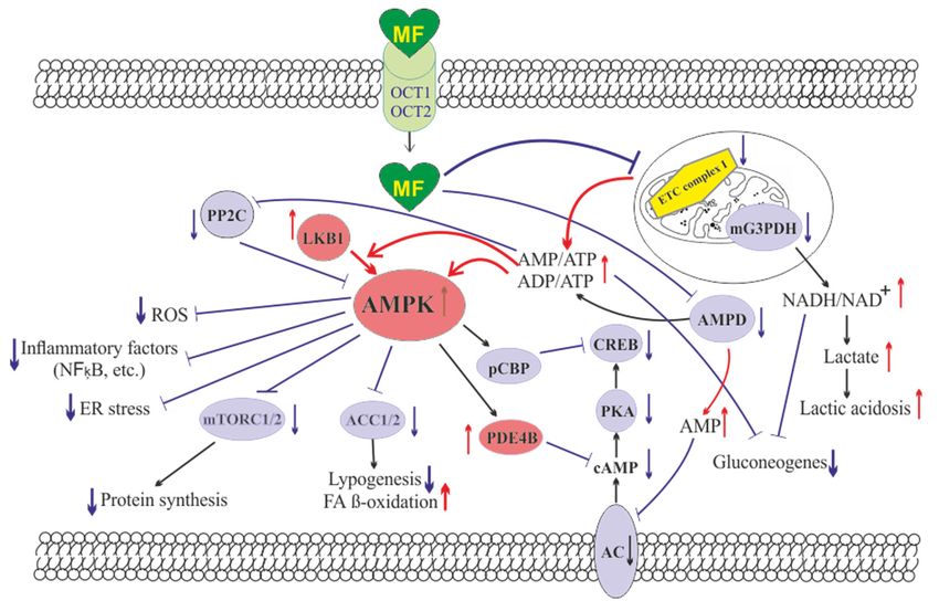
CC and MF in PCOS patients. The most profound potentiating effect of MF on the induction
of ovulation and pregnancy rates is found in patients with a pronounced resistance to
CC [144,146,150]. However, some PCOS patients may be also insensitive to MF, which
is due to many factors, including the polymorphisms and inactivating mutations in the
transmembrane proteins facilitating intracellular transport MF [151]. As a result, the
combined therapy is expected to benefit mainly PCOS patients with reduced sensitivity to
CC, pronounced obesity, IR and dyslipidemia, and high sensitivity to MF.
In recent years, the data have been obtained for the effectiveness of the combined use of
MF and letrozole, an aromatase inhibitor that is widely used to restore the ovarian cycle and
induction of ovulation and improves oocyte implantation and pregnancy rates in women
with PCOS, primarily those with reduced sensitivity to CC [144,148,150]. The combined
therapy with MF demonstrated enhancement for the improving effects of letrozole on
the pregnancy and live birth rates. Moreover, there are clinical results showing that the
combined use of letrozole and MF is more effective than the combined use of CC and
MF [145,147].
In PCOS patients, the efficiency of MF therapy is increased when MF is used with
oral estrogen-progestin contraceptives, both acting similar, by suppressing ovarian an-
drogen overproduction and normalizing menstrual cycle, most noticeably in obese PCOS
women [152,153]. On a contrary, when the same combined treatment (MF and oral con-
traceptives) is used for PCOS women with normal or reduced body weight, it results
in a decrease in their muscles mass, leads to the water retention and the formation of
an “osteosarcopenic” phenotype [154]. Two main reasons are behind the decrease in the
muscles mass during combined therapy. First, MF and oral contraceptives reduce the blood
androgen levels. It is known that in PCOS there is a significant positive correlation between
the blood level of androgens and the muscles mass [155]. Second, MF-induced activation
of AMPK and changes in mitochondrial energy status stimulate catabolic processes in the
muscles tissue, which leads to muscles atrophy, as shown in patients with T2DM [156]. In
this regard, it should be noted that MF treatment of T2DM patients leads to an increase in
the blood level of fibroblast growth factor 21 (FGF21), which is one of the specific markers
of muscles damage and degeneration [157]. Thus, it is highly recommended to take into
account the proportion of the muscle tissue and body mass index in PCOS women, as well
as the severity of HA when considering the option of using the combined therapy of MF
and oral contraceptives [154].Pharmaceuticals 2021, 14, 42 7 of 45
The agonists of glucagon-like peptide-1 (GLP-1) receptor and the inhibitors of dipep-
tidyl peptidase-4 are widely used to treat T2DM and MetS [158–161], but they can also be
used to correct the metabolic alterations and IR in PCOS women, as well as in pregnant
women with GDM and T2DM [162,163]. It is shown that MF enhances the beneficial
effect of liraglutide, a selective GLP-1 receptor agonist, on insulin sensitivity and glu-
cose homeostasis. The 12-week treatment of 30 obese PCOS women with a combination
of MF (1000 mg twice a day) and liraglutide (1.2 mg/day) causes a decrease in IR and
normalizes the sensitivity of patients to glucose, and the combined therapy was more
effective than monotherapy [164]. The treatment of premenopausal PCOS women with
MF (2000 mg/day), saxagliptin (5 mg/day), an inhibitor of dipeptidyl peptidase-4, or a
combination of MF and saxagliptin leads to normalization of glucose tolerance on average
of 56% of patients [165]. Moreover, in the group treated with MF alone or saxagliptin
alone, the improvement of glycemic control is demonstrated only in 25 and 55% of patients,
respectively, while the combined therapy restores glucose tolerance in 91% of women with
PCOS [165]. A high efficacy of the combined therapy was shown by other group of authors
who monitored the 16-week treatment of 38 women with pre-diabetes and PCOS using
the MF plus saxagliptin [166]. Weight loss and decrease in hyperglycemia and IR, which
are induced by treatment of obese PCOS patients with GLP-1 receptor agonists, lead to a
decrease in HA [167–169] and an improvement in menstrual frequency [167,169]. Liraglu-
tide, an analogue of GLP-1, normalizes the menstrual cycle and fertility in women with
HAIR-AN syndrome, which is due to a decrease in the levels of androgens and insulin [170].
Consequently, in PCOS patients, MF-induced potentiation of the metabolic-improving
effects of GLP-1 agonists may also increase their restorative effects on the menstrual cycle
and fertility.
3.4. The Mechanisms of Metformin Effects on Reproductive Functions in PCOS
3.4.1. Metformin-Induced Inhibition of Hyperandrogenism and Normalization of the
Steroid Hormones Balance
One of the main mechanisms of the restorative effect of MF on ovarian function, ovu-
lation and pregnancy in PCOS women mediates through the pronounced antiandrogenic
effect of MF, both in monotherapy and in combination with other drugs [141,152,171–179].
One-year treatment of overweight PCOS women with MF (1700 mg/day) reduced the
levels of free testosterone, dehydroepiandrosterone (DHEA) and androstenedione, and sig-
nificantly weakened the signs of hirsutism. This effect of MF was strongly associated with
a decrease in the homeostasis model assessment of insulin resistance index (HOMA-IR)
and an improvement in glucose tolerance, but was weakly associated with a decrease in the
body weight, which indicates a main contribution of a decrease in IR and hyperinsulinemia
to the antiandrogenic effect of MF [180]. An antiandrogenic effect was demonstrated in the
treatment of overweight and obese adolescents with MF (1000–2000 mg/day), and was
accompanied by a significant decrease in IR [152,173,174,176,177]. MF reduced both the
basal and gonadotropin-stimulated testosterone levels, and these effects were observed
even with short-term MF treatment. The administration of MF for two days to PCOS
women caused a decrease in their testosterone levels stimulated by luteinizing hormone
(LH). This effect was not due to a decrease in the body weight and the changes in metabolic
indices, pointing to potential direct influence of MF on steroidogenic activity in ovarian
cells [181].
In PCOS, the severity of IR is positively correlated with the severity of HA and
dysregulations of the ovulatory cycle. The PCOS women with oligomenorrhea and without
HA usually do not have IR, while the PCOS women with oligomenorrhea and HA often
show significant signs of IR [182]. In turn, in PCOS women with regular ovulatory cycle,
IR was less pronounced than in women with PCOS and irregular or no ovulation [183].
The inhibitory effect of MF on the production of steroid hormones by ovarian cells was
demonstrated in the in situ experiments using different cell lines [94,184–186]. Cultured
human ovarian cells grown in the presence of MF, showed a decrease in production of basal
and gonadotropin- or insulin-stimulated steroid hormones. Similar effects were shown forPharmaceuticals 2021, 14, 42 8 of 45
progesterone and estradiol in granulosa cells and androstenedione in theca cells. Inhibitory
effect of MF was dose-dependent and most pronounced in the measure of suppression of
hormone-stimulated steroidogenesis [185]. MF (10 mM) treatment of bovine granulosa
cells isolated from small follicles led to a decrease in both the basal and follicle-stimulating
hormone (FSH)- and IGF-1-stimulated production of progesterone and estradiol [186].
When deciphering the mechanisms of the inhibitory effect of MF on steroidogenic
activity in the ovaries, a key role is assigned to stimulation of AMPK, and the triggering of
AMPK-dependent pathways in ovarian cells [94,186,187] (Figure 2). The mechanisms of
AMPK activation in ovarian cells are the same as described in the Section 2 above, and are
triggered by MF-induced inhibition of electron transport chain in mitochondrial respiratory
complex I [188]. It is worth noticing that in humans, other mammals and birds (cows,
goats, sheep, pigs, rats, mice, chicken), AMPK is widely expressed in different types of
the ovarian cells (oocyte-cumulus complexes, granulosa cells, and theca cells) and in the
corpus luteum [186,187,189].
There is a large body of experimental data that AMPK is essential for the regulation of
folliculogenesis and meiotic activity, both control the maturation of oocytes [186,190–194],
and that AMPK is involved in the regulation of steroidogenesis in ovarian granulosa
cells [187,195]. Deletion of the AMPK α1-subunit in mouse oocytes leads to a 27% decrease
in litter size, and after IVF, the number of embryos in these mutant mice decreases by
68% [196]. In the ovaries of mutant mice, the levels of transmembrane connexin-37 and N-
cadherin, which mediate the intercellular communication and are involved in the formation
of the oocyte-cumulus complexes, were significantly reduced. The activity level within
cAMP-dependent cascade, which includes PKA and factor CREB, and the activity of
mitogen-activated protein kinase (MAPK) cascade are reduced, indicating weakening
of cAMP- and MAPK-mediated signal transduction [195,196]. The components of these
signaling pathways are involved in the junctional communication between the oocyte
and the cumulus/granulosa cells. The MII oocytes in mice lacking the α1-AMPK have
a significantly reduced intracellular ATP level and decreased levels of cytochrome c and
peroxisome proliferator-activated receptor γ coactivator 1-α (PGC1α), which indicates the
impaired mitochondrial biogenesis and the activation of apoptotic processes [196].
In ovarian cells, through the activation of AMPK, MF inhibits the cAMP signaling
pathways, decreases the expression and activity of the steroidogenic enzymes and the
production of androstenedione, a precursor of testosterone (Figure 2). When exposed to
cultured human theca cells, both in the basal and forskolin-stimulated state, application of
MF (50 and 200 µM) caused the AMPK stimulation and reduced androstenedione synthesis
in dose-dependent manner. In theca cells stimulated by forskolin, a non-hormonal AC
activator, MF suppressed the expression of StAR and Cyp171a genes encoding StAR protein,
which carries out cholesterol transport into mitochondria (the first, rate-limiting stage of
steroidogenesis), and cytochrome P450c17α, which catalyzes the synthesis of androstene-
dione [184]. The inhibitory effect of MF on steroidogenesis in the rat and bovine granulosa
cells was also due to AMPK activation, as indicated by an increase of Thr172 phosphoryla-
tion of AMPK α-subunit, as well as an increase of Ser79 phosphorylation and inhibition of
the main target of AMPK, the enzyme acetyl-CoA-carboxylase [186,197]. The involvement
of AMPK in the antiandrogenic action of MF is supported by the data on a similar effect of
5-aminoimidazole-4-carboxamide ribonucleotide (AICAR), the pharmacological activator
of AMPK [186]. The MF-induced stimulation of AMPK in granulosa cells results in a
decrease in the activity of MAPK cascade, primarily the kinases ERK1/2, and inhibition
of phosphorylation of MAPK-activated protein kinase-1, also referred to as ribosomal s6
kinase (p90RSK ) [186,197,198]. Therefore, through the AMPK-dependent mechanisms, MF
not only reduces the excessive steroidogenic activity in ovarian cells, but also suppresses
and/or modulates the cell growth and intracellular protein synthesis.cells results in a decrease in the activity of MAPK cascade, primarily the kinases ERK1/2,
and inhibition of phosphorylation of MAPK‐activated protein kinase‐1, also referred to as
Pharmaceuticals 2021, 14, 42 ribosomal s6 kinase (p90RSK) [186,197,198]. Therefore, through the AMPK‐dependent 9 of 45
mechanisms, MF not only reduces the excessive steroidogenic activity in ovarian cells, but
also suppresses and/or modulates the cell growth and intracellular protein synthesis.
Figure 2. The pathways involved in the inhibitory effect of metformin on hyperandrogenism in PCOS. Hyperinsulinemia
and HA are are among
among the
thekey
keypathogenetic
pathogeneticfactors
factorsininthe
thedevelopment
development ofof PCOS,
PCOS, which
which is why,
is why, their
their attenuation
attenuation by MF
by MF is
is the
the
mostmost important
important mechanism
mechanism for improving
for improving effecteffect
of thisofdrug
this on
drug on ovarian
ovarian function function
in PCOS PCOS women.
inwomen. In PCOS, InMF-induced
PCOS, MF‐
induced
increase increase
in insulininsensitivity
insulin sensitivity
leads to leads to a decrease
a decrease in the HOMA‐IR
in the HOMA-IR and a weakening
and a weakening of compensatory
of compensatory hyperinsu‐
hyperinsulinemia.
linemia. Another mechanism for lowering insulin levels may be an increase in the level of IGFBP‐1,
Another mechanism for lowering insulin levels may be an increase in the level of IGFBP-1, which specifically binds insulin which specifically
binds insulin and IGF‐1. In PCOS, the expression of IGFBP‐1 is generally reduced, and MF treatment may be one way to
and IGF-1. In PCOS, the expression of IGFBP-1 is generally reduced, and MF treatment may be one way to normalize it. A
normalize it. A reduced hyperinsulinemia and an increase in IGFB‐1 levels lead to a decrease in the stimulating effect of
reduced hyperinsulinemia and an increase in IGFB-1 levels lead to a decrease in the stimulating effect of insulin and IGF-1
insulin and IGF‐1 on the ovarian steroidogenesis and a weakening of HA. Hyperinsulinemia leads to a decrease in the
on the ovarian
production steroidogenesis
of SHBG, and a weakening
which provokes HA in PCOS. of HA. Hyperinsulinemia
MF‐induced reduction of leads to a decrease inleads
hyperinsulinemia the production of SHBG,
to the normalization
which
of the SHBG levels, thereby preventing excess androgen levels in the blood. By improving the functionality of the levels,
provokes HA in PCOS. MF-induced reduction of hyperinsulinemia leads to the normalization of the SHBG hypo‐
thereby preventing
thalamic excess androgen
signaling network responsible levels in the
for the blood.secretion
pulsatile By improving
of GnRH, thetreatment
functionalitywithofMF the hypothalamic
leads signaling
to the normalization
of blood LH
network levels and
responsible forthe
theLH/FSH
pulsatileratio, both of GnRH,
secretion which are increased
treatment within MF
PCOS.leadsA to
decrease in blood LHoflevels
the normalization bloodresults in a
LH levels
weakening of gonadotropin‐induced androgen production by the ovaries. A direct regulatory
and the LH/FSH ratio, both of which are increased in PCOS. A decrease in blood LH levels results in a weakening of effect of MF on ovarian
steroidogenesis
gonadotropin-inducedwas alsoandrogen
established. By inhibiting
production by thethe mitochondrial
ovaries. ETC complex
A direct regulatory I, stimulating
effect the LKB1
of MF on ovarian activity and,
steroidogenesis
was also established. By inhibiting the mitochondrial ETC complex I, stimulating the LKB1 activity and, asprevents
as a result, increasing the AMPK activity, MF reduces the synthesis of androstenedione in the ovarian cells and a result,
HA. It can be assumed that the prevalence of some mechanisms of the inhibitory effect of MF on HA is due to the charac‐
increasing the AMPK activity, MF reduces the synthesis of androstenedione in the ovarian cells and prevents HA. It can be
teristic features of PCOS pathogenesis and the metabolic and hormonal status of the ovaries. Details and bibliographic
assumed that
references are the prevalence
presented in the ofSection
some mechanisms of the inhibitory
3.4. Abbreviations: effect of MF onprotein
AMPK, AMP‐activated HA is due to the
kinase; FSH,characteristic features
follicle‐stimulating
of PCOS pathogenesis and the metabolic and hormonal status of the ovaries. Details and
hormone; HA, hyperandrogenism; HOMA‐IR, homeostasis model assessment of insulin resistance; IGF‐1, insulin‐like bibliographic references are
presented
growth in the Section
factor‐1; IGFBP‐1,3.4.insulin‐like
Abbreviations:
growthAMPK, AMP-activated
factor‐binding protein
protein‐1; LH,kinase; FSH, follicle-stimulating
luteinizing hormone; LKB1, liver hormone;
kinaseHA, B1;
hyperandrogenism;
SHBG, androgen andHOMA-IR, homeostasis
sex hormone‐binding model assessment of insulin resistance; IGF-1, insulin-like growth factor-1;
globulin.
IGFBP-1, insulin-like growth factor-binding protein-1; LH, luteinizing hormone; LKB1, liver kinase B1; SHBG, androgen
and sex hormone-binding globulin. An important role of AMPK‐dependent mechanisms in the antiandrogenic action of
MF in PCOS is supported by the data on the relationship between the androgen produc‐
An important
tion and the activityrole of AMPK-dependent
of LKB1 mechanisms
in the ovaries of mice in the antiandrogenic
with experimental action
HA [199]. The LKB1of
MF in PCOS is supported by the data on the relationship between the androgen
expression in the ovaries of hyperandrogenic mice is inhibited by high concentrations ofproduction
and the activity
androgens through of LKB1 in the
activation of ovaries of mice
intracellular with experimental
androgen receptors. InHA [199]. LKB1
opposite, The LKB1
acti‐
expression in the ovaries of hyperandrogenic mice is inhibited by high concentrations
vation leads to a decrease in the androgens production by theca cells, but increases the
of androgens through activation of intracellular androgen receptors. In opposite, LKB1
activation leads to a decrease in the androgens production by theca cells, but increases
the estrogen production by granulosa cells. Transgenic mice overexpressing LKB1 are
characterized by the increased resistance to the development of HA [199]. Mice with a
functionally inactive ovarian gene Lkb1 have significantly enlarged ovaries and activated
entire pool of primordial follicles, but without further maturation and ovulation, which
results in the premature ovarian failure and severely reduced fertility [200]. The dataPharmaceuticals 2021, 14, 42 10 of 45
indicates that MF-induced activation of LKB1/AMPK pathway in PCOS ovaries normalizes
ovarian steroidogenesis and counteracts HA (Figure 2).
Another, very important mechanism of antiandrogenic action of MF is largely due
to an MF-induced increase in insulin sensitivity and consequent weakening of compen-
satory hyperinsulinemia, the main pathogenic factor in PCOS, also closely associated with
HA [201–203] (Figure 2).
It is generally accepted that the stimulating effect of hyperinsulinemia on the pro-
duction of androgens by ovarian cells is based on low affinity binding of insulin with
IGF-1 receptors [203–205]. In the 1990s, it was shown that insulin in vitro and in vivo
activates the IGF-1 receptor in the ovaries, which leads to an increase in the synthesis
and secretion of androgens. This is supported by the data on the increased secretion of
androstenedione and the elevated basal and LH-stimulated production of testosterone in
cultured ovarian theca and stroma cells incubated in the presence of insulin [204,206]. A
decrease in insulin secretion induced by MF (500 mg three times daily) in obese PCOS
women led to inhibition of cytochrome P450c17α activity in the ovaries, decreasing the
basal levels of 17α-hydroxyprogesterone and the levels of this hormone stimulated by
leuprolide, a gonadotropin-releasing hormone (GnRH) analogue [171].
Along with the activation of IGF-1 receptor, hyperinsulinemia reduces the production
of insulin-like growth factor-binding protein-1 (IGFBP-1) by ovarian granulosa cells [204]
(Figure 2). This protein specifically binds IGF-1, decreasing the concentration of free IGF-1
and weakening its stimulating effect on the IGF-1 receptor and steroidogenesis in the
ovarian theca and stromal cells. The result of insulin-induced decrease in IGFBP-1 level is
overproduction of androgens and the impaired folliculogenesis and ovulatory cycle [207].
It should be noted that IGF-1, like insulin, reduces the IGFBP-1 production in ovarian cells,
while FSH stimulates the IGFBP-1 production, preventing IGF-1-induced stimulation of
androgen production [207]. The blood IGFBP-1 levels in PCOS women are significantly
lower compare to than in healthy women, which may indicate suppression of IGFBP-1
production under the conditions of hyperinsulinemia [201,203]. At the same time, there is
no strong correlation between the IGFBP-1 deficiency and the severity of hyperinsulinemia
and IR, which suggests the presence of additional mechanisms mediating the inhibition of
IGFBP-1 production in PCOS [203]. There are no data for the MF effect on blood IGFBP-1
levels in PCOS, but there is evidence of its significant increase in MF-treated women with
GDM [208]. MF caused an increase in both phosphorylated and non-phosphorylated forms
of IGFBP-1, thereby reducing the negative effect of IGFBP-1 deficiency on the course and
outcomes of pregnancy [208].
In PCOS, the blood levels of androgen and sex hormone-binding globulin (SHBG)
are reduced, which leads to an increase in free testosterone level and free androgen index.
As early as the 1990s, hyperinsulinemia was found to be an important factor for the
suppression of SHBG production in PCOS [171,209,210]. Overweight plays a key role in
this process, as supported by observation that weight loss in obese PCOS women induced
by a low-calorie diet leads to a restoration of SHBG levels, which may be due to a decrease
in IR and insulin levels [211]. MF administration increases the production of SHBG, by
reducing body weight and hyperinsulinemia, and, thereby, reduces the signs of HA in
PCOS women [171,212,213] (Figure 2). Blood SHBG levels are also increased in MF-treated
obese women without clear signs of PCOS [214]. The PCOS women with low SHBG levels
are more sensitive to MF therapy, while the effectiveness of MF in patients with normal
or high SHBG levels and, as a consequence, without signs of HA is significantly less
noticeable. The PCOS women with an average SHBG level in the blood of 37.5 nmol/L
respond well to MF treatment, while in PCOS women with an average SHBG level of
56.0 nmol/L, the response to MF was weak [215]. Therefore, the assessment of the blood
SHBG concentration is an important prognostic factor for predicting the effectiveness of
MF therapy in PCOS. Thus, a MF-induced decrease in the level of insulin should lead to
a weakening of the stimulating effect of insulin on the ovarian IGF-1 receptors, preventPharmaceuticals 2021, 14, 42 11 of 45
their stimulation by an excess of free IGF-1, and reduce the blood level of free androgens
by restoring the SHBG production.
Of great importance for normalization of the steroidogenic function in the ovaries can
be MF-induced decrease in the blood LH level and the LH/FSH ratio, which are signifi-
cantly increased in PCOS [216–219] (Figure 2). An increase in the LH/FSH ratio due to
abnormal gonadotropin pulsatility and hypersecretion of LH by the pituitary is a significant
factor responsible for the deterioration of folliculogenesis and oogenesis in PCOS [219–224].
In most cases, gonadotropin imbalance is found in PCOS women with obesity, and level
of increase in LH is correlated with the severity of obesity [220]. The restoration of this
ratio leads to normalization of the ovulatory cycle and triggers the development of the
dominant follicle, improving the rate and outcomes of pregnancy [222,225]. Eight-week
treatment of PCOS women with MF (1500 mg/daily) results in a 32% decrease in the blood
LH levels and a 42% decrease in the LH/FSH ratio [218]. There is reason to believe that, as
in the case of SHBG, the sensitivity of PCOS women to MF therapy depends on the LH
level and the LH/FSH ratio. The MF treatment of PCOS women with severely impaired
gonadotropin secretion and significantly increased LH level is more effective than the same
treatment of PCOS women without gonadotropin imbalance [226].
It is suggested that the restoration of normal LH secretion by the pituitary gland
may be due to MF-induced normalization of AMPK-dependent signaling in hypothalamic
neurons secreting GnRH [227] (Figure 2). This is due to the ability of MF to cross the blood-
brain barrier and reach the hypothalamus and the other brain regions [228]. The secretion
of GnRH is under the control of neuropeptides, such as kisspeptin, melanocortins, agouti-
related peptide and neuropeptide Y, as well as γ-aminobutyric acid and the other biogenic
amines [229], and the neurons producing these neurohormones may also be pharmaco-
logical targets for MF. The restoration of functional interaction between GnRH-expressing
neurons and the other components of the neuronal network responsible for hypothalamic
control of the HPG axis can prevent PCOS-associated HA and provide a balance between
steroid hormones, thereby normalizing the functionality of feedback loops in this axis. Our
group and other authors have demonstrated the restoring effect of MF on leptin signaling
pathways in the hypothalamus of animals with metabolic disorders and IR [230–233], and
the improvement of hypothalamic leptin signaling can also make a significant contribution
to the restoration the reproductive functions in PCOS. It should be noted that MF, both at
the periphery and in the CNS, acts on the signaling and effector systems synergistically
with leptin. It is generally accepted that leptin is the most important regulator of the
female and male reproductive systems, which stimulates the activity of hypothalamic
GnRH-expressing neurons, and affects the other links of the HPG axis [234,235].
3.4.2. Protective efFect of Metformin against Excess Androgens in PCOS
In addition to reducing HA in PCOS, MF is able to prevent the negative effect of
excess androgens on the ovarian cells [236,237]. It was shown in mice model of PCOS, that
treatment using MF (500 mg/kg, 20 days) after DHEA induction improved the quality
of oocytes and normalized the early stages of embryonic development. In the ovaries of
MF-treated mice, the restoration of the number of metaphase II oocytes, mitochondrial
membrane potential and ATP levels were shown. Along with this, MF attenuated oxidative
stress, as indicated by a decrease in reactive oxygen species levels and an increase in the
reduced form of glutathione [236].
The inhibition of endoplasmic reticulum (ER) stress and the prevention of MAPK
cascade hyperactivation make a significant contribution to protective effects of MF in
PCOS-associated HA. The activation of ER stress in the ovaries and the triggering of
signaling pathways induced by the unfolded protein response lead to impaired synthesis
and post-translational modification of proteins and the mitochondrial dysfunction, all of
which negatively affects folliculogenesis and meiotic maturation of oocytes [238–240]. The
effector components of MAPK cascade p38-MAPK is important for the activation of the
unfolded protein response signaling and apoptosis in ovarian cells [241,242]. More recently,Pharmaceuticals 2021, 14, 42 12 of 45
it was shown that an excess of androgens led to activation of ER stress and apoptosis
in human and mouse cumulus cells [237,243]. The MF treatment reduces ER stress and
inhibits p38-MAPK phosphorylation, which is significantly increased in cumulus cells of
PCOS women and in the granulosa cells and the oocyte-cumulus complexes in mice with
DHEA-induced PCOS [237]. There is every reason to believe that this effect of MF is based
on its ability to inhibit HA.
3.4.3. Effects of Metformin on FSH-Activated Signaling in the PCOS Ovaries
Another mechanism of the MF restoring effect on ovarian function in PCOS is the
inhibition of expression of the Cyp19a1 gene encoding aromatase. Reduced levels of
aromatase result in a decrease in estrogen response to FSH, insulin and IGF-1 in the
ovaries [185,244–246]. A large number of PCOS patients have increased sensitivity of
granulosa cells to stimulation with FSH, insulin, or IGF-1. This is due to the fact that in
granulosa cells of PCOS women, the expression of the FSH and IGF-1 receptors and the
IRS1 and IRS2 proteins are significantly increased [247–252]. In addition, the expression
of PTEN, a negative regulator of signaling pathway involving insulin/IGF-1 receptors,
IRS proteins, phosphatidylinositol 3-kinase (PI 3-K) and Akt-kinase, is reduced, which
leads to hyperactivation of Akt-kinase by insulin and IGF-1 [251]. In the PCOS ovaries,
the important mechanism of suppression of PTEN expression and hyperactivation of the
insulin and IGF-1 signaling pathways is an increase in the expression of two microRNAs
of miR-200 family, miR-200b and miR-200c, which negatively affect the expression of the
PTEN gene [252]. In addition, a decrease in the expression of miR-99a, a negative regulator
of IGF-1 receptor expression, leads to an increase in the sensitivity of granulosa cells to
IGF-1 [218]. An increase in the expression and activity of the receptor and postreceptor
components of the FSH-, insulin- and IGF-1-regulated signaling systems in PCOS results in
the accelerated growth and proliferation of the ovarian cells, primarily granulosa cells, in
the response to the stimulating effect of these hormones. Moreover, this potentiates already
pre-existing increased ovarian reactivity and premature luteinization [207,253,254].
MF reduces the expression of FSH receptors thereby weakening the stimulating effects
of FSH on steroidogenesis and proliferation of granulosa cells, increased in PCOS, which
leads to the normalization of folliculogenesis and ovulation. Under the conditions of ovar-
ian dysfunctions in PCOS, MF treatment postpones the triggering of processes that ensure
the normal growth of antral follicles, thus providing more appropriate window of time
required for their differentiation and development (on average about three months) [245].
By reducing the ovarian sensitivity to FSH, MF prevents the OHSS, the most common
complication of gonadotropin-stimulated induction of ovulation [135,136,138,255,256].
The inhibitory and modulating effects of MF on the effector components of gonadotropin-
stimulated cascades in ovarian cells can be realized through both AMPK-dependent and
AMPK-independent pathways, including the MAPK cascade [244,245]. Through AMPK-
independent pathways, MF reduces FSH-induced increases in aromatase activity and estra-
diol synthesis in granulosa cells, and this effect is not reproduced when using AICAR [245].
The inhibitory effect of MF on the expression and activity of aromatase can be elicited
through at least three well understood mechanisms.
The first mechanism is MF-induced inhibition of the expression of FSH receptor in
granulosa cells, which reduces the stimulatory effect of FSH on the intracellular signaling
pathways through which FSH controls the expression of aromatase and steroidogenic
enzymes [245]. As noted above, in granulosa cells of women with PCOS, the expression of
the Fshr gene is often significantly increased, which causes the elevated responsiveness of
the ovaries to FSH [247–249]. The polymorphisms in the Fshr gene can have a significant
role in modulating the responsiveness to FSH in both, positive and negative way, although
in PCOS the data on the interrelation between Fshr isoforms and the activity of FSH receptor
are contradictory [257]. At the same time, there is evidence that some polymorphisms can
lead to an increase in the sensitivity of FSH receptor to gonadotropin [258,259]. The second
mechanism of the inhibitory effect of MF on aromatase activity is due to a decrease in FSH-You can also read