Salmonella pSLT-encoded effector SpvB promotes RIPK3-dependent necroptosis in intestinal epithelial cells
←
→
Page content transcription
If your browser does not render page correctly, please read the page content below
www.nature.com/cddiscovery

                  ARTICLE                 OPEN

                  Salmonella pSLT-encoded effector SpvB promotes
                  RIPK3-dependent necroptosis in intestinal epithelial cells
                                   1,4                 1,4                                                                                                                         1✉
                  Kedi Dong          , Yuan Zhu          , Qifeng Deng1, Lanqing Sun              1
                                                                                                      , Sidi Yang2, Kai Huang3, Yu Cao1, Yuanyuan Li1, Shuyan Wu                        and
                                  1✉
                  Rui Huang

                  © The Author(s) 2022

                     Salmonella is one of the most important worldwide zoonotic pathogens. After invading a host orally, the bacteria break through the
                     intestinal epithelial barrier for further invasion. Intestinal epithelial cells (IECs) play a crucial role in maintaining the integrity of the
                     intestinal epithelial barrier. Necroptosis is considered one of the virulence strategies utilized by invasive Salmonella. Our previous
                     work has shown that SpvB, an effector encoded by S. Typhimurium virulence plasmid (pSLT), promotes bacterial translocation via
                     the paracellular route. However, it is still unknown whether SpvB could promote bacterial invasion through disrupting the integrity
                     of IECs. Here, we demonstrated that SpvB promoted necroptosis of IECs and contributed to the destruction of the intestinal barrier
                     during Salmonella infection. We found that SpvB enhanced the protein level of receptor-interacting protein kinase 3 (RIPK3)
                     through inhibiting K48-linked poly-ubiquitylation of RIPK3 and the degradation of the protein in an autophagy-dependent manner.
1234567890();,:

                     The abundant accumulation of RIPK3 upregulated the phosphorylation of MLKL, which contributed to necroptosis. The damage to
                     IECs ultimately led to the disruption of the intestinal barrier and aggravated infection. In vivo, SpvB promoted the pathogenesis of
                     Salmonella, favoring intestinal injury and colonic necroptosis. Our findings reveal a novel function of Salmonella effector SpvB,
                     which could facilitate salmonellosis by promoting necroptosis, and broaden our understanding of the molecular mechanisms of
                     bacterial invasion.

                     Cell Death Discovery (2022)8:44 ; https://doi.org/10.1038/s41420-022-00841-9

                  INTRODUCTION                                                                             and growth [5]. The spv gene consists of the positive regulatory
                  Salmonella is a common foodborne pathogen that poses an                                  spvR gene and the four structural spvABCD genes. Genetic analysis
                  urgent health-safety problem. The species of Salmonella are highly                       has demonstrated that the spvB gene contributes to the
                  diverse, and various serovars have different host specificity and                         pathogenesis of Salmonella infection [6]. As shown in our previous
                  clinical symptoms [1]. While most serovars cause mild gastro-                            work, spvB-encoded effector SpvB disrupts epithelial intercellular
                  enteritis, some serovars in individuals with weakened immune                             junctions, which is conducive to the paracellular translocation of
                  systems can lead to severe and invasive infections, such as enteric                      bacteria across the intestinal epithelial barrier [7]. The intestinal
                  fever and invasive nontyphoidal Salmonella disease, resulting in                         epithelial barrier consists of IECs and junctional complexes [8]. In
                  long-term health consequences and even death [2, 3]. Salmonella                          addition to the involvement of paracellular translocation, it
                  enterica serovar typhimurium (S. Typhimurium) is one of the most                         remains to be clarified whether SpvB could promote S.
                  common isolates that can infect both humans and animals. A                               Typhimurium invasion by disturbing the integrity of IECs.
                  comprehensive understanding of the molecular mechanisms of S.                               S. Typhimurium infection generally causes obvious intestinal
                  Typhimurium is crucial for expanding therapeutic strategies                              injury and cell death. Even though necrotic cell death was long
                  against infectious diseases.                                                             defined as a form of unregulated and uncontrollable accidental
                     After breaking through the mucosal epithelial barrier, S.                             cell death, recent studies have reported that a type of regulated
                  Typhimurium is mainly engulfed by immune cells such as                                   cell death (RCD) shows morphological features similar to necrosis,
                  macrophages. S. Typhimurium can replicate within phagocytic                              termed necroptosis. Necroptosis has been implicated in many
                  cells, facilitate further bacterial colonization, and cause systemic                     pathologies, such as inflammatory bowel disease. It is triggered by
                  infection. A wide range of virulence determinants and effectors                          the dysregulation of either extracellular or intracellular home-
                  encoded by these genes are crucial for S. Typhimurium to invade                          ostasis and requires the activity of mixed-lineage kinase domain-
                  the host and cause pathological changes [4]. Among them,                                 like protein (MLKL) and receptor-interacting protein kinase 3
                  Salmonella plasmid virulence (spv) is a highly conserved 8-kb-long                       (RIPK3). Phospho-MLKL (p-MLKL) mediates pore formation, which
                  region located on pSLT, which is crucial for intracellular survival                      is a molecular basis for this lytic form of programmed necrosis [9].

                  1
                   Department of Medical Microbiology, School of Biology & Basic Medical Sciences, Medical College of Soochow University, No. 199, Ren Ai Road, Suzhou, Jiangsu 215123, P. R.
                  China. 2Centre for Infection and Immunity Studies (CIIS), School of Medicine, Shenzhen Campus of Sun Yat-sen University, Shenzhen, Guangdong 518107, P. R. China.
                  3
                   Cambridge-Suda Genomic Resource Center, Jiangsu Key Laboratory of Neuropsychiatric Diseases, Medical College of Soochow University, Suzhou, Jiangsu 215123, P. R. China.
                  4
                   These authors contributed equally: Kedi Dong, Yuan Zhu. ✉email: wushuyan@suda.edu.cn; hruisdm@163.com

                  Received: 5 November 2021 Revised: 4 January 2022 Accepted: 20 January 2022

                  Official journal of CDDpress
K. Dong et al.
2
    In this study, we reported a novel function of the effector SpvB to      confirm these findings; the results showed that GSK’872 elimi-
    disrupt the integrity of the intestinal epithelial barrier by inducing   nated the significant difference in cell death among the three
    the necroptosis of IECs, thus promoting the invasion of Salmonella.      infected groups (Fig. 2D). Human cervical carcinoma cells (HeLa),
                                                                             deficient in RIPK3 expression [15], were used to investigate the
    Result 1 Salmonella pSLT-encoded effector SpvB induces                   role of RIPK3 in SpvB-mediated cell death. Interestingly, the ΔspvB-
    caspase‐independent cell death                                           infected cells showed a higher rate of cell death than the WT- and
    A number of genes that encode several important virulence                the ΔspvB/pspvB-infected HeLa cells, whereas the significant
    effectors are located on the pSLT plasmid [4]. Among them, the           difference in cell death was eliminated in the case of RIPK3
    spv is one of the most important virulence genes, and its encoded        overexpression (Fig. 2E). Moreover, western blot analysis showed
    effector SpvB is crucial for the virulence phenotype of the spv          that the level of MLKL phosphorylation was higher in cells co-
    locus [10]. To assess whether the spvB gene contributes to cell          transfected with pEGFP-N1-SpvB and pCMV-HA-RIPK3 plasmid,
    death, in the current study, human colon carcinoma cells (Caco-2)        relative to that of the control (Fig. 2F). Taken together, we can
    were infected with the wild-type (WT) S. Typhimurium or mutant           conclude that SpvB promotes necroptosis during Salmonella
    strains lacking pSLT (ΔpSLT), spv (Δspv), and spvB (ΔspvB)               infection.
    respectively. By detecting the release of lactate dehydrogenase
    (LDH), an established indicator of cell death, we found that ΔpSLT-,     Result 3 SpvB promotes IECs necroptosis in a manner
    Δspv-, and ΔspvB-infected cells showed a lower rate of cell death        independent of RIPK1
    than the WT-infected cells (Supplemental Fig. 1A). Consistent with       Receptor-interacting protein kinase 1 (RIPK1) is a key molecule in
    the observations from the LDH assays, we confirmed these                  mediating necroptosis, which recruits RIPK3 and leads to its
    findings using Ethidium Homodimer 1 (EthD-1), which is                    phosphorylation. However, the activation of RIPK3 could also
    considered a DNA staining marker of dead cells (Supplemental             occur in a manner independent of RIPK1 [9]. To determine
    Fig. 1B). These data suggested that the Salmonella pSLT-encoded          whether RIPK1 is involved in SpvB-mediated necroptosis, we
    effector SpvB has a crucial role in cell death. To confirm these          measured phospho-RIPK1 (p-RIPK1) level in Caco-2 cells after
    observations and further investigate the dynamic effect of SpvB,         Salmonella infection. Western blot analysis showed no significant
    we evaluated cell death at different time points after Salmonella        differences in the expression levels of RIPK1 and p-RIPK1 among
    infection. Although no significant difference in cell death was           cells infected with WT, ΔspvB, or ΔspvB/pspvB (Fig. 3A). To further
    observed among Caco-2 cells infected with the WT, ΔspvB, or spvB         confirm these observations, cells were treated with the RIPK1
    complemented (ΔspvB/pspvB) strain at 2 h postinfection, a lower          inhibitor necrostatin-1 (Nec-1) with or without the Z-VAD-FMK. In
    death rate was found in the ΔspvB-infected cells than in the WT- or      line with the data presented in Fig. 1D, the death rate was still
    the ΔspvB/pspvB-infected cells at 4, 16, and 24 h postinfection (Fig.    lower in cells infected with ΔspvB than infected with WT and
    1A). Several types of RCD occurred during the stage of infection,        ΔspvB/pspvB following solely employing Z-VAD-FMK. Importantly,
    including caspase-independent and caspase‐dependent RCD such             treatment with Nec-1 and Z-VAD-FMK reduced Salmonella-
    as apoptosis and pyroptosis [11]. It has been reported that SpvB         induced cell death but failed to reverse the effect of SpvB in
    causes apoptotic cell death in eukaryotic cells [12]. However, in        this biologic process (Fig. 3B). These data imply that SpvB induces
    the Salmonella-infected Caco-2 cells, we found that SpvB                 necroptosis in a RIPK1-independent manner. To further prove
    deficiency had no effect on the activity of caspase-3, a key              these observations, we performed transfection experiments using
    mediator of apoptosis (Fig. 1B). Recent studies have reported that       pEGFP-N1-SpvB overexpression plasmid [12]. In line with the
    Salmonella effectors could trigger pyroptosis [13]. To verify            findings obtained in Salmonella-infected Caco-2 cells, the over-
    whether SpvB-induced cell death was associated with pyroptosis,          expression of SpvB resulted in a significant increase in cell death
    we detected the cleavage of gasdermin D (GSDMD)—a crucial                which could not be inhibited by Z-VAD-FMK. Treatment with
    executor of pyroptosis. We found no significant difference among          either NSA or GSK’872 significantly reduced SpvB-mediated cell
    the WT-, ΔspvB-, or ΔspvB/pspvB-infected cells (Fig. 1C). To further     death, whereas no significant difference was observed between
    determine whether cell death induced by SpvB is dependent on             Nec-1-treated cells and corresponding control cells (Fig. 3C, D).
    caspases, the pan‐caspase inhibitor Z-VAD-FMK was used; we               These findings suggest that SpvB-mediated necroptosis is
    found that downregulating caspase activity failed to block SpvB-         independent of RIPK1.
    induced cell death (Fig. 1D). Together, these data suggest that
    SpvB induces cell death in a caspase-independent manner.                 Result 4 SpvB leads to epithelium necroptosis through
                                                                             inhibiting RIPK3 ubiquitination and autophagy-dependent
    Result 2 SpvB promotes necroptosis of IECs                               degradation
    Necroptosis is a form of caspase-independent RCD, which is               Given that RIPK3 is reserved predominantly for necroptosis in the
    associated with intestinal homeostasis and antibacterial defense         IECs [16–18], we further investigated the molecular mechanism
    [14]. Next, we focused on detecting the link between SpvB and            underlying SpvB-associated RIPK3 abundance observed in Fig. 2C.
    necroptosis in IECs. Necroptosis occurs when p-MLKL oligo-               We first explored whether SpvB regulates the RIPK3 transcription
    merizes, translocates to the plasma membrane and subsequently            and found no significant difference in the mRNA levels of RIPK3
    forms a lytic pore. In the Salmonella infected Caco-2 cells, we          among the WT-, ΔspvB-, and ΔspvBpspvB-infected groups (Fig. 4A).
    found that the phosphorylation of MLKL dramatically increased in         Western blot analysis showed that the cells transfected with SpvB
    the WT- and ΔspvB/pspvB-infected cells instead of ΔspvB-infected         displayed an increased expression level of RIPK3 (Fig. 4B).
    cells (Fig. 2A). As a complementary approach to confirm these             Accordingly, we focused on elucidating the post-translational
    findings, Caco-2 cells were pretreated with the MLKL inhibitor            modification of RIPK3 by SpvB. Caspase-8-mediated RIPK3
    necrosulfonamide (NSA). We found that the treatment with NSA             cleavage is a physiological approach to downregulating the level
    resulted in a decrease in SpvB-mediated cell death, and no               of necroptosis [19, 20]. However, we did not detect a significant
    significant difference was observed among cells infected with WT,         difference in caspase-8 activity among the three strains of
    ΔspvB, or ΔspvB/pspvB (Fig. 2B). The phosphorylation of RIPK3, an        Salmonella infection (Fig. 4C). It has been reported that RIPK3
    upstream molecule of MLKL, could recruit the MLKL and                    protein can undergo rapid degradation by proteasome and
    subsequently mediate its phosphorylation. As shown in Fig. 2C,           autophagolysosome [17, 21, 22]. MG132, an inhibitor of protea-
    the level of phospho-RIPK3 (p-RIPK3) was significantly higher in          some, failed to reverse SpvB-mediated RIPK3 accumulation (Fig.
    the WT- and the ΔspvB/pspvB-infected cells than in the ΔspvB-            4D). Interestingly, treatment with bafilomycin A1 (Baf A1)—an
    infected cells. GSK'872, an inhibitor targeting RIPK3, was used to       inhibitor of autophagy flux—resulted in no significant difference

                                                                                                    Cell Death Discovery (2022)8:44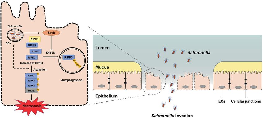
K. Dong et al.
                                                                                                                                                3

Fig. 1 Salmonella pSLT-encoded effector SpvB induces caspase‐independent cell death. A Caco-2 cells were infected with the WT, ΔspvB, or
ΔspvB/pspvB S. Typhimurium strain (MOI of 100) and incubated for 2, 4, 16, and 24 h. Aliquots of cellular supernatants were subjected to LDH
release assay. B, C Caco-2 cells were infected with the WT, ΔspvB, or ΔspvB/pspvB S. Typhimurium strain (MOI of 100) and incubated for 4 h. B
Cells were harvested and subjected to measurement of caspase-3 activity. C Western blot analysis of the expression of cleaved GSDMD. D
Caco-2 cells were treated with either vehicle (DMSO) or 20 μM Z-VAD-FMK for 1 h. The cells were then infected with WT, ΔspvB, or ΔspvB/pspvB
S. Typhimurium strain (MOI of 100) and incubated for 24 h. Aliquots of cellular supernatants were subjected to LDH release assay. Data were
analyzed with IBM SPSS Statistics 19 and presented as the mean ± SEM using Student’s t-test and ANOVA with S-N-K correction. *P < 0.05; ns,
not significant.

between cells transfected with pEGFP-N1 and pEGFP-N1-SpvB,              we found the WT-infected mice displayed a higher mortality and
suggesting a crucial role for autophagy in the regulation of RIPK3      lower body weight relative to the ΔspvB-infected mice (Fig. 5A, B).
degradation (Fig. 4E). To further confirm these findings, we              These observations suggested that SpvB aggravated the severity
detected the ratio of LC3-II/LC3-I and the expression level of p62,     of salmonellosis. Compared with ΔspvB-infected mice, WT-infected
two well-known autophagy biomarkers [23]. Western blot analysis         mice had a decreased colon length, which is indicative of severe
showed that the cells ectopically expressing SpvB displayed an          colitis (Fig. 5C). TUNEL staining is a nonspecific method for
increased expression level of p62 and a decreased ratio of LC3-II/      evaluating cell death that detects DNA fragments [25]. As shown
LC3-I (Fig. 4F). Ubiquitination is a versatile post-translational       in Fig. 5D, the number of TUNEL-positive epithelial cells in the
modification that determines selectivity in autophagy [24]. As           ΔspvB-infected colon was lower than that in the WT-infected
shown in Fig. 4G, the ubiquitination of RIPK3 was decreased in the      colon. These observations demonstrate that SpvB promotes
SpvB-transfected cells. We further detected the levels of K48- and      intestinal injury and cell death in colonic IECs of mice.
K63-type polyubiquitination of RIPK3 in these transfected cells.
Importantly, we found that K48-linked polyubiquitination, but not       Result 6 SpvB promotes necroptosis of IECs in vivo
K63-linked polyubiquitination, was considerably lower in the            To investigate the relationship between SpvB and colonic
presence of SpvB (Fig. 4G). Collectively, these data suggest that       necroptosis, the levels of p-MLKL and RIPK3 were examined by
SpvB restrains RIPK3 K48-linked polyubiquitination, thus down-          immunohistochemistry. We observed that the expression of
regulating RIPK3 degradation in an autophagy-dependent                  p-MLKL was much higher in the colon of the WT-infected mice
manner.                                                                 than that in the ΔspvB-infected mice (Fig. 6A). We also found an
                                                                        abundant accumulation of RIPK3 in the colon of WT-infected mice
Result 5 SpvB promotes the pathogenicity of Salmonella and              (Fig. 6B). We further isolated IECs from the colon of WT- and
induces colon cell death in vivo                                        ΔspvB-infected mice. Western blot analysis showed that WT-
Our in vitro studies suggested that SpvB promoted IECs                  infected IECs displayed a higher phosphorylated level of MLKL
necroptosis by regulating the degradation of RIPK3, so we next          than ΔspvB-infected mice. Consistently, we found a significant
sought to investigate whether SpvB contributes to promoting the         upregulation of RIPK3 in the WT-infected IECs (Fig. 6C, D). Taken
pathogenicity of Salmonella and inducing cell death in vivo. Mice       together, these findings demonstrate that SpvB promotes
were infected with the WT or ΔspvB S. Typhimurium strains, and          necroptosis of IECs in vivo.

Cell Death Discovery (2022)8:44
K. Dong et al.
4
Fig. 2 SpvB promotes necroptosis of IECs. A, C Caco-2 cells were infected with the WT, ΔspvB, or ΔspvB/pspvB S. Typhimurium strain (MOI of
100) and incubated for 4 h. Western blot analysis of the expression of A MLKL and p-MLKL (S358), C RIPK3 and p-RIPK3 (S227). B, D Caco-2 cells
were treated with either vehicle (DMSO), B 1 μM necrosulfonamide (NSA) or D 1 µM GSK’872 for 1 h. The cells were then infected with the WT,
ΔspvB, or ΔspvB/pspvB S. Typhimurium strain (MOI of 100) and incubated for 24 h. Aliquots of cellular supernatants were subjected to LDH
release assay. E HeLa cells were transiently transfected with pCMV-HA-RIPK3 for 24 h. Cells were then infected with WT, ΔspvB, or ΔspvB/pspvB
S. Typhimurium strain (MOI of 100) and cultured for 24 h. Aliquots of cellular supernatants were subjected to LDH release assay. F HeLa cells
were transiently transfected with pCMV-HA-RIPK3 and pEGFP-N1-SpvB for 24 h. Western blot analysis of the expression of p-MLKL. Data were
analyzed with IBM SPSS Statistics 19 and presented as the mean ± SEM using Student’s t-test and ANOVA with S-N-K correction. *P < 0.05; ns
not significant.
DISCUSSION we found that SpvB induced IECs cell death, and the existence of
Salmonella spreads to both humans and animals through the SpvB-associated cell death was captured at an early phase during
fecal–oral route. The gastrointestinal tract is the first site of Salmonella infection.
host–pathogen interaction after ingestion of Salmonella. IECs exert The pSLT-encoded SpvB has been shown to contain an ADP-
an influence on maintaining intestinal mucosal barrier function to ribosyltransferase domain in its C-terminus. Some researchers
resist the invasion by Salmonella. Our previous studies have believe that the type of cell death induced by SpvB is apoptosis
reported that SpvB promotes S. Typhimurium intracellular [30, 31], which is a complex multistep process driven by caspase-
replication by interfering with the host’s iron homeostasis dependent proteolytic cleavage cascades [32]. However, the SpvB
[26, 27]. In the current study, we further showed that SpvB deficiency had no effect on the apoptosis mediator caspase-3 in
contributes to the induction of necroptosis through downregulat- our study. Moreover, the treatment with the pan-caspase inhibitor
ing K48-Ub-mediated degradation of RIPK3, which is ultimately Z-VAD-FMK had no significant influence on SpvB-mediated cell
advantageous for Salmonella across the intestinal epithelial barrier death. These observations consistent with early research sug-
(Fig. 7). gested that SpvB-associated cell death is an unknown type of RCD
The intestinal mucosal barrier has functions to separate [12]. Necroptosis, which has morphological characteristics similar
intestinal lumen material, resist pathogen invasion, and maintain to necrosis, is typically considered a highly pro-inflammatory
the homeostasis of the organism [28, 29]. As the structural basis of mode of cell death due to the release of damage-associated
the intestinal mucosal barrier, the epithelial barrier including IECs molecular patterns [33]. Therefore, this type of RCD plays a role in
and intercellular junctions is of great significance for the host to many intestinal diseases associated with inflammation, such as
resist the invasion by Salmonella. Our findings demonstrated that inflammatory bowel disease and necrotizing enterocolitis [34–36].
SpvB promoted the pathogenicity of Salmonella, including an Necroptosis of the IECs often leads to uncontrolled translocation
increase in mortality and a decrease in body weight. We also of the bacteria and excessive inflammation [37]. Interestingly, we
found that SpvB resulted in a decrease in colon length and found that SpvB induced the phosphorylation of MLKL and RIPK3,
induced cell death in the colon. We further constructed the Caco-2 suggesting an increase in necroptosis. By employing various
cells infection model to confirm these observations in vitro. Hanna inhibitors, we further confirmed these observations. RIPK1, a key
et al. suggested that SpvB-delayed cell death may occur at a later molecule, can phosphorylate RIPK3 and then induce necroptosis
stage and cannot be captured [30, 31]. Interestingly, in this study, [9]. However, the inhibitor of RIPK1 failed to eliminate the
Cell Death Discovery (2022)8:44K. Dong et al.
5
Fig. 3 SpvB promotes IECs necroptosis in a manner independent of RIPK1. A Caco-2 cells were infected with WT, ΔspvB, or ΔspvB/pspvB S.
Typhimurium strain (MOI of 100) and incubated for 4 h. Western blot analysis of the expression of RIPK1 and p-RIPK1. B Caco-2 cells were
treated with either vehicle (DMSO), 20 µM Z-VAD-FMK with or without 10 µM necrostatin-1 (Nec-1) for 1 h. The cells were then infected with
the WT, ΔspvB, or ΔspvB/pspvB S. Typhimurium strain (MOI of 100) and incubated for 24 h. Aliquots of cellular supernatants were subjected to
LDH release assay. C, D Caco-2 cells were transiently transfected with pEGFP-N1 or pEGFP-N1-SpvB for 24 h. Cells transfected with pEGFP-N1-
SpvB were pretreated with either vehicle (DMSO), 20 µM Z-VAD-FMK with or without 10 µM Nec-1, 1 µM GSK’872, or 1 µM NSA for 1 h. C Cells
were subjected to cell viability assay. D Aliquots of cellular supernatants were subjected to LDH release assay. Data were analyzed with IBM
SPSS Statistics 19 and presented as the mean ± SEM using Student’s t-test and ANOVA with S-N-K correction. *P < 0.05, ***P < 0.001; ns not
significant.
significant effect of the SpvB on cell death. These results colonization of pathogens [19, 20]. In some diseases, such as
demonstrate that SpvB mediates the increased necroptosis in colon cancer and Cowpox virus infection, the expression of RIPK3
IECs in a RIPK3-dependent manner. could be regulated through transcription mechanisms [39, 40]. At
Previous investigations have demonstrated that RIPK3 accumu- the protein level, RIPK3 could also be regulated through post-
lation correlates with severe IECs necroptosis and colitis transcriptional modification, such as autophagy and proteasome
[16, 17, 38]. Interestingly, we found that the protein level of RIPK3 [17, 21, 22]. In the current study, SpvB had no obvious effect on
was substantially increased in the presence of SpvB both in vivo the activity of caspase-8. We also detected no significant effect of
and in vitro. These observations suggested that the RIPK3 SpvB on the RIPK3 transcription level in this infection model. Thus,
overexpression is the basis for SpvB to promote necroptosis. we focused on the hypothesis that SpvB-mediated increase in
RIPK3 is a critical mediator of necroptosis and could be regulated RIPK3 protein might indicate an inactive degradation mechanism.
at several levels. Previous studies have shown that caspase-8 In line with our hypothesis, treatment with the autophagy
could cleave RIPK3 to inhibit necroptosis of IECs during the inhibitor Baf A1 eliminated the effects of SpvB on RIPK3 and the
process of enteritis caused by Salmonella, thereby playing a role in K48-ubiquitination of RIPK3 was increased in the presence of
maintaining the intestinal barrier function and limiting the SpvB. Taking comprehensive consideration of Fig. 4, these results
Cell Death Discovery (2022)8:44K. Dong et al.
6
Fig. 4 SpvB leads to epithelium necroptosis through inhibiting RIPK3 ubiquitination and autophagy-dependent degradation. A, C Caco-2
cells were then infected with WT, ΔspvB, or ΔspvB/pspvB S. Typhimurium strain (MOI of 100) and incubated for 4 h. A RIPK3 levels were
determined by RT-qPCR. Values were normalized to those of the housekeeping gene β‐ACTIN and fold‐changes relative to the untreated
control were shown. C Cells were harvested and subjected to measurement of caspase-8 activity. B, D–G Caco-2 cells were transiently
transfected with pEGFP-N1-SpvB or pEGFP-N1 for 24 h. B Western blot analysis of the expression of RIPK3. D Cells were treated with 5 μM
MG132 for 24 h and western blot analysis of the expression of RIPK3. E Cells were treated with 100 nM bafilomycin A1 (Baf A1) and western
blot analysis of the expression of RIPK3. F Western blot analysis of the expression of LC3-I, LC3-II, and p62. G Cell lysates were
immunoprecipitated with anti-RIPK3 antibody or IgG, then immunoblotted with respective antibodies. Data were analyzed with IBM SPSS
Statistics 19 and presented as the mean ± SEM using Student’s t-test and ANOVA with S-N-K correction. *P < 0.05, **P < 0.005; ns not
significant.
are consistent with our assumption that SpvB upregulates colon injury. Taken together, Salmonella effector SpvB induces IECs
necroptosis through inhibiting the K48-Ub-dependent autophagy necroptosis in the early stage of the infection, which may be
degradation of RIPK3. considered the strategy of bacterial invasion.
In summary, we found a novel function of the effector SpvB to
aggravate the pathogenesis of Salmonella through inducing IECs
necroptosis, thereby promoting the bacteria to disrupt the integrity MATERIALS AND METHODS
of the intestinal epithelial barrier. By downregulating the K48-Ub- Cell culture
degradation of RIPK3 in an autophagy-dependent manner, SpvB Caco-2 cells were kindly provided by Professor Weiqi He (Soochow
mediates the abundant accumulation of RIPK3. The accumulation of University, Suzhou, China). HeLa cells were purchased from the American
RIPK3 increases the phosphorylation level of MLKL and upregulates Type Culture Collection (Manassas, VA, USA). The obtained cells were
necroptosis in IECs. The disturbed integrity of IECs ultimately leads to cultured in Dulbecco’s Modified Eagle’s medium (DMEM; HyClone
Laboratories, Logan, UT, USA) with 10% fetal bovine serum (FBS; Biological
the disruption of the intestinal epithelial barrier and aggravated Industries, Kibbutz Beit‐Haemek, Israel) and 1% penicillin–streptomycin
Salmonella infection. In vivo, SpvB promotes salmonellosis, mani- (Beyotime Biotechnology, Shanghai, China) solution at a temperature of
fested as increased mortality, decreased body weight, and severe 37 °C and 5% CO2.
Cell Death Discovery (2022)8:44K. Dong et al.
7
Fig. 5 SpvB promotes the pathogenicity of Salmonella and induces colon cell death in vivo. A, B C57BL/6 mice were treated with
streptomycin 24 h prior to oral infection with the WT or ΔspvB S. Typhimurium strain (1 × 10⁷ CFUs). A Survival curves, n = 10 mice per group. B
Body weight changes, n = 10 mice per group. C, D C57BL/6 mice were treated with streptomycin 24 h prior to oral infection with the WT or
ΔspvB S. Typhimurium strain (1 × 10⁸ CFUs) and analyzed at 48 h. C Colon length and representative photographs of colons, n = 5 mice per
group. D Immunofluorescence staining of colon sections from mice and representative images of TUNEL-positive cells were denoted by
arrows, counted ten high-power fields (TUNEL, green; DAPI, blue). Scale bars: 50 μm. Survival curves were analyzed with the log-rank test. Data
were analyzed with IBM SPSS Statistics 19 and presented as the mean ± SEM using Student’s t-test. *P < 0.05, **P < 0.005, ***P < 0.001; ns not
significant.
Bacterial strains and growth conditions Burlington, MA, USA) to prevent the growth of extracellular bacteria for 2 h.
The wild-type (WT) S. Typhimurium strain SL1344, mutants lacking spv Afterward, the infected cells were washed and subsequently incubated
(Δspv) or spvB (ΔspvB) and spvB complemented (ΔspvB/pspvB) in plasmid with DMEM-FBS (10%) and 10 µg/mL amikacin. Cells were subsequently
pBAD/gIIIA were used in the study as previously described [26]. The treated with DMSO (MilliporeSigma) vehicle, 5 µM MG123 (Selleck
mutant lacking pSLT was constructed by plasmid elimination assay with Chemicals, Houston, TX, USA) or 100 nM bafilomycin A1 (Baf A1; MCE)
sodium dodecyl sulfate (SDS) treatment and checked by means of PCR. For when appropriate.
SDS treatment, 30 μL of a bacterial suspension was added to 3 mL of 10%
SDS Luria‐Bertani (LB) broth (Hangwei, Hangzhou, China) and incubated at
37 °C with shaking overnight. A total of 30 μL of the bacterial suspension
Salmonella infection in vivo
treated with SDS was added to 3 mL LB broth and incubated at 37 °C with Female C57BL/6 mice (6–8 weeks) were bred and maintained at the
shaking for 14–16 h. After repeating the above two steps several times, experimental animal center of Soochow University. All animal experiments
30 μL of the bacterial suspension was grown on LB agar (Hangwei), and the were approved by the Ethics Committee of Soochow University, Suzhou,
elimination of the plasmid was identified by PCR with specific primers. China, and were conducted in accordance with the Guidelines for the Care
and Use of Research Animals established by Soochow University. For
Bacteria were grown on LB agar plates and then in LB broth at 37 °C with
experiments of mouse survival and body weight, the mice were treated
shaking for 14–16 h. Bacteria were diluted 1:100 with fresh LB medium
supplemented with 100 µg/mL ampicillin when appropriate and cultured with streptomycin (0.1 mL of a 200 mg/mL solution in sterile water) 24 h
until the logarithmic phase was reached. prior to oral infection with 1 × 10⁷ colony forming units (CFUs) of different
S. Typhimurium strains. For experiments that require the organs of mice,
the mice were treated with streptomycin (0.1 mL of a 200 mg/mL solution
Salmonella infection in vitro in sterile water) 24 h prior to oral infection with 1 × 10⁸ CFUs of different S.
The bacteria reaching the late‐logarithmic phase were washed three times Typhimurium strains and euthanized 48 h postinfection using CO2
with phosphate buffer saline (PBS) and then quantified using a spectro- asphyxiation. The control mice received only PBS.
photometer to determine optical density at 600 nm and calculate the
multiplicity of infection (MOI). Caco-2 and HeLa cells were infected with
bacteria at MOI of 100 in DMEM-10% FBS (v/v). One hour later, the cells Immunoblot analysis
were washed twice with warm PBS, and the medium was replaced with Cells were washed once with PBS, followed by lysis in RIPA buffer
DMEM-10% FBS (v/v) containing 100 µg/mL amikacin (MilliporeSigma, (Beyotime Biotechnology) containing protease and phosphatase inhibitors
Cell Death Discovery (2022)8:44K. Dong et al.
8
Fig. 6 SpvB promotes necroptosis of IECs in vivo. A, B C57BL/6 mice were treated with streptomycin 24 h prior to oral infection with the WT
or ΔspvB S. Typhimurium strain (1 × 10⁸ CFUs) and analyzed at 48 h. IHC staining evaluated the level of A p-MLKL and B RIPK3 in colon sections.
Scale bars: 50 μm. C, D Western blot analysis of purified colonic epithelial lysates with specific antibodies to C p-MLKL and D RIPK3, n = 3 mice
for control group, n = 4 mice for WT- and ΔspvB-infected group respectively. Data were analyzed with IBM SPSS Statistics 19 and presented as
the mean ± SEM using Student’s t-test. *P < 0.05.
(Beyotime Biotechnology) and sample loading buffer. Proteins were Cell transfection
separated by electrophoresis on 8–12% polyacrylamide SDS-PAGE gels HeLa cells were transiently transfected with pEGFP-N1-SpvB (fusion protein
and transferred onto polyvinylidene difluoride membranes (Millipore- with HA tag) or pCMV-HA-RIPK3 using ExFect2000 Transfection Reagent
Sigma). Nonspecific binding was blocked with either 5% nonfat dry milk (Vazyme, Nanjing, China) for 24 h in accordance with the manufacturer’s
powder or 5% bovine serum albumin in Tris-buffered saline containing instructions. Caco-2 cells were transiently transfected with pEGFP-N1-SpvB
0.1% Tween 20. Membranes were probed with primary antibodies at 4 °C or pEGFP-N1.
overnight, then washed and incubated with the appropriate horseradish
peroxidase-conjugated secondary antibodies including anti-rabbit IgG
(ab97051, Abcam, Cambridge, MA, USA) and anti-mouse IgG (Beyotime Immunohistochemistry (IHC) and TUNEL assays
Biotechnology) at room temperature for 1 h. Proteins were visualized using After the mice were euthanized, detached colons were fixed in 10%
an enhanced chemiluminescence luminescence reagent (Meilunbio, formalin, then processed and embedded in paraffin in line with the
Dalian, China). The intensities of the target bands were quantified using standard procedures. Formalin-fixed paraffin-embedded colons were cut
ImageJ Launcher broken symmetry software program (National Institutes into 5-μm-thick sections. IHC staining was performed using antibody
of Health, Bethesda, MD, USA). The antibodies used were as following: anti- directed against p-MLKL (phospho S345) and RIPK3. TUNEL staining was
GSDMD (ab210070, Abcam); anti-MLKL (YT2788, Immunoway, Plano, TX, performed using the Dead-End kit (Promega, Madison, WI, USA) in
USA); anti-p-MLKL (phospho S358) (ab187091, Abcam); anti-p-MLKL accordance with the manufacturer’s instructions.
(phospho S345) (ab196436, Abcam); anti-RIPK3 (17563-1-AP, Proteintech,
Rosemont, IL, USA); anti-p-RIPK3 (phospho S227) (ab209384, Abcam); anti-
p-RIPK1 (phospho S166) (YP1467, Immunoway); anti-RIPK1 (17519-1-AP, IECs isolation
Proteintech); anti-K63-Ubiquitin (ab179434, Abcam); anti-K48-Ubiquitin IECs were isolated as described previously [41]. In brief, the colons from the
(ab140601, Abcam); anti-Ubiquitin (#3936, Cell Signaling Technology, mice were opened longitudinally and washed in a solution containing
Danvers, Massachusetts, USA); anti-HA (#3724, Cell Signaling Technology); 0.154 M NaCl and 1 mM DTT to remove intestinal contents, and then
anti-GAPDH (BA2913, Boster, Wuhan, China); anti-LC3 (NBP100-2220, incubated in a solution containing 1.5 mM EDTA and 1 mM DTT at 37 °C for
Novus Biologicals, Littleton, Colorado, USA); anti-p62 (#5114, Cell Signaling 30 min. IECs were obtained by centrifugation for subsequent protein
Technology). extraction.
Cell Death Discovery (2022)8:44K. Dong et al.
9
Fig. 7 General summary of SpvB-mediated IECs necroptosis during Salmonella infection. SpvB, an effector encoded by pSLT,
downregulates the K48-Ub-degradation of RIPK3 in an autophagy-dependent manner. The abundant accumulation of RIPK3 upregulates
the phosphorylation level of MLKL and leads to necroptosis in IECs. The SpvB-mediated IECs necroptosis aggravates the pathogenesis of
Salmonella through promoting the bacteria to disrupt intestinal epithelial barrier integrity. IECs, intestinal epithelial cells; SCV, Salmonella-
containing vacuole; MLKL, mixed-lineage kinase domain-like protein; RIPK3, receptor-interacting protein kinase 3; RIPK1, receptor-interacting
protein kinase 1.
Lactate dehydrogenase (LDH) and cell viability assays were treated with 1 µg anti-RIPK3 antibody (17563-1-AP, Proteintech) for
Cells were seeded onto 96-well plates at a density of 30,000 cells per well. 1 h, followed by incubation with pellet beads on a shaker overnight.
The second day after seeding, the cells were counted and pretreated with Immunoprecipitated proteins and pellet beads were collected by
20 μM Z-VAD-FMK (Beyotime Biotechnology), 1 μM necrosulfonamide centrifugation for subsequent experiments.
(MCE, Monmouth Junction, NJ, USA), 1 μM GSK’872 (MCE), 10 μM
necrostatin-1 (MCE), or DMSO vehicle control for infection or transfection.
LDH released was determined by the LDH cytotoxicity assay detection kit Ethidium homodimer 1 (EthD-1) assay
(Beyotime Biotechnology). The cell culture supernatant was collected for Caco-2 cells were plated onto glass coverslips and then infected with
detection. The absorbance was read at 490 nm with Infinite® F50 respective bacteria at MOI of 100 for 24 h. In accordance with the
Absorbance Microplate Reader (Tecan, Switzerland). Cell viability was manufacturer’s instructions, EthD-1 (US EVERBRIGHT INC., Suzhou, China)
measured based on the intracellular ATP levels using CellTiter-Lumi™ Plus was diluted to 4 μΜ in PBS and incubated with the cells for 40 min at room
Luminescent Cell Viability Assay Kit (Beyotime Biotechnology). An equal temperature. Then, the cells were incubated with DAPI (MilliporeSigma) for
volume of Celltiter-LUMI™ Plus reagent was added to the cell culture 15 min at room temperature and observed under a Nikon Eclipse Ni-U
medium to induce cell lysis by oscillation. After incubation at room fluorescence microscope (Nikon, Tokyo, Japan). The percentage of the
temperature, the luminescence signal was measured by SynergyTM 2 Multi- EthD-1-positive cells was then determined.
Mode Microplate Reader (BioTek, Winooski, VT, USA).
Statistical analysis
Quantitative real-time PCR (qRT-PCR) Statistical analysis was performed using SPSS Statistics 19 software (IBM,
Total RNA was isolated from Caco-2 cells with trizol (Thermo Fisher Armonk, NY, USA). Survival curves were analyzed with log-rank (Mantel–Cox)
Scientific, Waltham, MA, USA) reagent and subjected to reverse transcrip- test. The Student’s t-test was used for data comparison between two groups
tion using the All-in-one RT MasterMix kit (Applied Biological Materials, and one-way analysis of variance (ANOVA) with S-N-K correction was used for
Richmond, BC, Canada). qRT-PCR was conducted using the ViiA7 real-time comparison between multiple groups after normality tests. These data were
PCR instrument (Applied Biosystems, Carlsbad, CA, USA) with EvaGreen expressed as mean ± SEM, and P < 0.05 was considered to be statistically
MasterMix-Low ROX (Applied Biological Materials) to analyze transcript significant. Values indicate the number of animals used for the experiments.
levels of target genes. The expression level of RIPK3 was normalized to No statistical methods were used to predetermine sample sizes, but our
β-ACTIN expression with the 2−ΔΔCT method. Each sample was detected in sample sizes are similar to those reported in previous publications [7]. No
triplicate. The primer sequences for RIPK3 and β-ACTIN were as follows: randomization was used for the data collection.
hRIPK3, 5'-GCTACGATGTGGCGGTCAAGAT-3' and 5'-TTGGTCCCAGT
TCACCTTCTCG-3' [42]; hβ-ACTIN, 5'-CACCATTGGCAATGAGCGGTTC-3' and
5'-AGGTCTTTGCGGATGTCCACGT-3'.
DATA AVAILABILITY
The datasets used and/or analyzed during the current study are available from the
Caspase activity assay corresponding author on reasonable request.
Caco-2 cells were plated in each well of a 12‐well plate and stimulated for
4 h with the indicated S. Typhimurium strains at MOI of 100. Caspase-3 and
-8 activities in cell lysates were analyzed using Caspase-3 and Caspase-8 REFERENCES
Activity Assay Kit (Beyotime Biotechnology) respectively in accordance 1. Gal-Mor O, Boyle EC, Grassl GA. Same species, different diseases: how and why
with the manufacturer’s protocol. typhoidal and non-typhoidal Salmonella enterica serovars differ. Front Microbiol.
2014;5:391.
Immunoprecipitation 2. Typhoid GBD, Paratyphoid C. The global burden of typhoid and paratyphoid
Cells were washed once with PBS, followed by lysis in RIPA buffer fevers: a systematic analysis for the Global Burden of Disease Study 2017. Lancet
(Beyotime Biotechnology) containing protease and phosphatase inhibitors. Infect Dis. 2019;19:369–81.
The whole-cell lysates were cleared for 30 min with 1 µg of rabbit IgG and 3. Collaborators GBD. N-T S I D. The global burden of non-typhoidal Salmonella
20 µL of Protein A/G PLUS-Agarose (Santa Cruz Biotechnology, Dallas, TX, invasive disease: a systematic analysis for the Global Burden of Disease Study
USA). After pellet beads were removed by centrifugation, the cell lysates 2017. Lancet Infect Dis. 2019;19:1312–24.
Cell Death Discovery (2022)8:44K. Dong et al.
10
4. Fabrega A, Vila J. Salmonella enterica serovar Typhimurium skills to succeed in the 32. McComb S, Chan PK, Guinot A, Hartmannsdottir H, Jenni S, Dobay MP, et al.
host: virulence and regulation. Clin Microbiol Rev. 2013;26:308–41. Efficient apoptosis requires feedback amplification of upstream apoptotic signals
5. Passaris I, Cambre A, Govers SK, Aertsen A. Bimodal expression of the Salmonella by effector caspase-3 or -7. Sci Adv. 2019;5:eaau9433.
Typhimurium spv operon. Genetics. 2018;210:621–35. 33. Frank D, Vince JE. Pyroptosis versus necroptosis: similarities, differences, and
6. Guiney DG, Fierer J. The role of the spv genes in Salmonella pathogenesis. Front crosstalk. Cell Death Differ. 2019;26:99–114.
Microbiol. 2011;2:129. 34. Wang R, Li H, Wu J, Cai ZY, Li B, Ni H, et al. Gut stem cell necroptosis by genome
7. Sun L, Yang S, Deng Q, Dong K, Li Y, Wu S, et al. Salmonella effector SpvB disrupts instability triggers bowel inflammation. Nature. 2020;580:386–90.
intestinal epithelial barrier integrity for bacterial translocation. Front Cell Infect 35. Zhou M, He J, Shi Y, Liu X, Luo S, Cheng C, et al. ABIN3 negatively regulates
Microbiol. 2020;10:606541. necroptosis-induced intestinal inflammation through recruiting A20 and
8. Barreau F, Hugot JP. Intestinal barrier dysfunction triggered by invasive bacteria. restricting the ubiquitination of RIPK3 in inflammatory bowel disease. J Crohns
Curr Opin Microbiol. 2014;17:91–98. Colitis. 2021;15:99–114.
9. Galluzzi L, Kepp O, Chan FK, Kroemer G. Necroptosis: mechanisms and relevance 36. Werts AD, Fulton WB, Ladd MR, Saad-Eldin A, Chen YX, Kovler ML, et al. A novel
to disease. Annu Rev Pathol. 2017;12:103–30. role for necroptosis in the pathogenesis of necrotizing enterocolitis. Cell Mol
10. Matsui H, Bacot CM, Garlington WA, Doyle TJ, Roberts S, Gulig PA. Virulence Gastroenterol. Hepatol. 2020;9:403–23.
plasmid-borne spvB and spvC genes can replace the 90-kilobase plasmid in 37. Kaczmarek A, Vandenabeele P, Krysko DV. Necroptosis: the release of damage-
conferring virulence to Salmonella enterica serovar Typhimurium in sub- associated molecular patterns and its physiological relevance. Immunity.
cutaneously inoculated mice. J Bacteriol. 2001;183:4652–8. 2013;38:209–23.
11. Tang D, Kang R, Berghe TV, Vandenabeele P, Kroemer G. The molecular 38. Wu T, Dai Y, Xue L, Sheng Y, Xu L, Xue Y. Expression of receptor interacting
machinery of regulated cell death. Cell Res. 2019;29:347–64. protein 3 and mixed lineage kinase domain-like protein-key proteins in
12. Kurita A, Gotoh H, Eguchi M, Okada N, Matsuura S, Matsui H, et al. Intracellular necroptosis is upregulated in ulcerative colitis. Ann Palliat Med. 2019;8:483–9.
expression of the Salmonella plasmid virulence protein, SpvB, causes apoptotic 39. Moriwaki K, Bertin J, Gough PJ, Orlowski GM, Chan FK. Differential roles of RIPK1
cell death in eukaryotic cells. Micro Pathog. 2003;35:43–48. and RIPK3 in TNF-induced necroptosis and chemotherapeutic agent-induced cell
13. Zhao Y, Yang J, Shi J, Gong YN, Lu Q, Xu H, et al. The NLRC4 inflammasome death. Cell Death Dis. 2015;6:e1636.
receptors for bacterial flagellin and type III secretion apparatus. Nature. 40. Liu Z, Nailwal H, Rector J, Rahman MM, Sam R, McFadden G, et al. A class of viral
2011;477:596–600. inducer of degradation of the necroptosis adaptor RIPK3 regulates virus-induced
14. Welz PS, Wullaert A, Vlantis K, Kondylis V, Fernandez-Majada V, Ermolaeva M, inflammation. Immunity. 2021;54:247–58 e247.
et al. FADD prevents RIP3-mediated epithelial cell necrosis and chronic intestinal 41. Nenci A, Becker C, Wullaert A, Gareus R, van Loo G, Danese S, et al. Epithelial
inflammation. Nature. 2011;477:330–4. NEMO links innate immunity to chronic intestinal inflammation. Nature.
15. Sun L, Wang H, Wang Z, He S, Chen S, Liao D, et al. Mixed lineage kinase domain- 2007;446:557–61.
like protein mediates necrosis signaling downstream of RIP3 kinase. Cell. 42. Xie Y, Zhu S, Zhong M, Yang M, Sun X, Liu J, et al. Inhibition of aurora kinase A induces
2012;148:213–27. necroptosis in pancreatic carcinoma. Gastroenterology. 2017;153:1429–43 e1425.
16. Gunther C, Martini E, Wittkopf N, Amann K, Weigmann B, Neumann H, et al.
Caspase-8 regulates TNF-alpha-induced epithelial necroptosis and terminal ileitis.
Nature. 2011;477:335–9. ACKNOWLEDGEMENTS
17. Xie Y, Zhao Y, Shi L, Li W, Chen K, Li M, et al. Gut epithelial TSC1/mTOR controls The authors thank Professor Ying Xu and Professor Weiqi He (Cambridge Suda
RIPK3-dependent necroptosis in intestinal inflammation and cancer. J Clin Invest. Genome Resource Center, Soochow University) for their excellent technical support.
2020;130:2111–28. The work is supported by the National Natural Science Foundation of China (No.
18. He GW, Gunther C, Thonn V, Yu YQ, Martini E, Buchen B, et al. Regression of 81971899, No. 31970132, No. 81671976, No. 31670140), Suzhou Municipal Science
apoptosis-resistant colorectal tumors by induction of necroptosis in mice. J Exp and Technology Foundation (SYS2019031), Project funded by China Postdoctoral
Med. 2017;214:1655–62. Science Foundation (2021M693668), and a project funded by the Priority Academic
19. Yang ZH, Wu XN, He P, Wang X, Wu J, Ai T, et al. A non-canonical PDK1-RSK signal Program Development (PAPD) of Jiangsu Higher Education Institutions.
diminishes pro-caspase-8-mediated necroptosis blockade. Mol Cell.
2020;80:296–310 e296.
20. Feng S, Yang Y, Mei Y, Ma L, Zhu DE, Hoti N, et al. Cleavage of RIP3 inactivates its
caspase-independent apoptosis pathway by removal of kinase domain. Cell AUTHOR CONTRIBUTIONS
Signal. 2007;19:2056–67. KD, QD, and RH designed the study. KD, YZ, and QD performed the experiments;
21. Moriwaki K, Chan FK. Regulation of RIPK3- and RHIM-dependent necroptosis by QD, SY, LS, KH, and YL contributed to development of methodology; KD, YZ, QD, LS,
the proteasome. J Biol Chem. 2016;291:5948–59. SY, and YC contributed to analysis and interpretation of data; KD, YZ,
22. Seo J, Lee EW, Sung H, Seong D, Dondelinger Y, Shin J, et al. CHIP controls and QD prepared figures and wrote the manuscript; YL, SW, and RH supervised
necroptosis through ubiquitylation- and lysosome-dependent degradation of the study.
RIPK3. Nat Cell Biol. 2016;18:291–302.
23. Deng Z, Lim J, Wang Q, Purtell K, Wu S, Palomo GM, et al. ALS-FTLD-linked
mutations of SQSTM1/p62 disrupt selective autophagy and NFE2L2/NRF2 anti-
oxidative stress pathway. Autophagy. 2020;16:917–31. COMPETING INTERESTS
24. Shaid S, Brandts CH, Serve H, Dikic I. Ubiquitination and selective autophagy. Cell The authors declare no competing interests.
Death Differ. 2013;20:21–30.
25. Tonnus W, Meyer C, Paliege A, Belavgeni A, von Massenhausen A, Bornstein SR, ETHICS APPROVAL AND CONSENT TO PARTICIPATE
et al. The pathological features of regulated necrosis. J Pathol. 2019;247:697–707. The animal experiments were reviewed and approved by the Ethics Committee of
26. Yang S, Deng Q, Sun L, Dong K, Li Y, Wu S, et al. Salmonella effector SpvB Soochow University (Grant 2111270).
interferes with intracellular iron homeostasis via regulation of transcription factor
NRF2. FASEB J. 2019;33:13450–64.
27. Deng Q, Yang S, Sun L, Dong K, Li Y, Wu S, et al. Salmonella effector SpvB ADDITIONAL INFORMATION
aggravates dysregulation of systemic iron metabolism via modulating the
Supplementary information The online version contains supplementary material
hepcidin-ferroportin axis. Gut Microbes. 2021;13:1–18.
available at https://doi.org/10.1038/s41420-022-00841-9.
28. Martens EC, Neumann M, Desai MS. Interactions of commensal and pathogenic
microorganisms with the intestinal mucosal barrier. Nat Rev Microbiol.
Correspondence and requests for materials should be addressed to Shuyan Wu or
2018;16:457–70.
Rui Huang.
29. Sanchez de Medina F, Romero-Calvo I, Mascaraque C, Martinez-Augustin O. Intestinal
inflammation and mucosal barrier function. Inflamm Bowel Dis. 2014;20:2394–404.
Reprints and permission information is available at http://www.nature.com/
30. Hilger H, Pust S, von Figura G, Kaiser E, Stiles BG, Popoff MR, et al. The long-lived
reprints
nature of clostridium perfringens iota toxin in mammalian cells induces delayed
apoptosis. Infect Immun. 2009;77:5593–601.
Publisher’s note Springer Nature remains neutral with regard to jurisdictional claims
31. Browne SH, Lesnick ML, Guiney DG. Genetic requirements for Salmonella-induced
in published maps and institutional affiliations.
cytopathology in human monocyte-derived macrophages. Infect Immun.
2002;70:7126–35.
Cell Death Discovery (2022)8:44K. Dong et al.
11
Open Access This article is licensed under a Creative Commons
Attribution 4.0 International License, which permits use, sharing,
adaptation, distribution and reproduction in any medium or format, as long as you give
appropriate credit to the original author(s) and the source, provide a link to the Creative
Commons license, and indicate if changes were made. The images or other third party
material in this article are included in the article’s Creative Commons license, unless
indicated otherwise in a credit line to the material. If material is not included in the
article’s Creative Commons license and your intended use is not permitted by statutory
regulation or exceeds the permitted use, you will need to obtain permission directly
from the copyright holder. To view a copy of this license, visit http://creativecommons.
org/licenses/by/4.0/.
© The Author(s) 2022
Cell Death Discovery (2022)8:44You can also read