Upregulated phospholipase D2 expression and activity is related to the metastatic properties of melanoma
←
→
Page content transcription
If your browser does not render page correctly, please read the page content below
ONCOLOGY LETTERS 23: 140, 2022

             Upregulated phospholipase D2 expression and activity
              is related to the metastatic properties of melanoma
     ARANTZA PEREZ‑VALLE1, BEGOÑA OCHOA2, KRUSHANGI N. SHAH3, GABRIEL BARREDA‑GOMEZ4,
          EGOITZ ASTIGARRAGA4, MARÍA DOLORES BOYANO1,5 and AINTZANE ASUMENDI1,5

                Departments of 1Cell Biology and Histology and 2Physiology, School of Medicine and Nursing,
         University of the Basque Country UPV/EHU, Leioa, 48940 Bizkaia, Spain; 3Department of Biochemistry and
              Molecular Biology, Wright State University, Dayton, OH 45435, USA; 4IMG Pharma Biotech S.L.,
             Bizkaia Technological Park, Zamudio, 48160 Bizkaia; 5Biocruces‑Bizkaia Health Research Institute,
                                Cruces University Hospital, Barakaldo, 48903 Bizkaia, Spain

                                      Received September 30, 2021; Accepted February 1, 2022

                                                       DOI: 10.3892/ol.2022.13260

Abstract. The incidence rates of melanoma have increased                 In addition, silencing of PLD2 in melanoma cells reduced the
steadily in recent decades and nearly 25% of the patients                metastatic potential of these cells. The present study provided
diagnosed with early‑stage melanoma will eventually develop              evidence that PLD2 is involved in melanoma malignancy and
metastasis, for which there is currently no fully effective              in particular, in its metastatic potential, and established a basis
treatment. The link between phospholipases and tumors                    for future studies evaluating PLD2 blockade as a therapeutic
has been studied extensively, particularly in breast and                 strategy to manage this condition.
colon cancers. With the aim of finding new biomarkers and
therapeutic options for melanoma, the expression of different            Introduction
phospholipases was assessed in 17 distinct cell lines in the
present study, demonstrating that phospholipase D2 (PLD2)                Phospholipase D (PLD) is a phospholipase that participates in
is upregulated in metastatic melanoma as compared to normal              the catabolism of glycerol‑based phospholipids. There are six
skin melanocytes. These results were corroborated by immu‑               different isoforms of the phospholipase D subclass (PLD1‑6)
nofluorescence and lipase activity assays. Upregulation of               and PLD1 and PLD2 are the best‑studied isoforms. The lipase
PLD2 expression and increased lipase activity were observed              activity of both these enzymes involves the preferential hydro‑
in metastatic melanoma relative to normal skin melanocytes.              lysis of phosphatidylcholines (PCs), to release phosphatidic
So far, the implication of PLD2 activity in melanoma malig‑              acid (PA) and choline (Cho), crucial lipid mediators of cell
nancies has remained elusive. To the best of our knowledge,              signaling pathways involved in cell proliferation, growth and
the present study was the first to demonstrate that the over‑            survival (1).
expression of PLD2 enhances lipase activity, and its effect to               Cho is an essential nutrient for humans and it has a wide
increase the proliferation, migration and invasion capacity of           range of vital physiological roles (2,3), although higher levels of
melanoma cells was assessed with XTT and Transwell assays.               Cho have been associated with an increased risk of melanoma
                                                                         and other cancer types (3‑5). However, as only small amounts
                                                                         of this molecule may be synthesized de novo in humans, it must
                                                                         be obtained from other sources, such as through the hydrolysis
                                                                         of the most abundant phospholipid, PC, by the lipase activity of
Correspondence to: Professor Aintzane Asumendi, Department
of Cell Biology and Histology, School of Medicine and Nursing,
                                                                         PLDs (3). PA is the other lipid mediator produced by the lipase
University of The Basque Country UPV/EHU, Barrio Sarriena s/n,           activity of PLDs, which may subsequently be converted into
Leioa, 48940 Bizkaia, Spain                                              essential second messengers such as lyso‑PA by phospholipase
E‑mail: aintzane.asumendi@ehu.eus                                        A2 (PLA2) or diacylglycerol (DAG) by PA‑phosphohydrolase.
                                                                         These products significantly expand the range of the effects in
Abbreviations: Cho, choline; DAG, diacylglycerol; GEF, guanine           which these enzymes may participate.
nucleotide exchange factor; PA, phosphatidic acid; PBut,                     Aside from the lipase activity of PLD2, a unique feature of
phosphatidylbutanol; PC, phosphatidylcholine; PLD, phospholipase D       this isoform is that it also acts as a guanine nucleotide exchange
                                                                         factor (GEF) (6,7). Certain small GTPases regulate PLD2 activity
Key words: melanoma, metastasis, phospholipases, phospholipase D2,       through a complex regulatory program of phosphorylation and
enzyme activity, phosphatidylcholine, lipidomics, cell migration, cell   dephosphorylation switching involving S6K, Grb2, SoS, WASp
invasion, cell proliferation                                             and Rac2 (7). The GEF activity of PLD2 has been associated
                                                                         with the migration of cancer cells, hypothesizing that PLD2
                                                                         constitutively activates Rac2, a GTPase known to participate
2 PEREZ-VALLE et al: UPREGULATED PLD2 RELATED TO METASTATIC PROPERTIES OF MELANOMA
in cancer cell metastasis (8). Furthermore, the GEF activity of RPMI‑7951, A2058 and HT144 lines were maintained in
PLD2 regulates the PA released by hydrolysis of PC, adding a DMEM culture medium (MilliporeSigma), while the Sk‑Mel‑28,
further layer of complexity to these regulatory circuits (6,9,10). WM‑266‑4 and Sk‑Mel‑2 lines were cultured in Eagle's Minimum
It is known that both the expression and activity of PLD1 Essential Medium (ATCC). RPMI 1640‑GlutaMAX™ (Thermo
and PLD2 are upregulated in several cancer types (1,10‑18), Fisher Scientific, Inc.) was used to culture the Mel‑Ho, Colo‑800
altering numerous signaling pathways involved in tumor and VMM1 cell lines, and McCoy's 5A was used for G361 and
progression and metastasis. Indeed, it has been suggested that Sk‑Mel‑3 cells (Thermo Fisher Scientific, Inc.). All the culture
PLD2 has a pivotal role in tumor cells, primarily in meta‑ media used for melanoma cell lines were supplemented with
static dissemination (16,19). In fact, PLD2 activity promotes 10% FBS (v/v; Cytiva), 100 UI/ml penicillin and 100 µg/ml
proliferation and invasiveness of lymphomas (20) and breast streptomycin (Thermo Fisher Scientific, Inc.). The cells were
cancer cells (12,21), as well as representing a marker of poor cultured at 37˚C in a humidified atmosphere with 5% CO2.
prognosis in colorectal and renal neoplasms (14‑16). PLD2 has All of the cultured cell lines tested negative for mycoplasma
also been observed to be overexpressed in melanoma along (Venor® GeM One Step Test Kit; Minerva Biolabs).
with enhanced lipase activity (22,23), although there is no
clear evidence of its specific role in this pathology. Western blot analysis. Cell protein extracts were obtained
Melanoma is a cancer that arises from the transforma‑ from cells incubated for 15 min in RIPA buffer (Thermo
tion of melanocytes and it mainly occurs in the skin. It is the Fisher Scientific, Inc.) containing protease inhibitor cock‑
seventh most common tumor type in Europe and although tail‑3 and a phosphatase inhibitor (MilliporeSigma), and then
the incidence of most cancers is declining, the number of centrifuged for 5 min at 15,000 x g at 4˚C. The proteins were
melanoma cases is expected to continue to rise in western quantified with a BCA assay (EMD Millipore) and equal
populations. While early diagnosis guarantees a 5‑year overall amounts (40 µg) of each protein extract were denatured for
survival of 99% for most melanomas, this rate falls to 25% in 30 min at 37˚C (22) and separated by 7.5% SDS‑PAGE elec‑
melanomas with distant metastases (24). Although numerous trophoresis under reducing conditions. The proteins were then
treatment options have been approved for metastatic tumors wet‑transferred to a nitrocellulose membrane (Cytiva) that was
in the past decade (25), there is still no effective therapy for probed overnight at 4˚C with primary antibodies against PLA2,
advanced melanoma. Thus, the aim of the present study was PLC, PLD1, PLD2 and tubulin. Subsequently, the membranes
to obtain novel insight into the contribution of PLD2 in the were incubated with HRP‑conjugated secondary antibodies
development and progression of melanoma, assessing its for 2 h at room temperature. The details of the antibodies are
value as a potential novel biomarker or therapeutic target for provided in Table I. The bands were visualized by enhanced
melanoma. It was indicated that an increase in the expres‑ chemiluminescence and digital images of the bands were
sion of PLD2 and its lipase activity contribute to increased obtained with a charge‑couple device camera‑based imager,
proliferation, migration and invasion of melanoma cells. the Syngene G box imaging system (Syngene).
Materials and methods Immunofluorescence staining. Cells were seeded on round cover‑
slips, incubated overnight, fixed in 4% paraformaldehyde and
Commercial cell lines. The HEMn‑LP, HEMn‑MP and permeabilized with 0.1% Triton X‑100. The cells were incubated
HEMn‑DP melanocyte cell lines were purchased from Thermo overnight at 4˚C with anti‑human PLD2 primary antibody (1:200
Fisher Scientific, Inc., while the A375, G361, Sk‑Mel‑28, dilution; Abgent), washed and then incubated for 2 h at room
HT144, Hs294t, A2058, Sk‑Mel‑2, Sk‑Mel‑3, WM‑266‑4 and temperature in the dark with an Alexa‑Fluor 594‑conjugated
VMM1 melanoma cell lines were obtained from the American secondary antibody (1:5,000 dilution; cat. no. ab150080; Abcam).
Type Culture Collection (ATCC). The Mel‑Ho, Colo‑800 Finally, the cells were incubated with Hoechst‑33342 (Thermo
and RPMI‑7951 melanoma cell lines were acquired from Fisher Scientific, Inc.) for 5 min and the coverslips were mounted
Innoprot S.L. with Fluoromount‑G (MilliporeSigma) on a microscope slide.
The cell lines used in the present study may be classified The cells were visualized by generating 20 z‑stacks on a Leica
into three different groups according to their origin: Normal SP8 confocal fluorescence microscope (Leica Microsystems
melanocytes (HEMn‑LP, HEMn‑MP and HEMn‑DP), primary GmbH) at an original magnification of x63.
melanomas (A375, G361, Sk‑Mel‑28, ME4405 and Mel‑Ho)
and metastatic melanomas (HT144, Hs294t, A2058, Sk‑Mel‑2, PLD lipase activity. PLD activity in melanoma cells was
Sk‑Mel‑3, WM‑266‑4, VMM1, RPMI‑7951 and Colo‑800). assessed using a protocol described previously (26). First, lipo‑
Table SI provides a brief description of these cell lines. somes were prepared from a short chain PC (8:0/8:0) (3.5 mM;
The Mel‑Ho cell line was authenticated by short‑tandem Avanti Polar Lipids) in HEPES (pH 7.8; 45 mM), phosphati‑
repeat DNA profiling analysis (TH01: 7; D21S11: 29; D5S818: dylinositol 4,5‑biphosphate (1 µM; Avanti Polar Lipids, Inc.)
12; D13S317: 11; D7S820: 10,11; D16S539: 11; CSF1P0: 12; and 1.0 µCi [3H]‑n‑butanol (American Radiolabeled Chemicals
AMEL: X; VWA: 14,18; TPOX: 8,10. The numbers after the Inc.). Pellets of the cell lines studied were resuspended in lysis
loci name indicate the number of repeats of the repeat unit for buffer [5 mM HEPES (pH 7.8), 100 µM Na orthovanadate,
that locus in the analysed cells' genome). 0.4% Triton X‑100, 2 µg/ml aprotinin and 5 µg/ml leupeptin],
sonicated on ice and the protein content of the sonicates was
Cell culture. Melanocyte cell lines were cultured in medium 254 measured with a BCA assay. The liposomes were incubated
supplemented with human melanocyte growth supplement for 20 min on a shaker with 50 µg of the cell sonicates at 30˚C
(Thermo Fisher Scientific, Inc.). The A375, ME4405, Hs294t, to achieve transphosphatidylation of PLD. This reaction wasONCOLOGY LETTERS 23: 140, 2022 3
Table I. Antibodies for western blot analysis.
Antibody Company (cat. no) Source Target species Dilution
PLD2 Abgent (AO2358a) Mouse Human 1/1500
PLD1 Cell Signaling Technology, Inc. (3832) Rabbit Human 1/1,000
PLA2 Santa Cruz Biotechnology, Inc. (Sc‑376563) Mouse Human 1/1,000
PLC Santa Cruz Biotechnology, Inc. (Sc‑5291) Mouse Human 1/1,000
α‑Tubulin MilliporeSigma (T9026) Mouse Human 1/3,000
Mouse IgGκ‑HRP Santa Cruz Biotechnology, Inc. (Sc‑516102) Goat Mouse 1/5,000
Mouse IgG‑HRP SouthernBiotech (1032‑05) Goat Mouse 1/8,000
Rabbit IgG‑HRP Abcam (Ab102279) Goat Rabbit 1/10,000
PLD, phospholipase D; IgG, immunoglobulin G; HRP, horseradish peroxidase.
stopped by adding ice‑cold chloroform/methanol (1:2) and the After 2 h in the incubator at 37˚C, the medium was replaced
lipids in the samples were isolated and resolved by thin‑layer with complete culture medium and 24 h later, XTT reagent
chromatography. The amount of [3H]‑phosphatidylbutanol (MilliporeSigma) was added to the cells. The cell absorbance
(PBut) that co‑migrated with the PBut standards was measured in each experimental condition was measured at 450 nm in
in a scintillation counter (PerkinElmer, Inc.). a Synergy HT spectrophotometer (BioTek Instruments, Inc.)
and the proliferation rate of the cells was expressed as a ratio
PLD2 overexpression. Melanoma cells were seeded in relative to their corresponding transfection controls.
6‑well plates and grown until 60‑70% confluence. They
were then transfected by incubation for 48 h with a transfec‑ Cell migration and invasion assay. Cell migration was assessed
tion mixture of 1 µg of plasmid DNA (cat. no. RC202042; using Boyden chambers with 8‑µm pores (Corning, Inc.).
Origene Technologies, Inc.), TransIT ® ‑2020 transfection Melanoma cells were incubated for 2 h at 37˚C in low‑FBS
reagent (Mirus Bio, LLC) and Opti‑MEM™ (Thermo Fisher medium (0.5%) and 400 µl of 5x10⁴ cells/ml were seeded in
Scientific, Inc.). Control cells were transfected with empty the upper chambers in medium containing 0.5% FBS. For
plasmid (pc‑DNA3.1‑myc; Origene Technologies, Inc.) invasion, 400 µl of 7.5x10⁵ cells/ml in 0.5% FBS medium were
following the same protocol and the overexpression efficiency seeded in the upper chambers of Matrigel®‑coated Boyden
was confirmed using western blot analysis. chambers (Corning, Inc.). Complete culture medium was added
to the bottom well and after 20 h of incubation at 37˚C, the
PLD2 silencing. When the melanoma cells cultured in 6‑well non‑migrated/invaded cells were removed by aspiration and
plates reached 60‑70% confluence, the culture medium was wiping the upper side of the membrane with a cotton bud, while
replaced with silencing mixture containing 50 nM small the migrated/invaded cells were fixed with 4% paraformalde‑
interfering (si)RNA (Ambion; Thermo Fisher Scientific, Inc.), hyde (Electron Microscopy Sciences, Inc.) and stained with
Opti‑MEM™ and Lipofectamine RNAiMax (Thermo Fisher 0.2% crystal violet at room temperature for 8 min. Finally, the
Scientific, Inc.), and the cells were incubated for 48 h prior filter was mounted and observed under a microscope at a magni‑
to performing the experiments. For control transfections, a fication of x20. Images of six different fields were acquired and
non‑targeting siRNA molecule suitable for negative controls the cells in each field were counted. The migratory/invasive
(cat. no. AM4611; Ambion; Thermo Fisher Scientific, Inc.) was capacity of each cell line was calculated as a ratio relative to
used following the same procedure and the correct silencing of their corresponding transfection control.
the cells was confirmed using western blot analysis.
Statistical analysis. Values are expressed as the mean ± stan‑
Cell proliferation assay. The relative proliferation capacity of dard error of the mean of at least three experiments and the
cells after PLD2 silencing and overexpression was measured statistical significance of the differences between the means
as the number of viable cells in that condition after 24 h in was calculated using one‑way ANOVA and Bonferroni's
culture, relative to their control. Viable cells were quantified post‑hoc correction for the comparison of more than two data
by XTT, which is based on dehydrogenase enzymes that sets, or Dunnett's multiple‑comparison test post‑hoc correc‑
reduce the tetrazolium salt to a highly coloured formazan dye. tion for comparison of multiple data sets with their control
The amount of the formazan produced is proportional to the as indicated in each experiment (GraphPad Prism 5.01;
number of viable cells. It was confirmed that the treatments GraphPad Software, Inc.). P4 PEREZ-VALLE et al: UPREGULATED PLD2 RELATED TO METASTATIC PROPERTIES OF MELANOMA Figure 1. Phospholipase expression in skin melanocytes and melanoma cell lines. (A) Western blots of PLD2 and PLD1 expression, using α‑tubulin as a loading control. Full‑length western blot images are provided in Fig S1. For each experiment, two gels were made and processed in the same way using the same washing and developing times. Melanocytes and primary melanoma lines were loaded in one gel and metastatic melanomas in the other gel. The figure is representative of four independent experiments. Quantification of (B) PLD2 and (C) PLD1 expression relative to α‑tubulin in each study group and cell line: Skin melanocytes, primary melanomas (squares) and metastatic melanomas (lines). Values are expressed as the mean ± standard error of the mean. **P
ONCOLOGY LETTERS 23: 140, 2022 5
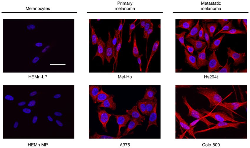
Figure 2. PLD2 expression in skin melanocytes, primary melanomas and metastatic melanoma cell lines as assessed by immunofluorescence. PLD2 is in red
and the nuclei in blue (scale bar, 50 µm; original magnification, x63). The images are representative of three different experiments. PLD, phospholipase D.
PLA2, a trait that was cell line‑specific as it was not common and raise the question of whether they have a role in melanoma,
to all the metastatic melanomas. PLC was expressed relatively particularly in the metastatic process.
homogeneously in the cell lines studied and no marked
differences were observed (Fig. S3). The enhanced presence of PLD2 overexpression and silencing in melanoma cell lines.
PLD2 in melanoma cells was specific to this isoform, since the After demonstrating that aggressive melanoma cells have
PLD1, PLA2 and PLC proteins remained relatively constant in stronger PLD2 expression and activity than skin melanocytes,
melanocytes and melanoma cells. modifications to the amounts of this enzyme were performed
Immunofluorescence analyses confirmed that PLD2 to shed light on the implication of PLD2 in specific carci‑
expression was upregulated in the malignant cell lines studied nogenic events. The effects of transient overexpression and
(Fig. 2). PLD2 was expressed more intensely in primary mela‑ silencing of PLD2 were examined in primary melanoma cells
nomas (A375, Mel‑Ho) and metastatic melanomas (Hs294t, (A375, Mel‑Ho) or metastatic melanoma cells (Colo‑800), as
Colo‑800), while its expression was faint in skin melanocytes confirmed in western blots (Figs. 4A and S4). Furthermore,
(HEMn‑LP, HEMn‑MP). By contrast, immunofluorescence these molecular alterations were also manifested through
analysis of the other phospholipases (PLA2, PLC and PLD1) enzymatic activity, as lipase activity was altered in accor‑
did not indicate any consistent differences between the cell dance with protein expression in all cases, although only
lines studied (data not shown). Indeed, both the western blot in the A375 line, the lipase activity exhibited statistically
and immunofluorescence data corroborated the enhanced significant differences compared to the transfection controls.
expression of PLD2 in primary and metastatic melanomas As indicated in Fig. 4B, PLD2 overexpression was associated
relative to normal skin melanocytes. with an increase in lipase activity when the ratio relative to
the transfection control was considered: A375 cells, 1.87;
Enhanced PLD lipase activity in melanoma cells. A comparative Mel‑Ho, 1.54; and Colo‑800, 1.4. By contrast, PLD2‑silenced
analysis of PLD activity was performed in melanocytes, primary cells exhibited weaker lipase activity: A375, 0.52; Mel‑Ho,
melanomas and metastatic melanomas (Fig. 3). The results 0.82; and Colo‑800, 0.68.
indicated that melanoma cells exhibited enhanced PLD activity
and that this was associated with melanoma progression. Indeed, PLD2 enhances the proliferation, migration and invasion
metastatic melanoma cells had significantly more lipase activity, of melanoma cells. The functional implications of the
while primary melanoma cell lines also had higher activity than modifications to PLD2 expression were analyzed by studying
skin melanocytes. However, the activity in the HEMn‑LP mela‑ the differences in proliferation, migration and invasion
nocyte cell line was comparable to that in primary melanomas of transformed cells. Although these changes were cell
and therefore, the differences were not statistically significant line‑specific, there was a clear trend towards an increase in prolif‑
between both groups. These results confirmed that, overall, the eration, migration and invasion of PLD2‑overexpressing cells,
lipase activity of PLD enzymes is increased in melanoma cells while these processes were downregulated in PLD2‑silenced6 PEREZ-VALLE et al: UPREGULATED PLD2 RELATED TO METASTATIC PROPERTIES OF MELANOMA Figure 3. PLD activity in melanocytes and melanoma cells. Summary and graphic representation of the PLD activity in (A) the three study groups and (B) in the individual cell lines. PLD activity is expressed as dpm/mg of protein and values are expressed as the mean ± standard error of the mean from three different experiments. ***P
ONCOLOGY LETTERS 23: 140, 2022                                                           7

                                                                                        Discussion

                                                                                        Phospholipases metabolize phospholipids, generating bioac‑
                                                                                        tive molecules that influence cell fate by regulating distinct cell
                                                                                        activities, including events that favor tumorigenesis and metas‑
                                                                                        tasis (11‑13,28). Deregulation of the expression and activity
                                                                                        of phospholipases has been implicated in different diseases,
                                                                                        including cancer. Indeed, the overexpression and enhanced
                                                                                        activity of PLA2 serves as a diagnostic marker of breast
                                                                                        cancer and is correlated with the tumor stage (29). Similarly,
                                                                                        the present analysis of the PLD2 protein demonstrated that it
                                                                                        accumulates at higher levels in metastatic melanoma than in
                                                                                        normal skin melanocytes, as indicated by the PLD2 expression
                                                                                        analyzed by western blot and immunofluorescence. However,
                                                                                        the immunohistochemical analysis performed was limited, as
                                                                                        neither the subcellular localization of PLD2 nor the changes in
                                                                                        PLD2 expression in the transfected cells was examined. The
                                                                                        results prompted us to analyze the activity of PLD, based on
                                                                                        the notion that not only is PLD expression enhanced in cancer
                                                                                        tissues but its activity is also increased (10,30). PLD activity
                                                                                        was enhanced particularly in murine melanoma cells, in asso‑
                                                                                        ciation with an acidic pH of the tumor microenvironment (31),
                                                                                        consistent with the enhanced PLD activity observed previ‑
                                                                                        ously in melanoma cells relative to skin melanocytes (22). This
                                                                                        trait was verified in the present study, as significantly stronger
                                                                                        PLD activity was observed in malignant cells. Furthermore,
                                                                                        the present study was the first, to the best of our knowledge,
                                                                                        to indicate that PLD activity in metastatic melanoma cells was
                                                                                        substantially higher than in primary melanoma cells.
                                                                                            One role of PLD2 is the hydrolysis of PC present in cell
Figure 5. Cell proliferation in PLD2‑overexpressing and ‑silenced melanoma              membranes to release free Cho and a PA molecule. The mole‑
cells. (A) Proliferation relative to the corresponding transfection controls.           cules liberated are known precursors and second messengers
(B) Graphs presenting the proliferation of the representative cell lines. No            that support a tumorigenic phenotype. Indeed, increased Cho
substantial differences were observed in the basal proliferation rates of the three
cell lines. Values are expressed as the mean ± SEM of at least three different trans‑
                                                                                        levels have been identified in cancer cells (3‑5). Cho is essential
fections. ***P
8 PEREZ-VALLE et al: UPREGULATED PLD2 RELATED TO METASTATIC PROPERTIES OF MELANOMA Figure 6. Cell migration and invasion of PLD2‑overexpressing and ‑silenced melanoma cells. (A) Summary of the migration relative to the corresponding transfection controls. (B) Summary of the invasion data represented as a ratio relative to the corresponding transfection controls. (C and D) Graphical represen‑ tation of the results from (C) the migration and (D) invasion assay. The three cell lines had a similar basal migration capacity, while the basal invasion capacity of Colo‑800 was significantly lower than that of the primary melanomas. Representative images for the migration and invasion assays for the three lines and all the conditions tested are provided in Figs. S5 and S6, respectively. Values are expressed as the mean ± SEM of at least three different transfections. **P
ONCOLOGY LETTERS 23: 140, 2022                                                          9

essential second messengers means that the range of effects of        function and analyzed the results of certain experiments.
the enzyme is broad and its effect on biological activities such as   KNS participated in the execution of PLD activity assays and
those studied may be cell line‑dependent.                             PLD2 transfection assays. EA and GBG obtained the financial
    Pharmacological inhibition or genetic downregulation of           support and performed parts of the statistical analysis. APV
PLD2 has been proposed as a potential treatment option for            and AA confirm the authenticity of all the raw data. All
certain cancer types. In breast cancer in particular, previous        authors read, reviewed and approved the final manuscript.
studies have indicated that the use of a PLD2‑specific inhibitor
markedly reduced the invasive capacity of cultured cell               Ethics approval and consent to participate
lines (21) and it promoted cancer cell apoptosis, while over‑
expression of PLD2 provided resistance to chemotherapeutic            Not applicable.
agents (54). Silencing of PLD2 or its pharmacological inhibi‑
tion in a mouse model repressed tumor progression, whereas            Patient consent for publication
overexpression of this enzyme translated into an increase in
tumorigenic potential (12). In lymphoma, catalytic inactivation       Not applicable.
of PLD2 reduced the formation of lung metastases (20) and
in colorectal cancer cells, the pharmacological inhibition and        Competing interests
silencing of PLD2 resulted in cancer cell autophagy (55).
    In conclusion, PLD2 contributes to different aspects of           The authors have no competing interests to declare.
cancer development including the production of survival
signals (10,46,56‑59), proliferation (60‑63), resistance to           References
apoptosis (10,64), angiogenesis (10,65), deregulated cellular
energetics (10), as well as matrix substrate degradation,              1. Bowling FZ, Frohman MA and Airola MV: Structure and
invadopodia formation, tumor‑cell migration, invasion and                 regulation of human phospholipase D. Adv Biol 79: 100783, 2021.
metastatic dissemination (10,19,31,43,46,47,66,67). However,           2. Zeisel SH and da Costa KA: Choline: An essential nutrient for
                                                                          public health. Nutr Rev 67: 615‑623, 2009.
in melanoma, a specific role for PLD2 has yet to be described          3. Onono FO and Morris AJ: Phospholipase d and choline metabo‑
in tumorigenesis. The present study was the first to report               lism. In: Lipid signaling in human diseases. Gomez‑Cambronero
increased PLD activity in metastatic melanoma cells relative              J and Frohman MA (eds). Vol 259. Springer International
                                                                          Publishing, Cham, pp205‑218, 2019.
to primary melanoma cells, as well as the influence of changes         4. Kosmopoulou M, Giannopoulou AF, Iliou A, Benaki D,
in PLD2 levels and activity in several properties of metastatic           Panagiotakis A, Velentzas AD, Konstantakou EG, Papassideri IS,
melanoma. In the light of the data on PLD2 from other cancers             Mikros E, Stravopodis DJ and Gikas E: Human melanoma‑cell
                                                                          metabolic profiling: Identification of novel biomarkers indicating
and the present in vitro results in melanoma, it is suggested that        metastasis. Int J Mol Sci 21: 2436, 2020.
PLD2 may be a promising therapeutic target and biomarker for           5. Glunde K, Bhujwalla ZM and Ronen SM: Choline metabolism
melanoma tumors, which merits further study.                              in malignant transformation. Nat Rev Cancer 11: 835‑848, 2011.
                                                                       6. Mahankali M, Henkels KM and Gomez‑Cambronero J: A
                                                                          GEF‑to‑phospholipase molecular switch caused by phosphatidic
Acknowledgements                                                          acid, Rac and JAK tyrosine kinase that explains leukocyte cell
                                                                          migration. J Cell Sci 126: 1416‑1428, 2013.
                                                                       7. Gomez‑Cambronero J, Morris AJ and Henkels KM: PLD
The authors would like to thank Dr Julian Gomez‑Cambronero                protein‑protein interactions with signaling molecules and modu‑
from the Department of Biochemistry and Molecular Biology                 lation by PA. Methods Enzymol 583: 327‑357, 2017.
of Wright State University (Dayton, OH, USA) for his support           8. Henkels KM, Mahankali M and Gomez‑Cambronero J: Increased
                                                                          cell growth due to a new lipase‑GEF (Phospholipase D2) fastly
with the PLD lipase activity measurements and PLD2                        acting on Ras. Cell Signal 25: 198‑205, 2013.
transfection in cultured cells.                                        9. Gomez‑Cambronero J: Phospholipase D in cell signaling: From
                                                                          a myriad of cell functions to cancer growth and metastasis. J Biol
Funding                                                                   Chem 289: 22557‑22566, 2014.
                                                                      10. Gomez‑Cambronero J: Phosphatidic acid, phospholipase D and
                                                                          tumorigenesis. Adv Biol Regul 54: 197‑206, 2014.
This study was supported by grants from the University of the         11. Brown HA, Thomas PG and Lindsley CW: Targeting phospholi‑
Basque Country/EHU (grant no. GIU17/066) and Ministerio                   pase D in cancer, infection and neurodegenerative disorders. Nat
                                                                          Rev Drug Discov 16: 351‑367, 2017.
de Economía y Competividad MINECO‑ONCOFINDER of                       12. Henkels KM, Boivin GP, Dudley ES, Berberich SJ and
the Spanish Government (grant no. RTC.2015‑3693‑1).                       Gomez‑Cambronero J: Phospholipase D (PLD) drives cell inva‑
                                                                          sion, tumor growth and metastasis in a human breast cancer
                                                                          xenograph model. Oncogene 32: 5551‑5562, 2013.
Availability of data and materials                                    13. Yao Y, Wang X, Li H, Fan J, Qian X, Li H and Xu Y: Phospholipase
                                                                          D as a key modulator of cancer progression. Biol Rev Camb
All data generated or analyzed during this study are included             Philos Soc 95: 911‑935, 2020.
                                                                      14. Oshimoto H, Okamura S, Yoshida M and Mori M: Increased
in this published article.                                                activity and expression of phospholipase D2 in human colorectal
                                                                          cancer. Oncol Res 14: 31‑37, 2003.
Authors' contribution                                                 15. Saito M, Iwadate M, Higashimoto M, Ono K, Takebayashi Y
                                                                          and Takenoshita S: Expression of phospholipase D2 in human
                                                                          colorectal carcinoma. Oncol Rep 18: 1329‑1334, 2007.
APV performed the experiments described in the study,                 16. Kandori S, Kojima T, Matsuoka T, Yoshino T, Sugiyama A,
collected the data and drafted the manuscript. MDB and AA                 Nakamura E, Shimazui T, Funakoshi Y, Kanaho Y and
                                                                          Nishiyama H: Phospholipase D2 promotes disease progression of
guided the design and execution of the study and revised                  renal cell carcinoma through the induction of angiogenin. Cancer
the manuscript. BO provided advice on lipid biology and                   Sci 109: 1865‑1875, 2018.
10 PEREZ-VALLE et al: UPREGULATED PLD2 RELATED TO METASTATIC PROPERTIES OF MELANOMA
17. McDermott MI, Wang Y, Wakelam MJO and Bankaitis VA: 39. Singh NK, Hansen DE III, Kundumani‑Sridharan V and
Mammalian phospholipase D: Function, and therapeutics. Prog Rao GN: Both Kdr and Flt1 play a vital role in hypoxia‑induced
Lipid Res 78: 101018, 2020. Src‑PLD1‑PKCγ‑cPLA2 activation and retinal neovasculariza‑
18. Noble AR, Hogg K, Suman R, Berney DM, Bourgoin S, tion. Blood 121: 1911‑1923, 2013.
Maitland NJ and Rumsby MG: Phospholipase D2 in prostate 40. Liu Y, Su Y and Wang X: Phosphatidic Acid‑Mediated Signaling.
cancer: Protein expression changes with Gleason score. Br In: Lipid‑mediated Protein Signaling. Capelluto DGS (ed).
J Cancer 121: 1016‑1026, 2019. Springer Netherlands, Dordrecht, pp159‑176, 2013.
19. Henkels KM, Farkaly T, Mahankali M, Segall JE and 41. Frohman MA: The phospholipase D superfamily as therapeutic
Gomez‑Cambronero J: Cell invasion of highly metastatic MTLn3 targets. Trends Pharmacol Sci 36: 137‑144, 2015.
cancer cells is dependent on phospholipase D2 (PLD2) and janus 42. Lee S and Lynch KR: Brown recluse spider (Loxosceles reclusa)
kinase 3 (JAK3). J Mol Biol 408: 850‑862, 2011. venom phospholipase D (PLD) generates lysophosphatidic acid
20. Knoepp SM, Chahal MS, Xie Y, Zhang Z, Brauner DJ, (LPA). Biochem J 391: 317‑323, 2005.
Hallman MA, Robinson SA, Han S, Imai M, Tomlinson S and 43. Wang Z, Zhang F, He J, Wu P, Tay LWR, Cai M, Nian W, Weng Y,
Meier KE: Effects of active and inactive phospholipase D2 on Qin L, Chang JT, et al: Binding of PLD2‑generated phosphatidic
signal transduction, adhesion, migration, invasion, and metas‑ acid to KIF5B promotes MT1‑MMP surface trafficking and lung
tasis in EL4 lymphoma cells. Mol Pharmacol 74: 574‑584, 2008. metastasis of mouse breast cancer cells. Dev Cell 43: 186‑197.
21. Scott SA, Selvy PE, Buck JR, Cho HP, Criswell TL, Thomas AL, e7, 2017.
Armstrong MD, Arteaga CL, Lindsley CW and Brown HA: 44. Borel M, Lollo G, Magne D, Buchet R, Brizuela L and Mebarek S:
Design of isoform‑selective phospholipase D inhibitors that Prostate cancer‑derived exosomes promote osteoblast differen‑
modulate cancer cell invasiveness. Nat Chem Biol 5: 108‑117, tiation and activity through phospholipase D2. Biochim Biophys
2009. Acta Mol Basis Dis 1866: 165919, 2020.
22. Riebeling C, Müller C and Geilen C: Expression and regulation 45. Muñoz‑Galván S, Lucena‑Cacace A, Perez M, Otero‑Albiol D,
of phospholipase D isoenzymes in human melanoma cells and Gomez‑Cambronero J and Carnero A: Tumor cell‑secreted PLD
primary melanocytes. Melanoma Res 13: 555‑562, 2003. increases tumor stemness by senescence‑mediated communica‑
23. Dogliotti G, Kullmann L, Dhumale P, Thiele C, Panichkina O, tion with microenvironment. Oncogene 38: 1309‑1323, 2019.
Mendl G, Houben R, Haferkamp S, Püschel AW and Krahn MP: 46. Zheng Y, Rodrik V, Toschi A, Shi M, Hui L, Shen Y and
Membrane‑binding and activation of LKB1 by phosphatidic Foster DA: Phospholipase D couples survival and migration
acid is essential for development and tumour suppression. Nat signals in stress response of human cancer cells. J Biol Chem 281:
Commun 8: 15747, 2017. 15862‑15868, 2006.
24. American Cancer Society: Cancer Facts & Figures 2020. 47. Williger BT, Ho WT and Exton JH: Phospholipase D mediates
American Cancer Society, Atlanta, GA, 2020. matrix metalloproteinase‑9 secretion in phorbol ester‑stimulated
25. Guterres AN, Herlyn M and Villanueva J: Melanoma. In: eLS. human fibrosarcoma cells. J Biol Chem 274: 735‑738, 1999.
John Wiley & Sons, Hoboken, NJ, pp1‑10, 2018. 48. Noble AR, Maitland NJ, Berney DM and Rumsby MG:
26. Gomez‑Cambronero J, Horwitz J and Sha'afi RI: Measurements Phospholipase D inhibitors reduce human prostate cancer cell
of phospholipases A2, C, and D (PLA2, PLC, and PLD): In vitro proliferation and colony formation. Br J Cancer 118: 189‑199, 2018.
microassays, analysis of enzyme isoforms, and intact‑cell assays. 49. Cerami E, Gao J, Dogrusoz U, Gross BE, Sumer SO, Aksoy BA,
Methods Mol Biol 218: 155‑176, 2003. Jacobsen A, Byrne CJ, Heuer ML, Larsson E, et al: The cBio
27. Henderson F, Johnston HR, Badrock AP, Jones EA, Forster D, cancer genomics portal: An open platform for exploring
Nagaraju RT, Evangelou C, Kamarashev J, Green M, multidimensional cancer genomics data. Cancer Discov 2:
Fairclough M, et al: Enhanced fatty acid scavenging and glyc‑ 401‑404, 2012.
erophospholipid metabolism accompany melanocyte neoplasia 50. Gao J, Aksoy BA, Dogrusoz U, Dresdner G, Gross B, Sumer SO,
progression in zebrafish. Cancer Res 79: 2136‑2151, 2019. Sun Y, Jacobsen A, Sinha R, Larsson E, et al: Integrative analysis
28. Park JB, Lee CS, Jang JH, Ghim J, Kim YJ, You S, Hwang D, of complex cancer genomics and clinical profiles using the cBio‑
Suh PG and Ryu SH: Phospholipase signalling networks in Portal. Sci Signal 6: pl1, 2013.
cancer. Nat Rev Cancer 12: 782‑792, 2012. 51. Powner DJ and Wakelam MJO: The regulation of phospholipase
29. Qu J, Zhao X, Wang J, Liu C, Sun Y, Cai H and Liu J: Plasma D by inositol phospholipids and small GTPases. FEBS Lett 531:
phospholipase A2 activity may serve as a novel diagnostic 62‑64, 2002.
biomarker for the diagnosis of breast cancer. Oncol Lett 15: 52. Santy LC and Casanova JE: Activation of ARF6 by ARNO
5236‑5242, 2018. stimulates epithelial cell migration through downstream
30. Gomez‑Cambronero J, Fite K and Miller TE: How miRs and activation of both Rac1 and phospholipase D. J Cell Biol 154:
mRNA deadenylases could post‑transcriptionally regulate 599‑610, 2001.
expression of tumor‑promoting protein PLD. Adv Biol Regul 68: 53. Chae YC, Kim JH, Kim KL, Kim HW, Lee HY, Heo WD,
107‑119, 2018. Meyer T, Suh PG and Ryu SH: Phospholipase D activity regu‑
31. Kato Y, Lambert CA, Colige AC, Mineur P, Noël A, Frankenne F, lates integrin‑mediated cell spreading and migration by inducing
Foidart JM, Baba M, Hata RI, Miyazaki K and Tsukuda M: Acidic gtp‑rac translocation to the plasma membrane. Mol Biol Cell 19:
extracellular pH induces matrix metalloproteinase‑9 expression 3111‑3123, 2008.
in mouse metastatic melanoma cells through the phospholipase 54. Hwang WC, Kang DW, Kang Y, Jang Y, Kim JA and Min DS:
D‑mitogen‑activated Protein Kinase Signaling. J Biol Chem 280: Inhibition of phospholipase D2 augments histone deacetylase inhib‑
10938‑10944, 2005. itor‑induced cell death in breast cancer cells. Biol Res 53: 34, 2020.
32. Jenkins GM and Frohman MA: Phospholipase D: A lipid centric 55. Hwang WC, Kim MK, Song JH, Choi KY and Min DS: Inhibition
review. Cell Mol Life Sci 62: 2305‑2316, 2005. of phospholipase D2 induces autophagy in colorectal cancer
33. Gomez‑Cambronero J: New concepts in phospholipase D cells. Exp Mol Med 46: e124, 2014.
signaling in inflammation and cancer. ScientificWorldJournal 10: 56. Rodrik V, Zheng Y, Harrow F, Chen Y and Foster DA: Survival
1356‑1369, 2010. signals generated by estrogen and phospholipase D in MCF‑7
34. Yoon MS, Rosenberger CL, Wu C, Truong N, Sweedler JV and breast cancer cells are dependent on myc. Mol Cell Biol 25:
Chen J: Rapid mitogenic regulation of the mTORC1 inhibitor, 7917‑7925, 2005.
DEPTOR, by phosphatidic acid. Mol Cell 58: 549‑556, 2015. 57. Hui L, Rodrik V, Pielak RM, Knirr S, Zheng Y and Foster DA:
35. Yin H, Gui Y, Du G, Frohman MA and Zheng XL: Dependence MTOR‑dependent suppression of protein phosphatase 2a is
of Phospholipase D1 multi‑monoubiquitination on its enzymatic critical for phospholipase D survival signals in human breast
activity and palmitoylation. J Biol Chem 285: 13580‑13588, 2010. cancer cells. J Biol Chem 280: 35829‑35835, 2005.
36. Jang YH and Min DS: The hydrophobic amino acids involved 58. Hui L, Zheng Y, Yan Y, Bargonetti J and Foster DA: Mutant p53
in the interdomain association of phospholipase D1 regulate the in MDA‑MB‑231 breast cancer cells is stabilized by elevated
shuttling of phospholipase D1 from vesicular organelles into the phospholipase D activity and contributes to survival signals
nucleus. Exp Mol Med 44: 571‑577, 2012. generated by phospholipase D. Oncogene 25: 7305‑7310, 2006.
37. Donaldson JG: Phospholipase D in endocytosis and endosomal 59. Chen Y, Rodrik V and Foster DA: Alternative phospholi‑
recycling pathways. Biochim Biophys Acta 1791: 845‑849, 2009. pase D/mTOR survival signal in human breast cancer cells.
38. Dall'Armi C, Hurtado‑Lorenzo A, Tian H, Morel E, Nezu A, Oncogene 24: 672‑679, 2005.
Chan RB, Yu WH, Robinson KS, Yeku O, Small SA, et al: The 60. Moolenaar WH, Kruijer W, Tilly BC, Verlaan I, Bierman AJ
phospholipase D1 pathway modulates macroautophagy. Nat and de Laat SW: Growth factor‑like action of phosphatidic acid.
Commun 1: 142, 2010. Nature 323: 171‑173, 1986.ONCOLOGY LETTERS 23: 140, 2022 11
61. Rizzo MA, Shome K, Vasudevan C, Stolz DB, Sung TC, 66. Park MH, Ahn BH, Hong YK and Min DS: Overexpression of
Frohman MA, Watkins SC and Romero G: Phospholipase D and phospholipase D enhances matrix metalloproteinase‑2 expression
its product, phosphatidic acid, mediate agonist‑dependent Raf‑1 and glioma cell invasion via protein kinase C and protein kinase
translocation to the plasma membrane and the activation of the A/NF‑κ B/Sp1‑mediated signaling pathways. Carcinogenesis 30:
mitogen‑activated protein kinase pathway. J Biol Chem 274: 356‑365, 2009.
1131‑1139, 1999. 67. Kang DW, Park MH, Lee YJ, Kim HS, Lindsley CW, Brown HA
62. Bruntz RC, Lindsley CW and Brown HA: Phospholipase D and Min DS: Autoregulation of phospholipase D activity is
signaling pathways and phosphatidic acid as therapeutic targets coupled to selective induction of phospholipase D1 expression to
in cancer. Pharmacol Rev 66: 1033‑1079, 2014. promote invasion of breast cancer cells. Int J Cancer 128: 805‑816,
63. Zhao C, Du G, Skowronek K, Frohman MA and Bar‑Sagi D: 2011.
Phospholipase D2‑generated phosphatidic acid couples EGFR This work is licensed under a Creative Commons
stimulation to Ras activation by Sos. Nat Cell Biol 9: 707‑712, Attribution-NonCommercial-NoDerivatives 4.0
2007.
64. Chen Y, Zheng Y and Foster DA: Phospholipase D confers International (CC BY-NC-ND 4.0) License.
rapamycin resistance in human breast cancer cells. Oncogene 22:
3937‑3942, 2003.
65. Ghim J, Moon JS, Lee CS, Lee J, Song P, Lee A, Jang JH, Kim D,
Yoon JH, Koh YJ, et al: Endothelial deletion of phospholipase
d2 reduces hypoxic response and pathological angiogenesis.
Arterioscler Thromb Vasc Biol 34: 1697‑1703, 2014.You can also read