The evolution of interdependence in a four-way mealybug symbiosis - Caltech Authors
←
→
Page content transcription
If your browser does not render page correctly, please read the page content below
bioRxiv preprint doi: https://doi.org/10.1101/2021.01.28.428658; this version posted January 29, 2021. The copyright holder for this preprint
(which was not certified by peer review) is the author/funder, who has granted bioRxiv a license to display the preprint in perpetuity. It is made
                                              available under aCC-BY-NC 4.0 International license.

                   The evolution of interdependence in a four-way
                                mealybug symbiosis
                 Arkadiy I. Garber1,2 , Maria Kupper1,2 , Dominik R. Laetsch3 , Stephanie R. Weldon1 ,
                       Mark S. Ladinsky4 , Pamela J. Bjorkman4 and John P. McCutcheon∗1,2
                         1
                           Division of Biological Sciences, University of Montana, Missoula, MT 59801
             2
              Biodesign Center for Mechanisms of Evolution and School of Life Sciences, Arizona State University,
                                                          Tempe, AZ 85287
                                 3
                                   Institute of Evolutionary Biology, University of Edinburgh, UK
             4
               Department of Biology and Biological Engineering, California Institute of Technology, Pasadena, CA
                                                               91125

                                                               January 2021

           Abstract
           Mealybugs are insects that maintain intracellular bacterial symbionts to supplement their nutrient-
           poor plant sap diets. Some mealybugs have a single betaproteobacterial endosymbiont, a Candidatus
           Tremblaya species (hereafter Tremblaya) that alone provides the insect with its required nutrients.
           Other mealybugs have two nutritional endosymbionts that together provide these nutrients, where
           Tremblaya has gained a gammaproteobacterial partner that resides in the cytoplasm of Tremblaya.
           Previous work had established that Pseudococcus longispinus mealybugs maintain not one but
           two species of gammaproteobacterial endosymbionts along with Tremblaya. Preliminary genomic
           analyses suggested that these two gammaproteobacterial endosymbionts have large genomes with
           features consistent with a relatively recent origin as insect endosymbionts, but the patterns of ge-
           nomic complementarity between members of the symbiosis and their relative cellular locations were
           unknown. Here, using long-read sequencing and various types of microscopy, we show that the
           two gammaproteobacterial symbionts of P. longispinus are mixed together within Tremblaya cells,
           and that their genomes are somewhat reduced in size compared to their closest non-endosymbiotic
           relatives. Both gammaproteobacterial genomes contain thousands of pseudogenes, consistent with
           a relatively recent shift from a free-living to endosymbiotic lifestyle. Biosynthetic pathways of key
           metabolites are partitioned in complex interdependent patterns among the two gammaproteobac-
           terial genomes, the Tremblaya genome, and horizontally acquired bacterial genes that are encoded
           on the mealybug nuclear genome. Although these two gammaproteobacterial endosymbionts have
           been acquired recently in evolutionary time, they have already evolved co-dependencies with each
           other, Tremblaya, and their insect host.

           Key words: endosymbionts, pseudogenes, transposases, genome reduction, metabolic interdepen-
           dence, mealybugs
                 ∗ Corresponding   author: john.mccutcheon@asu.edu

                                                                        1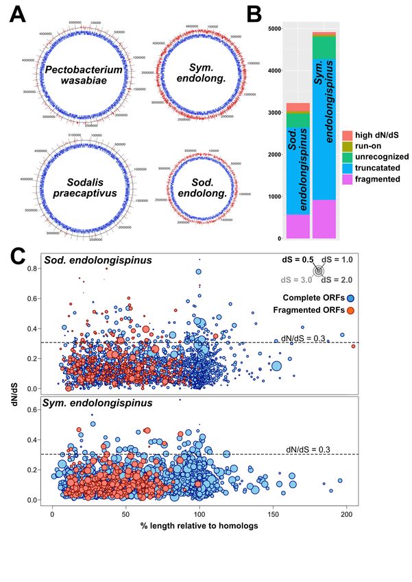
bioRxiv preprint doi: https://doi.org/10.1101/2021.01.28.428658; this version posted January 29, 2021. The copyright holder for this preprint
(which was not certified by peer review) is the author/funder, who has granted bioRxiv a license to display the preprint in perpetuity. It is made
                                              available under aCC-BY-NC 4.0 International license.

           Significance
           Mealybugs are sap-feeding insects that house between one and three bacterial endosymbionts to sup-
           plement their nutritionally poor diets. Many mealybug-bacteria relationships were established tens
           or hundreds of millions of years ago, and these ancient examples show high levels host-endosymbiont
           genomic and metabolic integration. Here, we describe the complete genomes and cellular loca-
           tions for two bacterial endosymbiont which have recently transitioned from a free-living to an
           intracellular state. Our work reveals the rapid emergence of metabolic interdependence between
           these two nascent endosymbionts, their partner bacterial co-symbiont in whose cytoplasm they
           reside, and their insect host cell. Our work confirms that intracellular bacteria rapidly adapt to
           a host-restricted lifestyle through breakage or loss of redundant genes.

           Introduction                                                     Dohlen et al., 2001). Many of these intra-
                                                                            Tremblaya endosymbionts are members of the
                                                                            Sodalis genus, which are commonly found as
           Insects with nutrient-poor diets (e.g. plant                     endosymbionts of insects (Oakeson et al., 2014;
           sap, blood, wood) retain nutritional symbionts                   Clayton et al., 2012; Hall et al., 2020; Toh et
           that supplement their diet with nutrients, such                  al., 2006 Husník and McCutcheon, 2016; Mc-
           as amino acids and vitamins (Baumann, 2005;                      Cutcheon et al., 2019).
           Douglas, 2006). Mealybugs (Figure 1A) are
           insects that exclusively consume phloem sap
           and maintain nutritional endosymbiotic bacte-                    Genomic studies of numerous insect-
           ria within specialized cells called bacteriocytes                endosymbiont systems have revealed strong
           (Buchner, 1965; von Dohlen et al., 2001; Bau-                    and consistent patterns of complementary gene
           mann et al., 2002). Mealybug bacteriocytes                       loss and retention among all members of the
           house between one and three different bacte-                     symbiosis (Shigenobu et al., 2000; van Ham et
           rial endosymbionts depending on the mealybug                     al., 2003; Wu et al., 2006; Gatehouse et al.,
           species (Kono et al., 2008; Koga et al., 2013;                   2012; Sloan and Moran, 2012; McCutcheon
           López-Madrigal et al., 2013; Husník and Mc-                      and Moran, 2010; Łukasik et al., 2018). While
           Cutcheon, 2016; Szabó et al., 2017; Gil et al.,                  in most cases, a single endosymbiont genome
           2017). These mealybug endosymbionts produce                      will retain complete or near-complete pathways
           essential amino acids and vitamins, which are                    for individual metabolites, mealybug endosym-
           present at low and variable levels in the in-                    bionts are unusual in that the reciprocal pat-
           sect’s specialized plant sap diet. While it is not               tern of gene loss and retention exists within
           uncommon for insects to simultaneously main-                     biochemical pathways (McCutcheon and von
           tain multiple endosymbionts (Buchner, 1965;                      Dohlen, 2011; Husník 2013; López-Madrigal et
           Fukatsu et al., 1998; Toh et al., 2006; Thao                     al., 2013; Husník 2016; Szabó et al., 2017; Gil
           et al., 2002; Moran et al., 2008; McCutcheon                     et al., 2017). Most of the previously published
           and Moran, 2010), the spatial organization of                    mealybug endosymbiont genomes were highly
           the dual mealybug endosymbionts is unusual:                      reduced in size (less than 1 Mb) and gene dense
           each bacteriocyte houses cells of Candidatus                     (containing few pseudogenes), which made dis-
           Tremblaya princeps (Betaproteobacteria; here-                    cerning these complementary gene loss and re-
           after referred to as Tremblaya), and inside each                 tention patterns relatively straightforward.
           Tremblaya cell reside tens to hundreds of cells
           of another endosymbiont from the gammapro-                       P. longispinus harbors the symbiont Tremblaya,
           teobacterial family Enterobacteriaceae (von                      but unlike Tremblaya in other mealybugs, the

                                                                        2
bioRxiv preprint doi: https://doi.org/10.1101/2021.01.28.428658; this version posted January 29, 2021. The copyright holder for this preprint
(which was not certified by peer review) is the author/funder, who has granted bioRxiv a license to display the preprint in perpetuity. It is made
                                              available under aCC-BY-NC 4.0 International license.

           P. longispinus strain of Tremblaya houses not                    at 25C, 77% relative humidity, and a 12h
           one but two species of gammaproteobacterial                      light/dark cycle in a Percival 136LL incuba-
           endosymbionts (Gatehouse et al., 2011; Rosen-                    tor.
           blueth et al., 2012; Husník and McCutcheon,
           2016). We previously reported draft genome                       RNA Fluorescence in situ Hybridization
           assemblies of these two gammaproteobacterial                     (RNA-FISH)
           endosymbionts, which suggested that their com-                          Whole P. longispinus individuals of the
           bined genome sizes were large, approximately                     second and third instar developmental stage
           8.2 megabase pairs (Mbp) in length (Husník                       were submerged in Ringer solution (3 mM
           and McCutcheon, 2016). Phylogenetic analysis                     CaCl2 * 2H2O, 182 mM KCl, 46 mM NaCl,
           showed that one of these gammaproteobacterial                    10 mM Tris base; adjusted to pH 7.2) and
           symbionts belonged to the Sodalis genus, and                     carefully opened for better buffer infiltration.
           the other was more closely related to members                    Samples were transferred into Carnoy’s fixative
           of the Pectobacterium genus. However, the poor                   (EtOH: chloroform: acetic acid; 6:3:1) and fixed
           quality of these draft genome assemblies made                    overnight at 4°C. Tissue samples were then de-
           detailed genomic analysis impossible. Light mi-                  hydrated in a graded ethanol series from 70%
           croscopy on P. longispinus (Gatehouse et al.,                    to 100% ethanol. Samples were transferred into
           2012) suggested that the gammaproteobacte-                       tissue bags and cassettes for paraffin embed-
           rial endosymbionts resided inside Tremblaya                      ding using a Leica ASP 300 Tissue Processor.
           cells, as is the case in other mealybugs (von                    Ethanol was exchanged for methyl salicylate,
           Dohlen et al., 2001). But it was unclear from                    and then incubated in 100% xylene before in-
           these data whether 1) one or both of these                       filtration with paraffin. Each individual sample
           gammaproteobacteria were restricted to Trem-                     was embedded in a single paraffin block, semi-
           blaya cells (that is, if they were also found                    thin sections (5-6µm) were prepared with a mi-
           in the cytoplasm of the host insect bacterio-                    crotome and mounted onto microscopy slides.
           cyte), 2) whether each gammaproteobacterial
                                                                                   Sections designated for RNA-FISH ex-
           species was restricted to particular Tremblaya
                                                                            periments were deparaffinized in xylene and
           cell types, or 3) whether the two gammapro-
                                                                            rehydrated in a graded ethanol series (100% to
           teobacterial symbionts were mixed together in-
                                                                            30%). Tissue sections were then prehybridized
           side undifferentiated Tremblaya cells. Here, we
                                                                            in hybridization buffer (900mM NaCl, 20mM
           add long-read data generated from P. longispi-
                                                                            Tris-HCl pH 7.5, 35% formamide). Hybridiza-
           nus bacteriome tissue to greatly improve the
                                                                            tion was performed by adding 1.5 – 2 µL of the
           gammaproteobacterial genome assemblies and
                                                                            probe targeting the Pectobacterium-related en-
           annotations. We describe the relative cellular
                                                                            dosymbiont (5’[Cy3]-ccacgcctcaagggcacaacctc;
           locations of the endosymbionts using fluores-
                                                                            100 µM) to each 100 µL hybridization buffer and
           cence and transmission electron microscopy,
                                                                            incubated at 40C. Samples were then briefly
           and report the genome evolutionary patterns
                                                                            rinsed in wash buffer (70 mM NaCl, 20 mM
           and metabolic contributions of the microbial
                                                                            Tris-HCl pH 7.5, 5 mM EDTA pH 8.0, 0.01%
           members of this unusual four-way symbiosis.
                                                                            SDS) before mounting the slides with hybridiza-
                                                                            tion buffer supplemented with 1.5 – 2 µL of each
                                                                            probe targeting the Sodalis-related endosym-
           Material and Methods                                             biont     (5’[Cy5]-aaagccacggctcaaggccacaacctt;
                                                                            100 µM) and Tremblaya (5’[fluorescein]-
           Insect rearing                                                   gccttagcccgtgctgccgtac; 100 µM) per 100µl
           Pseudococcus longispinus populations were                        buffer, followed by overnight incubation at 30C.
           reared on sprouted potatoes (Figure 1A)                          Slides were then washed in washing buffer at

                                                                        3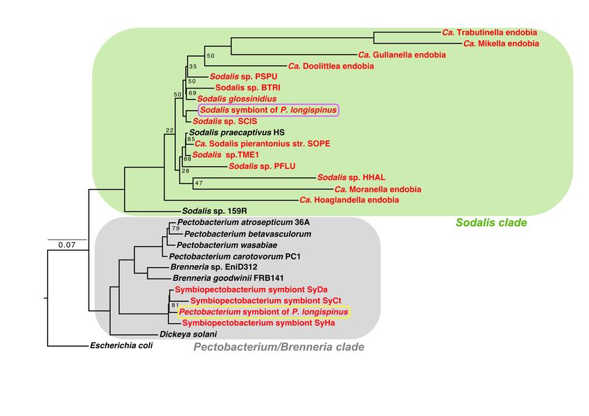
bioRxiv preprint doi: https://doi.org/10.1101/2021.01.28.428658; this version posted January 29, 2021. The copyright holder for this preprint
(which was not certified by peer review) is the author/funder, who has granted bioRxiv a license to display the preprint in perpetuity. It is made
                                              available under aCC-BY-NC 4.0 International license.

           30°C and counterstained with Hoechst in wash-                    1 mm slot grids (Electron Microscopy Sciences).
           ing buffer. After another washing step, sam-                     Grids were stained with 3% uranyl acetate and
           ple slides were rinsed in dH2O, mounted with                     lead citrate, then 10 nm colloidal gold particles
           FluorSave™ Reagent (Sigma Millipore), and                        were applied to both sides of the sections to
           analyzed by confocal laser scanning microscopy                   serve as fiducial markers for subsequent tomo-
           with a Zeiss LSM 880. Images were processed                      graphic image alignment.
           using Fiji version 1.0.
                                                                            Dual Axis Tomography
           Electron Microscopy                                              Grids were placed in a Dual-Axis tomography
           Bacteriomes were dissected from P. longispinus                   specimen holder (Model 2040; E.A. Fischione
           individuals as previously described (Bublitz et                  Instruments Inc., Export PA) and viewed with
           al., 2019). Isolated bacteriomes were pre-fixed                  a Tecnai TF-30ST transmission electron micro-
           with 3% glutaraldehyde, 1% paraformalde-                         scope at 300K eV. Dual-axis tilt-series were
           hyde, 5% sucrose in 0.1M sodium cacody-                          acquired automatically using the SerialEM
           late trihydrate for 12 – 24 h at 4°C, then                       software package (Mastronarde., 2005) and
           rinsed briefly with cacodylate buffer. Bacte-                    recorded digitally with a 2k x 2k CCD camera
           riomes were placed into brass planchettes (Ted                   (XP1000; Gatan, Inc. Pleasanton CA). Briefly,
           Pella, Inc.) pre-filled with cacodylate buffer                   sections were tilted +/- 64° with images taken
           + 10% 70kD Ficoll (extracellular cryoprotec-                     at 1° increments. The grid was then rotated 90°
           tant; Sigma) and ultra-rapidly frozen with a                     and a similar tilt-series was recorded around the
           HPM010 High Pressure Freezing machine (Bal-                      orthogonal axis. Tomograms were calculated,
           Tec/ABRA, Switzerland). Vitreously frozen                        joined and analyzed using the IMOD software
           samples were transferred under liquid nitrogen                   package (Mastronarde., 2008; Mastronarde and
           to Nunc cryovials (Thermo-Fisher Scientific)                     Held, 2017) on MacPro and iMac Pro comput-
           containing 2% OsO4 , 0.05% uranyl acetate in                     ers (Apple Inc.).
           acetone and placed into an AFS-2 Freeze Substi-
           tution Machine (Leica Microsystems, Austria).                    Sequencing and Assembly
           Samples were freeze-substituted at -90°C for 72                  Raw Illumina HiSeq 2000 reads published in
           h, warmed to -20°C over 12 h, held at -20°C for                  (Husník and McCutcheon, 2106; BioProject:
           an additional 12 h and then brought to room                      PRJEB12068) were downloaded from the Na-
           temperature. Samples were rinsed 4x with                         tional Center for Biotechnology Information
           acetone, infiltrated into Epon-Araldite resin                    (NCBI) Sequence Read Archive (SRA), using
           (Electron Microscopy Sciences, Port Wash-                        the SRA Toolkit v2.10.8 (SRA Toolkit Develop-
           ington PA), then flat-embedded between two                       ment Team). Reads were trimmed using Trim-
           Teflon-coated glass microscope slides. Resin                     momatic v0.36 (minimum length=36 bp, slid-
           was polymerized at 60°C for 24-48 h. Embed-                      ing window=4 bp, minimum quality score=15
           ded samples were observed by phase-contrast                      [ILLUMINACLIP:TruSeq3-PE:2:30:10 LEAD-
           microscopy to ascertain specimen quality and to                  ING:3 TRAILING:3 SLIDINGWINDOW:4:15
           select appropriate regions for EM study. Blocks                  MINLEN:36]) (Bolger et al., 2014). For PacBio
           of tissue (typically containing a single bacteri-                sequencing, genomic DNA was prepared from
           ome) were excised with a scalpel and glued                       pooled mealybugs using Qiagen Genomic Tip
           to plastic sectioning stubs. Serial semi-thick                   500 g extraction kits, size selected for fragments
           (150 – 300 nm) sections were cut with a UC6                      >20 kb using a BluePippen device, followed by
           ultramicrotome (Leica Microsystems) using a                      library preparation using a SMRTbell Template
           diamond knife (Diatome Ltd, Switzerland) and                     Prep Kit v1.0. The resulting libraries were se-
           collected onto Formvar-coated copper-rhodium                     quenced on 28 single molecule realtime (SMRT)

                                                                        4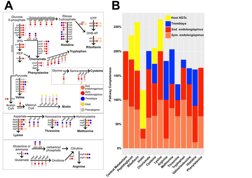
bioRxiv preprint doi: https://doi.org/10.1101/2021.01.28.428658; this version posted January 29, 2021. The copyright holder for this preprint
(which was not certified by peer review) is the author/funder, who has granted bioRxiv a license to display the preprint in perpetuity. It is made
                                              available under aCC-BY-NC 4.0 International license.

           PacBio cells using P6 version 2 chemistry and                    a hybrid SPAdes v3.13.0 (Bankevich et al.,
           reagents by Sci-Life labs in Uppsala, Sweden.                    2012) assembly (default k-mers). The two
           This sequencing effort resulted in 6,101,355                     gammaproteobacterial genomes were binned
           reads of average length 9,805 bases for a to-                    using a combination of metrics, including
           tal of 59,828,022,374 bases. These reads were                    coverage of Illumina reads mapped against
           error corrected and trimmed, resulting in 5.05                   the final assemblies. Coverage of Bowtie2-
           million reads with average sequence length of                    mapped Illumina reads was estimated us-
           9,318 bases. These corrected and trimmed reads                   ing the jgi_summarize_bam_contig_depths
           were then assembled in Canu v1.6 (correct-                       script from the MetaBAT package (Kang et al.,
           edErrorRate=0.45, genomesize=284m) (Ko-                          2019). Since the closest phylogenomic affilia-
           ren et al., 2017), which produced 3,049 con-                     tions of each gammaproteobacterial symbiont
           tigs spanning 438,113,873 bases. Preliminary                     are known (Husník and McCutcheon, 2016), we
           gammaproteobacterial contigs were extracted                      also used BLASTP (Camacho et al., 2009) to
           from the Canu assembly using the SprayN-                         compare the open reading frames (ORFs) from
           Pray software (https://github.com/Arkadiy-                       each contig against NCBI’s RefSeq database
           Garber/SprayNPray).          Briefly, SprayNPray                 (Pruitt et al., 2007). ORFs from each contig
           predicts open reading frames (ORFs) using                        were predicted using Prodigal v2.6.3 (Hyatt
           Prodigal (Hyatt et al., 2010), and then queries                  et al., 2010), and the phylogenetic affiliation
           each ORF against NCBI’s RefSeq database (re-                     of each contigs’ ORFs was inferred by its top
           lease 200) (Pruitt et al., 2007) using DIAMOND                   BLASTP hit from NCBI’s RefSeq database
           v2.0.4.142 (Buchfink et al., 2014, e-value 1E-                   (Pruitt et al., 2007).
           6). Putative endosymbiont contigs were then
           extracted from the larger assembly based on                      Phylogenomic analysis
           gene density, GC-content, and taxonomy of                        Phylogenomic analysis was carried out using
           top DIAMOND hits (to Sodalis- and Pectobac-                      GToTree v1.5.38 (Lee et al., 2019) and RAxML
           terium/Brenneria-related spp.) to each con-                      (Stamatakis, 2014). Briefly, single-copy genes
           tig. These contigs were then used to identify                    were identified using a set of HMMs for genes
           and extract all Illumina and PacBio reads as-                    common to Gammaproteobacteria (Lee, 2019).
           sociated with the gammaproteobacterial sym-                      As part of the GToTree pipeline, single-copy
           bionts. Identification of endosymbiont-affiliated                genes are identified using HMMER v3.2.1
           Illumina reads was performed using Bowtie2                       (Johnson et al., 2010), aligned with Muscle v3.8
           v2.3.4.1 (Langmead and Salzberg, 2012). Iden-                    (Edgar, 2004), and concatenated. These con-
           tification of endosymbiont PacBio reads was                      catenated alignments were then used to build
           performed using BLASR v5.1 (Chaisson and                         a phylogenomic tree with RAxML, with 100
           Tesler, 2012). About 3.2% of all PacBio reads                    bootstraps (-N 100), the PROTCAT model for
           mapped to the CANU-assembled contigs affili-                     amino acid substitution, and the BLOSUM 62
           ated with the gammaproteobacterial endosym-                      amino acid matrix (-m PROTCATBLOSUM62)
           bionts. Of the 124.5 million Illumina read pairs,                (Stamatakis, 2014).
           4.8% mapped to the crude gammaproteobacte-
           rial endosymbiont contigs.                                       Annotation and Biosynthetic Pathway
                                                                            Reconstruction
           Once these gammaproteobacterial subsets of                       Each endosymbiont was annotated using
           short and long reads were identified and                         Prokka v1.14.6 (Seemann, 2014). As part of
           extracted, Unicycler v0.4.8 (Wick et al.,                        Prokka’s pipeline, coding regions are detected
           2017) was used, with “normal” mode (min-                         using Prodigal; noncoding RNA sequences were
           imum bridge quality = 10) to carry out                           also identified: tRNAs and tmRNAs using

                                                                        5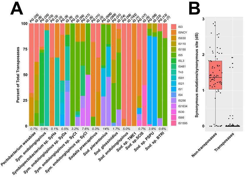
bioRxiv preprint doi: https://doi.org/10.1101/2021.01.28.428658; this version posted January 29, 2021. The copyright holder for this preprint
(which was not certified by peer review) is the author/funder, who has granted bioRxiv a license to display the preprint in perpetuity. It is made
                                              available under aCC-BY-NC 4.0 International license.

           Aragorn (Laslett and Canbeck, 2004), and                         Seq database, to identify pseudogenized ORFs
           rRNAs using RNAmmer (Lageson et al., 2007).                      that no longer appear as ORFs to Prodigal’s
           Prokka annotation also included identification                   algorithm. Non-genic regions required at least
           of transposases, using the ISfinder database of                  five DIAMOND matches to proteins in the Ref-
           insertion sequences (Siguier et al., 2006). Genes                Seq database (–intergenic_threshold 0.3) to be
           were also annotated using the GhostKOALA                         considered pseudogenes.
           v.2.2 web server, which uses the Kyoto En-
           cyclopedia of Genes and Genomes (KEGG)                           Using DIAMOND BLASTP, Pseudofinder also
           Orthology database (Kenehisa et al., 2016).                      compared ORFs from each endosymbiont to its
           Biosynthetic pathways for amino acids, vita-                     closest ancestor, inferred from phylogenomic
           mins, peptidoglycan, and translation-related                     analysis and average amino acid identity. We
           genes were manually identified from these an-                    identified Pectobacterium wasabiae as the clos-
           notations and organized into pathways. Hor-                      est free-living relative for one endosymbiont and
           izontal gene transfers (HGTs) present on the                     Sodalis praecaptivus HS for the other. Using
           mealybug genome were previously identified                       PAL2NAL v14 (Suyama et al., 2006), Pseud-
           (Husník and McCutcheon, 2016; Bublitz, et al.,                   ofinder generates codon alignments for each
           2019).                                                           ortholog pair, then, using Codeml v4.9j (Yang
                                                                            et al., 2007), calculates dN/dS values for each
           Pseudogene Prediction                                            pairwise comparison. We provide the control
           Candidate      pseudogenes     were     identi-                  file (codeml.ctl) containing the parameters used
           fied    with   the    Pseudofinder    software                   by Codeml in the following GitHub repository:
           (https://github.com/filip-husnik/pseudofinder),                  https://github.com/Arkadiy-Garber/PLON-
           using DIAMOND (–diamond) (Buchfink et al.,                       genome-paper. We required dS to be greater
           2014) to find each ORF’s closest homologs                        than 0.001 and lower than 3 for dN/dS calcula-
           NCBI’s RefSeq (Pruitt et al., 2007) database.                    tion (-m 0.001, -M 3). This allowed us to infer
           This allowed us to identify pseudogenes based                    cryptic pseudogenes, or genes that are likely un-
           on length and gene fragmentation due to early                    dergoing relaxed selection but have not acquired
           stop codons. ORFs that deviate more than 25%                     any obvious inactivating mutations (Clayton et
           (–length_pseudo 75) from the average length of                   al., 2012; Oakeson et al., 2014; Van Leuven
           the 15 top homologs (–hitcap 15, –evalue 1E-4)                   et al., 2014). We used a dN/dS cutoff of 0.3
           from RefSeq were flagged as potential pseudo-                    (Oakeson et al., 2014), flagging genes as pseu-
           genes. Additionally, ORFs with stop codons                       dogenes if their dN/dS values are higher than
           and frame-shift mutations were also flagged as                   this threshold (–max_dnds 0.3).
           pseudogenes. These fragmented ORFs were
           identified by Pseudofinder by finding adjacent                   Pseudogene calls from Pseudofinder were man-
           ORFs that have the same gene as their top DI-                    ually inspected, using AliView (Larsson, 2014)
           AMOND hit. For adjacent ORFs to be consid-                       to confirm gene fragmentation, dN/dS values,
           ered as fragmented parts of the same ancestral                   and other inactivating mutations.
           gene, we used a distance cutoff of 2000 bp (–
           distance 2000). The length of each gene and                      Identification of duplicated genes
           pairwise homology was also taken into consid-                    We used ParaHunter to identify gene dupli-
           eration to exclude intact adjacent ORFs that                     cates in the endosymbiont genomes (Miller et al,
           represent gene duplication events resulting in                   2020). This software uses MMseqs2 v12.113e3
           tandem-encoded duplicate ORFs. Non-genic                         (Steinegger and Söding, 2017) to identify ho-
           regions, in which Prodigal did not detect any                    mologous gene clusters within each genome.
           ORFs were also compared against NCBI’s Ref-                      For this analysis, we used a cutoff of 50% amino

                                                                        6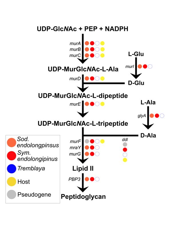
bioRxiv preprint doi: https://doi.org/10.1101/2021.01.28.428658; this version posted January 29, 2021. The copyright holder for this preprint
(which was not certified by peer review) is the author/funder, who has granted bioRxiv a license to display the preprint in perpetuity. It is made
available under aCC-BY-NC 4.0 International license.
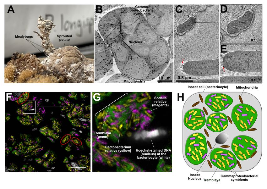
Figure 1: The structure of the P. longispinus symbiosis. A) Image of P. longispinus mealybugs on
a sprouted potato. B) Montaged TEM overview image of a bacteriocyte from P. longispinus. The 6-7 light
gray blobs are Tremblaya cells, surrounding a central eukaryotic nucleus. Within each Tremblaya cell reside
rod-shaped and more electron-dense gammaproteobacterial cells. Black-colored rods in between Tremblaya
are mitochondria within eukaryotic cytoplasm. The insect nucleus is at the center of the bacteriocyte in a
gray shade that’s similar to Tremblaya. C) Detail from an electron tomographic slice showing the boundary
of a Tremblaya cell, where a mitochondrion is visible near the Tremblaya cell envelope. D) Higher magni-
fication view of the mitochondrion shown in B. E) Tomographic slice of a gammaproteobacterial symbiont
that resides inside Tremblaya, showing numerous outer membrane vesicles (red arrows). The bacterial sym-
bionts are easily distinguished from eukaryotic mitochondria. F) Fluorescent in situ hybridization (FISH)
image of P. longispinus bacteriome tissue showing the localization of two different gammaproteobacterial
endosymbionts within Tremblaya cells. Fluorophore-labelled probes were used to localize Tremblaya cells
(green) and the two gammaproteobacterial endosymbionts (yellow and magenta). DNA and therefore insect
nuclei were counterstained with Hoechst (white). They appear to each be surrounded by several Tremblaya
cells per bacteriocyte. G) Zoomed in and annotated detail fluorescence microscopy image of a P. longispinus
bacteriocyte. H) Schematic representation of P. longispinus bacteriocytes.
7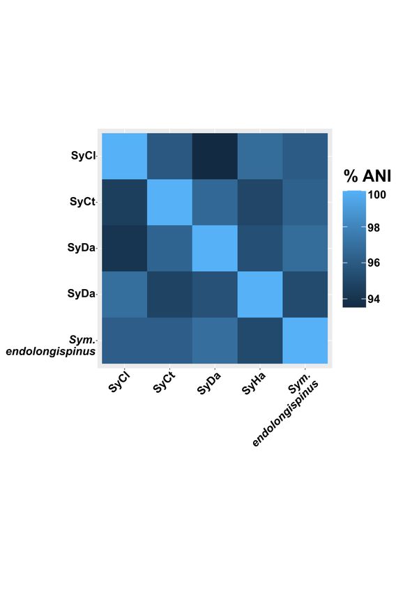
bioRxiv preprint doi: https://doi.org/10.1101/2021.01.28.428658; this version posted January 29, 2021. The copyright holder for this preprint
(which was not certified by peer review) is the author/funder, who has granted bioRxiv a license to display the preprint in perpetuity. It is made
                                              available under aCC-BY-NC 4.0 International license.

           acid identify (-m 0.5) over at least 50% of the                  Results
           length (-l 0.5) of target sequence. After clusters
           are identified, within-cluster analysis is carried               Gammaproteobacterial             endosymbionts
           out, where pairwise amino acid and nucleotide                    are located within Tremblaya
           alignments are converted to codon alignments                     Previous light microscopy suggested that at
           using PAL2NAL (Suyama et al., 2006), and                         least some, if not all, of the gammapro-
           then dN/dS is calculated using Codeml (Yang                      teobacterial endosymbionts of P. longispinus
           et al., 2007). Estimation of dN/dS required dS                   resided inside of Tremblaya (Gatehouse et al.,
           values greater than 0.001 and lower than 3.                      2011). However, these data lacked the reso-
                                                                            lution to clarify whether or not both species
           Additional scripts and plotting                                  of gammaproteobacterial endosymbionts were
                                                                            exclusively contained within Tremblaya, or
           Additional custom python scripts were used                       whether some might also live in the cytoplasm
           to process the data presented in this study.                     of bacteriocytes. We used transmission electron
           These scripts are all annotated and avail-                       microscopy (TEM) to identify the localization
           able in the following GitHub repository:                         of the gammaproteobacterial cells. Our TEM
           https://github.com/Arkadiy-Garber/PLON-                          data suggest that all gammaproteobacterial en-
           genome-paper. Many plots presented in this                       dosymbiont cells are contained within Trem-
           study were made in R (R Core Team, 2013),                        blaya and are not free in the cytoplasm of the
           using the following packages: ggplot2 (Wick-                     host insect cell (Figure 1B-E). At low magni-
           ham, 2009) and reshape (Wickham, 2007).                          fication, elongated cells of different shapes and
                                                                            sizes, which we presume to be the gammapro-
           Data Availability PacBio reads were de-                          teobacterial symbionts, can be seen inside of
           posited to the Sequence Read Archive (SRA),                      Tremblaya cells (Figure 1B). Structures of
           under NCBI BioProject PRJNAXXXXXX.                               similar size and electron density can be ob-
           Genome assemblies for the gammaproteobacte-                      served outside of Tremblaya cells, but these
           rial endosymbionts are available under NCBI                      were found to be mitochondria upon examina-
           BioProject PRJNAXXXXXX. Genome se-                               tion at higher magnification (Figure 1C-D).
           quences and annotation data for the two                          We note numerous outer-membrane vesicles
           gammaproteobacterial endosymbionts were                          (OMVs) apparently being extruded by the So-
           also made available via figshare: Genome se-                     dalis- or Pectobacterium-related endosymbiont
           quence and Prokka-annotation are available at                    cells (Figure 1E) (Toyofuku et al., 2019). The
           https://doi.org/10.6084/m9.figshare.13632407.v1                  function of these OMVs in the symbiosis, if any,
           for the Pectobacterium-related symbiont and                      is unknown.
           https://doi.org/10.6084/m9.figshare.13632398.v2
           for the Sodalis-related symbiont.         Pseu-                  Using the small subunit (SSU) ribosomal
           dogene     predictions   are    available     at                 RNA sequences reported in (Husník and Mc-
           https://doi.org/10.6084/m9.figshare.13632419.v1                  Cutcheon, 2016), we next performed fluores-
           for the Pectobacterium-related symbiont and                      cence in situ hybridization (FISH) targeting
           https://doi.org/10.6084/m9.figshare.13632416.v1                  SSU rRNA to establish the relative locations of
           for the Sodalis-related symbiont.          Files                 the two gammaproteobacterial endosymbionts.
           used in the analysis of other Sodalis-                           Here, we were testing whether there were two
           and Symbiopectobacterium-related endosym-                        different types of Tremblaya cells, each con-
           bionts are available at the following address:                   taining only one type of gammaproteobacterial
           https://doi.org/10.6084/m9.figshare.13661189.                    cell, or whether both gammaproteobacterial
                                                                            species were mixed together inside of one type of

                                                                        8
bioRxiv preprint doi: https://doi.org/10.1101/2021.01.28.428658; this version posted January 29, 2021. The copyright holder for this preprint
(which was not certified by peer review) is the author/funder, who has granted bioRxiv a license to display the preprint in perpetuity. It is made
available under aCC-BY-NC 4.0 International license.
Tremblaya cell. We find that both gammapro- ative appears highly abundant, with almost no
teobacterial endosymbionts are mixed together cells of the Sodalis relative present (red circles
in one type of Tremblaya cell (Figure 1F- in Figure 1F). Cells of the Pectobacterium
H). The overall distribution of endosymbionts relative appear to be longer than cells of the
within Tremblaya cells suggests that the Pecto- Sodalis relative. This mixture of long and short
bacterium relative is more abundant than the cells is consistent with what we see in the TEM
Sodalis relative (colored yellow and violet, re- images, although the identities of the two cell
spectively, in Figure 1F-G). We note that types cannot be discerned in TEM (Figure
there are some Tremblaya cells or regions of 1B).
Tremblaya cells where the Pectobacterium rel-
Table 1 | Assembly summary for the three endosymbionts and three putative plasmids assembled from
P. longispinus bacteriomes. The Pectobacterium and Sodalis symbionts are named Symbiopectobacterium
endolongispinus and Sodalis endolongispinus, respectively, as discussed in the section “Naming of the two
gammaproteobacterial symbionts” *Genome of Tremblaya taken from Husník and McCutcheon, 2016.
Endosymbionts Genome/plasmid Average read
Number of contigs
and plasmids size (bp) depth (Illumina)
Pectobacterium symbiont 4,343,494 43.08 9
Pectobacterium plasmid 148,954 47.04 1
Sodalis symbiont 3,638,256 22.33 3
Sodalis (possible plasmid) 89,872 56.70 1
Tremblaya* 144,042 1,565.37 1
Arsenophonus plasmid (unbinned) 64,583 160.98 1
Gammaproteobacterial endosymbionts have circular-mapping molecules.
large genomes similar to free-living bacteria
At 4.3 Mb and 3.6 Mb, both gammapro-
Previous efforts to assemble the genomes
teobacterial endosymbiont genomes are similar in
of the two gammaproteobacterial symbionts us-
size to genomes of many free-living bacteria. One
ing only short read Illumina technology resulted
circular-mapping contig of 150 kb was putatively
in highly fragmented genome assemblies (Husník
assigned as a plasmid to the Pectobacterium rela-
and McCutcheon, 2016). Our addition of PacBio
tive, based on its circular structure and its ORFs
reads greatly improved the quality of the Sodalis
showing high similarity to genes from other Pecto-
symbiont’s genome (3 contigs vs 200 contigs from
bacterium and Brenneria spp. One additional con-
short reads alone), likely due to their ability to
tig of 90 kb containing genes with high similarity to
span repetitive insertion sequences (IS) that ap-
other Sodalis spp. genes showed 3-fold higher cov-
pear to be abundant in the Sodalis endosymbiont
erage relative to the other Sodalis-related contigs;
genome (Supplemental File 1). The Pectobac-
this could either be a plasmid, or a large repeat
terium-related symbiont genome was also improved
region of the genome. Finally, we identified a plas-
by long-read sequencing (9 vs 40 contigs from short
mid seemingly related to other Arsenophonus plas-
reads alone). To aid in the binning of contigs for
mids, but we were unable to associate it to either
each symbiont genome, we relied on the differential
gammaproteobacterial endosymbiont. An overview
read coverage calculated from Illumina short reads
of the endosymbiont genomes is shown in Table 1.
for the two symbiont genomes as well as the similar-
ity of genes compared to non-endosymbiont Sodalis Mapping of Illumina sequence reads to the
and Pectobacterium genomes. Despite numerous endosymbiont genomes indicates that the Pecto-
computational and PCR-based experiments, we bacterium endosymbiont is twice as abundant as
were unable to close either genome into complete the Sodalis endosymbiont. This is consistent with
9bioRxiv preprint doi: https://doi.org/10.1101/2021.01.28.428658; this version posted January 29, 2021. The copyright holder for this preprint
(which was not certified by peer review) is the author/funder, who has granted bioRxiv a license to display the preprint in perpetuity. It is made
available under aCC-BY-NC 4.0 International license.
FISH images where the Pectobacterium-related cells 2).
appear more abundant than the Sodalis-related The second gammaproteobacterial endosym-
cells (Figure 1B). Illumina read mapping to the biont in P. longispinus is more closely affiliated to
genome of Tremblaya suggests that it is 25 times Pectobacterium and Brenneria spp., and appears to
more abundant than its gammaproteobacterial co- fall within a newly proposed group of nematode and
symbionts. It is likely that Tremblaya’s genome insect endosymbionts named Symbiopectobacterium
is present in hundreds or thousands of copies per (Martinson et al., 2020). BLAST-based comparison
cell, consistent with previous reports of extreme of open-reading frames confirms that these Sym-
polyploidy in ancient endosymbionts with tiny biopectobacterium-clade symbionts are very closely
genomes, such as Candidatus Hodgkinia cicadi- related, sharing 94-97% average nucleic acid iden-
cola (Van Leuven et al., 2014), Candidatus Sulcia tity (ANI) across their genomes (Supplemental
muelleri (Woyke et al., 2010), and Buchnera aphidi- Figure 1).
cola (Komaki et al., 1999).
Naming of the two gammaproteobacterial en-
Gammaproteobacterial symbionts are related dosymbionts
to opportunistic pathogens known to infect For the Sodalis relative, we propose the name
insects Candidatus Sodalis endolongispinus (hereafter, Sod.
The closest sequenced non-endosymbiont rel- endolongispinus). This name highlights its close
atives of the P. longispinus gammaproteobacterial phylogenetic relationship with other bacteria in
symbionts are Pectobacterium wasabiae (average the Sodalis genus (Figure 2) and its localization
amino acid identity = 76.1%) and Sodalis praecap- inside P. longispinus bacteriomes. We propose
tivus HS (average amino acid identity = 86.0%; the name Candidatus Symbiopectobacterium en-
hereafter, Sodalis HS) (Figure 2). Sodalis HS dolongispinus (hereafter, Sym. endolongispinus)
was isolated from a human infection (Clayton et for the Pectobacterium relative, reflecting its close
al., 2012; Chari et al., 2015), and its genome sug- phylogenetic relationship with the new Symbiopec-
gests that this bacterium may be an opportunis- tobacterium group (Martinson et al., 2020) along
tic pathogen capable of infecting animal and plant with its localization inside P. longispinus bacteri-
cells (Clayton et al., 2012). P. wasabiae is a known omes.
pathogen of plants and has been identified as the
causative agent of potato soft rot (Gardan et al., Pseudogenes abound in the gammaproteobac-
2003; Yuan et al., 2014; Pasanen et al., 2013). Phy- terial endosymbiont genomes
logenomic analysis, using a concatenated set of 172 Newly established endosymbionts contain un-
single-copy genes common to Gammaproteobacte- usually high numbers of pseudogenes compared to
ria (Lee et al., 2019), confirms the affiliation of one most bacterial genomes (Toh et al., 2006; Burke
endosymbiont squarely within the Sodalis genus, and Moran, 2011; McCutcheon and Moran, 2012;
closely related to other recently established en- Clayton et al., 2012; Oakeson et al., 2014). Pseu-
dosymbionts such as Ca. S. glossinidius (hereafter, dogenes are thought to form as a bacterium transi-
S. glossinidius) and S. pierantonius str. SOPE tions to a strict intracellular lifecycle because many
(hereafter, SOPE) (Husník and McCutcheon, 2016) previously essential genes are no longer required
(Figure 2). Of note, SOPE was estimated to have in the intracellular environment (Toh et al., 2006;
been established as an insect endosymbiont from Burke and Moran, 2011; McCutcheon and Moran
a Sodalis HS relative very recently, approximately et al., 2012). Additionally, rapid pseudogeniza-
28,000 years ago (Clayton et al., 2012). Using the tion of some genes coding for immune-stimulating
GC-content among 4-fold degenerate sites in the So- compounds, such as lipopolysaccharide, is likely to
dalis endosymbiont of P. longispinus (in compari- be adaptive for bacteria that have recently tran-
son to SOPE), and assuming a clock-like reduction sitioned to an intracellular lifestyle (D’Souza and
in GC-content following host restriction, we esti- Kost, 2016; McCutcheon et al., 2019).
mate that its divergence from Sodalis HS occurred The genomes of both gammaproteobacterial
roughly 67,000 years ago, although we stress that endosymbionts of P. longispinus contain thousands
this is a very rough estimate (Supplemental File of pseudogenes (Figure 3, Table 2, Supplemen-
10bioRxiv preprint doi: https://doi.org/10.1101/2021.01.28.428658; this version posted January 29, 2021. The copyright holder for this preprint
(which was not certified by peer review) is the author/funder, who has granted bioRxiv a license to display the preprint in perpetuity. It is made
available under aCC-BY-NC 4.0 International license.
Figure 2: The gammaproteobacterial endosymbionts are from two different groups. A phyloge-
nomic tree constructed with a concatenated set of 172 single-copy genes designed for Gammaproteobacte-
ria (Lee, 2019), of Sodalis- and Pectobacterium-related endosymbionts (colored red) and the closest non-
endosymbiotic relatives (colored black). Escherichia coli genome is used as the outgroup. This tree reveals
two distinct clades: one containing the Pectobacterium/Brenneria-related bacteria and one containing the
Sodalis-related bacteria. The two endosymbionts residing within P. longispinus bacteriocytes are emphasized
in yellow (Pectobacterium-related) and violet (Sodalis-related) boxes. Unlabeled nodes have bootstrap support
values greater than 90%.
tal Files 3-4). The coding densities of both of many predicted pseudogenes are shorter, longer, or
these genomes are approximately 50%, much lower fragmented relative to their closest, presumably
than average for most other free-living bacteria functional, homologs in non-endosymbiotic bacte-
(Ochman and Davalos, 2006). Pseudogenes in ria (Figure 3C). Many putative pseudogenes or
Sod. endolongispinus and Sym. endolongispinus pseudogene fragments were unrecognizable to the
are found in nearly all gene categories, including prokaryotic gene-finding program Prodigal (Hy-
membrane transport, amino acid metabolism, en- att et al., 2010) likely due to missing start/stop
ergy generation, secretion systems, transcriptional codons and/or frameshifts, and were only iden-
regulation, and motility. Several regions which ap- tified by performing BLASTX (Camacho et al.,
pear to be remnants of prophages are also largely 2009) searches of intergenic regions against NCBI’s
pseudogenized. Pseudogenes have been formed in RefSeq database. We also detected cryptic pseudo-
a variety of ways, and some genes show multi- genes, or genes that are structurally intact but are
ple signs of pseudogenization [e.g. truncations and likely experiencing relaxed purifying selection, in-
dN/dS values > 0.3 (Oakeson et al., 2014)] (Figure ferred from dN/dS ratios greater than 0.3 (Oakeson
3B). A substantial proportion of pseudogenes were et al., 2014). However, we find that only a small
formed by nonsense mutations, resulting in early proportion of predicted genes have elevated dN/dS
stop codons or partial gene deletions. Consequently, values (0.5% of genes in Sym. endolongispinus and
11bioRxiv preprint doi: https://doi.org/10.1101/2021.01.28.428658; this version posted January 29, 2021. The copyright holder for this preprint
(which was not certified by peer review) is the author/funder, who has granted bioRxiv a license to display the preprint in perpetuity. It is made
available under aCC-BY-NC 4.0 International license.
Figure 3: The features of gammaproteobacterial pseudogenes. A) Genome maps showing the po-
sitions of candidate pseudogenes in the two P. longispinus gammaproteobacterial endosymbiont genomes
and their closest free-living relatives. Input genes (i.e. open reading frames [ORFs] predicted using
Prokka/Prodigal) are on the inner tracks and colored blue. Predicted pseudogenes are on the outer tracks
and colored red. B) Summary of the types of gene disruptions occurring in each of the gammaproteobacterial
symbionts. The total number of disruptions is greater than the total number of pseudogenes in each genome
because many pseudogenes have more than one type of disruption. C) Plots showing gene degradation of
endosymbiont genes. Each circle represents an endosymbiont ORF; the x-axis represents the length of each
gene relative to its ortholog in the reference genome (HS or P. wasabiae); the y-axis represents dN/dS
of each gene relative to its ortholog in the reference genome; ORFs that have truncating stop codons and
appear fragmented relative to orthologs in free-living genomes are colored red; finally, the size of each circle
represents dS, a proxy for evolutionary divergence.
12bioRxiv preprint doi: https://doi.org/10.1101/2021.01.28.428658; this version posted January 29, 2021. The copyright holder for this preprint
(which was not certified by peer review) is the author/funder, who has granted bioRxiv a license to display the preprint in perpetuity. It is made
available under aCC-BY-NC 4.0 International license.
2.2% in Sod endolongispinus) suggesting that most 2004; Miller et al., 2011). In contrast to Sod. en-
genes are still experiencing strong purifying selec- dolongispinus, we find that Sym. endolongispinus
tion (Figure 3C). does not appear to have undergone an expansion of
transposases.
Transposases recently proliferated within the Many of the identified transposase genes ap-
genome of Sod. endolongispinus pear to have been pseudogenized in some way. In
Sym. endolongispinus and Sod. endolongispi- Sod. endolongispinus and Sym. endolongispinus,
nus were screened for insertion sequences (ISs), 26% and 70%, respectively, of all identified trans-
which are types of mobile genetic elements in bacte- posases have been flagged as pseudogenes nu the
ria. ISs are typically made up of transposase genes Pseudofinder software (Supplemental Figure 3).
along with other accessory and passenger genes The vast majority of these pseudogene predictions
(Mahillon and Chandler, 1998), and have previ- are based on the shorter length of each transposase
ously been suggested to proliferate during the early relative to the closest homologs available in NCBI.
stages of host restriction in endosymbionts (Plague There are also some transposases that appear to
et al., 2008; Gil et al., 2008; Belda et al., 2010; have acquired nonsense mutations, and exist as
Schmitz-Esser et al., 2011; Clayton et al., 2012; multiple fragments on the genome. The fact that
Oakeson et al., 2014). Sod. endolongispinus en- many transposases have become pseudogenized is
codes at least 220 transposase genes, 96% of which not unique to the P. longispinus endosymbionts.
are part of the IS3 family (Supplemental Figure Other Sodalis and Symbiopectobacterium-related
2A). The rest of the transposases are part of the IS- symbionts show similar levels of pseudogenization
NCY transposase family. Both of these IS families among their transposases.
are encoded by the close relative Sodalis HS, but
in smaller numbers and different proportions. The Both gammaproteobacterial endosymbionts
expansion of IS3 family transposases in Sod. endo- show complementary patterns of gene pseu-
longispinus appears to have occurred very recently, dogenization and loss in amino acid and vi-
because the vast majority of these transposases are tamin biosynthesis
part of two distinct clusters of paralogs, where each While the genomes of the gammaproteobac-
cluster contains about 80 nearly identical copies of terial symbionts of P. longispinus are still large,
the same transposase that has proliferated through- the pseudogenization of nearly half of their genes
out the genome (Supplemental File 1, Supple- allows us to ask whether gene inactivation events
mental Figure 3). Only a handful of transposase show nascent signals of the interdependency that is
duplications have a dS value (proxy for evolution- common in more established endosymbionts (Mar-
ary divergence) greater than 0.5, which is the av- tin and Herrmann, 1998; Shigenobu, 200l; Wu
erage dS of homologs between Sod. endolongispi- et al., 2006; Gosalbes et al., 2008; McCutcheon
nus and Sodalis HS, suggesting that most transpo- and Moran 2010; Lamelas et al., 2011; Sloan and
sition events occurred after divergence of the two Moran, 2012; Husník et al., 2013; López-Madrigal
species. In contrast, non-transposase gene dupli- et al., 2013; Bennett et al., 2014; Santos-Garcia
cates, which comprise 101 genes, have an average dS et al., 2014; Luan et al., 2015; Husník and Mc-
of 1.3, suggesting that they likely duplicated prior Cutcheon, 2016; Szabó et al., 2017; Ankrah et al.,
to host restriction. Gene duplication prior to diver- 2020). Clear patterns of complementary gene loss
gence is also supported by the fact that orthologs and retention have been observed in other mealybug
to most non-transposase duplicated genes are also symbioses that host intra-Tremblaya gammapro-
encoded as duplicates on the genome of Sodalis HS. teobacterial symbionts, but in these other cases, the
Long-term maintenance of gene duplicates is con- gammaproteobacterial endosymbionts have highly
sidered rare in prokaryotic genomes (Hooper and reduced and gene-dense genomes of less than 1 Mb,
Berg, 2003); however, in certain bacterial species, consistent with much longer periods of host restric-
gene duplicates do persist, and can accumulate to a tion (Szabó et al., 2016; Husník and McCutcheon,
considerable fraction of the genome (Gevers et al., 2016).
13bioRxiv preprint doi: https://doi.org/10.1101/2021.01.28.428658; this version posted January 29, 2021. The copyright holder for this preprint
(which was not certified by peer review) is the author/funder, who has granted bioRxiv a license to display the preprint in perpetuity. It is made
available under aCC-BY-NC 4.0 International license.
Figure 4: Distribution of metabolic genes in the P. longispinus symbiosis. A) Presence, absence,
and pseudogenes among the various biosynthetic pathways in P. longispinus. Also shown are the central
metabolism pathways (glycolysis, pentose phosphate, and acetate node). Pseudogenes are colored gray. The
presence of a gene on the host genome (either native or from HGT) is shown as a filled yellow circle. B)
Barplot showing percent completion of various metabolic pathways among the P. longispinus endosymbionts
and host-encoded genes.
14bioRxiv preprint doi: https://doi.org/10.1101/2021.01.28.428658; this version posted January 29, 2021. The copyright holder for this preprint
(which was not certified by peer review) is the author/funder, who has granted bioRxiv a license to display the preprint in perpetuity. It is made
available under aCC-BY-NC 4.0 International license.
Table 2 | Summary of pseudogene counts and coding densities in Sym. endolongispinus and Sod. endo-
longispinus in comparison to other Sodalis- and Symbiopectobacterium-related endosymbionts. Endosym-
bionts are arranged in order of decreasing genome size as a proxy for the age of the symbiosis, as ordered as
newest to oldest. 1 Yuan et al., 2014; 2 Martinson et al., 2020; 3 Clayton et al., 2012; 4 Oakeson et al., 2014;
5
Toh et al., 2006; 6 GenBank accession: GCA_001879235.1; 7 Meseguer et al., 2017; 8 Koga and Moran, 2014;
9
GenBank accession: GCF_001602625.1; 10 GenBank accession: GCF_900161835.1; 11 GenBank accession:
GCA_003668825.1; 12 Husník and McCutcheon, 2016; 12 Szabó et al., 2017.
Closest Total Candidate
Genome Coding
Genome Status sequenced predicted pseudogenes/
size density
relative ORFs intact genes
Pectobacterium Opportunistic Pectobacterium
5.04 Mbp 81% 4,627 332/4316
wasabiae 1 pathogen atrosepticum
Ca. Sym. Pectobacterium
Endosymbiont 4.65 Mbp 51% 5,864 2885/2993
endolongispinus wasabiae
Pectobacterium
Ca. Sym. sp. SyHa2 Endosymbiont 4.56 Mbp 57% 5,510 2313/3237
wasabiae
Pectobacterium
Ca. Sym. sp. SyDa2 Endosymbiont 3.64 Mbp 72% 3,795 842/3022
wasabiae
Pectobacterium
Ca. Sym. sp. SyCt2 Endosymbiont 1.50 Mbp 54% 1,739 840/1013
wasabiae
2 Pectobacterium
Ca. Sym. sp. SyCl Endosymbiont 246 Kbp 64% 289 107/197
wasabiae
Ca. Sodalis Opportunistic
Sodalis sp. 159R 5.16 Mbp 77% 4505 314/4204
praecaptivus HS3 pathogen
Ca. Sodalis
pierantonius Endosymbiont Sodalis HS 4.51 Mbp 46% 5,288 3296/2418
str. SOPE1,4
Ca. Sodalis
Endosymbiont Sodalis HS 4.31 Mbp 52% 5,750 3091/3025
glossinidius 5
Ca. Sodalis
Endosymbiont Sodalis HS 3.59 Mbp 53% 4,368 1959/2453
endolongispinus
Ca. Sodalis sp. TME16 Endosymbiont Sodalis HS 3.42 Mbp 53% 3,803 1596/2423
Ca. Sodalis sp. SCIS9 Endosymbiont Sodalis HS 3.08 Mbp 68% 2,041 382/1681
Ca. Sodalis sp. PSPU8 Endosymbiont Sodalis HS 2.23 Mbp 68% 2,041 382/1681
Ca. Sodalis sp. PFLU9 Endosymbiont Sodalis HS 2.17 Mbp 37% 2,582 1896/1249
Ca. Sodalis sp. HHAL10 Endosymbiont Sodalis HS 1.62 Mbp 37% 795 92/750
Ca. Sodalis sp. BTRI11 Endosymbiont Sodalis HS 1.58 Mbp 38% 2,462 1285/1218
Ca. Gullenella endobia12 Endosymbiont Sodalis HS 938 Kbp 48% 479 45/456
Ca. Doolittlea endobia12 Endosymbiont Sodalis HS 847 Kbp 58% 658 160/570
Ca. Hoaglandella endobia12 Endosymbiont Sodalis HS 637 Kbp 78% 530 33/510
Ca. Moranella endobia12 Endosymbiont Sodalis HS 538 Kbp 76% 438 43/410
Ca. Mikella endobia12 Endosymbiont Sodalis HS 353 Kbp 74% 282 26/267
Ca. Trabutinella endobia13 Endosymbiont Sodalis HS 298 Kbp 77% 247 31/232
We find that Sym. endolongispinus and Sod. endo- Sym. endolongispinus (e.g. ribC, bioA, and bioB )
longispinus show signs of nascent complementarity are encoded either on the Tremblaya or on the
in gene loss and retention. This pattern is most host genome as bacterial HGTs (Husník and Mc-
clear in key host-required pathways used to build Cutcheon, 2016). There are also many pathway
essential amino acids and vitamins (Figure 4A). components that remain redundant in the system,
For example, the pathways for biosynthesis of the with multiple gene copies present between the sym-
amino acids histidine, cysteine, arginine, threonine, biotic partners (Figure 4B). For example, three
methionine, and others, show signs of partition- of the genes responsible for lysine biosynthesis are
ing between both gammaproteobacterial genomes encoded on the host as HGTs, but these genes
through reciprocal pseudogene formation and gene are also retained in both Sod. endolongispinus and
loss. Some of the biosynthetic genes missing or Sym. endolongispinus. There are also two instances
pseudogenized from both Sod. endolongispinus and where a required gene (argA [arginine] and bioC
15bioRxiv preprint doi: https://doi.org/10.1101/2021.01.28.428658; this version posted January 29, 2021. The copyright holder for this preprint
(which was not certified by peer review) is the author/funder, who has granted bioRxiv a license to display the preprint in perpetuity. It is made
available under aCC-BY-NC 4.0 International license.
[biotin]) is missing completely from the symbiosis.
These genes are also missing in older symbioses,
and it is possible that their roles have been taken
over by host proteins of eukaryotic origin (Husník Discussion
et al., 2013).
Gammaproteobacterial endosymbionts in P.
longispinus are of recent origin
Core metabolic and cell structural genes in
We conclude that the gammaproteobacterial
gammaproteobacterial genomes are strongly
endosymbionts in P. longispinus mealybugs have
retained.
been introduced into a host-restricted lifestyle rel-
Contrary to the pattern of complementary atively recently, on a timescale roughly similar to
degradation in pathways for amino acid and vita- other young endosymbionts in insects and nema-
min biosynthesis, genes that are part of the core todes (Toh et al., 2006; Burke and Moran, 2011;
metabolic and cell structural pathways show strong Clayton et al., 2012; Oakeson et al., 2016; Boyd
retention in both Sod. endolongispinus and Sym. et al., 2016; Martinson et al., 2020). We base this
endolongispinus (Figure 4). Specifically, genes conclusion on three features of their genomes. First,
for glycolysis, pentose phosphate, and the acetate their genome sizes are large, comparable to those of
node, as well as the other essential pathways (e.g. free-living bacteria (Table 1, Figure 3) (Husník
iron-sulfur cluster biosynthesis, tRNA modifica- and McCutcheon, 2016), showing that they have
tion), are completely intact on both of the young not yet undergone most of the genome reduction
endosymbiont genomes in P. longispinus. seen in more established bacterial endosymbionts
Finally, we investigated the pathway for pep- (McCutcheon and Moran, 2012). Second, they fall
tidoglycan (PG) biosynthesis. PG is an important on relatively short branch lengths on phylogenomic
component of the bacterial cell envelope; it provides trees relative to their non-endosymbiont relatives
rigidity and shape to most bacterial cells (Otten et (Figure 2), indicating that they have not yet expe-
al., 2018). We have previously shown that in a rienced the rapid sequence evolution typical of older
related mealybug species, Planococcus citri, PG is endosymbiotic bacteria (Moran, 1996). Third, their
produced by a biosynthetic pathway split between GC contents at 4-fold degenerate sites in coding re-
horizontally acquired genes encoded on the host gions remains relatively high (Supplemental File
genome and genes on the gammaproteobacterial 2), whereas older endosymbionts typically show
endosymbiont genome (Bublitz et al., 2019). How- pronounced AT biases at these sites (Wernegreen,
ever, in P. citri, Tremblaya harbors an ancient and 2002; Van Leuven and McCutcheon, 2012).
long-established gammaproteobacterial symbiont, We attempted to infer which gammapro-
Ca. Moranella endobia (hereafter, Moranella), teobacterial endosymbiont might have been estab-
which has a highly reduced genome with many lished first within P. longispinus bacteriocytes. The
deleted PG-related genes. P. citri and P. longispi- lower average dS and shorter branch length relative
nus are somewhat closely related mealybugs and to its closest non-endosymbiotic relative (Figure 2)
share the same PG-related bacterial HGTs on their suggests that Sod. endolongispinus is the younger
nuclear genomes (Husník and McCutcheon, 2016). of the two gammaproteobacterial endosymbionts in
Here, we find that the core PG biosynthesis path- P. longispinus. This is consistent with our rough
way is intact in Sym. endolongispinus (Figure estimate of 68,000 years as the divergence time be-
5A). In Sod. endolongispinus, however, two PG- tween Sod. endolongispinus and Sodalis HS, com-
related genes, murF and ddl, have acquired non- pared with >100,000 years for the symbiosis events
sense substitutions which fragment each gene into in the Symbiopectobacterium clade, as estimated by
two separate ORFs (Supplemental File 3). In- Martinson et al. (2020). It is important to em-
terestingly, these two genes are present as HGTs phasize that these dates are extremely speculative.
on the P. longispinus genome (Husník and Mc- Additionally, it is possible that the longer branch
Cutcheon, 2016), but it is unclear if these HGTs length of Sym. endolongispinus (as well as the
somehow complement the early loss of PG genes in other Symbiopectobacterium symbionts) relative to
Sod. endolongispinus in a manner similar to that the non-endosymbiotic Pectobacterium/Brenneria
in P. citri (Bublitz, et al., 2019). spp. is due to the fact that a close relative to
16You can also read