The pesticide chlorpyrifos promotes obesity by inhibiting diet-induced thermogenesis in brown adipose tissue
←
→
Page content transcription
If your browser does not render page correctly, please read the page content below
ARTICLE
                  https://doi.org/10.1038/s41467-021-25384-y                 OPEN

                  The pesticide chlorpyrifos promotes obesity by
                  inhibiting diet-induced thermogenesis in brown
                  adipose tissue
                  Bo Wang1,2,3, Evangelia E. Tsakiridis1,2, Shuman Zhang1,2, Andrea Llanos1,4, Eric M. Desjardins1,2,
                  Julian M. Yabut1,2, Alexander E. Green 1,2, Emily A. Day 1,2, Brennan K. Smith1,2, James S. V. Lally1,2,
                  Jianhan Wu1,2, Amogelang R. Raphenya 1,5, Krishna A. Srinivasan1,5, Andrew G. McArthur 1,5,
1234567890():,;

                  Shingo Kajimura 6, Jagdish Suresh Patel 7,8, Michael G. Wade9, Katherine M. Morrison 1,10,
                  Alison C. Holloway 1,4 & Gregory R. Steinberg 1,2,5 ✉

                  Obesity results from a caloric imbalance between energy intake, absorption and expenditure.
                  In both rodents and humans, diet-induced thermogenesis contributes to energy expenditure
                  and involves the activation of brown adipose tissue (BAT). We hypothesize that environ-
                  mental toxicants commonly used as food additives or pesticides might reduce BAT ther-
                  mogenesis through suppression of uncoupling protein 1 (UCP1) and this may contribute to
                  the development of obesity. Using a step-wise screening approach, we discover that the
                  organophosphate insecticide chlorpyrifos suppresses UCP1 and mitochondrial respiration in
                  BAT at concentrations as low as 1 pM. In mice housed at thermoneutrality and fed a high-fat
                  diet, chlorpyrifos impairs BAT mitochondrial function and diet-induced thermogenesis, pro-
                  moting greater obesity, non-alcoholic fatty liver disease (NAFLD) and insulin resistance. This
                  is associated with reductions in cAMP; activation of p38MAPK and AMPK; protein kinases
                  critical for maintaining UCP1 and mitophagy, respectively in BAT. These data indicate that the
                  commonly used pesticide chlorpyrifos, suppresses diet-induced thermogenesis and the
                  activation of BAT, suggesting its use may contribute to the obesity epidemic.

                  1 Centre for Metabolism, Obesity and Diabetes Research, McMaster University, Hamilton, ON, Canada. 2 Division of Endocrinology and Metabolism,

                  Department of Medicine, McMaster University, Hamilton, ON, Canada. 3 State Key Laboratory of Animal Nutrition, College of Animal Science and
                  Technology, China Agricultural University, Beijing, PR China. 4 Department of Obstetrics and Gynecology, McMaster University, Hamilton, ON, Canada.
                  5 Department of Biochemistry and Biomedical Sciences, McMaster University, Hamilton, ON, Canada. 6 Beth Israel Deaconess Medical Center and Harvard

                  Medical School, Boston, MA, USA. 7 Institute for Modeling Collaboration and Innovation, University of Idaho, Moscow, ID, USA. 8 Department of Biological
                  Sciences, University of Idaho, Moscow, ID, USA. 9 Environmental Health Science & Research Bureau, Health Canada, Ottawa, ON, Canada. 10 Department of
                  Pediatrics, McMaster University, Hamilton, ON, Canada. ✉email: gsteinberg@mcmaster.ca

                  NATURE COMMUNICATIONS | (2021)12:5163 | https://doi.org/10.1038/s41467-021-25384-y | www.nature.com/naturecommunications                              1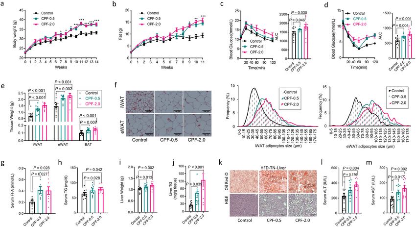
ARTICLE                                                                  NATURE COMMUNICATIONS | https://doi.org/10.1038/s41467-021-25384-y

O
          besity is a major risk factor for type 2 diabetes (T2D),            processing, and/or packaging (Table 1) on the expression of Ucp1
          non-alcoholic fatty liver disease (NAFLD), and cardio-              in immortalized brown adipocytes generated from Ucp1-lucifer-
          vascular disease1 that arises from a caloric surplus of as          ase reporter mice32. Of the 34 chemicals tested, only the pesticide
little as 10–30 kcal per day2. And while increased consumption of             CPF significantly decreased Ucp1 promoter activity and mRNA
energy-dense foods and reduced physical activity are commonly                 expression by at least 25% across six orders of magnitude of
thought to be the major contributors to this caloric imbalance1,              concentrations tested down to 1 pM (Fig. 1a, b, Supplementary
diet-induced thermogenesis is a quantitatively important com-                 Fig. 1a–c).
ponent of the energy balance equation3. In adult humans, recent                  CPF is an organophosphate insecticide with broad-spectrum
studies have indicated that diet-induced thermogenesis requires               activity against many foliar and soil insects and is commonly used
the activation of brown adipose tissue (BAT)4–6, however, the                 for pest control in a wide range of field crops and fruit33. Previous
determinants regulating this process and why they may differ                  studies in animals have found that high doses of CPF, which
between individuals are not fully understood.x                                inhibits plasma butyryl cholinesterase and brain acet-
    A key protein regulating BAT thermogenesis is uncoupling protein          ylcholinesterase activity, can promote obesity and glucose dys-
1 (UCP1). Genetic removal of Ucp1 in mice promotes obesity and                regulation through mechanisms that are poorly defined but may
insulin resistance when mice are fed a diet high in fat and housed            involve increases in energy intake and/or alterations in the gut
under thermoneutral conditions (29–30 °C), indicating a vital role for        microbiome34–39. However, whether doses of CPF below those
this protein in diet-induced thermogenesis7. UCP1 is also important           that inhibit acetylcholinesterase and closer to human-relevant
for diet-induced thermogenesis in humans, as a genetic loss of                exposures promote obesity in rodents or humans is currently
function mutation reduces postprandial thermogenesis in response to           unknown. We found that consistent with the acute suppression of
a high-fat meal8. While genetic polymorphisms in Ucp1 have been               Ucp1 promoter activity and Ucp1 mRNA, chronic CPF (6 days)
linked to obesity in some studies9–11, this has not been observed in          also suppressed UCP1 protein in brown adipocytes at a con-
large genome-wide association studies12, suggesting that other factors        centration of 1 pM (Fig. 1c and Supplementary Fig. 1c). To
besides genomic alterations in Ucp1 may be contributing to reduced            further characterize the effects of CPF, we completed global RNA-
BAT activity in obese humans13.                                               sequencing after just 4 h of CPF treatment and found that RNA
    Over the last two decades, a number of environmental tox-                 transcripts related to mitochondrial function/metabolism were
icants have been linked to the development of obesity through                 most affected (Fig. 1d). Notably, in addition to Ucp1, RNA
their effects on gut microbiota14, energy intake, or                          transcripts are critical for regulating fatty acid oxidation (Cpt1a,
adipogenesis15–18 (for review see refs. 19,20), however, only a few           Cpt1b, Acat3), and Cytochrome C oxidase assembly factor
of these studies have examined a role for these agents to con-                (Cox16) (Fig. 1e) were reduced with CPF treatment. Consistent
tribute to obesity by inhibiting energy expenditure21–23 and/or               with previous studies in UCP1 null mice40,41, compensatory
BAT thermogenesis24,25 (reviewed in refs. 18,20). We hypothesized             upregulation of some mitochondrial transcripts was also observed
that environmental toxicants commonly present in food might                   (Fig. 1e). Importantly, consistent with alterations in transcrip-
reduce diet-induced thermogenesis through suppression of                      tional programs regulating mitochondrial function, chronic
UCP1, the defining protein of human BAT thermogenesis26–31.                    treatment with CPF-lowered respiration in the presence of the
Through a step-wise screening of several common food con-                     mitochondrial ATP synthase inhibitor oligomycin and the
taminants or additives, we discovered that the organophosphate                mitochondrial uncoupler FCCP; indicating impaired mitochon-
insecticide chlorpyrifos (CPF) potently suppressed the expression             drial leak and maximal respiration, respectively (Fig. 1f). CPF also
of UCP1 and mitochondrial respiration in brown adipocytes at                  lowered cytochrome c oxidase activity (Fig. 1g) and mitochon-
concentrations as low as 1 pM. Chloropyrifos-induced suppres-                 drial membrane potential (Fig. 1h). These reductions in mito-
sion of diet-induced thermogenesis in BAT has also been                       chondrial function were not due to impairments in BAT
observed in mice fed a diet high in fat and housed at thermo-                 differentiation as Oil Red O staining and key transcription fac-
neutrality where it promoted greater obesity, NAFLD, and insulin              tors/markers of BAT differentiation (Prdm16, Ppargc1a, Pparg,
resistance. Reductions in BAT thermogenesis by CPF were                       Cidea) were not altered by CPF (Supplementary Fig. 1d, e).
associated with reductions in cAMP and protein kinases critical               Impaired mitochondrial function was not observed following
for regulating UCP1 and mitophagy. These data indicate that the               acute CPF exposure (4 h) (Supplementary Fig. 1f, g) as had pre-
commonly used pesticide CPF, at very low concentrations, sup-                 viously been observed at high concentrations of CPF (40 µM)42.
presses the activation of BAT, suggesting that its use may con-               These data indicate that concentrations of CPF significantly
tribute to the obesity epidemic.                                              below those causing toxicity in animal studies and more aligned
                                                                              with real-world human exposures43 inhibit transcriptional pro-
Results                                                                       grams crucial for regulating BAT mitochondrial function.
We examined the effects of 34 chemicals—selected based on their                  CPF is readily detected in a wide range of foods including
widespread presence in food due to agriculture practices, food                fruits, vegetables, grains, beans, nuts, legumes, dairy, meat, fish,

 Table 1 List of tested chemicals.

 Pesticides                      Pesticide metabolites                      Food-packaging compounds                        Food processing compounds
 2,4-D                           AMPA                                       BPA                                             Zearalanone
 Atrazine                        Atrazine mercapturate                      BPAF                                            Allura red
 Chlorpyrifos                    6-Chloronicotinic acid                     BPB                                             Brilliant blue
 Deltamethrin                    Chlorpyrifos oxon                          BPS                                             Cresidinesulfonic acid
 Glyphosate                      Desethyl atrazine                          BPF                                             Sulfanilic acid
 Imidacloprid                    Diaminochlorotriazine                      BPA-b-D-glucuronide                             Sunset yellow
 Metam sodium                    MITC                                       MINCH                                           Tartrazine yellow
 Permethrin                      3-Phenoxybenzoic acid                      PFOA
 S-Metolachlor                   TCP-y                                      TBPA

2                 NATURE COMMUNICATIONS | (2021)12:5163 | https://doi.org/10.1038/s41467-021-25384-y | www.nature.com/naturecommunications
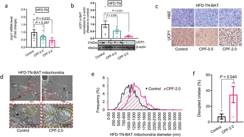
NATURE COMMUNICATIONS | https://doi.org/10.1038/s41467-021-25384-y                                                                           ARTICLE

Fig. 1 CPF inhibits the thermogenic gene program and mitochondrial respiration in cultured brown adipocytes. a Ucp1 promoter activity of brown
adipocytes treated with 34 different chemical compounds at the dose of 1 pM for 16 h. b Ucp1 mRNA levels of brown adipocytes treated with compounds at 1 pM
for 4 h. c UCP1 protein concentration in brown adipocytes treated with compounds at 1 pM for 6 days. d and e Gene Set Enrichment Analysis of mRNA-sequencing
data (d) of brown adipocytes treated with 1 pM chlorpyrifos (CPF) for 4 h and relative expression of genes within gene sets (e, Whiskers: min to max). f Oxygen
consumption rate of brown adipocytes treated with different doses of CPF for 6 days. g Cytochrome C oxidase activity of brown adipocytes treated with 1 pM CPF
for 6 days. h Representative images of mitochondria stained by MitoTracker Green and quantification of mitochondria potential reflected by MitoTracker Red
CMXRos in brown adipocytes treated with 1 pM CPF for 6 days. Significant differences between 3 or more mean values were determined by one-way ANOVA with
the post hoc Bonferroni’s multiple comparisons test; differences between 2 mean values were determined by a two-tailed Student’s t-test. Data presented are
mean ± SEM, n = 6, *p < 0.05. Scale bar = 200 μm.

and eggs, at concentrations ranging from 0.002 to 436 μg/kg44.                   better mimics a condition in which humans reside45 and as such
We examined the effects of CPF in male C57BL6J mice that were                    recent studies have indicated that it more accurately models the
fed a control diet (CD, 10 kcal% fat) or high-fat diet (HFD,                     development of metabolic diseases including obesity, insulin
45 kcal% fat) supplemented with two doses of CPF (5 or 20 mg/kg                  resistance, and NAFLD46,47. Therefore, mice were housed at
diet, which equated to an exposure of ~0.5 or 2 mg/kg body                       either RT or TN. To the best of our knowledge, this is the first
weight in mice fed a control diet housed at room temperature                     time an environmental toxicant has been tested in rodents
(CD-RT) with slightly lower exposure in HFD fed mice housed at                   housed at TN.
thermoneutrality (TN, Supplementary Fig. 2a). In contrast to an                     On a control diet, CPF supplementation did not influence body
acute gavage of CPF (100 mg/kg) which resulted in dramatic                       weight (Supplementary Fig. 3a, b), fat mass (Supplementary Fig. 3c,
inhibition of ChE activity in the serum, we found that CPF in the                d), glucose tolerance (Supplementary Fig. 3e, f), or insulin sensitivity
diet did not inhibit acetyl-cholinesterase activity in skeletal                  (Supplementary Fig. 3g, h) in RT or TN conditions. However, when
muscle (Supplementary Fig. 2b and c), which is consistent to a                   mice were housed at TN and fed an HFD, CPF treatment increased
previous study34.                                                                body mass (Fig. 2a), adiposity (Fig. 2b), and reduced glucose toler-
   Mice housed at room temperature (RT, 21–23 °C), common in                     ance (Fig. 2c), and insulin sensitivity (Fig. 2d) compared to non-CPF
most animal vivarium, are under cold stress and have increased                   treated mice. Importantly, these effects were blunted in mice housed
sympathetic tone that upregulates basal metabolic rate through                   at RT such that only the highest dose of CPF (2.0 mg/kg) promoted
adaptive thermogenesis7,45. This elevation in basal metabolic rate               weight gain, glucose intolerance, and insulin resistance (Supple-
masks the effects of diet-induced thermogenesis mediated                         mentary Fig. 4a–e). Consistent with increased adiposity under TN-
through β-adrenergic driven activation of BAT UCP1 compared                      HFD-fed conditions (Fig. 2b, e), CPF increased the size of WAT lipid
to housing mice at thermoneutrality (TN, 29–30 °C) as evidenced                  droplets (Fig. 2f) and elevated serum free fatty acids (FFA, Fig. 2g)
by the development of obesity in mice lacking UCP1 at TN but                     and triglycerides (TGs, Fig. 2h). Consistent with increased obesity,
not RT. Thus TN housing is a temperature range for mice that                     insulin resistance and FFAs HFD-fed mice treated with CPF and

NATURE COMMUNICATIONS | (2021)12:5163 | https://doi.org/10.1038/s41467-021-25384-y | www.nature.com/naturecommunications                                     3
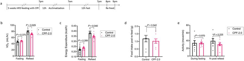
ARTICLE                                                                    NATURE COMMUNICATIONS | https://doi.org/10.1038/s41467-021-25384-y

Fig. 2 CPF promotes obesity and metabolic dysfunction in mice fed a high-fat diet and at thermoneutrality. C57BL/6J male mice were fed a HFD
(45 kcal% fat) supplemented with 0 (Control), 0.5 mg/kg/BW (CPF-0.5) or 2.0 mg/kg/BW (CPF-2.0) chlorpyrifos and housed at thermoneutrality (TN,
30 °C) for 14 weeks. a Bodyweight. b Fat mass. c Glucose tolerance test. d Insulin tolerance test (ITT). e Adipose tissues weight. f Representative images of
H&E stained inguinal white adipose tissue (iWAT) and epididymal white adipose tissue (eWAT). g Serum-free fatty acid (FFA) level. h Triacylglycerol (TG)
level. i Liver weight. j Liver TG concentration. k Representative images of Oil Red O-stained liver lipids and H&E-stained liver. l Serum alanine
aminotransferase (ALT) level. m Aspartate aminotransferase (AST) level. Significant differences between mean values were determined by one-way
ANOVA with the post hoc Bonferroni’s multiple comparisons test. Data presented are mean ± SEM, n = 10, *p < 0.05, **p < 0.01, ***p < 0.001. Scale
bar = 100 μm.

housed at TN had increased liver weights (Fig. 2i), liver TGs (Fig. 2j,         these data suggest that CPF reduces energy expenditure, con-
k) and serum ALT (Fig. 2l) and AST (Fig. 2m) indicative of greater              tributing to the increased weight gain observed at TN.
NAFLD than HFD controls. Importantly, these effects of CPF were                    Our data demonstrating reductions in oxygen consumption at
blunted when fed an HFD at RT and were not observed when mice                   TN, but not at RT with CPF, are similar to observations in UCP1
were fed with CD indicating these markers of NAFLD/liver toxicity               null mice, which also only develop HFD-induced obesity when
were likely secondary to the increased weight rather than toxicity to           housed at TN; an effect attributed to impairments in BAT diet-
CPF (Supplementary Figs. 5 and 6, respectively). These data indicate            induced thermogenesis7, We found that in HFD-fed mice housed
that low concentrations of CPF, which do not inhibit AchE activity,             at TN, CPF reduced Ucp1 mRNA and protein expression (Fig. 4a
when combined with TN housing and an HFD promote obesity,                       and b) and increased the size and number of lipid droplets
insulin resistance, and NAFLD.                                                  (Fig. 4c) within the BAT. In addition to suppressing UCP1,
   To investigate the mechanisms by which CPF promoted HFD-                     transmission electron microscopy revealed an increased abun-
induced weight gain, caloric intake and energy expenditure were                 dance of large mitochondria (Fig. 4d and e) with disrupted cristae
assessed in metabolic cages. Consistent with previous studies mice              (Fig. 4d and f). These effects of CPF on UCP1 and BAT mor-
housed at TN had lower caloric intake compared to mice housed                   phology and mitochondria were not evident when mice were
at RT48 and regardless of housing temperature, CPF increased                    housed at RT (Supplementary Fig. 7). These data indicate that
caloric intake36,49 (Fig. 3a). No differences in physical activity              consistent with increased weight gain and metabolic dysfunction
were observed between groups (Fig. 3b). As anticipated, TN                      when mice are housed at TN, CPF reduces BAT UCP1 expression
lowered absolute (not corrected for body mass) resting oxygen                   and mitochondrial morphology.
consumption compared to RT housing (Fig. 3c). Importantly,                         To directly test the hypothesis that CPF was suppressing diet-
CPF suppressed oxygen consumption by ~15% in mice housed at                     induced thermogenesis we conducted a subsequent study in which
TN but not RT (Fig. 3c). These data indicate that CPF suppresses                mice were housed at TN and fed an HFD with or without CPF for
energy expenditure at TN but not RT. To further examine whe-                    5 weeks (Fig. 5a), a time point preceding any differences in body
ther reductions in energy expenditure were important for the                    mass or adiposity (Supplementary Fig. 8a, b). Mice were then placed
weight gain observed in HFD-fed mice housed at TN, in separate                  in metabolic cages and fasted overnight before refeeding. CPF-treated
experiments mice were fed with CPF and allowed ad libitum                       mice had similar oxygen consumption (Fig. 5b) and energy expen-
access to food or were pair-fed to controls (Fig. 3d). Importantly,             diture (Fig. 5c) during fasting. However, CPF-treated mice had an
even in the pair-fed group CPF treated mice had a strong ten-                   ~10% reduction in oxygen consumption and energy expenditure
dency for higher body weight and increased adiposity after                      during refeeding (Fig. 5b and c) indicative of impaired diet-induced
9 weeks of HFD feeding when the amount of food intake was                       thermogenesis. Importantly, this was not due to differences in food
matched with that of the control mice (Fig. 3e and f). Collectively             intake during the refeed (Fig. 5d) or activity levels (Fig. 5e). These

4                  NATURE COMMUNICATIONS | (2021)12:5163 | https://doi.org/10.1038/s41467-021-25384-y | www.nature.com/naturecommunications

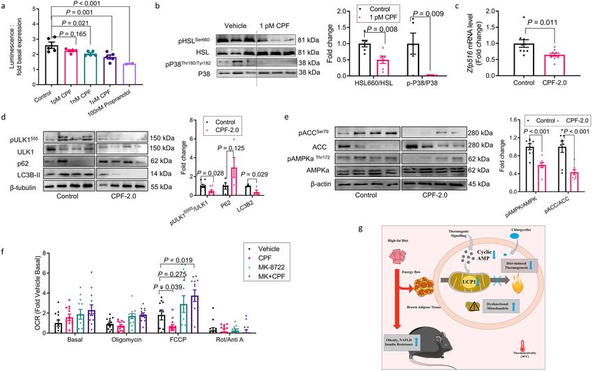
NATURE COMMUNICATIONS | https://doi.org/10.1038/s41467-021-25384-y                                                                     ARTICLE

Fig. 3 Dietary exposure to chlorpyrifos promotes weight gain in HFD-fed mice housed at thermoneutrality even in the absence of increased food
intake. a–c C57BL/6J male mice were fed a HFD (45 kcal% fat) supplemented with 0 (Control), 0.5 mg/kg/BW (CPF-0.5) or 2.0 mg/kg/BW (CPF-2.0)
chlorpyrifos and housed at thermoneutrality (TN, 30 °C) for 14 weeks. Daily food intake (a) was recorded weekly; animal activity (b) and resting O2
consumption rate (c) were measured in CLAMS; n = 10. d–f C57BL/6J male mice were treated with HFD (45 kcal% fat) supplemented with 0 (Control) or
2.0 mg/kg BW CPF for 9 weeks, the food provided to one group of the CPF mice (CPF/Pair-fed) was matched with that of the control group. The food
intake (n = 4, d), body weight (n = 8, e), and fat tissue weight (n = 8, f) were measured. Significant differences between mean values were determined by
one-way ANOVA with the post hoc Bonferroni’s multiple comparisons test. Data presented are mean ± SEM, *p < 0.05.

Fig. 4 Dietary exposure to chlorpyrifos impairs mitochondrial function and thermogenesis in brown adipose tissue in vivo. C57BL/6J male mice were
fed a HFD (45 kcal% fat) supplemented with 0 (Control), 0.5 mg/kg/BW (CPF-0.5), or 2.0 mg/kg/BW (CPF-2.0) chlorpyrifos and housed at
thermoneutrality (TN, 30 °C) for 14 weeks. a mRNA expression level of Ucp1 in BAT, n = 10. b UCP1 protein in BAT of mice at TN, n = 8. c Representative
immunohistochemistry images showing UCP1 in BAT and H&E images, Scale bar = 100 μm. d Representative electron micrographs for mitochondria, scale
bar = 2 μm. e Diameter distribution of mitochondria. f Quantification of disrupted mitochondria cristae, n = 4. Significant differences between 3 or more
mean values were determined by one-way ANOVA with the post hoc Bonferroni’s multiple comparisons test; differences between 2 mean values were
determined by a two-tailed Student’s t-test. Data presented are mean ± SEM.

data suggest that CPF reduces energy expenditure by suppressing                 which results in activation of adenylyl cyclase, and subsequent
diet-induced thermogenesis.                                                     increases in cAMP and PKA activity50,51. To examine whether this
   In mice diet-induced thermogenesis is mediated through nor-                  pathway may be altered with CPF, we measured cAMP in response
epinephrine stimulated β3-adrenergic receptor (β3-AR) activation,               to the pan beta-agonist isoproterenol and found that CPF reduced

NATURE COMMUNICATIONS | (2021)12:5163 | https://doi.org/10.1038/s41467-021-25384-y | www.nature.com/naturecommunications                              5
ARTICLE                                                                   NATURE COMMUNICATIONS | https://doi.org/10.1038/s41467-021-25384-y

Fig. 5 Chlorpyrifos inhibits diet-induced thermogenesis. C57BL/6J male mice were treated with an HFD (45 kcal% fat) supplemented with 0 (control) or
2.0 mg/kg/BW (CPF-2.0) chlorpyrifos for 5 weeks, followed by 12 h fasting and 2 h-refeeding. a A timeline showing treatment procedure. b Oxygen
consumption. c Energy expenditure. d Food intake during 1 h refeed. e Physical activity levels from 6–7 p.m. (fasting) and from 7–8 p.m. (refeed). n = 10.
Significant differences between mean values were determined by a two-tailed Student’s t-test. Data presented are mean ± SEM.

Fig. 6 Chlorpyrifos inhibits diet-induced thermogenesis and β-adrenergic signaling in brown adipose tissue. a Mature brown adipocyte treated with
vehicle or chlorpyrifos (CPF) or propranolol for 7 days. Fold change of cAMP levels in response to 30 min isoproterenol stimulations, n = 5. b–e
Phosphorylation of HSL and p38 MAPK (b, n = 6); Zfp516 mRNA expression (c, n = 10), pULK1555, ULK, p62, LC3B-II, pACC, ACC, pAMPKaThr172 and
AMPK proteins (d, e, n = 8) in BAT of C57BL/6J mice fed a HFD with 0 (control) or 2.0 mg/kg/BW (CPF-2.0) CPF and housed at thermoneutrality (TN,
30 °C) for 14weeks. f Oxygen consumption rate of brown adipocytes treated with MK-8722 and/or CPF for 6 days, n = 12. g Graphical abstract showing
CPF induced obesity, non-alcoholic fatty liver disease (NAFLD), and insulin resistance by inhibiting diet-induced thermogenesis. Significant differences
between three or more mean values were determined by one-way ANOVA with the post hoc Bonferroni’s multiple comparisons test; differences between
two mean values were determined by a two-tailed Student’s t-test. Data presented are mean ± SEM.

cAMP in isolated brown adipocytes (Fig. 6a). To examine potential              bonds with S200. (Supplementary Fig. 9a) Surprisingly, the docking
mechanisms which might be contributing to this reduction in cAMP               score of the most populated CPF docking pose was higher (−7.4 kcal/
several experiments were completed. We first hypothesized that CPF              mol, lower the better) than cyanopindolol (−8.8 kcal/mol), a potent
may directly inhibit the β3-AR. We built a homology model of the β3-           β-adrenergic receptor antagonist, suggesting a weaker binding. To
AR which was used as a starting structure to carry out classical               validate the modeling observations, studies using a radioligand
molecular dynamic simulations (MD). Snapshots obtained from MD                 competition assay of CPF or its metabolite CPF-oxon with cyano-
simulation were then used to dock CPF. The most populated docked               pindolol were conducted. However, in contrast to the docking stu-
pose shows that CPF is predicted to bind to the known pocket in the            dies, we found that neither CPF nor CPF-oxon competed with
β3-AR and predominantly forms hydrophobic contacts and hydrogen                cyanopindolol for β3-AR binding (Supplemental Fig. 9b). These data

6                  NATURE COMMUNICATIONS | (2021)12:5163 | https://doi.org/10.1038/s41467-021-25384-y | www.nature.com/naturecommunications
NATURE COMMUNICATIONS | https://doi.org/10.1038/s41467-021-25384-y                                                                 ARTICLE

suggest that neither CPF nor its metabolite CPF-oxon directly bind              cholinesterase activity or toxicity and is more aligned with real-
the β3-AR.                                                                      world exposures in humans39,59.
   We subsequently interrogated the hypothesis that CPF could                      When mice are housed at TN, but not RT, a low concentration
be reducing cAMP by altering the expression of β-ARs (Adrb),                    of CPF (0.5 mg/kg body weight) promotes weight gain, insulin
adenylyl cyclase (Adcy), or phosphodiesterases (Pde) but found                  resistance, and NAFLD. To the best of our knowledge, this is the
no change in isolated BAT cells (Supplementary Fig. 9c–e). In                   first toxicological study conducted in rodents that have been
addition to direct binding or alterations in expression, cAMP                   completed under TN conditions. The findings demonstrating
production is dependent on membrane localization of adenylate                   effects of low concentrations of CPF at TN, but not RT, are
cyclase therefore we subsequently examined whether this might                   consistent with recent studies that have highlighted that RT does
be reduced by CPF treatment. Interestingly, CPF lead to a                       not allow for accurate modeling of human disease due to
reduction of membrane-bound ADCY3 with a corresponding                          increased thermogenic demands that mask biological processes
increase in cytoplasmic localization (Supplementary Fig. 9f).                   that occur in humans who predominately reside at TN60. A key
These data suggest that CPF may reduce cAMP by reducing                         reason for this is because at RT mice require adaptive thermo-
membrane localization, however, the exact mechanisms that                       genesis to maintain their body temperature which in turn masks
contribute to this effect require further investigation.                        diet-induced thermogenesis. Therefore, the observation of
   To examine the downstream consequences of this reduction in                  increased weight gain at TN but not RT, despite increases in
cAMP we measured the phosphorylation and activation of hor-                     caloric consumption under both housing conditions, suggested
mone sensitive lipase (HSL), p38MAPK, and AMP-activated                         that low dose CPF may be inhibiting diet thermogenesis and this
protein kinase (AMPK), which are important for increasing                       could be critical for mediating the increased weight gain. Con-
lipolysis, UCP1 expression, and mitophagy respectively50–53.                    sistent with this hypothesis pair-feeding experiments indicated
Reductions in cAMP in isolated brown adipocytes (Fig. 6a) were                  that CPF mice housed at TN who were calorically matched to the
associated with reduced activating phosphorylation of HSL                       lower food intake of controls still gained more weight suggesting
(Ser660) and p38MAPK (Fig. 6b); effects similar to the pan-beta                 reductions in energy expenditure were important. To further
antagonist propranolol (Supplementary Fig. 10a). Reductions in                  examine whether diet-induced thermogenesis was critical for the
the phosphorylation of PKA, HSL, and p38 MAPK were also                         weight gain mice were housed at TN and fed an HFD containing
observed in BAT of CPF-treated mice fed an HFD and housed at                    CPF for 4 weeks, and before any differences in adiposity emerged,
TN (Supplementary Fig. 10b–d). Concomitant with these                           were fasted and refed in metabolic cages. These experiments
reductions in cAMP the expression of Zfp516, a cAMP-responsive                  demonstrated that despite similar activity levels and food con-
transcription factor regulating UCP154, was also reduced                        sumption, oxygen consumption was reduced in CPF-treated mice
(Fig. 6c).                                                                      during refeeding. These findings indicating that CPF suppresses
   Treatment of mouse and human BAT cells with nor-                             diet-induced thermogenesis indicate a mechanism by which CPF
epinephrine increases cAMP and AMPK activity55. Similar to                      promotes obesity and metabolic dysfunction. They also suggest
CPF-treated mice, mice lacking AMPK in BAT have an increased                    that since to date all environmental toxicants have been tested in
abundance of large mitochondria with disrupted cristae, an effect               rodents housed at RT, the effects of these chemicals on human
which is mediated through AMPK-induced increases in                             health may have been underestimated and therefore additional
mitophagy55. We found that consistent with mice genetically                     toxicological testing at TN may be important.
lacking AMPK in BAT, CPF-treated mice and BAT cells have                           BAT and UCP1 are important for diet-induced thermogenesis
reduced activating phosphorylation of AMPK and ULK1 and                         in both mice and humans50. Increased weight gain and metabolic
reduced accumulation of LC3B-II and increased p62, the latter                   dysfunction in mice treated with low doses of CPF housed at TN
being two markers of reduced autophagic flux (Fig. 6d and                        were associated with a reduction in UCP1 and alterations in BAT
Supplementary Fig. 10e). There was also reduced phosphorylation                 mitochondria morphology. Interestingly, this effect was not
of ACC, another downstream substrate of AMPK important for                      observed when mice are housed at RT potentially because of the
regulating fatty acid oxidation and fatty acid synthesis (Fig. 6e               increased adrenergic drive which may have overridden the sup-
and Supplementary Fig. 10e). To test whether this inhibition of                 pressive effects of CPF. These findings are in agreement with our
AMPK was important for mediating the inhibitory effects of CPF                  observations in vitro that low pM concentrations of CPF suppress
on mitochondrial function, brown adipocytes were treated with                   cAMP and downstream signaling pathways in BAT cells, effects
the highly selective AMPK agonist MK-872256 which eliminated                    associated with reductions in UCP1 expression, mitochondrial
the ability of CPF to suppress mitochondrial uncoupling by FCCP                 function, and oxygen consumption. In rodents, diet-induced
(Fig. 6f). These data indicate that CPF reduces BAT cAMP and                    thermogenesis is dependent on norepinephrine-induced activa-
downstream signaling leading to reductions in UCP1 and path-                    tion of the β3-AR and UCP1 and in the current study, CPF
ways critical for controlling mitochondrial function.                           exposure led to reductions in both cAMP and UCP1. We hypo-
                                                                                thesized that this may be due to binding of CPF to the β3-AR or
                                                                                reductions in β3-AR expression levels, however, we found no
Discussion                                                                      effect of CPF on either of these parameters. Previous studies in
A small imbalance between energy expenditure and energy intake/                 developing neurons found that CPF may reduce the activity of
absorption increases adiposity and over time can lead to the devel-             adenyl cyclase61 and consistent with this observation we observed
opment of obesity. Due to the lipophilicity of environmental tox-               a reduction in membrane localization. The exact mechanisms
icants, adipose tissue is the major site of environmental toxicant              contributing to this effect are unknown however one possibility is
accumulation57,58. In agreement with previous studies35–38, we found            that given that CPF and its active metabolite CPF oxon has been
that treatment of HFD-fed mice with 2.0 mg/kg body weight of CPF                shown to disrupt microtubule networks this may be affecting
increased caloric intake and promoted obesity, insulin resistance, and          intracellular localization62. Additional studies are needed to fur-
NAFLD when mice were housed at RT. Interestingly no effect of this              ther investigate the mechanisms by which CPF reduces adre-
dose was observed on metabolic parameters when this dose of CPF                 nergic signaling in BAT.
was combined with the control low-fat diet indicating important                    One of the limitations of our study is that we only examined
interactions between CPF and dietary fat. Importantly, this dose of             the effects of CPF in male mice. It is known that CPF interacts
CPF is below that associated with the inhibition of acetyl-                     with sex hormones63 and steroid hormone receptors64. In terms

NATURE COMMUNICATIONS | (2021)12:5163 | https://doi.org/10.1038/s41467-021-25384-y | www.nature.com/naturecommunications                         7
ARTICLE NATURE COMMUNICATIONS | https://doi.org/10.1038/s41467-021-25384-y
of the neurotoxic effects, males are more susceptible to organo- reduced cytochrome c, and ddH2O, and cell lysate. Absorbance was immediately
phosphates than females65. However, as reviewed66, women may read at 550 nm wavelength for 3 min using SpectraMax M5 plate reader. The
reaction was performed at 37 °C and cytochrome c oxidase activity was calculated
be more vulnerable to environmental pollution than men mainly as Eq. (1):
because of the physiological differences caused by sex steroids.
With a larger relative fat mass, women have a larger distribution 4OD=reaction time reaction volume
Activity ¼ ´
protein concentration ´ sample volume exctinction coefficient ´ path length
volume for lipophilic substances than men66. Indeed, in aged
ð1Þ
human beings, concentrations of organochlorine pesticides are
elevated in females67. Females may also have higher levels of An extinction coefficient of 18.5 mmol−1 cm−1 (reduced cytochrome c), a path
length of 0.625 cm, and a reaction volume of 0.0002 L were used. Protein was
thermogenic BAT than males68. Upon adrenergic activation, quantified during final calculations to determine the activity of enzyme per µg of
women have more potential to induce browning of perirenal protein. Each sample was run in triplicate on a 96-well plate.
adipose tissue than men due to sex-specific intrinsic factors69 and
have higher lipolytic activities70. Interestingly, females exhibit In vitro respiration assay of brown adipocytes. The immortalized brown pre-
higher DIT than males at 20 °C but lower DIT than males at adipocytes cell line were fully differentiated in a 75 cm2 flask, then transferred to a
96-well flat-bottomed OxoPlate (OP96C, PreSens Precision Sensing) at a dilution
TN71. Future studies examining the effects of CPF in female mice of 1:8 by culture area. Before the assay, cell culture media was replaced with 200 μl
are needed. respiration medium (minimal DMEM-D5030 supplemented with 10 mM HEPES,
In conclusion, our study modeled the effects of an environ- 25 mM glucose, 31 mM NaCl, 2 mM sodium pyruvate, 2 mL L-glutamine, and 4%
mental toxicant under TN housing conditions. These studies have BSA, pH = 7.4), covered by 75 μl heavy mineral oil and prewarmed at 37 °C in a
revealed that in addition to promoting food intake, CPF sup- SpectraMax M5 plate reader (Molecular Devices, San Jose, CA, USA) for 45 min.
For calibration, each plate contained six wells of 0% O2 standard (H2O with 10 mg/
presses diet-induced thermogenesis in BAT and this exacerbates mL sodium sulfite) or 100% O2 standard (respiration media). The plate was
the development of obesity, NAFLD, and insulin resistance even kinetically read prior (basal) and following the addition of 2.5 μM oligomycin,
at low doses which have no effect in mice housed at RT. These 1 μM FCCP or 2 μM Rotenone +2 μM Antimycin A injections for 20 min. Oxygen
studies suggest that the effects of environmental toxicants on the levels were calculated according to the manufacturer’s manual.
development of obesity may have been underestimated as all
RNA-sequencing analysis. TMCON brown pre-adipocytes were differentiated in 24-
studies to date have been conducted in mice housed at RT. Future well plates and treated with 1 pM CPF on day 9 of differentiation. Cells were
studies examining the mechanisms driving reductions in β-AR treated for 4 h in Krebs–Ringer bicarbonate buffer (Sigma-Aldrich) supplemented
signaling and whether there are associations between BAT with 10 mM HEPES (Janssen Pharmaceuticals, 3a2440 Geel, Belgium), 1 mM
metabolic activity and CPF in humans will be important. CaCl2, and 1% fatty-acid free Bovine Serum Albumin (BSA) (EquiTech Bio Inc.,
TX, USA) at pH 7.4. Cell lysates were collected by administering 175 µL per well of
RNeasy Lysis Buffer (RLT) (Qiagen, Hilden, Germany) supplemented with
Methods 143 mM beta-mercaptoethanol (Sigma-Aldrich) and vortexing until cells lifted
In-vitro analysis from plates. Once cells were lifted from plates, 175 µL of 70% ethanol was added to
Immortalized brown adipocyte cell line generation. Cell lines were created from each well, and lysates were transferred to a High Pure Filter tube from Roche RNA
either FVB/N mice (wildtype immortalized brown adipocytes) or UCP-1-Luc2- Isolation Kit (Roche Diagnostics, Mannheim, Germany). RNA isolation was done
TdTomato reporter mouse (immortalized UCP-1 reporter brown adipocytes), also as outlined by manufacturers' specifications.
known as the “ThermoMouse”32 using reported techniques72. Briefly, BAT tissue Raw sequencing reads (~12.5 million paired-end per sample; 50 base pairs each)
was harvested from 4-day-old pups and subsequently digested with collagenase II, were assessed for quality (such as GC content, PHRED scores, synthetic aptamer
filtered, and plated. Isolated cells were then infected with a retrovirus created by content, and sequence length) using FastQC (https://www.bioinformatics.
transfecting Phoenix-ECO cells with a pBABE-SV40 plasmid from Addgene. Cells babraham.ac.uk/projects/fastqc/). Trimmomatic73 (default parameters) was used to
positive for SV40 were selected for 7 days using 2 µg/ml puromycin antibiotic and trim low-quality reads, with the final set of reads aligned to the Ensembl Mus
underwent a 14-day treatment with the antibiotic ciprofloxacin (10 µg/ml) in order musculus (GRCm38.92) genome using HISAT274 (default parameters). The
to eliminate any mycoplasma contamination in the cells. UCP-1 Luc2-TdTomato number of reads mapping to each gene was identified using htseq-count, with
(UCP-1 reporter line) was used for all luciferase assays to quantify UCP-1 pro- stranded set to “reverse”, minimum alignment quality set to 10, feature type set to
moter activity. For the cyclic AMP assay wildtype immortalized brown adipocytes “exon” and “simple” advanced options. Mouse GTF gene annotation reference files
were transfected with Promega pGloSensor-22F which can detect dynamic changes were obtained from Ensembl. DESeq275 (default parameters) was used to
in cyclic AMP from concentration 0.003–10 µM in living cells. normalize these data and a custom python script (McArthur) was used to convert
the DESeq2 output into a ranked list of genes. The ranked list was used as input for
Gene Set Enrichment Analysis (GSEA)76,77, which was conducted using
Immortalized murine brown adipocyte culture. Immortalized brown adipocytes Cytoscape78. GSEA was performed using 1000 permutations, a false discovery rate
were cultured in high glucose DMEM (GibcoTM) containing 10% fetal bovine cut-off of 25%, and a mouse gene set database from the Bader Lab at the University
serum and 1% antibiotic–antimycotic solution. For differentiation, confluent cells of Toronto (http://download.baderlab.org/EM_Genesets/current_release/Mouse/).
were placed in induction media (0.5 mM IBMX; 0.125 mM indomethacin; 0.2 μg ml
−1 dexamethasone; 1 nM T3, 20 nM insulin) for 2 days and subsequently main-
Oil red O staining and quantification. Cells were rinsed with PBS and fixed with
tained on differentiation media (1 nM T3, 20 nM insulin) for 7 days. For all cell
10% formalin for 40 min at RT. Fixed cells were incubated with Oil-Red O staining
experiments, cells were fully differentiated prior to compound or vehicle (≤0.1%
solution (Sigma Aldrich) for 10 min and then rinsed in 60% isopropanol to remove
dimethyl sulfoxide; DMSO) treatment.
excessive Oil-Red O. Following imaging, Oil Red O stain was solubilized in iso-
propanol, and absorbance measures were taken at 510 nm utilizing Asys UVM340
Cyclic AMP assay. Cells were grown to confluence and fully differentiation as microtiter plate reader.
described above. Fully differentiated cells were seeded into 96-well assay plates at a
1:1 dilution based on well area. Following 48 h post-seeding, cells were treated with
Radioligand ADRB3-binding competition assays. Radioligand-binding competition
vehicle, CPF, or positive control propranolol for 7 days. Fresh media containing
activity was tested on the recombinant adrenergic β3 receptor for CPF and CPF
compound concentrations was added every 48 h. On day 7 a kinetic luciferase assay
oxon utilizing filtration binding assays by Epics and Euroscreen FAST (Belgium).
was performed. A 30 min basal read was first obtained, followed by a 100 nM
Assays were performed in a 96-well plate (Master Block, Greiner, 786201), in
isoproterenol injection and a 30 min stimulated kinetic read. Cyclic AMP activity is
duplicate. Binding buffer (25 mM Hepes pH 7.4, 1 mM EDTA, 0.5% BSA, 10 µg/ml
expressed as luminescence (RLU). Data are expressed as fold basal change (sti-
saponin), membranes prepared from β3-receptor-expressing CHO-K1 cells (2 µg
mulated read (5–20 min)/basal read (5–30 min)).
protein/well), radiotracer 125I-Cyanopindolol (0.165 nM final assay concentration)
and test compound were utilized. A total of 9 concentrations were assessed per test
MitoTracker staining. After washing with PBS, cells were treated with 200 nM compound between 0.0001 and 10,000 nM in duplicate. The assay was performed
MitoTracker Red and 100 nM MitoTracker Green staining solutions in DMEM using reference competitor ZD 7114 (Tocris Pharmaceuticals) a β3 agonist. Non-
(serum-free) for 25 min at 37 °C. Cells were then washed with PBS and stained with specific binding was determined by co-incubation with 200-fold excess of a cold
1 µg/ml DAPI in DMEM. Images were taken using fluorescence microscopy and competitors. The samples were incubated in a final volume of 0.1 ml at 25 °C and
quantified using Image J. for 30 min and then filtered over GF/B Unifilter plates presoaked for 2 h in 0.5%
BSA. Filters are washed nine times with 0.5 ml of ice-cold washing buffer (25 mM
Cytochrome c oxidase activity. Wildtype immortalized murine brown adipocytes Hepes pH 7.4, 0.5 M NaCl) and 50 µl of Microscint 20 (Packard) are added to each
were differentiated in 12-well plates. Cytochrome c oxidase activity was determined well. The plates were incubated for 15 min on an orbital shaker and then counted
calorimetrically. The reaction mixture containing potassium phosphate buffer, with a TopCountTM for 1 min/well. Dose–response data from test compounds
8 NATURE COMMUNICATIONS | (2021)12:5163 | https://doi.org/10.1038/s41467-021-25384-y | www.nature.com/naturecommunicationsNATURE COMMUNICATIONS | https://doi.org/10.1038/s41467-021-25384-y ARTICLE
were analyzed with XLfit (IDBS) software using nonlinear regression applied to a AChE activity assay. Acetylcholine esterase activity was measured in mouse skeletal
sigmoidal dose–response model. muscle (hindlimb) and serum using Acetylcholinesterase Activity Assay Kit
(Sigma-Aldrich Cat# MAK119). Skeletal muscle samples were homogenized in
0.1 M phosphate buffer (pH 7.5), centrifuged at 12,100×g for 5 min, and the
In-vivo analysis supernatant was obtained. Diluted serum samples or supernatant from skeletal
Housing and diet. All experiments were approved by the McMaster University muscle samples was utilized as per the manufacturer’s instruction. AChE activity
Animal Ethics Committee and conducted under the Canadian guidelines for ani- was then normalized to protein content in the homogenates determined using the
mal research. All mice used in this study were ordered at 7-week-old C57BL/6J PierceTM BCA Protein Assay Kit. The assay utilizes the Ellman method in which
males (JAX®, The Jackson Laboratory). They were housed in specific pathogen-free thiocholine reacts with 5,5-dithiobis (2-nitrobenzoic acid) to produce a colori-
microisolator cages located in a room maintained at a constant temperature of 22 metric product indicative of AChE activity.
(room temperature, RT) or 30 °C (thermoneutrality, TN) on a 12-h light–dark
cycle with lights on at 7:00 a.m., the humidity was 40–60%. The mice were fed a Immunoblotting analysis. Proteins were extracted from tissue or cells using cell lysis
control diet (CD, 10 kcal% fat; D12450H research diet) or a high-fat diet (HFD, buffer (50 mM HEPES pH 7.4, 150 mM NaCl, 100 mM NaF, 10 Napyrophosphate,
45 kcal% fat; D12451 research diet) for 14 weeks. To avoid AChE inhibition78 and 5 EDTA mM, 250 mM sucrose, 1 mM DTT, and 1 mM Na-orthovanadate, 1%
mimic the level of environmental CPF exposure, the low dose CPF diet was sup- Triton X and Complete protease inhibitor cocktail (Roche)). Samples (1 μg/μL)
plemented with 2 mg/kg/DT (per kg diet) CPF for the first week; 3 mg/DT diet CPF were prepared in 4× SDS sample buffer and boiled at 95 °C for 5 min. Sample
(Toronto Research Chemicals, C425300, ON) for the second and third weeks and proteins were separated in a 10% or 12% SDS–PAGE gel and transferred to a
5 mg/kg/DT CPF for 11 weeks, to provide 0.5 mg/kg/BW (per kg body weight) PVDF membrane at 100 V for 90 min. Membranes were blocked with 5% skim
CPF. High dose CPF was 4 times the low dose. Based on food intake the actual CPF milk in TBST (50 mM Tris–HCl, 150 mM NaCl, 0.05 % Tween 20) for 1 h. Sub-
intake of each group was lower than the designed doses (Supplementary Fig. 2a). sequently, membranes were subject to primary antibody (1:1000 dilution) in
Diets and water were provided to the animal’s ad libitum. TBST (with 5% BSA) overnight at 4 °C and then incubated with secondary anti-
body (1:1000 dilution) at room temperature for 1 h. Protein bands were imaged
Body composition measures. Bodyweight (per mouse, n = 10) and body composi- using electrochemiluminescence and analyzed using Image J software (National
tion via Bruker’s minispec Whole Body Composition Analyzer software and Institutes of Health, Bethesda, USA). Antibodies against pACCS79 (#3661), ACC
(Bruker-LF90-MRI) were monitored weekly. (#3662), pAMPKαT172 (#2535), AMPKα (#2532), pULK1S555 (#5869), ULK1
(#8054), p-HSLS660 (#4126), HSL (#4107), LC3B (#2775), p38 (#8690), p-p38
Metabolic testing. Glucose- and insulin-tolerance tests (GTT and ITT) were performed (#9211), p62 (#5114), pPKA substrate (#9621) and p62 (#5114) were purchased
after 12 and 13 weeks of treatment respectively. Mice were fasted for 6 h and intra- from Cell Signaling (Danvers, MA). Antibody against UCP1 (#UCP11-A) was
peritoneally injected with glucose (1 g/kg in saline) or insulin (0.7 U/kg for CD mice and purchased from Alpha Diagnostic International. Antibody against β-tubulin (#32-
1 U/kg for HFD mice). Blood samples were collected by tail vein bleed and analyzed 2600) was purchased from Invitrogen.
using a glucometer (Accu-Chek®, Roche, 04680448003) immediately before and at 20,
40, 60, 90, and 120 min after injection of glucose or insulin. Tissue and serum triglyceride determination. Approximately 50 mg of tissue was
Metabolic cages, Columbus Instruments Comprehensive Lab Animal homogenized in chloroform:methanol (2:1) and a portion of the organic phase was
Monitoring System (CLAMS), and Oxymax software (Columbus Instruments) dried down and suspended in isopropanol. Serum or tissue samples were assayed
were used to obtain VO2, VCO2, food intake, and activity data. for triglyceride amount using a Triglyceride Colorimetric Assay Kit (Cayman
Chemical, MI).
Diet-induced thermogenesis measures. Mice were fed with HFD (45 kcal% fat)
supplemented with or without 20 mg/kg CPF for 5 weeks. Prior to 12 h of fasting, RNA isolation and quantitative real-time PCR (qRT-PCR). Total RNA was isolated
mice were placed in the CLAMS for 12 h for acclimatization and motoring the using TRIzol (Life Technologies, Grand Island, NY, USA) and purified using an
basal metabolic rate and activities from 7 p.m. to 7 a.m. Mice were then fasted from RNeasy kit (QIAGEN) column. cDNA was synthesized using iScript™ cDNA
7 a.m. to 7 p.m. and re-fed with 2 g of respective diet at 7 p.m. the following day. Synthesis Kit (Bio-Rad, Hercules, CA). qRT-PCR was performed in a qPCR
thermocycler (Corbett Rotor Gene 6000, MBI, QC, Canada) using TaqMan primers
Tissue processing, histological examination, and transmission electron microscopy purchased from Invitrogen (Supplementary Table 1). Relative gene expression was
(TEM). Tissues for histology were fixed in 10% neutral buffered formalin for 12 h at calculated using the comparative Ct (2−ΔΔCt) method, where values were nor-
4 °C, embedded in paraffin, and sectioned (5 µm thickness). Following depar- malized to a housekeeping gene (Ppia).
affinization, tissue sections were used for Hematoxylin-eosin (H&E) staining or
immunostaining. The size of adipocytes was measured (three images per mouse)
using Image J (NIH), representative images were selected according to the average In-silico analysis
adipocyte size. For immunostaining, sections were heated in citrate buffer for Docking of CPF to β3-AR. To predict the binding pocket and binding mode of CPF
20 min, blocked with 5% goat serum in TBS containing 0.3% Triton X-100 for 2 h, in human beta-3 adrenergic receptors (β3-AR), we employed an approach com-
then incubated sequentially with primary antibodies overnight and secondary bining molecular dynamics (MD) simulations and docking calculations. Since the
antibody for 1 h. The color was developed using a VECTOR® NovaREDTM Per- experimental 3-D structure of β3-AR is unavailable, we used the multiple sequence
oxidase (HRP) Substrate Kit (#SK-4800, Vector Laboratories). Sections were then alignment approach to build a homology model. a β3-AR homology model was
mounted with a mounting medium (Vector Lab, Burlingame, CA). For liver used used as a starting structure to carry out classical MD simulation. The MD simu-
for lipid staining, tissues were infiltrated with 30% sucrose for 24 h after formalin lations were performed using GROMACS v5.1.279. AutoDock Vina80 was then
fixation, then embed with OTC and frozen in isopentane cooled in liquid nitrogen. used to dock CPF to the 100 snapshots each of β3AR extracted from the MD
Cryo-sections of 10-µm-thick were rinsed with PBS, then stained with Oil Red O simulation to determine the pocket and high scoring pose.
for 10 min, washed with 60% isopropanol for three times then mounted with an
aqueous mounting media for further imaging. β3-AR multiple sequence alignment and model building. The amino acid sequence of
Cell fractions of BAT tissue were isolated using a Membrane and Cytosol β3-AR was downloaded from UniProt (http://www.uniprot.org/) using accession
Protein Extraction kit according to the manufacturer’s instructions (Beyotime, Cat. number P13945. A template structure search was first carried out using SWISS-
No. P0033, Shanghai, China). MODEL (https://swissmodel.expasy.org/). The template structures with a sequence
For transmission electron microscopy (TEM), four interscapular BAT in each identity > 50% and high GMQE score was selected for building the three-
group were randomly selected and fixed in 2% glutaraldehyde (2% v/v) in 0.1 M dimensional (3-D) structure of β3-AR. The selected template structures were chain
sodium cacodylate buffer (pH 7.4) for 24 h. Tissues were sectioned on a Leica UCT B of activated turkey β1-AR (56.33%; PDB ID: 6H7J)81 and chain A of human β2-
ultramicrotome and picked up onto Cu grids. Sections were post-stained with AR (50%; PDB ID: 3NY8)82. MODELLER v9.2083 was then used to perform
uranyl acetate and lead citrate. The sectioning was performed by the electron multiple sequence alignment and generate the 3-D structure of β3-AR. Prior to
microscopy group at McMaster University Medical Center. Mitochondria sizes was multiple sequence alignment, the G protein-like antibody, T4-lysozyme, seen in the
analyzed using Image J (NIH). The number of total mitochondria and template structure of human β2-AR was removed. Five 3-D structures of β3-AR
mitochondria with disrupted cristae were counted. Cristae with any observable were generated. Stereochemical checks were performed using the SWISS-MODEL
disorganization, vacuolization, or dissolution of cristae within mitochondria were structure assessment tool (https://swissmodel.expasy.org/) on all five structures and
categorized as disrupted. For each animal, more than 20 images were taken and the best was chosen based on minimal stereochemical deviation and low Mol-
analyzed. Researchers were blinded during image capturing and quantification. probity score. The final selected model had 96.74% of amino acid residues in the
Representative images were selected according to the quantification data. Ramachandran favored region with no outlier. Molprobity score, which combines
the clash score, rotamer, and Ramachandran evaluations into a single score, nor-
Pair-feeding experiments. Food intake of the control animals was measured every malized to be on the same scale as X-ray resolution for the model was 2.80. root
3 days and the amount of food was provided to the pair-fed group in the following mean square deviation (RMSD) between backbone atoms of final β3-AR model and
3 days. For most of the time, the pair-fed animals treated with CPF ingested all the β2-AR experimental structure was 3.6 Å. The final modeled structure of β3-AR
provided food. The difference in cumulative food intake of control and pair-fed lacked N-terminal amino acid residues 1–32, intracellular loop 237–284, and
mice wasARTICLE NATURE COMMUNICATIONS | https://doi.org/10.1038/s41467-021-25384-y
System setup and molecular dynamics simulation. The homology model of β3-AR 7. Feldmann, H. M., Golozoubova, V., Cannon, B. & Nedergaard, J. UCP1
was used as a starting structure to carry out MD simulation. The structure file was ablation induces obesity and abolishes diet-induced thermogenesis in mice
first uploaded on the web server, Prediction of Proteins in Membranes (http:// exempt from thermal stress by living at thermoneutrality. Cell Metab. 9,
opm.phar.umich.edu/server.php). Membrane boundaries provided by this server 203–209 (2009).
along with the protein model were then uploaded onto the CHARMM-GUI server 8. Nagai, N., Sakane, N., Ueno, L. M., Hamada, T. & Moritani, T. The -3826
(http://charmm-gui.org/) for further processing. Protonation states of amino acid A -> G variant of the uncoupling protein-1 gene diminishes postprandial
residues were assigned at the physiological pH of 7.4. The protein was embedded in thermogenesis after a high fat meal in healthy boys. J. Clin. Endocr. Metab. 88,
1-palmitoyl-2-oleoyl-sn-glycero-3-phosphocholine. The replacement method was 5661–5667 (2003).
used to pack the receptor within the lipid bilayer. The lipid layer thickness was 9. Jia, J. J. et al. The polymorphisms of UCP1 genes associated with fat
chosen to be 1.6 (80 lipids in a top leaflet and 78 lipids in the bottom leaflet). This metabolism, obesity and diabetes. Mol. Biol. Rep. 37, 1513–1522 (2010).
system was then placed in a rectangular solvent box, and a 15 Å TIP3P water layer 10. Yoneshiro, T. et al. Impact of UCP1 and beta3AR gene polymorphisms on
was added to solvate intra-and extra-cellular space. Charge neutrality of the system age-related changes in brown adipose tissue and adiposity in humans. Int. J.
was achieved by adding Na+ and Cl− ions at a concentration of 0.15 mol/L to the Obes. 37, 993–998 (2013).
water layers. 10 ns MD simulation of this system was then performed using the 11. Sramkova, D. et al. The UCP1 gene polymorphism A-3826G in relation to
protocol described in Patel et al.84. During the 10 ns production simulation, DM2 and body composition in Czech population. Exp. Clin. Endocrinol.
snapshots were saved every 100 ps giving 100 snapshots of β3-AR to be used for
Diabetes 115, 303–307 (2007).
docking calculations.
12. Tang, L. et al. Meta-analyses between 18 candidate genetic markers and
overweight/obesity. Diagn. Pathol. 9, 56 (2014).
Docking of CPF to β3-AR. Each of the 100 snapshots obtained during MD simulations 13. Leitner, B. P. et al. Mapping of human brown adipose tissue in lean and obese
was used to dock CPF using AutoDock Vina80. The docking program allows the ligand young men. Proc. Natl Acad. Sci. USA 114, 8649–8654 (2017).
to be completely flexible during the conformational search. However, only a few 14. Liang, Y. et al. Organophosphorus pesticide chlorpyrifos intake promotes
restricted side chains on the protein can be assigned as flexible. The use of 100 snap- obesity and insulin resistance through impacting gut and gut microbiota.
shots from MD simulation allows us to at least partly overcome this limitation. Microbiome 7, 19 (2019).
CPF was sketched in 2-D using the ChemDraw tool of the ChemBio office
15. Baillie-Hamilton, P. F. Chemical toxins: a hypothesis to explain the global
package (http://www.cambridgesoft.com). The 2-D structure was converted to 3-D
obesity epidemic. J. Altern. Complement. Med. 8, 185–192 (2002).
using the Chem3D tool. Ligand geometry was then optimized by performing an
16. Singh, K. & Chan, H. M. Persistent organic pollutants and diabetes among
energy minimization run using MMFF94force-field. Lastly, the optimized 3-D
Inuit in the Canadian Arctic. Environ. Int. 101,183–189 (2017).
structure of CPF was converted to the PDBQT format using AutoDockTools
17. Kassotis, C. D. & Stapleton, H. M. Endocrine-mediated mechanisms of
(http://mgltools.scripps.edu/) with the default identification of rotatable bonds and
Gasteiger partial charges. metabolic disruption and new approaches to examine the public health threat.
Ligand docking was then carried out by creating a grid box of size Front. Endocrinol. 10, 39 (2019).
28 Å × 28 Å × 28 Å, centered at a known ligand-binding site, with a grid spacing of 18. Di Gregorio, I. et al. Environmental pollutants effect on brown adipose tissue.
1 Å. This grid was large enough to allow exploration of all possible CPF-binding Front. Physiol. 9, 1891 (2018).
orientations surrounding known AR-binding pocket in an unbiased manner. The 19. Heindel, J. J. History of the obesogen field: looking back to look forward.
input exhaustiveness parameter for the docking was set to 500. The number of top Front. Endocrinol. 10, 14 (2019).
docking orientations with high docking scores was fixed to 15. This docking 20. Gutgesell, R. M., Tsakiridis, E. E., Jamshed, S., Steinberg, G. R. & Holloway, A.
protocol was similarly applied to all 100 snapshots of β3-AR. CPF docking pose C. Impact of pesticide exposure on adipose tissue development and function.
with the highest docking score was extracted and analyzed. Biochem. J. 477, 2639–2653 (2020).
21. Castriota, F. et al. Chronic arsenic exposure impairs adaptive thermogenesis in
Statistical analysis. All data were found to be normally distributed. Results were male C57BL/6J mice. Am. J. Physiol. Endocrinol. Metab. 318, E667–E677 (2020).
analyzed using Student’s t-test or ANOVA where appropriate, using GraphPad Prism 22. Bae, J. et al. Arsenite exposure suppresses adipogenesis, mitochondrial
software. A repeated-measures ANOVA was used for all bodyweight plots, fed blood biogenesis and thermogenesis via autophagy inhibition in brown adipose
glucose, and GTT and ITT data. A Bonferroni post hoc test was used to test for tissue. Sci. Rep. 9, 14464 (2019).
significant differences revealed by the ANOVA. Significance was accepted at p ≤ 0.05. 23. Zuo, Z. et al. Prolonged inorganic arsenic exposure via drinking water impairs
brown adipose tissue function in mice. Sci. Total Environ. 668, 310–317
(2019).
Reporting summary. Further information on research design is available in the Nature
24. Xu, C.-X. et al. Aryl hydrocarbon receptor deficiency protects mice from diet-
Research Reporting Summary linked to this article.
induced adiposity and metabolic disorders through increased energy
expenditure. Int. J. Obes. 39, 1300 (2015).
Data availability 25. La Merrill, M. et al. Perinatal exposure of mice to the pesticide DDT impairs
RNA-sequencing data are deposited in Gene Expression Omnibus (GEO) under energy expenditure and metabolism in adult female offspring. PLoS ONE 9,
accession number GSE178366. All data supporting the findings of this study are provided e103337 (2014).
within the paper and its supplementary information. All additional information will be 26. Virtanen, K. A. et al. Functional brown adipose tissue in healthy adults. N.
made available upon reasonable request to the corresponding author. Source data are Engl. J. Med. 360, 1518–1525 (2009).
provided with this paper. 27. Ouellet, V. et al. Outdoor temperature, age, sex, body mass index, and diabetic
status determine the prevalence, mass, and glucose-uptake activity of 18F-FDG-
detected BAT in humans. J. Clin. Endocrinol. Metab. 96, 192–199 (2011).
Received: 11 August 2020; Accepted: 2 August 2021; 28. Saito, M. et al. High incidence of metabolically active brown adipose tissue in
healthy adult humans: effects of cold exposure and adiposity. Diabetes 58,
1526–1531 (2009).
29. van der Lans, A. A. et al. Cold acclimation recruits human brown fat and
increases nonshivering thermogenesis. J. Clin. Investig. 123, 3395–3403
(2013).
References 30. Harms, M. & Seale, P. Brown and beige fat: development, function and
1. Malik, V. S., Willett, W. C. & Hu, F. B. Global obesity: trends, risk factors and therapeutic potential. Nat. Med. 19, 1252–1263 (2013).
policy implications. Nat. Rev. Endocrinol. 9, 13–27 (2013). 31. Bartelt, A. & Heeren, J. Adipose tissue browning and metabolic health. Nat.
2. Hill, J. O. Can a small-changes approach help address the obesity epidemic? A Rev. Endocrinol. 10, 24–36 (2014).
report of the Joint Task Force of the American Society for Nutrition, Institute 32. Galmozzi, A. et al. ThermoMouse: an in vivo model to identify modulators of
of Food Technologists, and International Food Information Council. Am. J. UCP1 expression in brown adipose tissue. Cell Rep. 9, 1584–1593 (2014).
Clin. Nutr. 89, 477–484 (2009). 33. Solomon, K. R. et al. Properties and uses of chlorpyrifos in the United States.
3. Rothwell, N. J. & Stock, M. J. A role for brown adipose tissue in diet-induced Rev. Environ. Contam. Toxicol. 231, 13–34 (2014).
thermogenesis. Nature 281, 31 (1979). 34. Huff, R. A., Abu-Qare, A. W. & Abou-Donia, M. B. Effects of sub-chronic
4. Vosselman, M. J. et al. Brown adipose tissue activity after a high-calorie meal in vivo chlorpyrifos exposure on muscarinic receptors and adenylate cyclase of
in humans. Am. J. Clin. Nutr. 98, 57–64 (2013). rat striatum. Arch. Toxicol. 75, 480–486 (2001).
5. M, U. D. et al. Postprandial oxidative metabolism of human brown fat 35. Akhtar, N., Srivastava, M. K. & Raizada, R. B. Assessment of chlorpyrifos
indicates thermogenesis. Cell Metab. 28, 207–216 e203 (2018). toxicity on certain organs in rat, Rattus norvegicus. J. Environ. Biol. 30,
6. Hibi, M. et al. Brown adipose tissue is involved in diet-induced thermogenesis 1047–1053 (2009).
and whole-body fat utilization in healthy humans. Int. J. Obes. 40, 1655–1661 36. Peris-Sampedro, F. et al. Chronic exposure to chlorpyrifos triggered body
(2016). weight increase and memory impairment depending on human apoE
10 NATURE COMMUNICATIONS | (2021)12:5163 | https://doi.org/10.1038/s41467-021-25384-y | www.nature.com/naturecommunicationsYou can also read


