VERIFIED MULTIOMYX MULTIPLEXING PANELS - IMMUNE PANEL (TIL PANEL) - 12 MARKERS - NEOGENOMICS ...
←
→
Page content transcription
If your browser does not render page correctly, please read the page content below
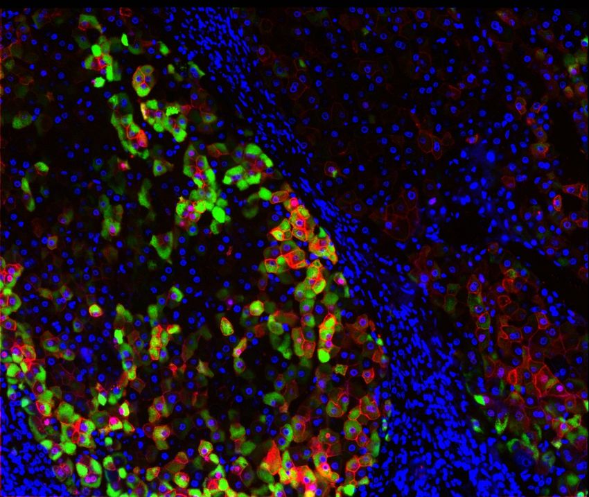
Verified MultiOmyx™ Multiplexing Panels

Immune Panel (TIL panel) – 12 markers
Tested in multiple cancer indications (melanoma, lung, colorectal, prostate, breast)
TIL Panel       Co-expression                 Phenotypes
CD3             CD3+CD4+                      T helper
CD4             CD3+CD4+FoxP3+                T regulatory
CD8             CD3+CD4+CD45RO+               Memory T helper
CD45RO          CD3+CD4+PD1+                  Immune modulation
FoxP3           CD3+CD8+                      T cytotoxic
CD20            CD3+CD8+CD45RO+               Memory T cytotoxic
CD68            CD3+CD8+PD1+                  Immune modulation
CD56            CD68+                         Macrophage                                                   PanCK+ CD8+ PD-L1+ CD68+ FoxP3+
                                                                    Au Q. et al. (April, 2016). MultiOmyx™ multiplexed tumor infiltrating lymphocyte
CTLA-4          CD68+PDL1+                    Macrophage PD-L1      panel provides comprehensive immunophenotyping from a single FFPE slide.
                                                                    Poster presented at AACR Annual Meeting, New Orleans, LA.
PD-1            CD20+                         B cell
PD-L1           CD20+PDL1+                    B cell PD-L1
PanCK           CD3-CD56+                     Natural Killer cell
                PanCK+PDL1+                   Tumor cell PD-L1

Myeloid Panel – 10 markers
Tested in PDAC tissue

Myeloid Panel   Co-expression                 Phenotypes
 CD11b          CD11b+CD33+                   Myeloid cells
                                                                                                                      M1 TAM
 CD14           CD11b+CD33+HLADR-             MDSC
 CD15           CD11b+CD33+HLADR-CD14+CD15-   M-MDSC
 CD16           CD11b+CD33+HLADR-CD14-CD15+   G-MDSC
 CD33           CD68+                         TAM
 CD68           CD68+HLADR+CD163-             M1 TAM
 CD163          CD68+HLADR-CD163+             M2 TAM
 HLA-DR         CD11b+HLADR-CD16+             Neutrophil/G-MDSC                                                 PanCK+ CD68+ CD11b+ HLA-DR+
                                                                    Juncker-Jensen A. et al. (April, 2018). Tumor-Infiltrating Myeloid Cells – Using
 Arginase1                                                          MultiOmyx™ to Distinguish between MDSCs, TAMs and TANs in the Pancreatic
                                                                    Tumor Microenvironment. Poster presented at AACR Annual Meeting, Chicago, IL.
 PanCK

                                                                                                             Verified MultiOmyx™ Multiplexing Panels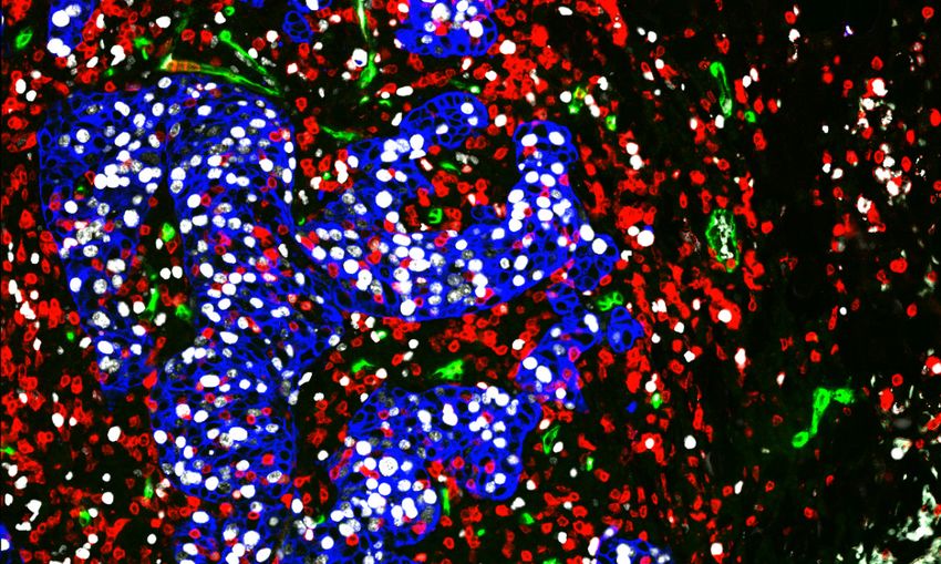
TIL & Myeloid Panel #1 – 16 markers
Tested in NSCLC tissue

 TIL/TAM Panel #1                 Co-expression                 Phenotypes
  CD3                             CD3+CD4+                      T helper                                                            LAG3+/PD1+ CTL
  CD4                             CD3+CD4+FoxP3+                T regulatory
  CD8                             CD3+CD4+PD1+                  Immune modulation
  FoxP3                           CD3+CD8+                      T cytotoxic
  CD20                            CD8+PD1+                      Immune modulation
  CD68                            CD3+TIM3+                     Immune modulation
  CD163                           CD3+LAG3+                     Immune modulation
                                  CD3+ICOS+                     Immune modulation                                                        PanCK+ LAG-3+ PD-1+ CD8+
  CD11b
                                                                                       Juncker-Jensen A. et al. (2019, December). PD-1 and LAG-3 synergize to drive
  CD15                            CD3+OX40+                     Immune modulation      tumor-infiltration of T cytotoxic cells in NSCLC tumors. Poster presented at ESMO
                                                                                       I/O Annual Meeting, Geneva, Switzerland.
  TIM-3                           CD20+                         B cell
  LAG-3                           CD11b+CD15+                   Granulocyte
  OX40                            CD68+                         TAM
  ICOS                            CD68+CD163+                   M2 TAM
  PD-1                            CD68+PDL1+                    Macrophage PD-L1
  PD-L1                           CD68+TIM3+                    Macrophage TIM-3
  PanCK                           PanCK+PDL1+                   Tumor cell PD-L1

TIL & Myeloid Panel #2 – 19 markers
Tested in PDAC tissue

 TIL/Myeloid Panel               Co-expression                  Phenotypes
 #2
                                  CD3+CD4+                      T helper
  CD3
                                  CD3+CD4+FoxP3+                T regulatory
                                                                                                                          G-MDSC
  CD4
                                  CD3+CD4+PD1+                  Immune modulation
  CD8
                                  CD3+CD8+                      T cytotoxic
  CD45RO
                                  CD8+PD1+                      Immune modulation
  FoxP3
                                  CD8+GranzymeB+                Effector T cytotoxic
  CD11b
                                  CD3+CD45RO                    Memory T cells
  CD14                                                                                                                        PanCK+ CD11b+ CD3+ CD15+ FoxP3+
                                  CD68+                         TAM
                                                                                       Juncker-Jensen A. et al. (October, 2018). Using MultiOmyx™ to Analyze
  CD15
                                  CD68+HLADR+CD163-             M1 TAM                 Correlations between Immunosuppressive Cells and Tumor-Infiltrating
                                                                                       Lymphocytes in the Pancreatic Tumor Microenvironment. Poster presented at
  CD16
                                  CD68+HLADR-CD163+             M2 TAM                 ESMO Annual Meeting, Munich, Germany.
  CD33
                                  CD68+PDL1+                    Macrophage PD-L1
  CD68
                                  CD11b+CD33+                   Myeloid cells
  CD163
                                  CD11b+CD33+HLADR-             MDSC
  HLA-DR
                                  CD11b+CD33+HLADR-CD14+CD15-   M-MDSC
  Arginase1
                                  CD11b+CD33+HLADR-CD14-CD15+   G-MDSC
  GranzymeB
                                  PanCK+Ki67+                   Proliferating tumor
  Ki67
                                  PanCK+PDL1+                   Tumor cell PD-L1
  PD-1
  PD-L1
  PanCK

2 | Verified MultiOmyx™ Multiplexing Panels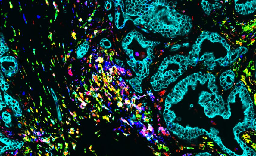
Dendritic Cell Panel – 15 markers
Tested in melanoma tissue
Dendritic Panel Co-expression Phenotypes
CD3 CD3+CD4+ T helper pDC
CD4 CD3+CD8+ T cytotoxic
CD8 CD68+ TAM
CD11c CD68+HLADR+CD163- M1 TAM
CD14 CD68+HLADR-CD163+ M2 TAM
CD40 CD123+HLADR+ pDC
CD68 CD123+HLADR+CD40+ CD40 positive pDC
CD123 CD123+HLADR+DCLAMP+ Activated pDC
CD141 CD11c+Clec9A+CD141+HLADR+ cDC
CD163 CD11c+Clec9A+CD141+CD40+ CD40 positive cDC SOX10+CD123+HLADR+CD11c+
Gozo M. et al. (2020, June). Distinguishing dendritic cell subtypes in the
HLA-DR CD11c+Clec9A+CD141+DCLAMP+ Activated cDC tumor microenvironment using MultiOmyxTM. Poster presented at AACR
Annual Meeting, Virtual.
Clec9A CD11c+CD14+DCSIGN+HLADR+ Monocyte derived DC
DC-SIGN
DC-LAMP
SOX10/PanCK
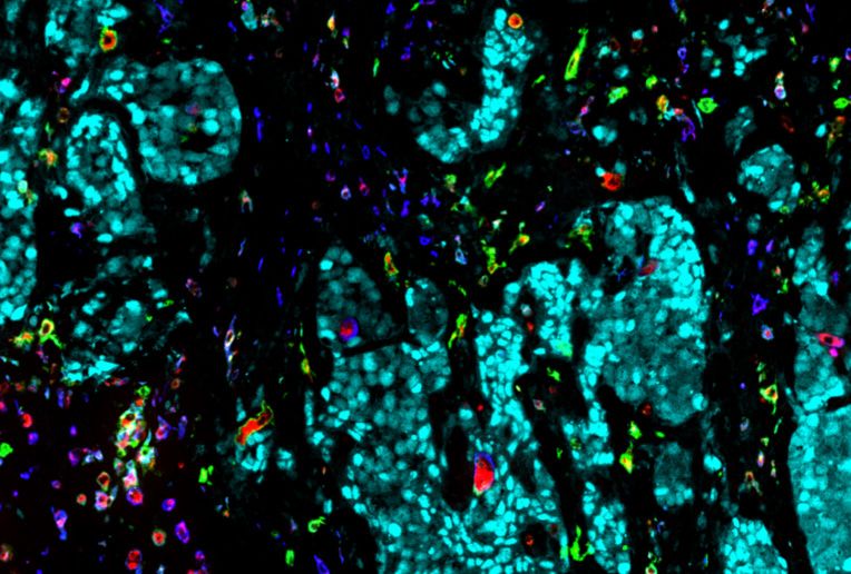
Tertiary Lymphoid Structure (TLS) Panel – 14 markers
Tested in bladder cancer tissue
TLS Panel Co-expression Phenotypes
CD3 CD3+CD4+ T helper
CD4 CD3+CD4+FoxP3+ T regulatory
CD8 CD3+CD8+ T cytotoxic
FoxP3 CD20+ B cell
CD11b CD11b+HLADR+ APC myeloid cell
CD14 CD11b+HLADR- MDSC
CD15 CD11b+HLADR-CXCR2+ MDSC positive for CXCR2
CD20 CD11b+HLADR-iNOS MDSC positive for iNOS
CD68 CD11b+HLADR-CD14+CD15- M-MDSC HEV
PNAd CD11b+HLADR-CD14+CD15- G-MDSC
CXCR2 CD68+ TAM
iNOS CD68+HLADR+ M1 TAM CD20+ PNAd+ CD3+
Image of TLS in a bladder cancer sample from an unpublished academic
HLA-DR CD68+iNOS+ M1 TAM collaboration study with University of Notre Dame.
PanCK PNAd+ High Endothelial Venule
PanCK+iNOS+ iNOS positive tumor cells
Verified MultiOmyx™ Multiplexing Panels | 3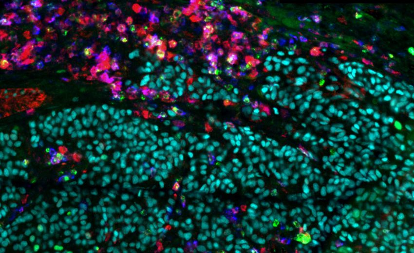
TIL, TAM & Vessel Panel #1 – 10 markers
Tested in breast cancer tissue

 TIL/TAM Panel #1                 Co-expression       Phenotypes

 CD3                              CD3+CD4+            T helper

 CD4                              CD3+CD4+FoxP3+      T regulatory

 CD8                              CD3+CD8+            T cytotoxic

 FoxP3                            CD68+               TAM

 CD68                             CD68+HLADR+CD163-   M1 TAM

 CD163                            CD68+HLADR-CD163+   M2 TAM
                                                                                                                               PanCK+ CD3+ CD34+ Ki67+
 HLA-DR                           CD34+               Vessels               Juncker-Jensen A. et al. (April, 2019). Pro-Tumorigenic Mechanisms of M2 Tumor-
                                                                            Associated Macrophages in Triple-Negative Breast Cancer. Poster presented at
 Ki67                             PanCK+Ki67+         Proliferating tumor   AACR Annual Meeting, Chicago, IL.

 CD34

 PanCK

TIL, TAM & Vessel Panel #2 – 16 markers
Tested in ovarian tumor tissue

 TIL/TAM Panel #2                 Co-expression       Phenotypes                             M1 TAM
 CD3                              CD3+CD4+            T helper

 CD4                              CD3+CD4+FoxP3+      T regulatory
                                                                                         M2 TAM
 CD8                              CD3+CD4+PD1+        Immune modulation

 FoxP3                            CD4+CTLA4+          Immune modulation

 CD20                             CD3+CD8+            T cytotoxic

 CD68                             CD8+PD1+            Immune modulation

 CD163                            CD8+CTLA4+          Immune modulation

 HLA-DR                           CD20+               B cell
                                                                                                                          S100+ CD68+ HLA-DR+ CD163+
 PD-1                             CD68+               TAM                   Juncker-Jensen A. et al. (2019, October). An Integrated Multiplexing Approach for
                                                                            the Immunoprofiling of the Tumor Microenvironment of Ovarian Granulosa Cell
 PD-L1                            CD68+HLADR+CD163-   M1 TAM                Tumors. Poster presented at AACR-NCI-EORTC Annual Meeting, Boston, MA.

 CTLA-4                           CD68+HLADR-CD163+   M2 TAM

 CD34                             CD68+PDL1+          Macrophage PD-L1

 Vimentin                         CD34+               Vessels

 S100                             PanCK+Ki67+         Proliferating tumor

 Ki67                             PanCK+PDL1+         Tumor cell PD-L1

 PanCK

4 | Verified MultiOmyx™ Multiplexing Panels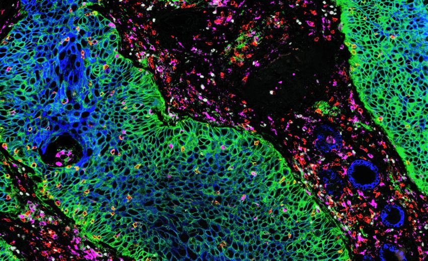
TIGIT Immune Cell Panel – 15 markers
Tested in NSCLC & melanoma tissue

TIGIT Immune Panel   Co-expression                     Phenotypes

TIGIT                CD3+CD4+                          T helper

CD226                CD3+CD4+FoxP3+                    T regulatory

CD155                CD3+CD4+PD1+                      Immune modulation

CD3                  CD3+CD8+                          T cytotoxic

CD4                  CD8+PD1+                          Immune modulation

CD8                  CD8+GranzymeB+                    Effector T cytotoxic
                                                                                                                                 SOX10 + TIGIT+ LAG3+ PD1+
FoxP3                CD68+                             TAM
                                                                              Au Q. et al. (April, 2019). Characterization of TIGIT Expression Using
                                                                              MultiOmyx™ Hyperplexed Immunofluorescence Assay in NSCLC and melanoma.
CD56                 CD68+HLADR+CD163-                 M1 TAM                 Poster presented at AACR Annual Meeting, Atlanta, GA.

CD45RO               CD68+HLADR-CD163+                 M2 TAM

CD11b                CD68+PDL1+                        Macrophage PD-L1

CD11c                CD11b+CD33+                       Myeloid cells

PD-1                 CD11b+CD33+HLADR-                 MDSC

LAG-3                CD11b+CD33+HLADR-CD14+CD15-       M-MDSC
TIM-3                CD11b+CD33+HLADR-CD14-CD15+       G-MDSC
PanCK/SOX10          PanCK+Ki67+                       Proliferating tumor

                     PanCK+PD1+                        Tumor cell PD-L1

Myeloid Leukemia Immune Panel – 14 markers
Tested in AML/HL/DLBCL

Leukemia Panel       Co-expression                         Phenotypes
CD3                  CD3+CD4+                              T helper                                     G-MDSC

CD4                  CD3+CD4+FoxP3+                        T regulatory
CD8                  CD3+CD8+                              T cytotoxic
                                                                                                                           G-MDSC
FoxP3                CD68+                                 TAM
                                                                                G-MDSC
CD68                 CD68+HLADR+CD163-                     M1 TAM
CD163                CD68+HLADR-CD163+                     M2 TAM
HLA-DR               CD11b+CD33+                           Myeloid cells
                                                                                                                             CD11b+CD33+CD15+CD34+
CD11b                CD11b+CD33+HLADR-                     MDSC
                                                                              Au Q. et al. (2019, December). Phenotypic Characterization of the Immune
CD33                 CD11b+CD33+HLADR-CD14+CD15-           M-MDSC             Landscape in the Bone Marrow of Patients with Acute
                                                                              Myeloid Leukemia (AML) Using MultiOmyx™ Hyperplexed Immunofluorescence
CD14                 CD11b+CD33+HLADR-CD14-CD15+           G-MDSC             Assay. Poster presented at ASH Annual Meeting, Orlando, FL.

CD15                 CD11b+CD33+HLADR-CD14-CD15+Arg1       Arg1+ G-MDSC
CD34
Arginase1
Tumor marker

                                                                                                                 Verified MultiOmyx™ Multiplexing Panels | 5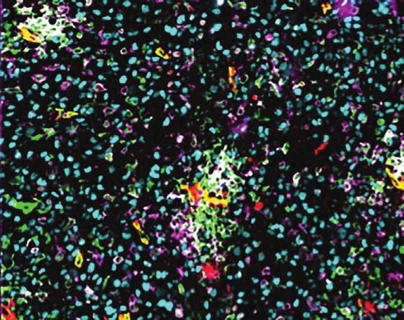
Hepatitis B (HBV) Panel – 19 markers
Tested in liver tissue

 HBV Panel                        Co-expression                  Phenotypes
  HBcAG                           CD3+CD4+                       T helper
  HBsAG                           CD3+CD4+FoxP3+                 T regulatory
  NaKATPase                       CD3+CD4+PD1+                   Immune modulation
  CD3                             CD3+CD8+                       T cytotoxic
  CD4                             CD3+CD8+PD1+                   Immune modulation
  CD8                             CD68+                          TAM
  FoxP3                           CD68+HLADR+                    M1 macrophage
                                  CD68+CD163+                    M2 macrophage                      HBcAG+ HBsAG+ DAPI+
  CD56
  CD20                            CD68+PDL1+                     Macrophage PD-L1
  CD11b                           CD68+DCSIGN+                   Macrophage DC-SIGN
  HLA-DR                          CD11b+HLADR-                   MDSC
  CD11c                           CD11c+HLADR+                   Dendritic cell (cDC)
  DC-SIGN                         CD11c+HLADR+DCSIGN+            Dendritic cell
  CD68                            CD20+                          B cell
                                                                                            Dendritic cell
  CD163                           CD20+HLADR+                    B cell HLA-DR
  PD-1                            CD56+CD3-                      NK cell
  PD-L1                           CD56+CD3-HLADR+                NK cell HLA-DR                  CD11c+ HLADR+ CD68+ CD20+

  IDO1
  Ki67

NASH Liver Panel – 17 markers
Tested in liver tissue

 NASH Panel                      Co-expression                   Phenotypes
  CD11b                           CD11b+CD11c+CD14-HLADR+         cDC
  HLA-DR                          CD11c+HLADR+CD14-IDO1+          cDC IDO1+
  CD68                            CD11b+CD11c+HLADR+CD14-Ki67+    cDC proliferating
  CD163                           CD68+CD163+CD16                 Kupffer cell             Kupffer cell
  CD11c                           CD68+CD163+CD16+DCSIGN+         Kupffer cell DC-SIGN+
  DC-SIGN                         CD68+CD163+CD16+PDL1+           Kupffer cell PD-L1+
  CD66b                                                           Kupffer cell prolifer-
                                  CD68+CD163+CD16+Ki67+          ating                                    CD68+ CD16+ CD163+
  CD9
                                  CD68+CD163+CD16+CD33+           Kupffer cell CD33+
  CD14
                                  CD11b+CD14+HLADR+               Tmo
  CD16
                                  CD11b+CD14+HLADR+CD16+          TMo CD16+
  CD33
                                  HLADR+CD9+CD14+                 SAM
  SMA
                                  HLADR+CD9+CD14+TREM2+           SAM TREM2+
  TREM2
                                  HLADR+CD9+CD14+PDL1+            SAM PD-L1+
  MNDA
                                  HLADR+CD9+CD14+DCSIGN+          SAM DC-SIGN+
  IDO1
                                  HLADR+CD9+CD14+CD33+            SAM CD33+
  PD-L1
                                  HLADR+CD9+CD14+MNDA+            SAM MNDA+
  Ki67                                                                                             HLADR+ CD16+ CD14+ CD9+
                                  HLADR+CD9+CD14+Ki67+            SAM proliferating
  PD-L1
  PanCK

6 | Verified MultiOmyx™ Multiplexing Panels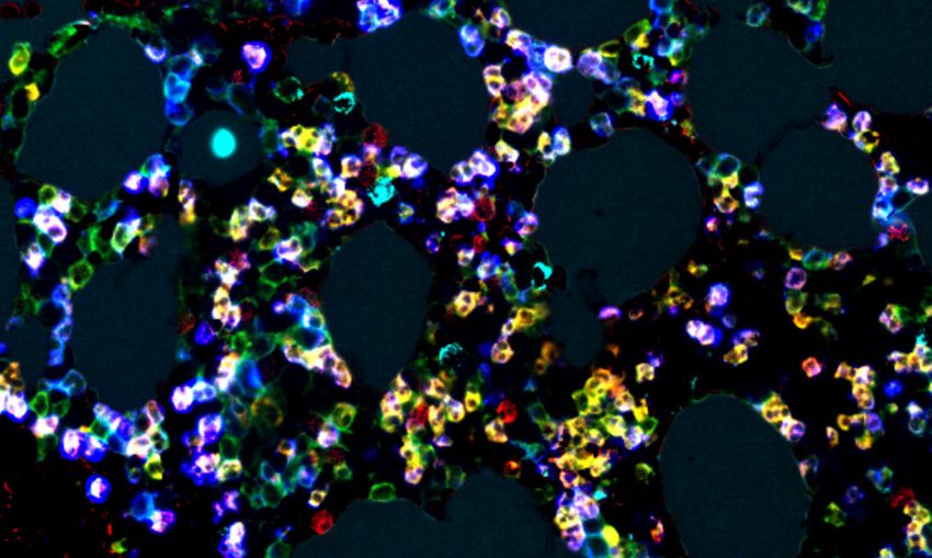
Verified MultiOmyx™ Multiplexing Panels | 7

About NeoGenomics
NeoGenomics is an industry-leading cancer diagnostics and pharma services company. We enable advanced
oncology patient care leveraging the most extensive diagnostic solid tumor and hematological test menus,
sub-specialized pathology, and expertise in biomarker development. We partner closely with our pharma and
research clients to meet program objectives and delivery from biomarker discovery through companion CDx
validation and commercialization.

About Neogenomics Pharma Services
NeoGenomics’ Pharma Services unifies several innovative companies’ scientific and medical leadership under
one leading brand, offering one of the most comprehensive laboratory services menu available for biomarker
testing supporting oncology clinical trials globally.

We provide our clients with an unparalleled level of expertise, service, flexibility, and scalability. Additionally,
we offer alternative business models and solutions across the continuum of development from pre-clinical
research and development through commercialization.

To learn more about NeoGenomics Pharma Services, visit us online at https://neogenomics.com/pharma-
services. NeoGenomics Pharma Services can be your research partner with NGS or other innovative
services.

Please contact NeoGenomics Pharma Services at 800.720.4363 or email at
pharmaservices@neogenomics.com.

NeoGenomics Laboratories is a specialized oncology reference laboratory providing the latest technologies, testing, partnership
opportunities, and interactive education to the oncology and pathology communities. We offer the complete spectrum of diagnostic
services in molecular testing, FISH, cytogenetics, flow cytometry, and immunohistochemistry through our nation-wide network of CAP-
accredited, CLIA-certified laboratories.
Committed to research as the means to improve patient care, we provide Pharma Services for pharmaceutical companies, in vitro
diagnostic manufacturers, and academic scientist-clinicians. We promote joint publications with our client physicians. NeoGenomics
welcomes your inquiries for collaborations. Please contact us for more information.

                                                                       12701 Commonwealth Dr., Suite 9                          NeoGenomics Europe, S.A.
                                                                       Fort Myers, FL 33913 United States                       A-One Business Center
                                                                       Phone: 866.776.5907                                      Bâtiment A5, 2nd Floor
                                                                       Fax: 239.690.4237                                        Z.A. La Pièce / Route de l’Etraz 1
                                                                                                                                1180 Rolle, Switzerland
                                                                       31 Columbia
                                                                                                                                Phone: +41.21.721.06.00
                                                                       Aliso Viejo, CA 92656 United States
                                                                                                                                Fax: +41.(0)21.826.00.73
                                                                       Phone: 800.720.4363
                                                                       Fax: 949.425.5865                                        NeoGenomics Singapore Pte. Ltd.
                                                                       7256 S. Sam Houston Pkwy W., Suite 300                   61 Science Park Road, #02-11
                                                                       Houston, TX 77085 United States                          Singapore 117525
                                                                       Phone: 800.720.4363 / 713.528.4363                       Phone: +65.69115202
                                                                       Fax: 713.668.3565
                                                                                                                                Suzhou NeoGenomics Pharmaceutical
                                                                       NeoGenomics Laboratories, Inc.                           Research Co., Ltd
                                                                       4570 Executive Drive., Suite 250                         Building 6, Block 19, Yong’An Road
                                                                       San Diego, CA 92121                                      Huguan Industrial Park,
                                                                       Phone: 949.445.7300 x5056                                Suzhou New District, China
                                                                       © 2021 NeoGenomics Laboratories, Inc. All Rights Reserved.
                                                                       All other trademarks are the property of their respective owners. Rev.012621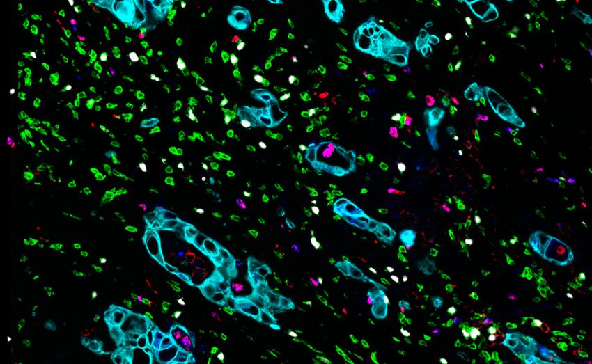
You can also read


